Embed Size (px)
Citation preview
BioMed Central
World Journal of Surgical Oncology
ss
Open AcceTechnical innovationsStereotaxic gamma knife surgery in treatment of critically located pilocytic astrocytoma: preliminary resultRaef FA Hafez*Address: International Medical Center, Gamma Knife Center, Cairo- Egypt
Email: Raef FA Hafez* - [email protected]
* Corresponding author
AbstractBackground: Low-grade gliomas are uncommon primary brain tumors, located more often in theposterior fossa, optic pathway, and brain stem and less commonly in the cerebral hemispheres.
Case presentations: Two patients with diagnosed recurrent cystic pilocytic astrocytomacritically located within the brain (thalamic and brain stem) were treated with gamma knife surgery.Gamma knife surgery (GKS) did improve the patient's clinical condition very much which remainedstable later on. Progressive reduction on the magnetic resonance imaging (MRI) studies of the solidpart of the tumor and almost disappearance of the cystic component was achieved within thefollow-up period of 36 months in the first case with the (thalamic located lesion) and 22 months inthe second case with the (brain stem located lesion).
Conclusion: Gamma knife surgery represents an alternate tool in the treatment of recurrent and/or small postoperative residual pilocytic astrocytoma especially if they are critically located
BackgroundLow-grade gliomas are uncommon primary brain tumorsclassified as histological grades I or II in the World HealthOrganization (WHO) classification. The most commonvariants are pilocytic astrocytoma, low-grade astrocyto-mas, oligodendrogliomas, and mixed oligo-astrocytomaslocated more often in the posterior fossa, optic pathway,and brain stem and less commonly in the cerebral hemi-spheres. Prognostic factors that predict progression-freeand overall survival include young age, pilocytic histol-ogy, good Karnofsky performance status, gross total resec-tion, lack of enhancement on imaging, and smallpreoperative tumor volumes. Edema and vasogenic effectsare typically managed with corticosteroids. The rationalefor open craniotomy depends on the need for immediatepalliation of symptoms by reduction of intracranial pres-
sure or focal mass effect, and/or improved oncologic con-trol. [1,2]
Gross total resection of low-grade glioma is generallydefined as the absence of residual enhancement on con-trast-enhanced postoperative MRI scan. Most retrospec-tive studies suggest that patients who have undergone agross total resection of tumor have improved survival.Depending upon the proximity of the tumor to eloquentbrain areas, gross total resection may or may not be possi-ble. In these cases a stereotactic biopsy is required to pro-vide the histological diagnosis. Adjuvant radiotherapy isrecommended for patients with incompletely resectedgrade II tumors or for patients older than age 40 regardlessof extent of resection, it may be considered for any pilo-cytic astrocytoma from which a biopsy has been made.Radiosurgery and/or experimental chemotherapy may
Published: 29 March 2007
World Journal of Surgical Oncology 2007, 5:39 doi:10.1186/1477-7819-5-39
Received: 22 August 2006Accepted: 29 March 2007
This article is available from: http://www.wjso.com/content/5/1/39
© 2007 Hafez; licensee BioMed Central Ltd. This is an Open Access article distributed under the terms of the Creative Commons Attribution License (http://creativecommons.org/licenses/by/2.0), which permits unrestricted use, distribution, and reproduction in any medium, provided the original work is properly cited.
Page 1 of 6(page number not for citation purposes)
World Journal of Surgical Oncology 2007, 5:39 http://www.wjso.com/content/5/1/39
provide some measure of local control in the recurrentdisease setting [3-6]
Stereotactic radiotherapy provides excellent local controlfor small, localized low-grade glial tumors. Marginal fail-ures have not been observed, supporting the use of lim-ited margins radiation dose to minimize late sequelaeusing stereotactic leksell gamma knife technique [7,8]
Case presentationsCase 1A 10 years old girl presented with right side weakness,with vomiting attacks and blurring of vision. MRI deline-ated a large cystic lesion at left thalamic region with solidpart. Stereotactic biopsy from the solid portion and aspi-ration of the cyst revealed pilocytic astrocytoma. Post aspi-ration the child improved clinically, but three monthslater the symptoms recurred and MRI revealed recurrenceof the cystic part of the tumor that was treated again withstereotactic aspiration of the fluid, followed with station-ary course. There was no history of patient receiving radi-otherapy.
The child was referred to our Gamma Knife Center inOctober 2003; 6 months post second stereotactic aspira-tion with right side weakness more in the upper limb,with right facial palsy, and headache. MRI revealed largenonenhanced left thalamic cystic lesion with small twohyperintense intracystic solid parts.
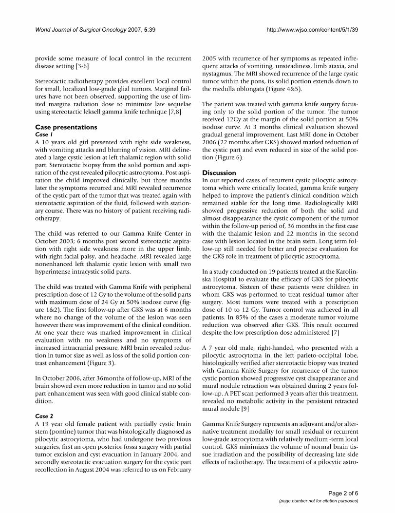
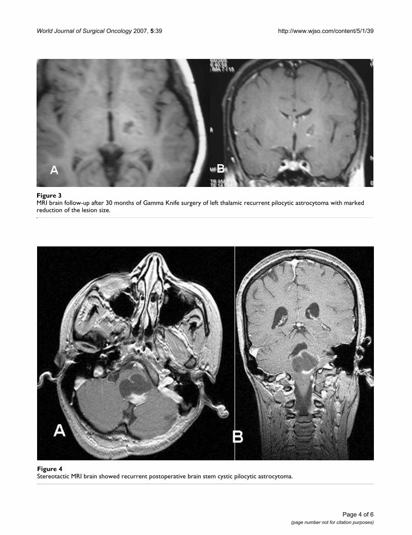
The child was treated with Gamma Knife with peripheralprescription dose of 12 Gy to the volume of the solid partswith maximum dose of 24 Gy at 50% isodose curve (fig-ure 1&2). The first follow-up after GKS was at 6 monthswhere no change of the volume of the lesion was seenhowever there was improvement of the clinical condition.At one year there was marked improvement in clinicalevaluation with no weakness and no symptoms ofincreased intracranial pressure, MRI brain revealed reduc-tion in tumor size as well as loss of the solid portion con-trast enhancement (Figure 3).
In October 2006, after 36months of follow-up, MRI of thebrain showed even more reduction in tumor and no solidpart enhancement was seen with good clinical stable con-dition.
Case 2A 19 year old female patient with partially cystic brainstem (pontine) tumor that was histologically diagnosed aspilocytic astrocytoma, who had undergone two previoussurgeries, first an open posterior fossa surgery with partialtumor excision and cyst evacuation in January 2004, andsecondly stereotactic evacuation surgery for the cystic partrecollection in August 2004 was referred to us on February
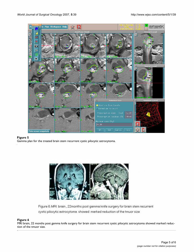
2005 with recurrence of her symptoms as repeated infre-quent attacks of vomiting, unsteadiness, limb ataxia, andnystagmus. The MRI showed recurrence of the large cystictumor within the pons, its solid portion extends down tothe medulla oblongata (Figure 4&5).
The patient was treated with gamma knife surgery focus-ing only to the solid portion of the tumor. The tumorreceived 12Gy at the margin of the solid portion at 50%isodose curve. At 3 months clinical evaluation showedgradual general improvement. Last MRI done in October2006 (22 months after GKS) showed marked reduction ofthe cystic part and even reduced in size of the solid por-tion (Figure 6).
DiscussionIn our reported cases of recurrent cystic pilocytic astrocy-toma which were critically located, gamma knife surgeryhelped to improve the patient's clinical condition whichremained stable for the long time. Radiologically MRIshowed progressive reduction of both the solid andalmost disappearance the cystic component of the tumorwithin the follow-up period of, 36 months in the first casewith the thalamic lesion and 22 months in the secondcase with lesion located in the brain stem. Long term fol-low-up still needed for better and precise evaluation forthe GKS role in treatment of pilocytic astrocytoma.
In a study conducted on 19 patients treated at the Karolin-ska Hospital to evaluate the efficacy of GKS for pilocyticastrocytoma. Sixteen of these patients were children inwhom GKS was performed to treat residual tumor aftersurgery. Most tumors were treated with a prescriptiondose of 10 to 12 Gy. Tumor control was achieved in allpatients. In 85% of the cases a moderate tumor volumereduction was observed after GKS. This result occurreddespite the low prescription dose administered [7]
A 7 year old male, right-handed, who presented with apilocytic astrocytoma in the left parieto-occipital lobe,histologically verified after stereotactic biopsy was treatedwith Gamma Knife Surgery for recurrence of the tumorcystic portion showed progressive cyst disappearance andmural nodule retraction was obtained during 2 years fol-low-up. A PET scan performed 3 years after this treatment,revealed no metabolic activity in the persistent retractedmural nodule [9]
Gamma Knife Surgery represents an adjuvant and/or alter-native treatment modality for small residual or recurrentlow-grade astrocytoma with relatively medium -term localcontrol. GKS minimizes the volume of normal brain tis-sue irradiation and the possibility of decreasing late sideeffects of radiotherapy. The treatment of a pilocytic astro-
Page 2 of 6(page number not for citation purposes)
World Journal of Surgical Oncology 2007, 5:39 http://www.wjso.com/content/5/1/39
Page 3 of 6(page number not for citation purposes)
Gamma Knife treatment planning for the solid parts of the recurrent cystic pilocytic astrocytomaFigure 1Gamma Knife treatment planning for the solid parts of the recurrent cystic pilocytic astrocytoma.
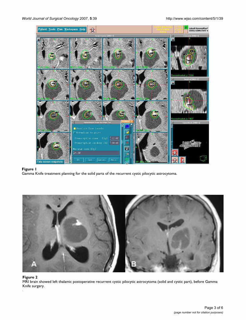
MRI brain showed left thalamic postoperative recurrent cystic pilocytic astrocytoma (solid and cystic part), before Gamma Knife surgeryFigure 2MRI brain showed left thalamic postoperative recurrent cystic pilocytic astrocytoma (solid and cystic part), before Gamma Knife surgery.
World Journal of Surgical Oncology 2007, 5:39 http://www.wjso.com/content/5/1/39
Page 4 of 6(page number not for citation purposes)
MRI brain follow-up after 30 months of Gamma Knife surgery of left thalamic recurrent pilocytic astrocytoma with marked reduction of the lesion sizeFigure 3MRI brain follow-up after 30 months of Gamma Knife surgery of left thalamic recurrent pilocytic astrocytoma with marked reduction of the lesion size.
Stereotactic MRI brain showed recurrent postoperative brain stem cystic pilocytic astrocytomaFigure 4Stereotactic MRI brain showed recurrent postoperative brain stem cystic pilocytic astrocytoma.
World Journal of Surgical Oncology 2007, 5:39 http://www.wjso.com/content/5/1/39
Page 5 of 6(page number not for citation purposes)
MRI brain, 22 months post gamma knife surgery for brain stem recurrent cystic pilocytic astrocytoma showed marked reduc-tion of the tmuor sizeFigure 6MRI brain, 22 months post gamma knife surgery for brain stem recurrent cystic pilocytic astrocytoma showed marked reduc-tion of the tmuor size.
Gamma plan for the treated brain stem recurrent cystic pilocytic astrocytomaFigure 5Gamma plan for the treated brain stem recurrent cystic pilocytic astrocytoma.
World Journal of Surgical Oncology 2007, 5:39 http://www.wjso.com/content/5/1/39
Publish with BioMed Central and every scientist can read your work free of charge
"BioMed Central will be the most significant development for disseminating the results of biomedical research in our lifetime."
Sir Paul Nurse, Cancer Research UK
Your research papers will be:
available free of charge to the entire biomedical community
peer reviewed and published immediately upon acceptance
cited in PubMed and archived on PubMed Central
yours — you keep the copyright
Submit your manuscript here:http://www.biomedcentral.com/info/publishing_adv.asp
BioMedcentral
cytoma located in a functional area can be performedusing GKS [10,11]
ConclusionGamma knife surgery represents an alternate tool in thetreatment of recurrent and/or small postoperative residualpilocytic astrocytoma especially if they are criticallylocated minimizes the volume of normal brain tissue irra-diation and the possibility of decreasing late side effects ofradiotherapy.
Competing interestsThe author(s) declare that they have no competing inter-ests.
Authors' contributionsRFH: conceived and prepared the manuscript.
AcknowledgementsThe written consent was obtained form the patient for publication of this case report.
References1. Simonová G, Novotny J Jr, Liscák R: Low-grade gliomas treated
by fractionated gamma knife surgery. J Neurosurg 2005,102(Suppl):19-24.
2. Heppner PA, Sheehan JP, Steiner LE: Gamma knife surgery forlow-grade gliomas. Neurosurger 2005, 57:1132-1139.
3. Keles GE, Lamborn KR, Berger MS: Low-grade hemispheric glio-mas in adults A critical review of extent of resection as a fac-tor influencing outcome. J Neurosurg 2001, 95:735-745.
4. Novotný J Jr, Novotný J, Simonová G, Lissck R, Vladyka V: Fraction-ated stereotactic radiotherapy with Leksell Gamma Knife.Radiosurgery 1997 Radiosurgery. Basel, Karger 1998, 2:197-205.
5. Saran FH, Baumert BG, Khoo VS, Adams EJ, Garre ML, WarringtonAP, Brada M: Stereotactically guided conformal radiotherapyfor progressive low-grade gliomas in childhood. Int J RadiatOncol Biol Phys 2002, 53:43-51.
6. Stieber VW: Low-grade gliomas. Curr Treat Options Oncol 2001,2:495-506.
7. Boethius J, Ulfarsson E, Rahn T, Lippitz B: Gamma knife radiosur-gery for pilocytic astrocytomas. J Neurosurg 2002, 97(5Suppl):677-680.
8. Marcus KJ, Goumnerova L, Billett AL, Lavally B, Scott RM, Bishop K,Xu R, Young Poussaint T, Kieran M, Kooy H, Pomeroy SL, Tarbell NJ:Stereotactic radiotherapy for localized low-grade gliomas inchildren: final results of a prospective trial. Int J Radiat Oncol BiolPhys 2005, 61:374-379.
9. Proust F, Coche-Dequeant B, Carpentier P, Laquerriere A, DerlonJM, Blond S, Christiaens J, Freger P: Combination treatment forpilocytic astrocytoma: stereotaxic radiosurgery and endo-cavitary radiotherapy. Neurochirurgie 1998, 44:50-54.
10. Graab PA, Lunsford LD, Albright AL, Kondziolka D, Flickinger JC:Stereotactic radiosurgeryfor glial neoplasms of childhood.Neurosurgery 1996, 38:696-702.
11. Wowra B, Muacevic A, Muller-Schunk S, Tonn JC: Special indica-tions in gamma knife surgery. Acta Neurochir 2004,91(Suppl):89-102.
Page 6 of 6(page number not for citation purposes)