Embed Size (px)
Citation preview
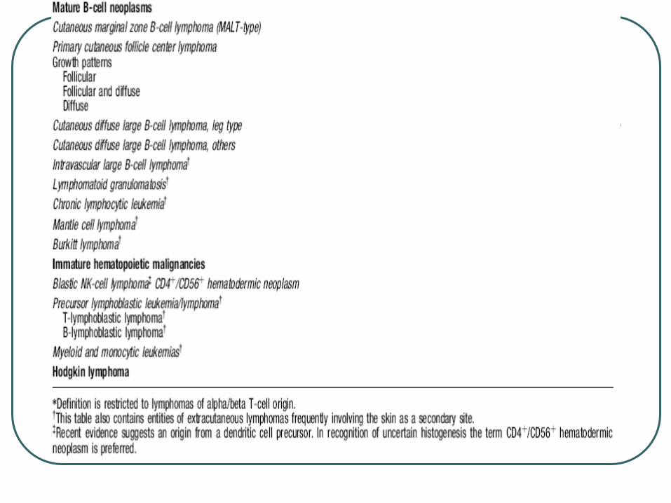
WHO/EORTC
Classification of
Cutaneous Lymphomas
2005
Introduction
Lymphoma in skin, in the past were considered to be manifestations of systemic processes.
A variety of T- and B-cell neoplasms can involve the skin, either primarily or secondarily.
After GI, skin is the 2nd most common site of extranodal non-Hodgkin lymphoma, annual incidence of 1:100,000.
Primary cutaneous lymphomas, completely different clinical behavior and prognosis from histologically similar systemic lymphomas, which may involve the skin secondarily
Treatment for primary vs secondary is usually different.
Introduction 2
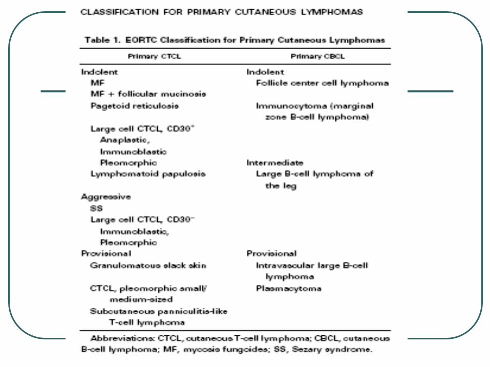
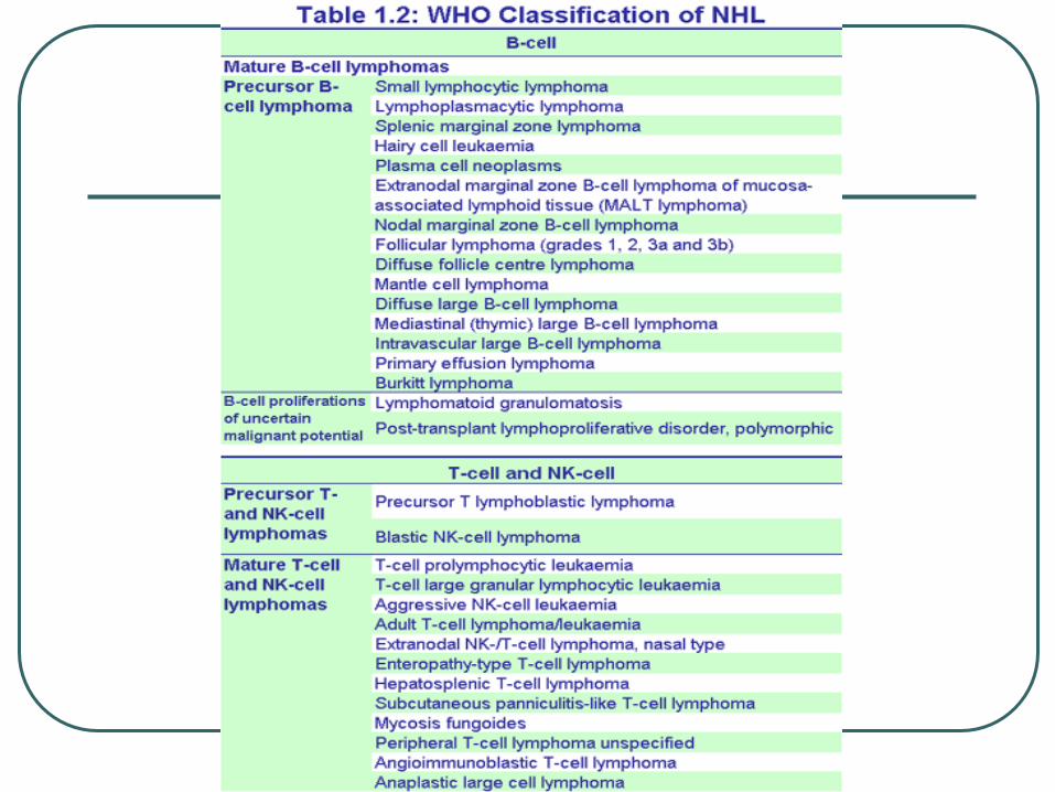
EORTC – Classification 1997
WHO – Classification 2001
Concensus meetings on 2003 and 2004
lead to the 2005 WHO/EORTC
classification.
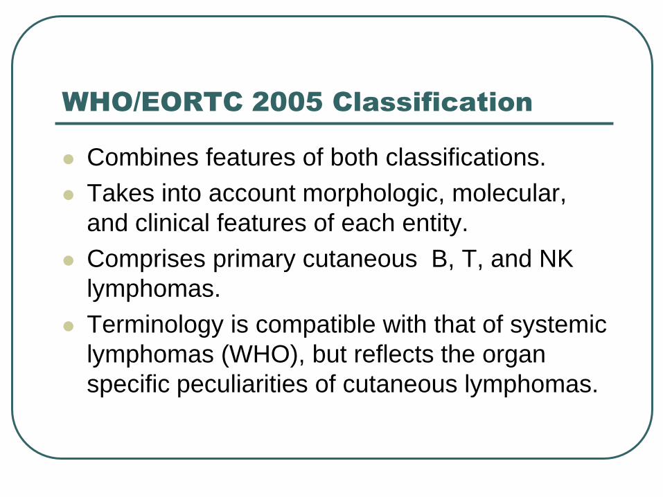
WHO/EORTC 2005 Classification
Combines features of both classifications.
Takes into account morphologic, molecular,
and clinical features of each entity.
Comprises primary cutaneous B, T, and NK
lymphomas.
Terminology is compatible with that of systemic
lymphomas (WHO), but reflects the organ
specific peculiarities of cutaneous lymphomas.
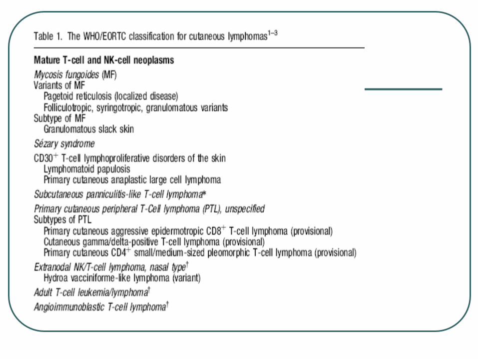
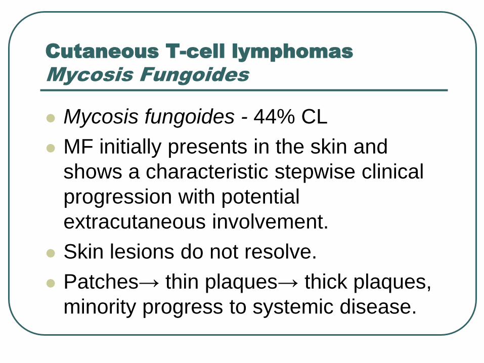
Cutaneous T-cell lymphomas
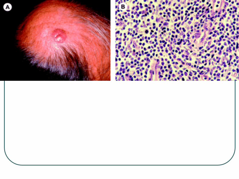
Mycosis Fungoides
Mycosis fungoides - 44% CL
MF initially presents in the skin and
shows a characteristic stepwise clinical
progression with potential
extracutaneous involvement.
Skin lesions do not resolve.
Patches→ thin plaques→ thick plaques,
minority progress to systemic disease.
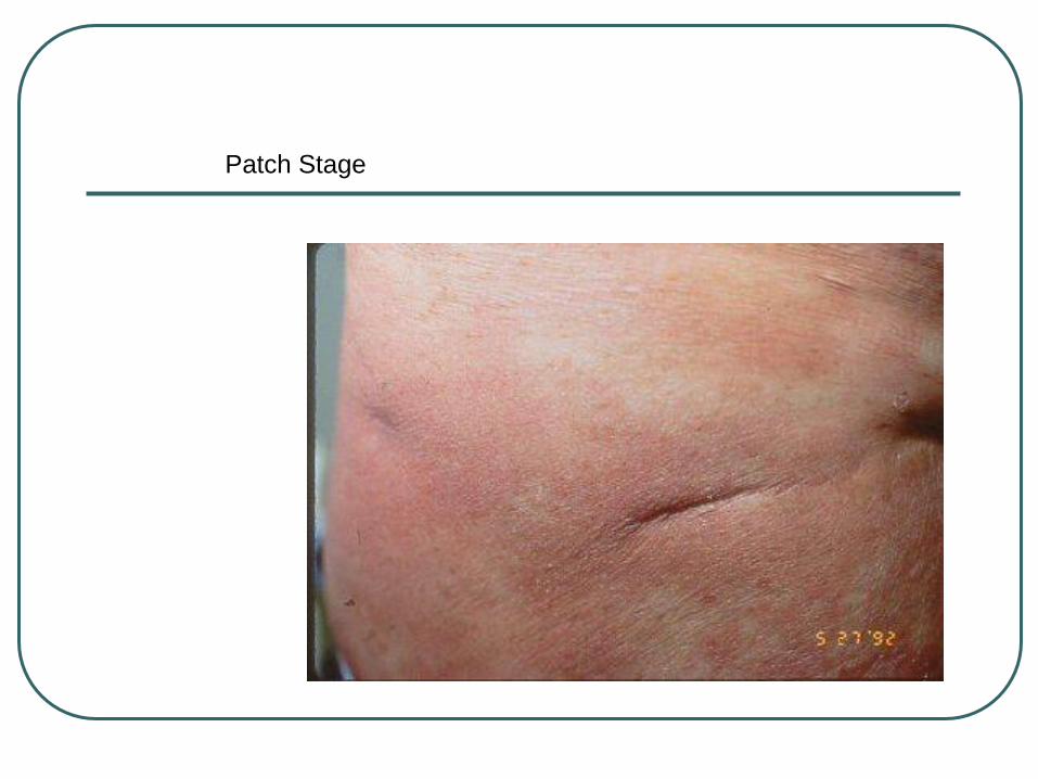
Patch Stage
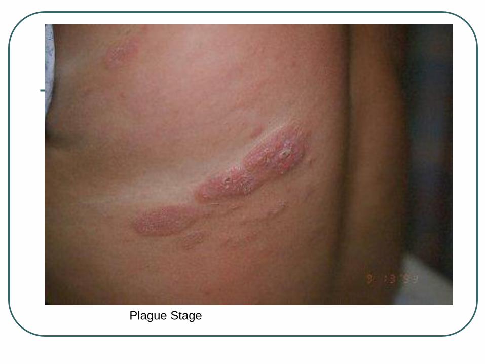
Plague Stage
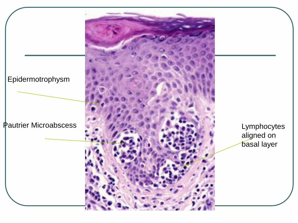
Pautrier Microabscess Lymphocytes
aligned on
basal layer
Epidermotrophysm
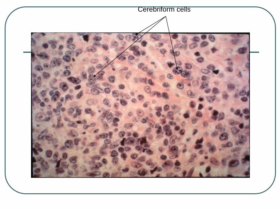
Cerebriform cells
Histologic features of MF
Pautrier microabscessess – 10%
Psoriasiform – lichenoid pattern
Lymphocytes aligned in the basal layer
(at tips of rete ridges)
Epidermotrophism 95%
Cerebbriform cells seen in 50% of cases
Eosinophils, plasma cells, and
macrophages may be admixed.
Mycosis Fungoides
Immunohistochemistry is helpful but not necessary for dx.
CD2 +, CD3 +, CD4+, CD5+ TCRbeta+
CD8 -, Cd 30 –
Rare cases may be CD8 + or CD30+
5yr survival 90%
Tx Skin-targeted therapies as photo (chemo)–therapy PUVA, topical nitrogen mustard or chlormustine (BCNU), or radiotherapy, including total skin electron beam irradiation.
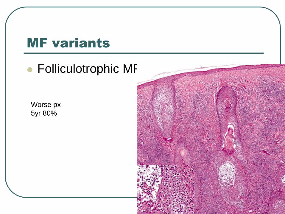
MF variants
Folliculotrophic MF
Worse px
5yr 80%
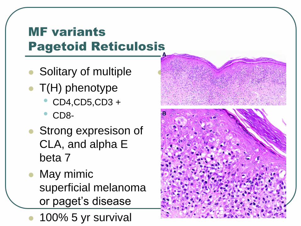
MF variants
Pagetoid Reticulosis
Solitary of multiple
T(H) phenotype
• CD4,CD5,CD3 +
• CD8-
Strong expresison of
CLA, and alpha E
beta 7
May mimic
superficial melanoma
or paget’s disease
100% 5 yr survival
Pagetoid reticulosis
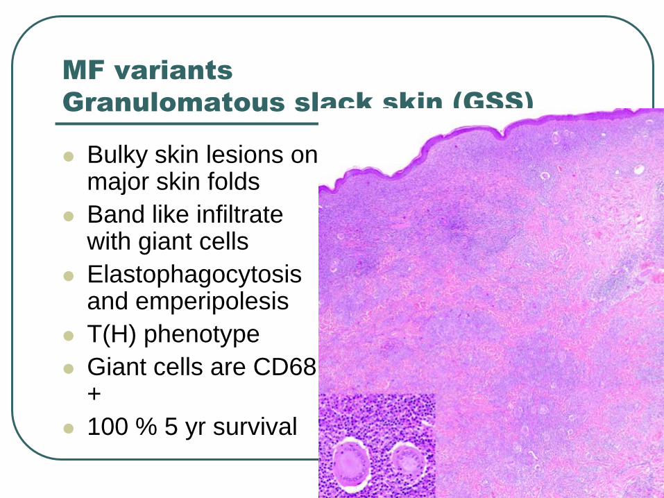
MF variants
Granulomatous slack skin (GSS)
Bulky skin lesions on major skin folds
Band like infiltrate with giant cells
Elastophagocytosis and emperipolesis
T(H) phenotype
Giant cells are CD68 +
100 % 5 yr survival
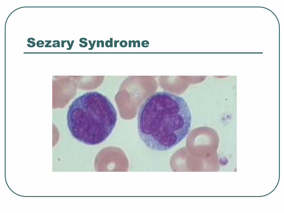
Sezary Syndrome
Sezary Syndrome
ISCL (International society of cutaneous lymphoma
• Absolute sezary cell count of 1000 cells/mm
• Expanded CD4 population resulting in CD4/CD8 ratio of > 10
• Loss of any of the T cell antigens (CD2, CD3, CD4, and CD5) or demonstration of T cell clonality by molecular studies.
20% 5 yr survival
Tx Extracorporeal photopheresis (ECP), either alone or in combination with other treatment modalities ( interferon alpha, methotrexate, campath)
CD 30 + T cell lymphoproliferative
disorders of the skin
Lymphomatoid papulosis
Primary cutaneous anaplastic large cell
lymphoma
Hallmark is CD 30 +, however both
entities differ in clinical and histologic
presentations.
Represent 30% of cutaneous
lymphomas
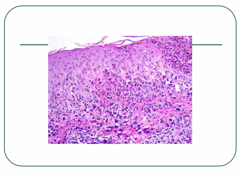
Lymphomatoid Papulosis
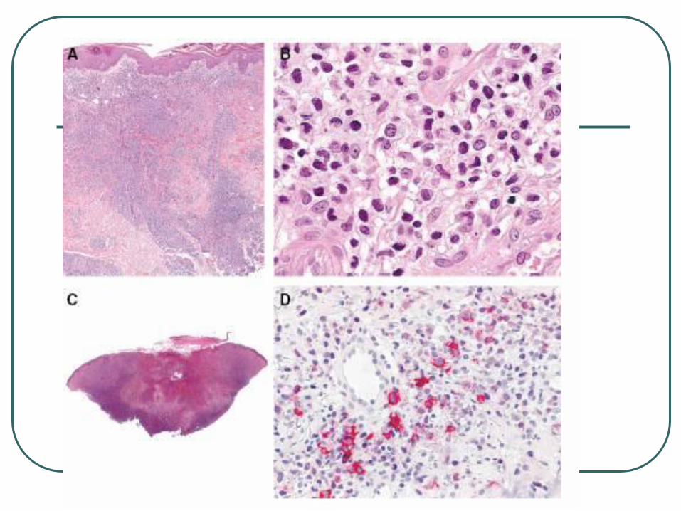
Three histologic subtypes of LyP (types A, B, and C) have been described, which represent a spectrum with overlapping features
LyP type A lesions, scattered or small clusters of large, sometimes multinucleated or Reed-Sternberg–like, CD30+ cells are intermingled with numerous inflammatory cells, such as histiocytes, small lymphocytes, neutrophils, and/or eosinophils.
LyP type C lesions demonstrate a monotonous population or large clusters of large CD30+ T cells with relatively few admixed inflammatory cells.
LyP type B is uncommon (less than 10%) and is characterized by an epidermotropic infiltrate of small atypical cells with cerebriform nuclei similar to that observed in MF.
The large atypical cells in the LyP type A and type C lesions have the same phenotype as the tumor cells in C-ALCL.
The atypical cells with cerebriform nuclei in the LyP type B lesions have a CD3+, CD4+, CD8- phenotype and do not express CD30 antigen.
Clonally rearranged T-cell receptor genes have been detected in approximately 60%-70% of LyP lesions.
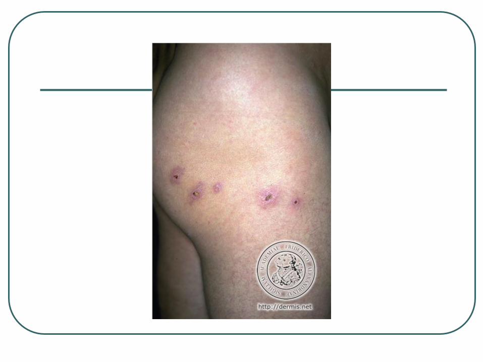
Lymphomatoid Papulosis Chronic recurrent, self-healing papulonecrotic or
papulonodular skin eruption with histologic features of a malignant lymphoma.
Histologically malignant but clinically benign
Considered a low-grade cutaneous T-cell lymphoma
Adults, median age 45, M:F 1.5, trunks and limbs
Individual lesion disappear within 3 to 12 weeks
10-15% of the cases may be followed by a cutaneous lymphoma, usually MF
4% develop systemic lymphomas
Px - 98% 5yr survival
Tx – Long term follow up without active tx
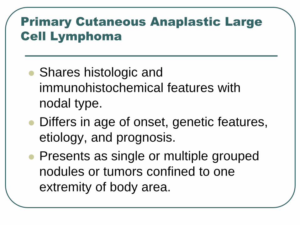
Primary Cutaneous Anaplastic Large
Cell Lymphoma
Shares histologic and
immunohistochemical features with
nodal type.
Differs in age of onset, genetic features,
etiology, and prognosis.
Presents as single or multiple grouped
nodules or tumors confined to one
extremity of body area.
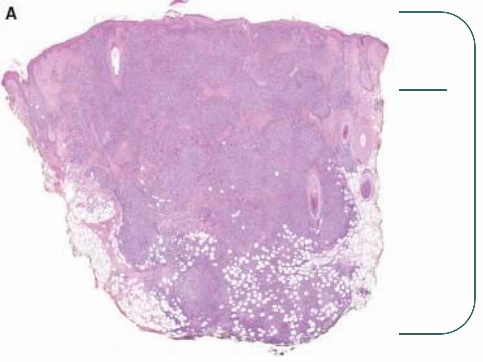
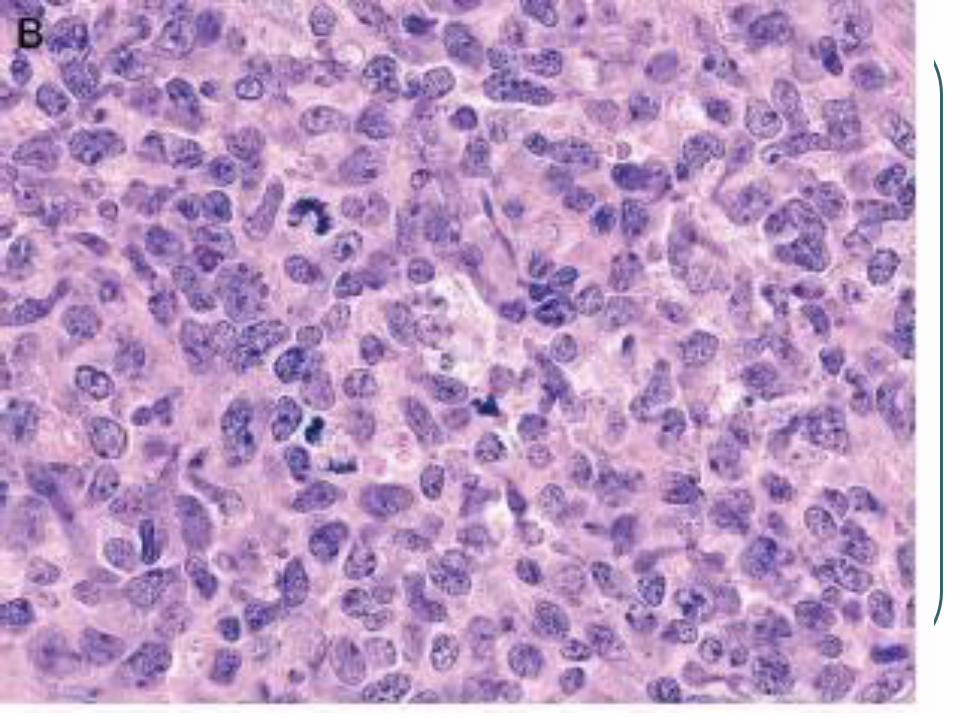
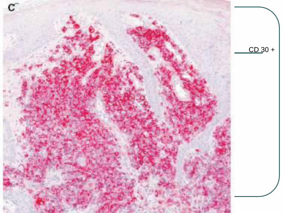
CD 30 +
Large cells with an anaplastic, pleomorphic, or immunoblastic cytomorphology
There is no clinical evidence or history of LyP, MF, or another type of CTCL
CD30 antigen by more than 75%of tumor cells.
Activated T cell phenotype, CD4+ , CD25, CD71, HLADR.
Variable loss of CD2, CD3, and CD5
CLA +, EMA -, ALK-
Lack translocation 2:5, 90% have clonal rearrangement of TCR.
Px is excellent, 10 yr survival of over 90%
Tx radiotherapy or surgery
Primary Cutaneous Anaplastic Large
Cell Lymphoma
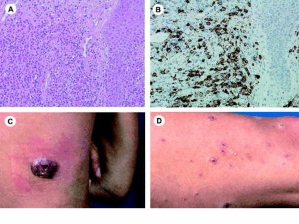
Subcutaneous panniculitis like T-cell
lymphoma
Adults and children, no sex predilection
Most often on legs
Systemic symptoms such as fever,
fatigue, and weight loss may be present.
Can be associated with hemophagocytic
syndrome, rapidly progressive course.
Subcutaneous panniculitis like T-cell
lymphoma
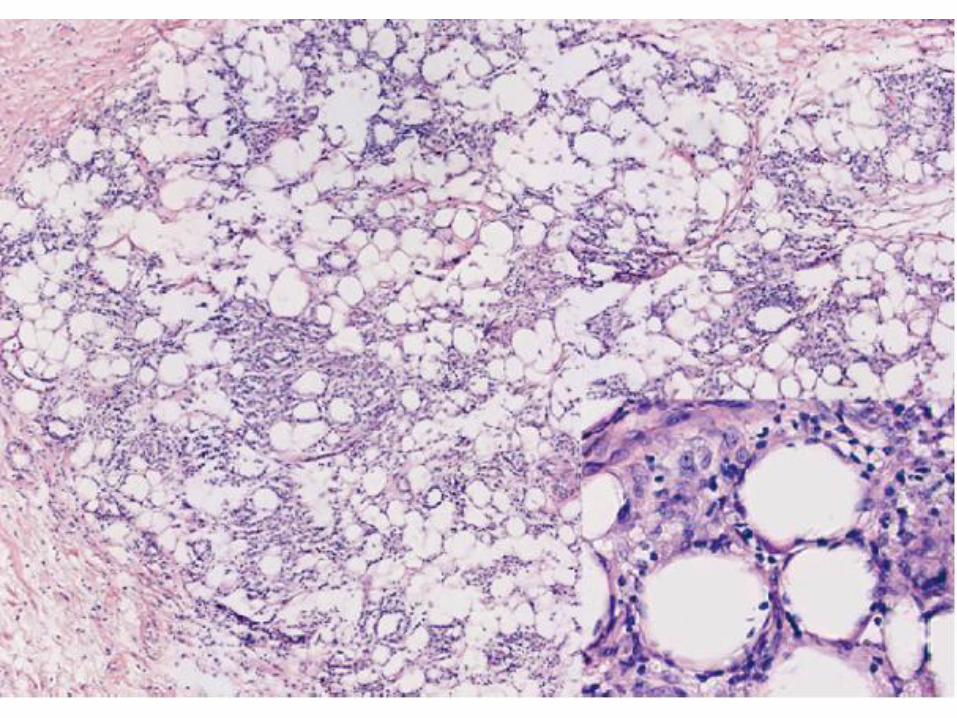
Panniculitis like growth of T cells with hyperchromatic nuclei and often many macrophages.
Overlying epidermis and dermis are typically uninvolved.
Rimming of individual fat cells by neoplastic T cells
Necrosis, karyorrhexis, and cytophagocytosis are common findings
Plasma cells and reactive lymphoid follicles are generally absent, in contrast to lupus profundus, and other forms of lobular panniculitis.
Cytotoxic T-cell lymphoma, subcutaneous infiltrates of pleomorphic T cells.

α/β T cells, CD 2+, CD3+, CD5+, CD4-,
CD8 +, CD43 +, TIA-1 + granzyme B +,
perforin +
Neoplastic T cells show clonal T-cell
receptor (TCR) gene rearrangements
5-year survival of 80%
Tx Doxorubicin-based chemotherapy
and radiotherapy
Subcutaneous panniculitis like T-cell
lymphoma
Primary cutaneous peripheral T-cell
lymphoma, unspecified
T-cell neoplasms that do not fit into any of the
better defined subtypes of T-cell
lymphoma/leukemia .
10% of CTLs
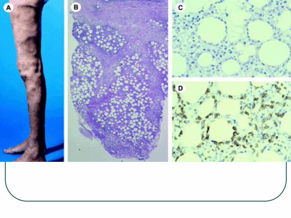
Cutaneous gamma/delta T-cell lymphoma
Primary cutaneous aggressive epidermotropic
CD8+ cytotoxic T-cell lymphoma
Primary cutaneous CD4+ small/medium-sized
pleomorphic T-cell lymphoma
Cutaneous gamma/delta T-cell
lymphoma
Clonal proliferation of mature, activated γ/δ T cells expressing a cytotoxic phenotype.
Epidermotropic, dermal, and subcutaneous.
Dermal and epidermal involvement often coexists with subcutaneous disease, in contrast to SPTCL of α/β origin.
Apoptosis and necrosis are common, often with vascular invasion.
CD3+, CD2+, CD43+, CD45RO+,
CD15-, CD30-, CD20-, CD25-
CD4-, CD8 –
Positive for TIA-1 and the cytotoxic proteins granzyme B, granzyme M, and perforin.
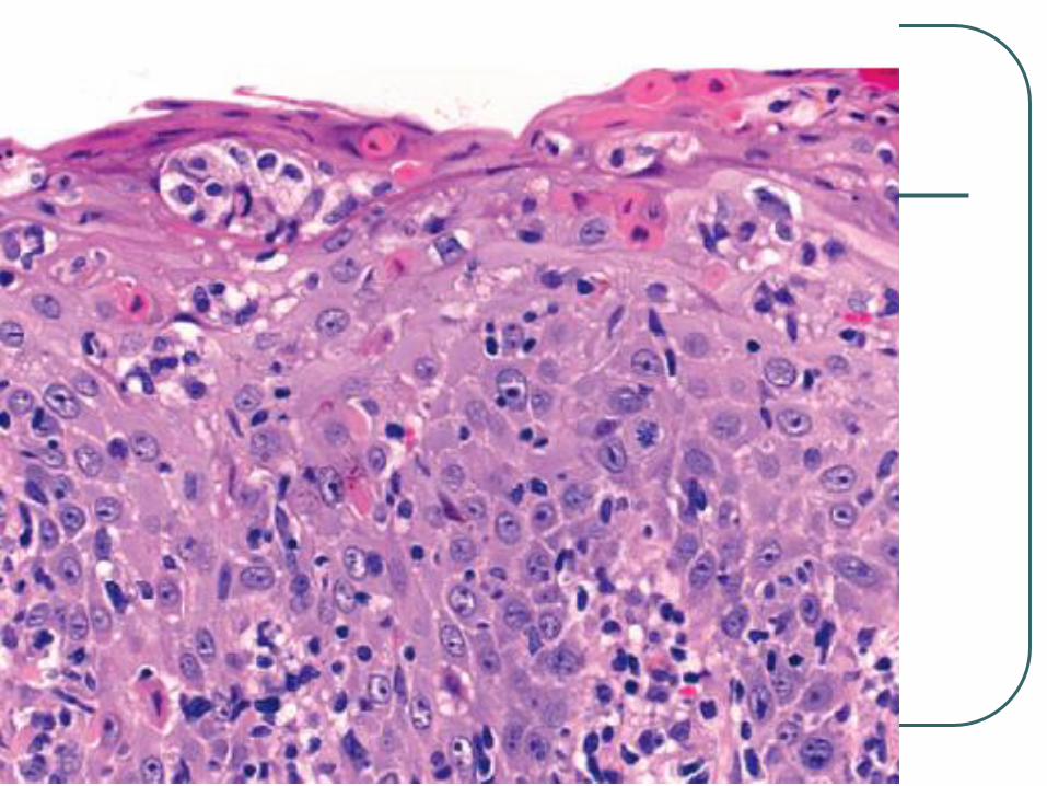
Primary cutaneous aggressive
epidermotropic CD8+ cytotoxic T-cell
lymphoma
Clinically, this form of CD8+ cutaneous lymphoma differs from the
slowly progressive CD8+ form similar to classic MF. It presents with
erosive plaques rather than patches. It exhibits an unfavorable
prognosis with rapid course .
The tumor cells have a CD3+, CD8+, granzyme B+, perforin+, TIA-
1+, CD45RA+, CD45RO-, CD2-, CD4-, CD5-
The neoplastic T cells show clonal TCR gene rearrangements.
Specific genetic abnormalities have not been described. Median survival of 32 months
Patients are generally treated with doxorubicin-based multiagent
chemotherapy.
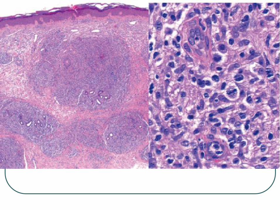
Primary cutaneous CD4+
small/medium-sized pleomorphic T-
cell lymphoma
This is a non-cytotoxic CTCL characterized by a predominance of
small to medium-sized CD4+ pleomorphic T cells with clinical
features not compatible with MF.
Diffuse or nodular lymphoid infiltrate, predominantly perivascular
and periadnexal and shows a tendency to extend to the
subcutaneous tissue.
It consists of small-to-medium-sized pleomorphic lymphoid cells with
irregular hyperchromatic nuclei and a pale scanty cytoplasm Patients are commonly adults, who present with solitary, localized,
or more frequently generalized nodules or tumors. No sites of
predilection have been recorded
Cutaneous B-cell lymphomas
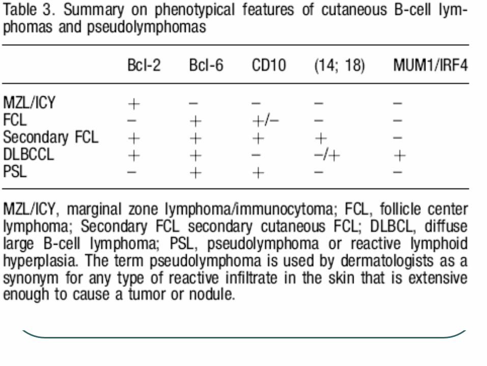
Cutaneous marginal zone B cell lymphoma
Primary cutaneous follicle center lymphoma
Cutaneous large B cell lymphoma, leg type
Cutaneous large B cell lymphoma, other
Cutaneous intravascular large B cell lymphoma
Cutaneous marginal zone B-cell
lymphoma (MZL)
Adults trunk or extremities, especially the arms
The nodular or diffuse infiltrate
Small to medium-sized lymphocytes
Irregular nuclei, dispersed chromatin, inconspicuous nucleoli and an abundant pale cytoplasm
Monocytoid appearance (reniform nuclei) or plasma cell differentiation.
Typified by darker centers surrounded by brighter zones of pale-staining cells.
Dutcher bodies and intracytoplasmic PAS+ globular inclusions may be seen
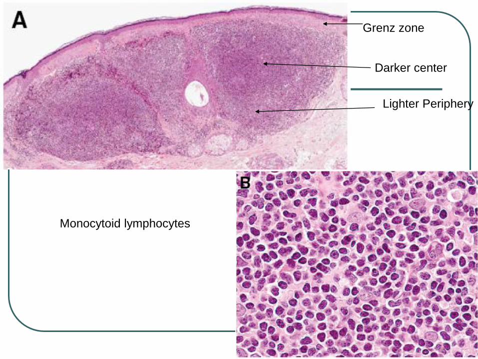
Grenz zone
Darker center
Lighter Periphery
Monocytoid lymphocytes
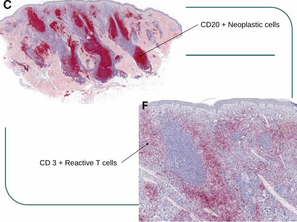
CD20 + Neoplastic cells
CD 3 + Reactive T cells
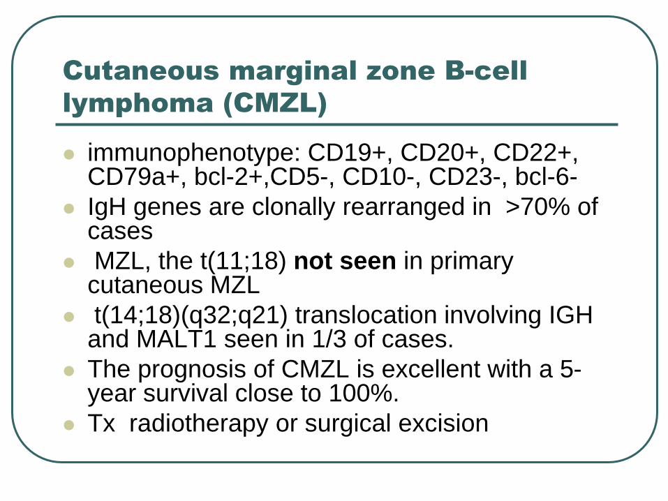
Cutaneous marginal zone B-cell
lymphoma (CMZL)
immunophenotype: CD19+, CD20+, CD22+, CD79a+, bcl-2+,CD5-, CD10-, CD23-, bcl-6-
IgH genes are clonally rearranged in >70% of cases
MZL, the t(11;18) not seen in primary cutaneous MZL
t(14;18)(q32;q21) translocation involving IGH and MALT1 seen in 1/3 of cases.
The prognosis of CMZL is excellent with a 5-year survival close to 100%.
Tx radiotherapy or surgical excision
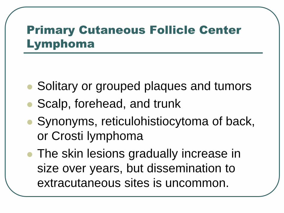
Primary Cutaneous Follicle Center
Lymphoma
Solitary or grouped plaques and tumors
Scalp, forehead, and trunk
Synonyms, reticulohistiocytoma of back,
or Crosti lymphoma
The skin lesions gradually increase in
size over years, but dissemination to
extracutaneous sites is uncommon.
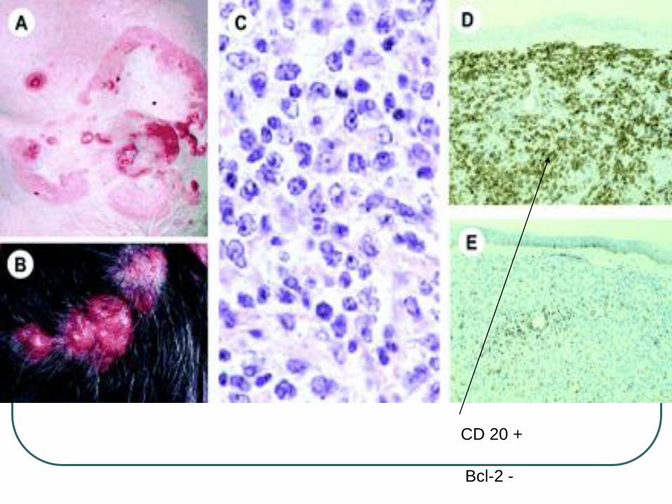
CD 20 +
Bcl-2 -
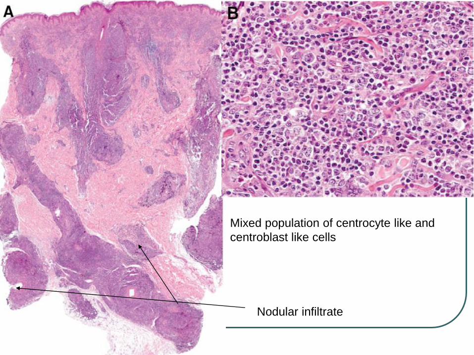
Nodular infiltrate
Mixed population of centrocyte like and
centroblast like cells
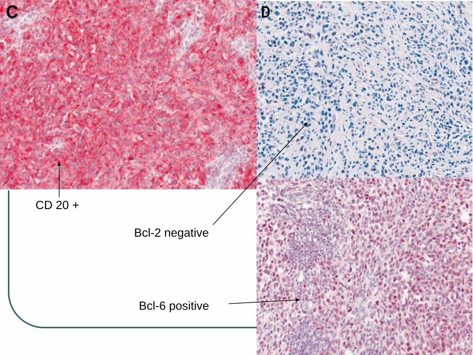
CD 20 +
Bcl-2 negative
Bcl-6 positive
Primary Cutaneous Follicle Center
Lymphoma
Neoplasm of follicle center cells (centrocytes and centroblasts)
Follicular, follicular and diffuse or a diffuse growth pattern
Grading of primary cutaneous FCL as in its nodal counterpart based is not prognostically relevant.
Few mitosis, and no tingible body macrophages seen (present in pseudolymphoma)
CD19+, CD20+, CD22+, CD79a+, CD5-, CD43 -, bcl-2 -, bcl-6 +
Bcl-2 gene rearrangement and t(14;18) chromosomal translocation are absent in most cases.
5-year survival of more than 95%
Tx radiotherapy or surgical excision
Cutaneous diffuse large B-cell
lymphoma (DLBCL)
composed of large B cells (centroblasts and
immunoblasts)
DLBCL, leg-type and DLBCL, other
DLBCL, leg type, is the most common variant,
occurs on the leg and less frequently at other
sites.
DLCL other include;Tcell/histiocyte-rich
DLBCL, plasmablastic lymphoma and others
that do not fulfill the criteria for a DLBCL, leg-
type
Diffuse large B-cell lymphoma
(DLBCL), leg-type Histology
Diffuse growth pattern, monomorphous infiltrate, entire dermis involvement, adnexal structures are usually destroyed
The epidermis is often spared, Grenz zone
Centrocytes are absent
Mitotic figures can frequently be detected
Nuclei are round with coarsely clumped chromatin.
Minimal inflammatory component and little stromal reaction.
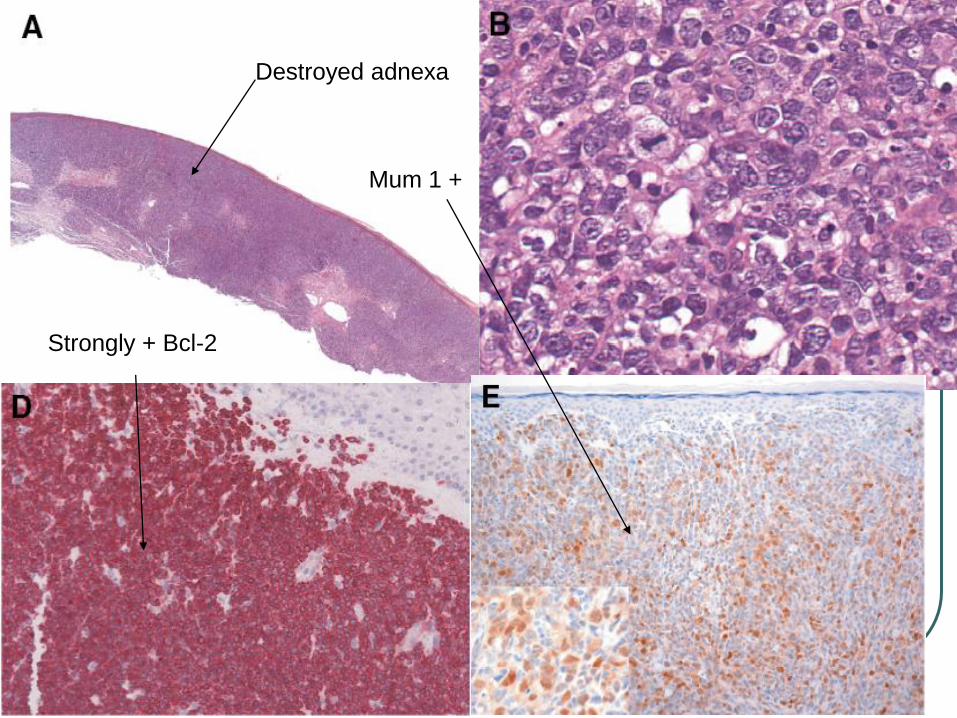
Destroyed adnexa
Strongly + Bcl-2
Mum 1 +
Diffuse large B-cell lymphoma
(DLBCL), leg-type
CD19+, CD20+, CD22+, and CD79a+ CD10-, Bcl-6 + in most cases.
Combination + Bcl-2, and + MUM-1/IRF4 is characteristic regardless of site and distinguish from FCL diffuse type.
The t(14;18) can be detected in secondary cutaneous large B-cell lymphomas but not in primary cutaneous diffuse large B-cell lymphomas.
The 5-year survival 55%
PCLBCLs on the leg have an inferior prognosis compared to PCLBCLs presenting at other
Tx Radiotherapy and Rituximab
Diffuse large B-cell lymphoma, other
Neoplastic large B-cells that lack the typical features of DLBCL, leg-type and do not conform to the definition of primary cutaneous FCL with diffuse growth pattern.
T cell rich, histiocyte rich, plasmablastic (HIV) variants
CD19+, CD22 +, and CD79a+, with light-chain restriction, negative for CD15 and CD30, which excludes Hodgkin lymphoma.
5 yr survival 65%
Tx Radiotherapy and Rituximab
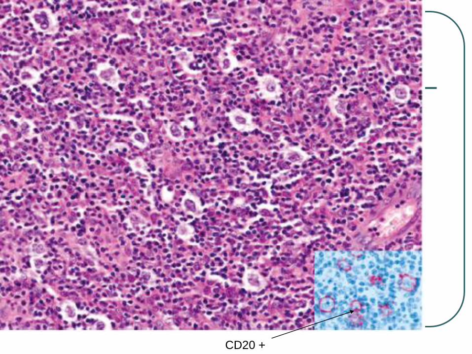
CD20 +
Intravascular large B-cell lymphoma
(IVL)
Rare highly malignant large-cell lymphoma with systemic spread
Tumor cells in the lumina of small vessels, particularly capillaries and venules.
Skin and the nervous system are preferential sites of primary manifestation.
The tumor cells express B-cell markers in the vast majority of cases; rarely a T-cell phenotype is found
Patients with disease limited to the skin (cutaneous variant) have a significantly better outcome than the other patients with IVL. 3-year overall survival: 56% versus 22%
Tx Multiagent chemotherapy

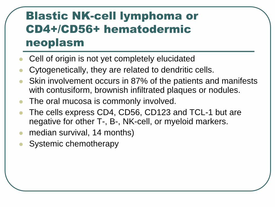
Blastic NK-cell lymphoma or
CD4+/CD56+ hematodermic
neoplasm
Cell of origin is not yet completely elucidated
Cytogenetically, they are related to dendritic cells.
Skin involvement occurs in 87% of the patients and manifests with contusiform, brownish infiltrated plaques or nodules.
The oral mucosa is commonly involved.
The cells express CD4, CD56, CD123 and TCL-1 but are negative for other T-, B-, NK-cell, or myeloid markers.
median survival, 14 months)
Systemic chemotherapy
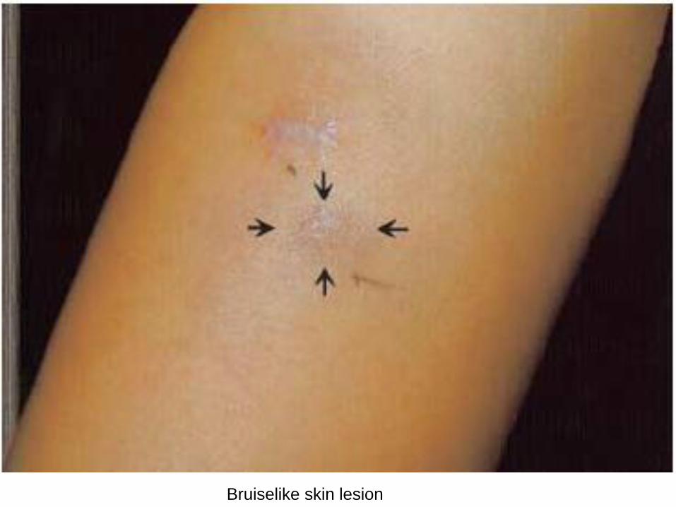
Bruiselike skin lesion
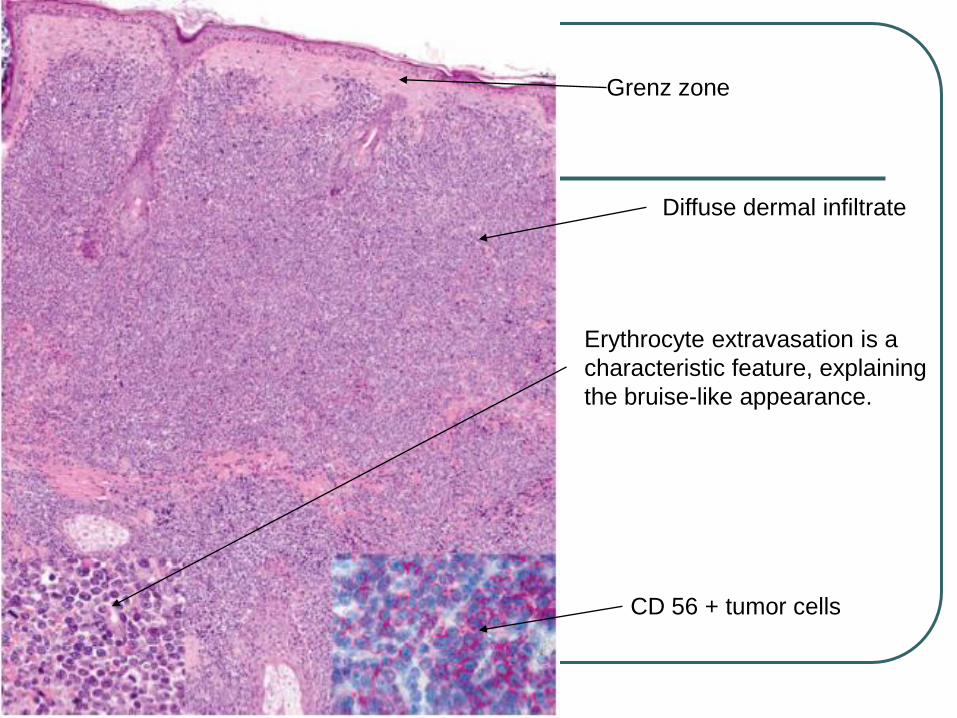
Grenz zone
Diffuse dermal infiltrate
Erythrocyte extravasation is a
characteristic feature, explaining
the bruise-like appearance.
CD 56 + tumor cells
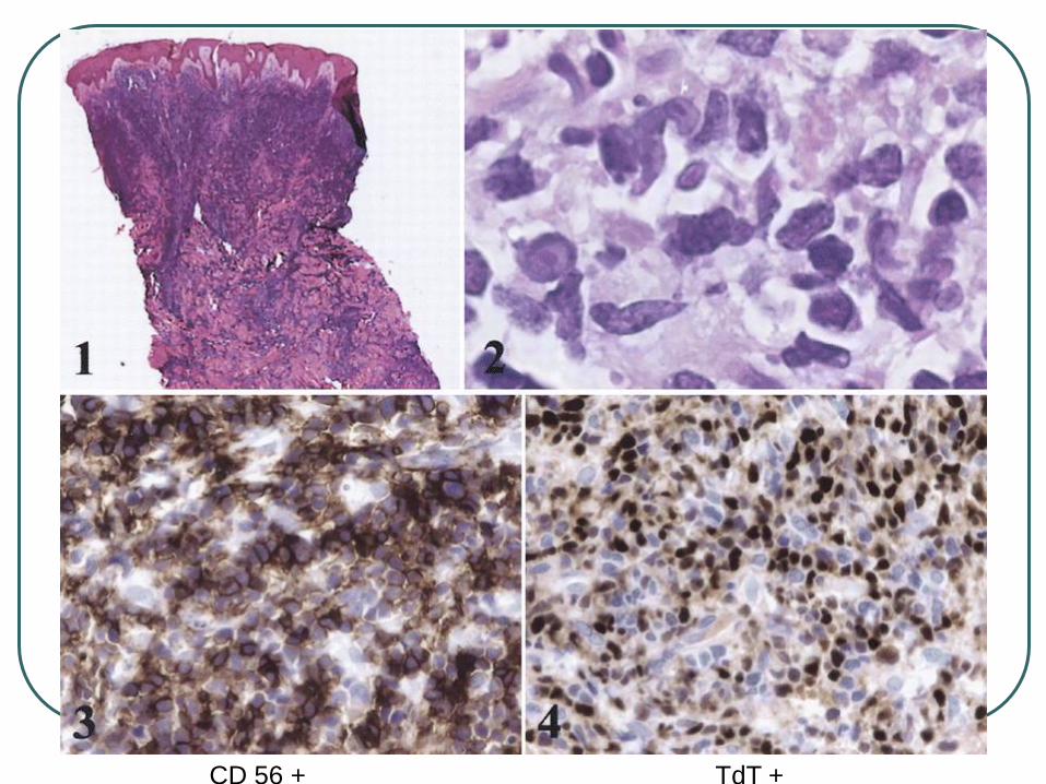
CD 56 + TdT +
Extranodal NK/T-cell lymphoma,
nasal type
Adults, males, Asia, Central America, and South America.
Multiple plaques or tumors preferentially on the trunk and extremities, ulceration is common.
Dense infiltrates in dermis and often the subcutis.
Epidermotropism, prominent angiocentricity and angiodestruction and extensive necrosis.
Small to large cells with irregular or oval nuclei, moderately dense chromatin, and pale cytoplasm, histiocytes, plasma cells, and eosinophils can be seen.
ExpressCD2, CD56, cytoplasmic CD3 , and cytotoxic proteins (TIA-1, granzyme B, perforin), but lack surface CD3.
In rare CD56- cases detection of EBV by in situ hybridization and expression of cytotoxic proteins are required for diagnosis.
EBV is expressed almost in all cases, suggesting a pathogenetic role of this virus
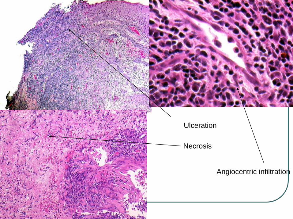
Ulceration
Necrosis
Angiocentric infiltration
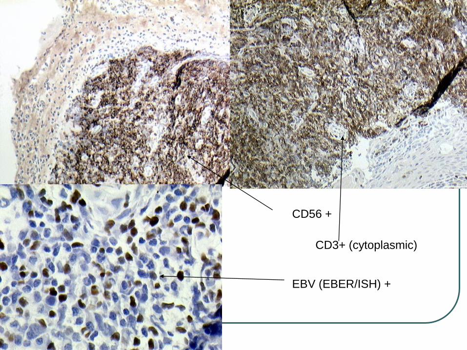
CD56 +
CD3+ (cytoplasmic)
EBV (EBER/ISH) +
Extranodal NK/T-cell lymphoma,
nasal type
Nearly allways EBV+ lymphoma of small, medium, or large cells usually with an NK-cell, or more rarely a cytotoxic T-cell, phenotype.
The skin is the second most common site of involvement after the nasal cavity/nasopharynx
Skin involvement may be a primary or secondary manifestation
Since both groups show an aggressive clinical behavior and require the same type of treatment, distinction is not useful.
median survival of 27 months was reported, compared with 5 months for patients presenting with cutaneous and extracutaneous disease.
Tx systemic hemotherapy
Conclusion
The new WHO/EORTC classification of
cutaneous lymphomas, employs a
terminology compatible with systemic
lymphomas but also reflects the organ-
specific pecularities of cutaneous
lymphomas.
References
1)WHO-EORTC classification for
cutaneous lymphomas Blood, 15 May 2005,
Vol. 105, No. 10, pp. 3768-3785.
2)WHO/EORTC classification of cutaneous
lymphomas 2005: histological and
molecular classification Journal of
Cutaneous Pathology
Volume 32 Page 647 - November 2005