Embed Size (px)
Citation preview
ORIGINAL ARTICLE
Which flap method should be preferred for the treatmentof pilonidal sinus? A prospective randomized study
K. Arslan • S. Said Kokcam • H. Koksal •
E. Turan • A. Atay • O. Dogru
Received: 8 November 2012 / Accepted: 21 January 2013
� Springer-Verlag Italia 2013
Abstract
Background Although many methods, either surgical or
non-surgical, are being used for the treatment of pilonidal
sinus disease (PSD), there is still no consensus as to what
constitutes the most appropriate method of treatment. The
aim of this study was to compare the outcomes of the
Limberg flap (LF), modified Limberg flap (MLF), and
Karydakis flap (KF) procedures.
Methods A prospective, randomized study was conducted
on 295 patients scheduled for surgical treatment for PSD at
the General Surgery Clinic of the Konya Training and
Research Hospital in January 2009–May 2010. Patients
with recurrent disease, an ASA score higher than III,
obesity (BMI [ 35 kg/m2), insulin-dependent diabetes, or
a drug or alcohol addiction were excluded. The procedures
performed were as follows: LF (n = 96), MLF (n = 108),
and KF (n = 91).
Results The patients were followed up for a median of
33 months (range 24–41 months). There were more female
patients in the LF group. The rate of seroma formation was
higher in the KF group (19.8 %) compared to the LF and
MLF groups (5.2 and 7.4 %, respectively; p = 0.027). The
rate of wound dehiscence was higher in the KF group
(15.4 %) compared to the LF and MLF groups (2.1 and
3.7 %, respectively; p \ 0.001) as was the incidence of
flap maceration (11 % in the KF vs. 1 % in the LF and
3.7 % in the MLF; p = 0.004). The incidence of PSD
recurrence was also higher in the KF group (11 %) com-
pared to the LF and MLF groups (6.3 and 1.9 % respec-
tively; p = 0.027). In a multivariate analysis, the presence
of seroma, hematoma, and wound infection were inde-
pendent predictors of recurrence.
Conclusions In our study, LF and MLF procedures were
associated with a lower recurrence and complication rate
compared to KF. However, more randomized studies
comparing different reconstruction methods after PSD
excision are needed.
Keywords Pilonidal sinus disease � Limber flap �Modified Limberg flap � Karydakis flap
Introduction
Pilonidal sinus disease (PSD) is a fairly common condition
associated with significant morbidity. The disease most
frequently occurs in the sacrococcygeal region of the body.
PSD, which was considered congenital at first but is
recently thought to be acquired, is more prevalent among
young males and leads to time loss from school or work.
The true prevalence of PSD is unknown. All races can
develop PSD; however, it is more common in those with
dark, stiff, or auburn hair. In a Norwegian study, the
incidence of disease was estimated to be 25 per 100,000
[1]. Approximately 70,000 diagnoses are established
annually in the United States [2]. Risk factors include male
gender, obesity, sedentary lifestyle, occupations requiring
Presented at the XVI. Annual meeting of the European Society of the
Surgery—ESS 2012. Istanbul,Turkey, November 22–24, 2012.
K. Arslan � S. Said Kokcam � H. Koksal � E. Turan �A. Atay � O. Dogru
Department of General Surgery, Konya Training
and Research Hospital, Konya, Turkey
K. Arslan (&)
Genel Cerrahi Bolumu, Konya Egitim ve Arastirma Hastanesi,
Hacı Saban Mahallesi Meram, Yeniyol Caddesi No: 97,
42040 Meram, Konya, Turkey
e-mail: [email protected]
123
Tech Coloproctol
DOI 10.1007/s10151-013-0982-2
sitting, family history, hirsute body habitus, trauma or
irritation to the gluteal cleft skin, and poor hygiene [2–4].
Surgical and non-surgical methods are used for the
treatment of the PSD. For the surgical management of
chronic PSD, many options ranging widely from simple
excision with or without primary closure to complex flap
reconstruction are available [2, 3]. There is no consensus yet
on a standard surgical method, which has proven to be
superior or is widely accepted. To the best of our knowledge,
there is no study comparing Limberg flap (LF), modified
Limberg flap (MLF), and Karydakis flap (KF) procedures in
the literature; thus, we aimed to compare the outcomes of
these three surgeries in a prospective randomized study.
Materials and methods
The present prospective randomized study was performed
between January 2009 and May 2010 at the General Sur-
gery Clinic of Konya Training and Research Hospital. The
study was approved by the Ethics Committee of the Selcuk
University Meram Medical Faculty (2009/0038), and
written informed consent was obtained from all patients.
Patients scheduled for surgical intervention because of PSD
were included in the present study. If the patients admitted
to our hospital with the diagnosis of sacrococcygeal PSD
had abscesses, the abscesses were drained first and the
patients were operated on at least 10 days later; those with
active infection were operated on after antibiotic treatment.
Exclusion criteria of the study were as follows: recurrent
cases, ASA group higher than III, obesity (body mass index
[35 kg/m2), insulin-dependent diabetes, drug and alcohol
addiction.
The patients were divided into the following three
groups by drawing lots from an envelope: (1) LF group, (2)
MLF group, and (3) KF group. For randomization, 120
envelopes were prepared for each group. An envelope
designating the surgery type was selected just prior to the
operation by the surgical nurse for each patient. The ran-
domization was performed in the outpatient clinic. All
patients underwent surgery by or under the supervision of
the same surgeon (OD).
To guarantee adequate statistical power, a priori sample
size calculation was performed. Because the reported rate
of postoperative complications associated with flap proce-
dures varies between 1 and 13 %, it was determined that
112 patients per treatment arm would allow for the detec-
tion of a 10 % difference in the rate of complications with
80 % power and 5 % confidence. Eligible patients were
randomized to the LF, MLF, or KF repair. Block ran-
domization was done to ensure an equal number of patients
assigned to each of the three treatment arms. All patients
received inpatient treatment.
Surgical procedures
Prior to the surgical procedure, hair at the surgical site was
removed using hair removal cream and 1 g cefazolin
sodium was intravenously administered while the patients
were on the operating table. The surgical procedure was
performed in the jackknife position under spinal anesthesia.
A rhomboid excision including the sinus was performed.
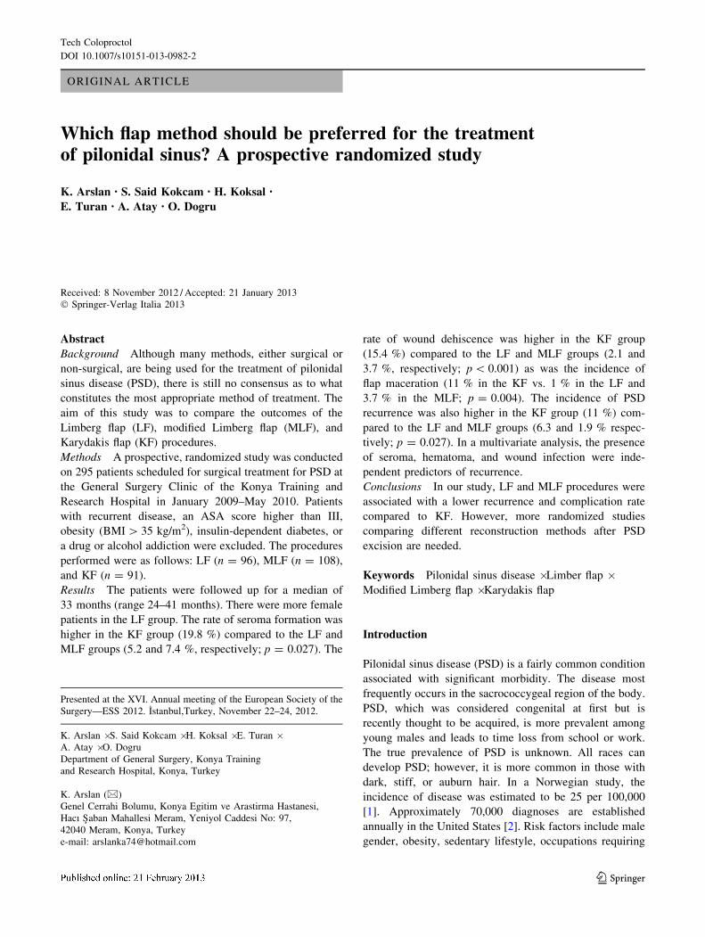
Limberg flap surgery was performed as defined in the
study by Azab et al. [5]. A rhomboid excision was carried
out. A right- or left-sided fasciocutaneous Limberg trans-
position flap, incorporating the gluteal fascia, was fully
mobilized on its inferior edge and transposed medially to
fulfill the rhomboid defect. The defect in the gluteal region
was closed primarily. The subcutaneous layers were
approximated with 2/0 or 3/0 vicryl sutures over a Jackson-
Pratt drain, and the skin was closed with 3/0 polypropylene
sutures or skin staplers (Fig. 1).
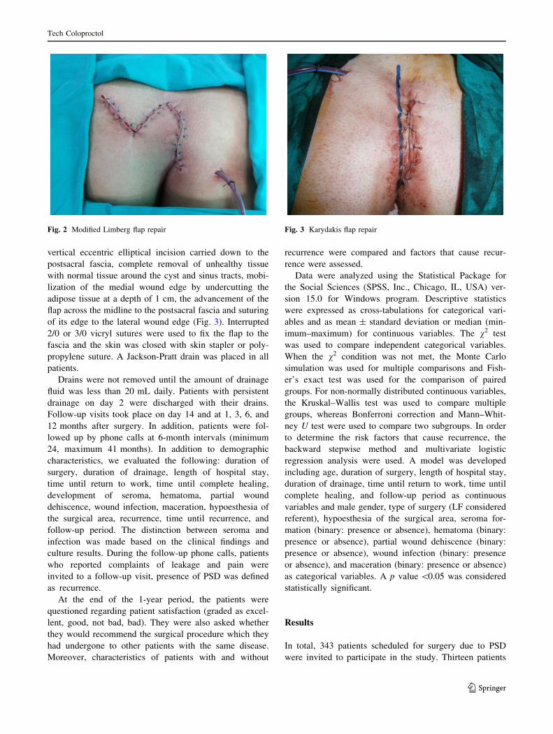
MLF surgery was performed as defined in the study by
Mentes et al. [6]. The inferior apices of the rhomboid
excisions were extended laterally 2 cm to the inferior
midline. The entire sinus tract and diseased area were
resected with a rhomboid excision. The flap, incorporating
the gluteal fascia, and anatomic bands between the rectum
and dermis of the midline sulcus were fully mobilized on
the inferior edge and transposed medially to fill the Lim-
berg defect. To remove the midline gap and to transpose
the flap to the contralateral side rather than to the midline,
the lower pole of the incision was placed on the contra-
lateral side of the elevated flap. In this way, there is no
incision on the lower intergluteal sulcus. The subcutaneous
layers were approximated with 2/0 or 3/0 vicryl sutures
over a Jackson-Pratt drain, and the skin was closed with 3/0
polypropylene sutures or skin staplers (Fig. 2).
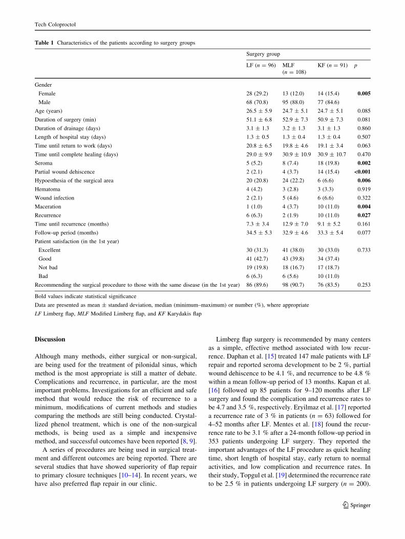
Karydakis flap surgery was performed as defined in the
study by Karydakis [7]. The technique consisted of a
Fig. 1 Limberg flap repair
Tech Coloproctol
123
vertical eccentric elliptical incision carried down to the
postsacral fascia, complete removal of unhealthy tissue
with normal tissue around the cyst and sinus tracts, mobi-
lization of the medial wound edge by undercutting the
adipose tissue at a depth of 1 cm, the advancement of the
flap across the midline to the postsacral fascia and suturing
of its edge to the lateral wound edge (Fig. 3). Interrupted
2/0 or 3/0 vicryl sutures were used to fix the flap to the
fascia and the skin was closed with skin stapler or poly-
propylene suture. A Jackson-Pratt drain was placed in all
patients.
Drains were not removed until the amount of drainage
fluid was less than 20 mL daily. Patients with persistent
drainage on day 2 were discharged with their drains.
Follow-up visits took place on day 14 and at 1, 3, 6, and
12 months after surgery. In addition, patients were fol-
lowed up by phone calls at 6-month intervals (minimum
24, maximum 41 months). In addition to demographic
characteristics, we evaluated the following: duration of
surgery, duration of drainage, length of hospital stay,
time until return to work, time until complete healing,
development of seroma, hematoma, partial wound
dehiscence, wound infection, maceration, hypoesthesia of
the surgical area, recurrence, time until recurrence, and
follow-up period. The distinction between seroma and
infection was made based on the clinical findings and
culture results. During the follow-up phone calls, patients
who reported complaints of leakage and pain were
invited to a follow-up visit, presence of PSD was defined
as recurrence.
At the end of the 1-year period, the patients were
questioned regarding patient satisfaction (graded as excel-
lent, good, not bad, bad). They were also asked whether
they would recommend the surgical procedure which they
had undergone to other patients with the same disease.
Moreover, characteristics of patients with and without
recurrence were compared and factors that cause recur-
rence were assessed.
Data were analyzed using the Statistical Package for
the Social Sciences (SPSS, Inc., Chicago, IL, USA) ver-
sion 15.0 for Windows program. Descriptive statistics
were expressed as cross-tabulations for categorical vari-
ables and as mean ± standard deviation or median (min-
imum–maximum) for continuous variables. The v2 test
was used to compare independent categorical variables.
When the v2 condition was not met, the Monte Carlo
simulation was used for multiple comparisons and Fish-
er’s exact test was used for the comparison of paired
groups. For non-normally distributed continuous variables,
the Kruskal–Wallis test was used to compare multiple
groups, whereas Bonferroni correction and Mann–Whit-
ney U test were used to compare two subgroups. In order
to determine the risk factors that cause recurrence, the
backward stepwise method and multivariate logistic
regression analysis were used. A model was developed
including age, duration of surgery, length of hospital stay,
duration of drainage, time until return to work, time until
complete healing, and follow-up period as continuous
variables and male gender, type of surgery (LF considered
referent), hypoesthesia of the surgical area, seroma for-
mation (binary: presence or absence), hematoma (binary:
presence or absence), partial wound dehiscence (binary:
presence or absence), wound infection (binary: presence
or absence), and maceration (binary: presence or absence)
as categorical variables. A p value \0.05 was considered
statistically significant.
Results
In total, 343 patients scheduled for surgery due to PSD
were invited to participate in the study. Thirteen patients
Fig. 2 Modified Limberg flap repair Fig. 3 Karydakis flap repair
Tech Coloproctol
123

refused to participate; thus, 330 patients were included in
the study. These patients were divided into the three groups
by drawing lots (110 patients in each surgical group).
Thirty-five patients (14 patients from the LF group, 2
patients from the MLF group, and 19 patients from the KF
group) were lost to follow-up; they either did not attend the
follow-up visits or could not be reached. Accordingly, data
of 295 patients were analyzed (Fig. 4). The median age of
the 295 patients was 24 years (range 18–39 years). Fifty-
five (18.6 %) of these patients were females and 240
(81.4 %) were males. Ninety-six (32.5 %) were in the LF
group, 108 (36.6 %) were in the MLF group, and 91
(30.8 %) were in the KF group. Patient demographics and
results are shown in Table 1. The patients were followed
up for a median of 33 months (range 24–41 months). There
were more female patients in the LF group. The rate of
seroma formation was higher in the KF group (19.8 %)
compared to the LF and MLF groups (5.2 and 7.4 %,
respectively; p = 0.027). The rate of wound dehiscence
was higher in the KF group (15.4 %) compared to the LF
and MLF groups (2.1 and 3.7 %, respectively; p \ 0.001)
as was the incidence of flap maceration (11 % in the KF vs.
1 % in the LF and 3.7 % in the MLF; p = 0.004). The
incidence of PSD recurrence was also higher in the KF
group (11 %) compared to the LF and MLF groups (6.3
and 1.9 %, respectively; p = 0.027). The rate of patient
satisfaction (reported as excellent or good) at the end of
1 year was 74.2 %. No significant difference was found
between the surgery groups in terms of the rate of
satisfaction.
Recurrence was identified in 18 (6.1 %) patients. The
median time until recurrence was 8 months (range
3–24 months). The rates of seroma formation, partial
wound dehiscence, hematoma development, wound infec-
tion, and maceration were significantly higher in the group
with recurrence than in the group without recurrence. The
rate of patient satisfaction was found to be significantly
higher in the group without recurrence. Additionally, in the
group without recurrence, 94.7 % of the patients stated that
they would recommend the surgical procedure, which they
had undergone, to other patients with the same disease
compared to only 32.3 % of the group with recurrence
(Table 2). In a multivariate binary logistic regression
model, the presence of seroma, hematoma, and wound
infection were determined as statistically significant risk
factors for recurrence (Table 3).
Fig. 4 Treatment flow chart
Tech Coloproctol
123
Discussion
Although many methods, either surgical or non-surgical,
are being used for the treatment of pilonidal sinus, which
method is the most appropriate is still a matter of debate.
Complications and recurrence, in particular, are the most
important problems. Investigations for an efficient and safe
method that would reduce the risk of recurrence to a
minimum, modifications of current methods and studies
comparing the methods are still being conducted. Crystal-
lized phenol treatment, which is one of the non-surgical
methods, is being used as a simple and inexpensive
method, and successful outcomes have been reported [8, 9].
A series of procedures are being used in surgical treat-
ment and different outcomes are being reported. There are
several studies that have showed superiority of flap repair
to primary closure techniques [10–14]. In recent years, we
have also preferred flap repair in our clinic.
Limberg flap surgery is recommended by many centers
as a simple, effective method associated with low recur-
rence. Daphan et al. [15] treated 147 male patients with LF
repair and reported seroma development to be 2 %, partial
wound dehiscence to be 4.1 %, and recurrence to be 4.8 %
within a mean follow-up period of 13 months. Kapan et al.
[16] followed up 85 patients for 9–120 months after LF
surgery and found the complication and recurrence rates to
be 4.7 and 3.5 %, respectively. Eryilmaz et al. [17] reported
a recurrence rate of 3 % in patients (n = 63) followed for
4–52 months after LF. Mentes et al. [18] found the recur-
rence rate to be 3.1 % after a 24-month follow-up period in
353 patients undergoing LF surgery. They reported the
important advantages of the LF procedure as quick healing
time, short length of hospital stay, early return to normal
activities, and low complication and recurrence rates. In
their study, Topgul et al. [19] determined the recurrence rate
to be 2.5 % in patients undergoing LF surgery (n = 200).
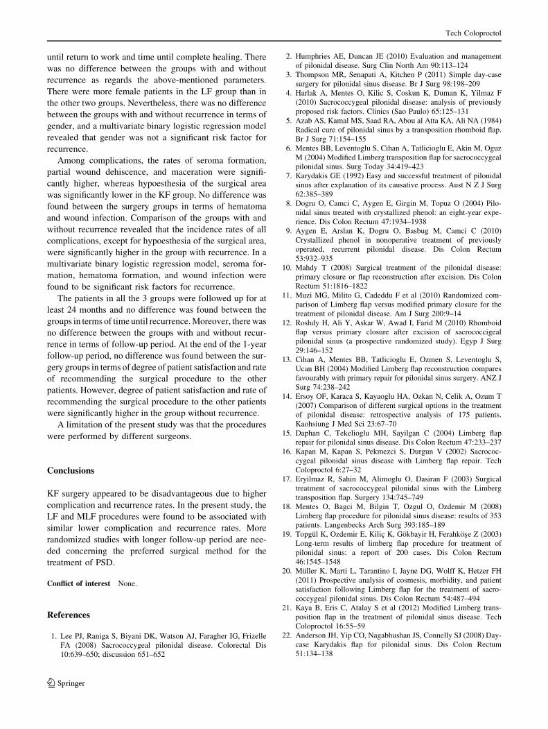
Table 1 Characteristics of the patients according to surgery groups
Surgery group
LF (n = 96) MLF
(n = 108)
KF (n = 91) p
Gender
Female 28 (29.2) 13 (12.0) 14 (15.4) 0.005
Male 68 (70.8) 95 (88.0) 77 (84.6)
Age (years) 26.5 ± 5.9 24.7 ± 5.1 24.7 ± 5.1 0.085
Duration of surgery (min) 51.1 ± 6.8 52.9 ± 7.3 50.9 ± 7.3 0.081
Duration of drainage (days) 3.1 ± 1.3 3.2 ± 1.3 3.1 ± 1.3 0.860
Length of hospital stay (days) 1.3 ± 0.5 1.3 ± 0.4 1.3 ± 0.4 0.507
Time until return to work (days) 20.8 ± 6.5 19.8 ± 4.6 19.1 ± 3.4 0.063
Time until complete healing (days) 29.0 ± 9.9 30.9 ± 10.9 30.9 ± 10.7 0.470
Seroma 5 (5.2) 8 (7.4) 18 (19.8) 0.002
Partial wound dehiscence 2 (2.1) 4 (3.7) 14 (15.4) <0.001
Hypoesthesia of the surgical area 20 (20.8) 24 (22.2) 6 (6.6) 0.006
Hematoma 4 (4.2) 3 (2.8) 3 (3.3) 0.919
Wound infection 2 (2.1) 5 (4.6) 6 (6.6) 0.322
Maceration 1 (1.0) 4 (3.7) 10 (11.0) 0.004
Recurrence 6 (6.3) 2 (1.9) 10 (11.0) 0.027
Time until recurrence (months) 7.3 ± 3.4 12.9 ± 7.0 9.1 ± 5.2 0.161
Follow-up period (months) 34.5 ± 5.3 32.9 ± 4.6 33.3 ± 5.4 0.077
Patient satisfaction (in the 1st year)
Excellent 30 (31.3) 41 (38.0) 30 (33.0) 0.733
Good 41 (42.7) 43 (39.8) 34 (37.4)
Not bad 19 (19.8) 18 (16.7) 17 (18.7)
Bad 6 (6.3) 6 (5.6) 10 (11.0)
Recommending the surgical procedure to those with the same disease (in the 1st year) 86 (89.6) 98 (90.7) 76 (83.5) 0.253
Bold values indicate statistical significance
Data are presented as mean ± standard deviation, median (minimum–maximum) or number (%), where appropriate
LF Limberg flap, MLF Modified Limberg flap, and KF Karydakis flap
Tech Coloproctol
123

Muller et al. [20] found a complication rate of 25.7 % and a
recurrence rate of 1.6 % within a median follow-up period
of 1.4 years (range 1–2.8 years) in 57 patients undergoing
LF surgery. In the present study, the recurrence rate was
determined to be 6.3 % in the LF group (n = 96) within a
median follow-up of 35.5 months.
Mentes et al. [6] performed LF surgery on 40 patients
and then modified the method and performed MLF surgery
on 198 patients. While they observed three recurrences in
patients undergoing LF surgery within a follow-up period
of 12–38 months, they noted no recurrence in those
undergoing MLF surgery. In the study by Kaya et al. [21],
the recurrence rate was reported to be 4.2 % in 94 patients
undergoing MLF surgery within a mean follow-up period
of 30 months (range 12–54 months). In the same study,
they reported the mean operative time to be 38.95 ±
6.77 min, wound dehiscence to be 1.1 %, seroma devel-
opment to be 2.1 %, wound infection to be 5.3 %,
maceration of the surgical incision site to be 8.5 %, and
hypoesthesia to be 9.6 %. In the present study, recurrence
rate was found to be 1.9 % in the MLF group (n = 108)
within a median follow-up period of 34 months.
KF surgery has been reported to be feasible, safe, and
effective as a day case surgical procedure and is preferred
in some centers. Anderson et al. [22] prospectively fol-
lowed up 51 patients and observed no recurrence after KF
surgery. They also reported that 95 % of the patients
returned to their normal daily activities within a month. In
their study, Keshava et al. [23] performed KF surgery on
70 patients and determined a recurrence rate of 4.2 %
Table 2 Characteristics of the groups with and without recurrence
Recurrence
No (n = 277) Yes (n = 18) p
Gender
Female 53 (19.1) 2 (11.1) 0.145
Male 224 (80.9) 16 (88.9)
Age (years) 25.2 ± 5.7 25.5 ± 4.9 0.582
Duration of surgery (min) 47.9 ± 11.0 43.7 ± 12.6 0.073
Duration of drainage (days) 3.1 ± 1.3 3.1 ± 1.3 0.833
Length of hospital stay (days) 1.3 ± 0.5 1.2 ± 0.4 0.468
Time until return to work (days) 17.7 ± 5.5 16.6 ± 4.2 0.338
Time until complete healing (days) 30.7 ± 10.7 27.1 ± 8.6 0.108
Seroma 22 (7.9) 9 (50.0) <0.001
Partial wound dehiscence 16 (5.7) 4 (22.2) 0.025
Hypoesthesia of the surgical area 46 (16.6) 4 (22.2) 0.526
Hematoma 8 (2.9) 2 (11.1) 0.013
Wound infection 10 (3.6) 3 (16.7) 0.001
Maceration 11 (3.9) 4 (22.2) 0.002
Follow-up period (months) 33.2 ± 5.0 32.7 ± 4.6 0.580
Patient satisfaction (in the 1st year)
Excellent 97 (36.7) 4 (12.9) <0.001
Good 115 (43.6) 3 (9.7)
Not bad 45 (17.0) 9 (29.0)
Bad 7 (2.7) 15 (48.4)
Recommending the surgical procedure to those with the same disease (in the 1st year) 250 (94.7) 10 (32.3) <0.001
Bold values indicate statistical significance
Table 3 Risk factors that affect the development of recurrence
p Odds ratio (95 % CI
min–max)
Time until complete
healing
0.099 0.964 (0.922–1.007)
Seroma <0.001 7.920 (3.057–20.520)
Hematoma 0.009 7.690 (1.680–35.189)
Infection <0.001 14.609 (4.086–52.237)
Bold values indicate statistical significance
CI confidence interval, min minimum, max maximum
Tech Coloproctol
123

within a follow-up period ranging from 1 to 79 months.
Kitchen [24] followed up 69 % of 114 patients who
underwent KF surgery for more than 18 months and found
the recurrence rate to be 4 %. Kulacoglu et al. [25] used
modified KF in 14 patients and reported no recurrence. In
the present study, recurrence rate within a median follow-
up period of 31 months was found to be 11 % in 91
patients undergoing KF surgery.
In the literature, there are studies that compare different
types of surgery. While some studies generally compare
primary closure and flap procedures, some compare dif-
ferent types of flap. Akin et al. [26] retrospectively eval-
uated 416 patients and compared the outcomes of classic
LF (n = 211) and MLF (n = 205) repairs. They found no
significant difference between the groups in terms of age,
gender, follow-up period, length of hospital stay, and
hypoesthesia. They reported that the MLF group had better
clinical outcomes and a lower recurrence rate (4.73 % in
the LF group and 0.97 % in the MLF group, p = 0.03). In
addition, they determined that time until return to work was
shorter and the rates of maceration and wound infection
were lower in MLF group. In a randomized prospective
study, Can et al. [27] compared MLF (n = 72) and KF
(n = 73) procedures and reported longer operation times in
the MLF group. In addition, they determined no significant
difference between the groups in terms of complication
rate, length of hospital stay, recurrence rate, degree of
satisfaction, and the rate of patients recommending the
same surgical procedure to other patients. Cihan et al. [13]
retrospectively evaluated LF (n = 40), MLF (n = 44), and
primary closure (n = 78) methods. For the surgical treat-
ment of sacrococcygeal PSD, they suggested that LF or
MLF reconstruction was superior to primary closure with
respect to infection, time until mobilization, discharge from
hospital and time off work. Moreover, they determined that
MLF reconstruction was associated with a statistically
lower recurrence rate as compared with primary closure.
Four-year recurrence rates were reported to be 17.9 % in
the primary closure group, 7.5 % in the LF group, and 0 %
in the MLF group. In their study, Ates et al. [28] compared
the results of 135 KF and 134 LF procedures in a pro-
spective randomized study. The mean duration of surgery
was significantly shorter in the KF group than in the LF
group (42.32 ± 8.64 vs. 50.14 ± 6.96 min). The compli-
cation rate was 11.1 % for KF and 20.8 % for LF
(p = 0.02), whereas the recurrence rate was 3 % for KF
and 6.9 % for LF (p = 0.15). Petersen et al. [29] compared
the patients who underwent KF surgery (n = 97) and
excision only (n = 91) and reported the overall compli-
cation rates to be 21 and 2 %, respectively. They concluded
that in the majority of patients in the KF group, there was
primary healing despite the considerable wound-related
complication rate. They reported no association between
the complication rate and degree of contamination in the
KF group; thus, they suggested KF surgery as a potential
alternative to simple excision in infected PSD. In the study
by Polat et al. [30], KF (n = 15) and primary closure
techniques (n = 33) were compared and the rates of early
recurrence were found to be 6.7 and 3 %, respectively.
They reported primary closure to be an advantageous and
preferable method. In the study by Saylam et al. [31], four
methods including primary closure, D-flap, KF, and LF
were compared. Although they observed the lowest recur-
rence rate in the primary closure group (7.5 %) and the
highest recurrence rate in the KF group (13.5 %), they did
not note a statistically significant difference between the
groups in terms of recurrence rates. Horwood et al. [32]
conducted a meta-analysis of randomized controlled stud-
ies that compared primary closure and LF procedures. They
concluded that the use of rhomboid flap excision and
LF-repair procedures rather than primary midline suture
techniques are supported by the current published literature
for the elective management of primary PSD. They also
suggested that further high-quality studies comparing flap
with off-midline repairs need to be conducted.
Different rates of reported recurrence are associated
with various variables. Postoperative remnants may cause
the recurrence of a disease, and the degree of inflammation
influences the treatment and recurrence. Since recurrence is
a time-dependent condition, a prolonged follow-up period
may increase the rate of observed recurrence [33]. It has
been reported that 60 % of recurrences occur within the
first 5 years and 12–15 % of recurrences occur within the
first year [33]. In most of the studies, the follow-up period
is approximately 1–2 years. Further studies that compare
different methods within longer follow-up periods are
needed to obtain a more accurate assessment of recurrence.
In the present study, we compared LF, MLF, and KF
procedures. The overall recurrence rate was found to be
6.1 % and was higher in the KF group than in the LF
(p = 0.105) and MLF (p = 0.07) groups. Although the
reason was not clear, we found the recurrence rate to be
slightly higher than that reported in the literature. The
recurrence rate was lower in the MLF group than in the LF
group; however, the difference was not statistically sig-
nificant (1.9 vs. 6.3 %; p = 0.105). Since operative time
(51.1 ± 6.8 min for the LF group and 52.9 ± 7.3 min for
the MLF group; p = 0.753) and complication rates
(35.4 % for the LF group and 44.4 % for the MLF group;
p = 0.121) were similar in the LF and MLF groups and
recurrence is the most important parameter after PSD
treatment, MLF could be the treatment of choice. It is our
opinion that modification of the LF procedure is of
importance and we recommend its use. No difference was
found between the surgery groups in terms of age, opera-
tive time, duration of drainage, length of hospital stay, time
Tech Coloproctol
123
until return to work and time until complete healing. There
was no difference between the groups with and without
recurrence as regards the above-mentioned parameters.
There were more female patients in the LF group than in
the other two groups. Nevertheless, there was no difference
between the groups with and without recurrence in terms of
gender, and a multivariate binary logistic regression model
revealed that gender was not a significant risk factor for
recurrence.
Among complications, the rates of seroma formation,
partial wound dehiscence, and maceration were signifi-
cantly higher, whereas hypoesthesia of the surgical area
was significantly lower in the KF group. No difference was
found between the surgery groups in terms of hematoma
and wound infection. Comparison of the groups with and
without recurrence revealed that the incidence rates of all
complications, except for hypoesthesia of the surgical area,
were significantly higher in the group with recurrence. In a
multivariate binary logistic regression model, seroma for-
mation, hematoma formation, and wound infection were
found to be significant risk factors for recurrence.
The patients in all the 3 groups were followed up for at
least 24 months and no difference was found between the
groups in terms of time until recurrence. Moreover, there was
no difference between the groups with and without recur-
rence in terms of follow-up period. At the end of the 1-year
follow-up period, no difference was found between the sur-
gery groups in terms of degree of patient satisfaction and rate
of recommending the surgical procedure to the other
patients. However, degree of patient satisfaction and rate of
recommending the surgical procedure to the other patients
were significantly higher in the group without recurrence.
A limitation of the present study was that the procedures
were performed by different surgeons.
Conclusions
KF surgery appeared to be disadvantageous due to higher
complication and recurrence rates. In the present study, the
LF and MLF procedures were found to be associated with
similar lower complication and recurrence rates. More
randomized studies with longer follow-up period are nee-
ded concerning the preferred surgical method for the
treatment of PSD.
Conflict of interest None.
References
1. Lee PJ, Raniga S, Biyani DK, Watson AJ, Faragher IG, Frizelle
FA (2008) Sacrococcygeal pilonidal disease. Colorectal Dis
10:639–650; discussion 651–652
2. Humphries AE, Duncan JE (2010) Evaluation and management
of pilonidal disease. Surg Clin North Am 90:113–124
3. Thompson MR, Senapati A, Kitchen P (2011) Simple day-case
surgery for pilonidal sinus disease. Br J Surg 98:198–209
4. Harlak A, Mentes O, Kilic S, Coskun K, Duman K, Yilmaz F
(2010) Sacrococcygeal pilonidal disease: analysis of previously
proposed risk factors. Clinics (Sao Paulo) 65:125–131
5. Azab AS, Kamal MS, Saad RA, Abou al Atta KA, Ali NA (1984)
Radical cure of pilonidal sinus by a transposition rhomboid flap.
Br J Surg 71:154–155
6. Mentes BB, Leventoglu S, Cihan A, Tatlicioglu E, Akin M, Oguz
M (2004) Modified Limberg transposition flap for sacrococcygeal
pilonidal sinus. Surg Today 34:419–423
7. Karydakis GE (1992) Easy and successful treatment of pilonidal
sinus after explanation of its causative process. Aust N Z J Surg
62:385–389
8. Dogru O, Camci C, Aygen E, Girgin M, Topuz O (2004) Pilo-
nidal sinus treated with crystallized phenol: an eight-year expe-
rience. Dis Colon Rectum 47:1934–1938
9. Aygen E, Arslan K, Dogru O, Basbug M, Camci C (2010)
Crystallized phenol in nonoperative treatment of previously
operated, recurrent pilonidal disease. Dis Colon Rectum
53:932–935
10. Mahdy T (2008) Surgical treatment of the pilonidal disease:
primary closure or flap reconstruction after excision. Dis Colon
Rectum 51:1816–1822
11. Muzi MG, Milito G, Cadeddu F et al (2010) Randomized com-
parison of Limberg flap versus modified primary closure for the
treatment of pilonidal disease. Am J Surg 200:9–14
12. Roshdy H, Ali Y, Askar W, Awad I, Farid M (2010) Rhomboid
flap versus primary closure after excision of sacrococcigeal
pilonidal sinus (a prospective randomized study). Egyp J Surg
29:146–152
13. Cihan A, Mentes BB, Tatlicioglu E, Ozmen S, Leventoglu S,
Ucan BH (2004) Modified Limberg flap reconstruction compares
favourably with primary repair for pilonidal sinus surgery. ANZ J
Surg 74:238–242
14. Ersoy OF, Karaca S, Kayaoglu HA, Ozkan N, Celik A, Ozum T
(2007) Comparison of different surgical options in the treatment
of pilonidal disease: retrospective analysis of 175 patients.
Kaohsiung J Med Sci 23:67–70
15. Daphan C, Tekelioglu MH, Sayilgan C (2004) Limberg flap
repair for pilonidal sinus disease. Dis Colon Rectum 47:233–237
16. Kapan M, Kapan S, Pekmezci S, Durgun V (2002) Sacrococ-
cygeal pilonidal sinus disease with Limberg flap repair. Tech
Coloproctol 6:27–32
17. Eryilmaz R, Sahin M, Alimoglu O, Dasiran F (2003) Surgical
treatment of sacrococcygeal pilonidal sinus with the Limberg
transposition flap. Surgery 134:745–749
18. Mentes O, Bagci M, Bilgin T, Ozgul O, Ozdemir M (2008)
Limberg flap procedure for pilonidal sinus disease: results of 353
patients. Langenbecks Arch Surg 393:185–189
19. Topgul K, Ozdemir E, Kilic K, Gokbayir H, Ferahkose Z (2003)
Long-term results of limberg flap procedure for treatment of
pilonidal sinus: a report of 200 cases. Dis Colon Rectum
46:1545–1548
20. Muller K, Marti L, Tarantino I, Jayne DG, Wolff K, Hetzer FH
(2011) Prospective analysis of cosmesis, morbidity, and patient
satisfaction following Limberg flap for the treatment of sacro-
coccygeal pilonidal sinus. Dis Colon Rectum 54:487–494
21. Kaya B, Eris C, Atalay S et al (2012) Modified Limberg trans-
position flap in the treatment of pilonidal sinus disease. Tech
Coloproctol 16:55–59
22. Anderson JH, Yip CO, Nagabhushan JS, Connelly SJ (2008) Day-
case Karydakis flap for pilonidal sinus. Dis Colon Rectum
51:134–138
Tech Coloproctol
123
23. Keshava A, Young CJ, Rickard MJ, Sinclair G (2007) Karydakis
flap repair for sacrococcygeal pilonidal sinus disease: how
important is technique? ANZ J Surg 77:181–183
24. Kitchen PR (1996) Pilonidal sinus: experience with the Karydakis
flap. Br J Surg 83:1452–1455
25. Kulacoglu H, Dener C, Tumer H, Aktimur R (2006) Total sub-
cutaneous fistulectomy combined with Karydakis flap for sacro-
coccygeal pilonidal disease with secondary perianal opening.
Colorectal Dis 8:120–123
26. Akin M, Leventoglu S, Mentes BB et al (2010) Comparison of
the classic Limberg flap and modified Limberg flap in the treat-
ment of pilonidal sinus disease: a retrospective analysis of 416
patients. Surg Today 40:757–762
27. Can MF, Sevinc MM, Hancerliogullari O, Yilmaz M, Yagci G
(2010) Multicenter prospective randomized trial comparing
modified Limberg flap transposition and Karydakis flaprecon-
struction in patients with sacrococcygeal pilonidal disease. Am J
Surg 200:318–327
28. Ates M, Dirican A, Sarac M, Aslan A, Colak C (2011) Short and
long-term results of the Karydakis flap versus the Limberg flap
for treating pilonidal sinus disease: a prospective randomized
study. Am J Surg 202:568–573
29. Petersen S, Aumann G, Kramer A, Doll D, Sailer M, Hellmich G
(2007) Short-term results of Karydakis flap for pilonidal sinus
disease. Tech Coloproctol 11:235–240
30. Polat N, Albayrak D, Ibis AC, Altan A (2008) Comparison
between Karydakis flap repair and primary closure for surgical
treatment of sacrococcygeal pilonidal sinus. Trakya Univ Tıp Fak
Derg 25:87–94
31. Saylam B, Balli DN, Duzgun AP, Ozer MV, Coskun F (2011)
Which surgical procedure offers the best treatment for pilonidal
disease? Langenbecks Arch Surg 396:651–658
32. Horwood J, Hanratty D, Chandran P, Billings P (2012) Primary
closure or rhomboid excision and Limberg flap for the manage-
ment of primary sacrococcygeal pilonidal disease? A metaanal-
ysis of randomized controlled trials. Colorectal Dis 14:143–151
33. Doll D (2010) 5- and 10-year recurrence rate is the new gold
standard in pilonidal sinus surgery benchmarking. Med Princ
Pract 19:216–217
Tech Coloproctol
123


![Umbilical metastasis as a primary presentation in ... · endometriosis, hypertrophic scar, umbilical granuloma, pilonidal sinus, mycosis psoriasis, and eczema [2, 6]. Tissue diagnosis](https://img.dokumen.tips/doc/110x75/5d641e7488c993204a8b582b/umbilical-metastasis-as-a-primary-presentation-in-endometriosis-hypertrophic.jpg)