Embed Size (px)
Citation preview
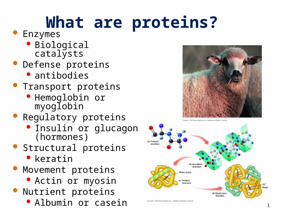
What are proteins? Enzymes
Biological catalysts Defense proteins
antibodies Transport proteins
Hemoglobin or myoglobin
Regulatory proteins Insulin or glucagon
(hormones) Structural proteins
keratin Movement proteins
Actin or myosin Nutrient proteins
Albumin or casein 1

What are amino acids?
Amino acids • are the building blocks of proteins.• contain a carboxylic acid group and an
amino group on the alpha () carbon.• are ionized in solution.• each contain a different side group (R).
R side chain R │ + │H2N—C —COOH H3N—C —COO−
│ │ H H ionized form
2
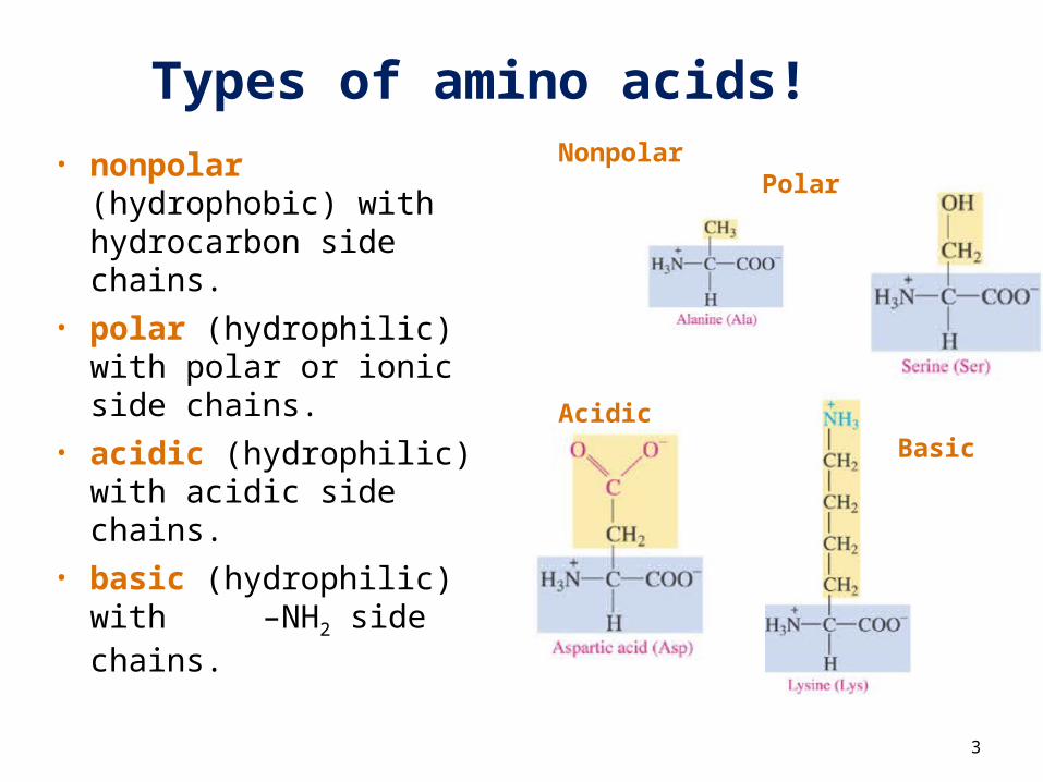
Types of amino acids!• nonpolar
(hydrophobic) with hydrocarbon side chains.
• polar (hydrophilic) with polar or ionic side chains.
• acidic (hydrophilic) with acidic side chains.
• basic (hydrophilic) with –NH2 side chains.
3
Nonpolar Polar
AcidicBasic
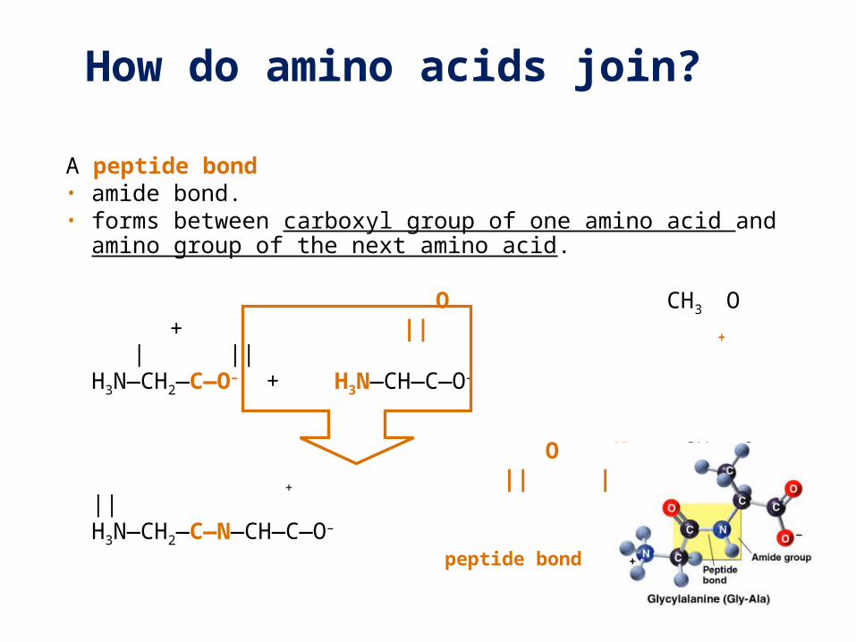
How do amino acids join?
A peptide bond• amide bond. • forms between carboxyl group of one amino acid
and amino group of the next amino acid.
O CH3 O
+ || + | ||H3N—CH2—C—O– + H3N—CH—C—O–
O H CH3 O + || | | ||
H3N—CH2—C—N—CH—C—O– peptide bond
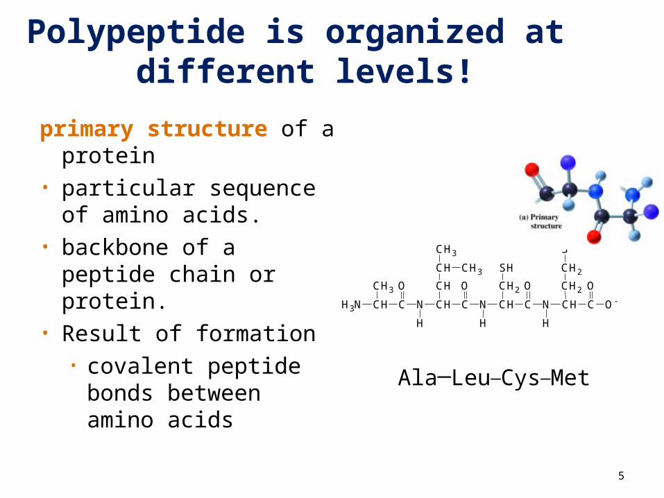
Polypeptide is organized at different levels!
primary structure of a protein
• particular sequence of amino acids.
• backbone of a peptide chain or protein.
• Result of formation • covalent peptide
bonds between amino acids
CH3
SH
CH2
CH3
S
CH2
CH2CH O
O-CCH
H
N
O
CCH
H
N
O
CCH
H
N
O
CCHH3N
CH3
CH3CH
5
Ala─Leu─Cys─Met
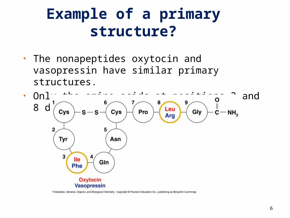
Example of a primary structure?
• The nonapeptides oxytocin and vasopressin have similar primary structures.
• Only the amino acids at positions 3 and 8 differ.
6
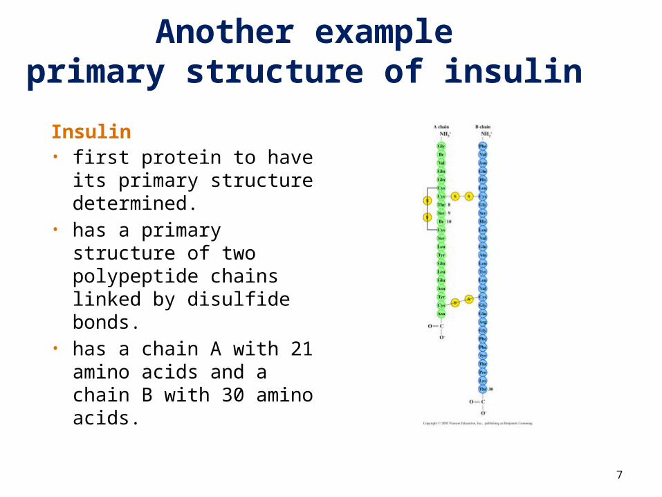
Another exampleprimary structure of insulin
Insulin• first protein to have its
primary structure determined.
• has a primary structure of two polypeptide chains linked by disulfide bonds.
• has a chain A with 21 amino acids and a chain B with 30 amino acids.
7
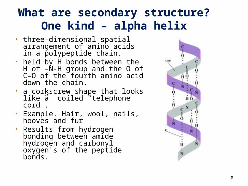
What are secondary structure?One kind – alpha helix
• three-dimensional spatial arrangement of amino acids in a polypeptide chain.
• held by H bonds between the H of –N-H group and the O of C=O of the fourth amino acid down the chain.
• a corkscrew shape that looks like a coiled “telephone cord”.
• Example. Hair, wool, nails, hooves and fur
• Results from hydrogen bonding between amide hydrogen and carbonyl oxygen's of the peptide bonds.
8
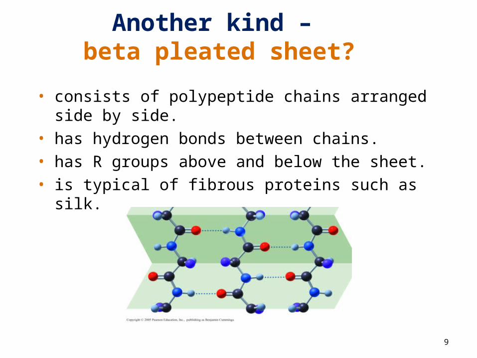
Another kind – beta pleated sheet?
9
• consists of polypeptide chains arranged side by side.• has hydrogen bonds between chains.• has R groups above and below the sheet.• is typical of fibrous proteins such as silk.
10
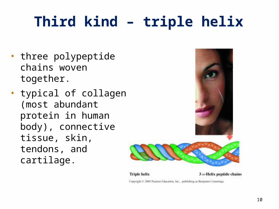
Third kind – triple helix
• three polypeptide chains woven together.
• typical of collagen (most abundant protein in human body), connective tissue, skin, tendons, and cartilage.
11
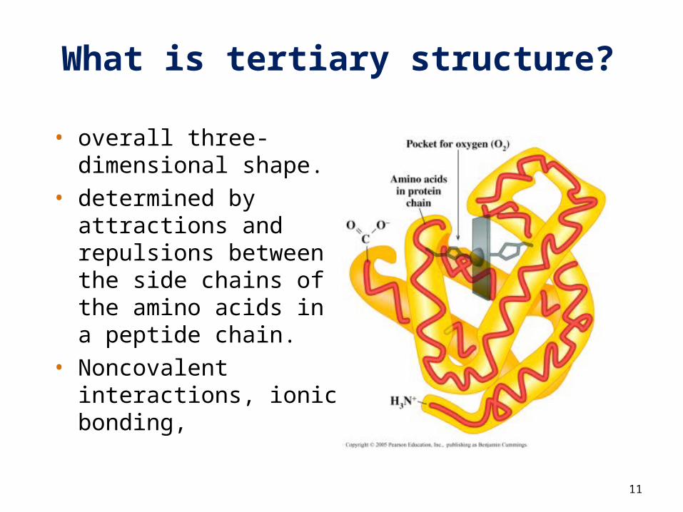
What is tertiary structure?
• overall three-dimensional shape.
• determined by attractions and repulsions between the side chains of the amino acids in a peptide chain.
• Noncovalent interactions, ionic bonding,
12
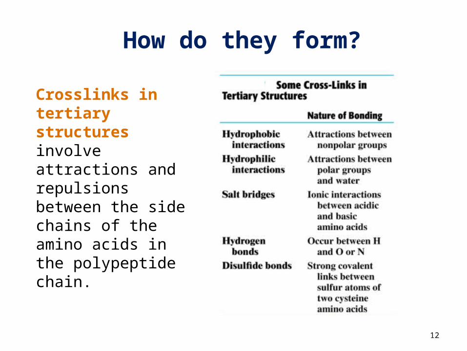
How do they form?
Crosslinks in tertiary structures involve attractions and repulsions between the side chains of the amino acids in the polypeptide chain.
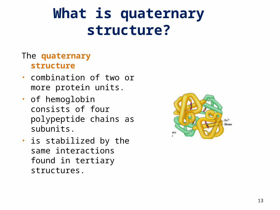
What is quaternary structure?
The quaternary structure
• combination of two or more protein units.
• of hemoglobin consists of four polypeptide chains as subunits.
• is stabilized by the same interactions found in tertiary structures.
13
14
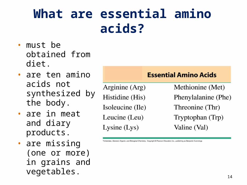
• must be obtained from diet.
• are ten amino acids not synthesized by the body.
• are in meat and diary products.
• are missing (one or more) in grains and vegetables.
What are essential amino acids?
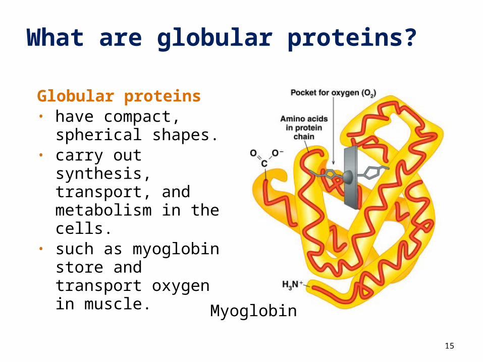
What are globular proteins?
Globular proteins • have compact,
spherical shapes.• carry out synthesis,
transport, and metabolism in the cells.
• such as myoglobin store and transport oxygen in muscle.
15
Myoglobin
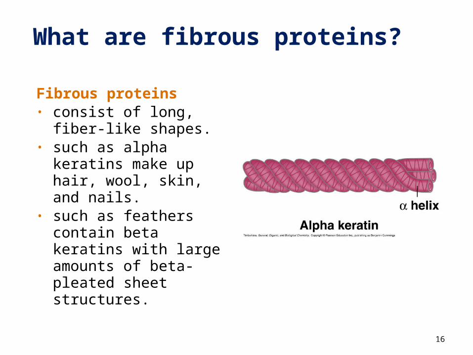
What are fibrous proteins?
Fibrous proteins• consist of long, fiber-
like shapes.• such as alpha
keratins make up hair, wool, skin, and nails.
• such as feathers contain beta keratins with large amounts of beta-pleated sheet structures.
16
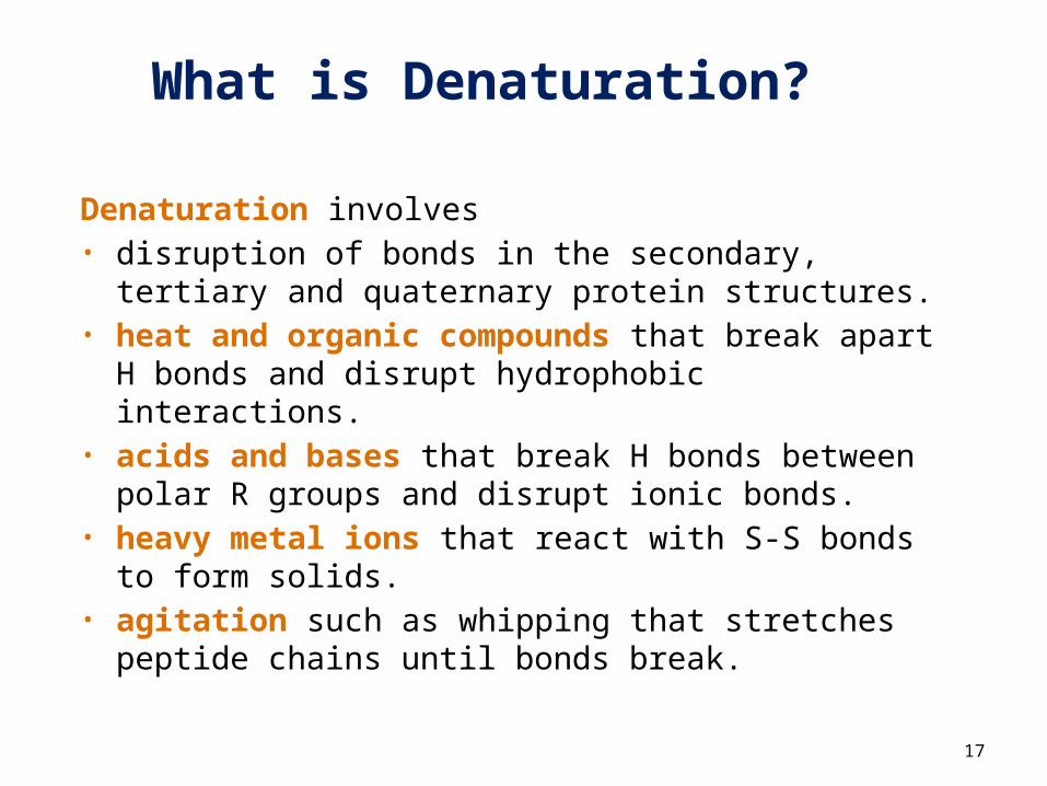
What is Denaturation?
Denaturation involves • disruption of bonds in the secondary, tertiary
and quaternary protein structures.• heat and organic compounds that break
apart H bonds and disrupt hydrophobic interactions.
• acids and bases that break H bonds between polar R groups and disrupt ionic bonds.
• heavy metal ions that react with S-S bonds to form solids.
• agitation such as whipping that stretches peptide chains until bonds break.
17
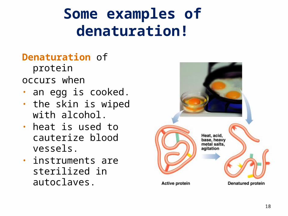
Some examples of denaturation!
Denaturation of protein
occurs when• an egg is cooked. • the skin is wiped
with alcohol.• heat is used to
cauterize blood vessels.
• instruments are sterilized in autoclaves.
18
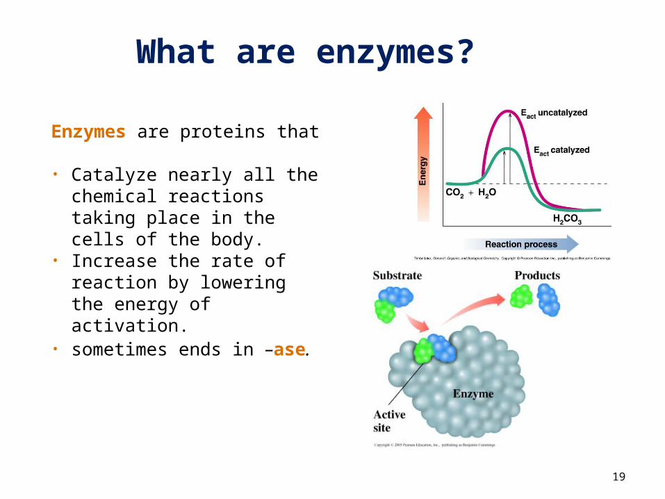
What are enzymes?
Enzymes are proteins that
• Catalyze nearly all the chemical reactions taking place in the cells of the body.
• Increase the rate of reaction by lowering the energy of activation.
• sometimes ends in –ase.
19
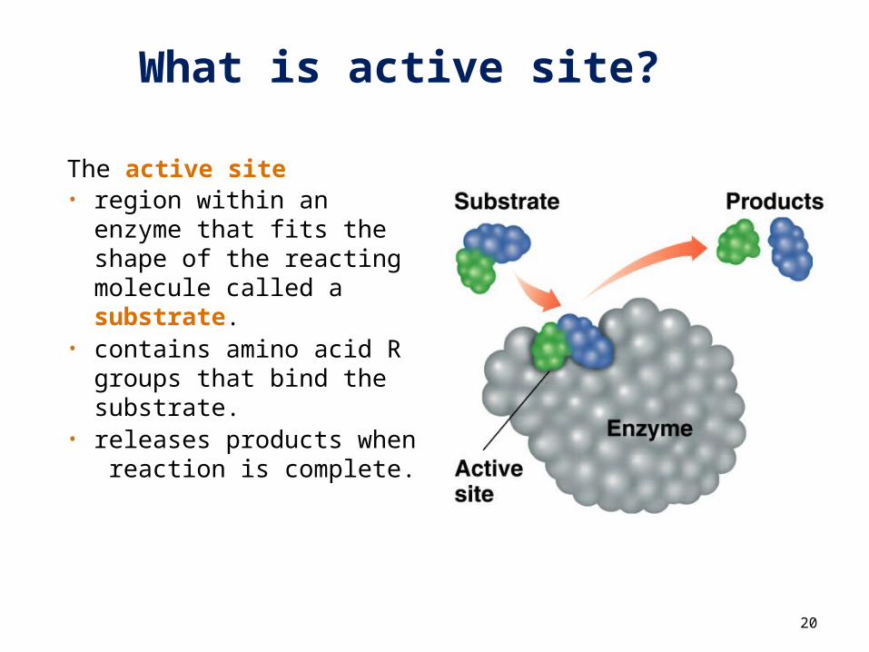
What is active site?
The active site • region within an enzyme
that fits the shape of the reacting molecule called a substrate.
• contains amino acid R groups that bind the substrate.
• releases products when reaction is complete.
20
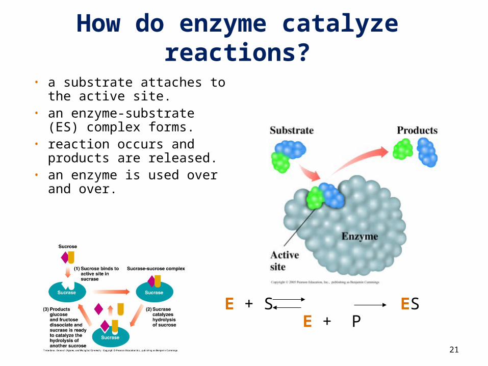
How do enzyme catalyze reactions?
• a substrate attaches to the active site.
• an enzyme-substrate (ES) complex forms.
• reaction occurs and products are released.
• an enzyme is used over and over.
21
E + S ES E + P
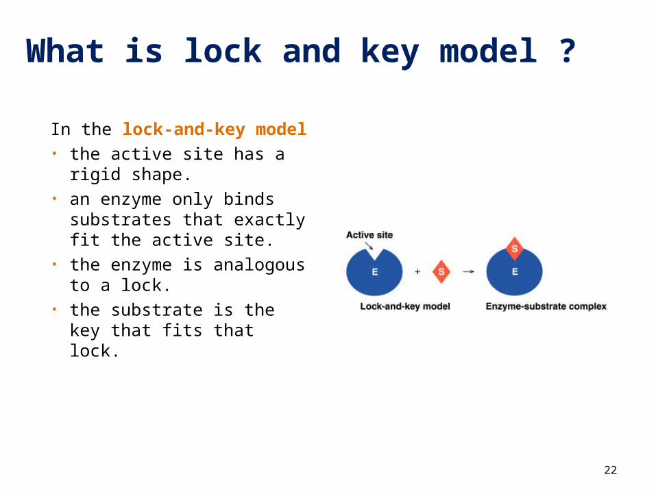
What is lock and key model ?
In the lock-and-key model• the active site has a rigid
shape.• an enzyme only binds
substrates that exactly fit the active site.
• the enzyme is analogous to a lock.
• the substrate is the key that fits that lock.
22
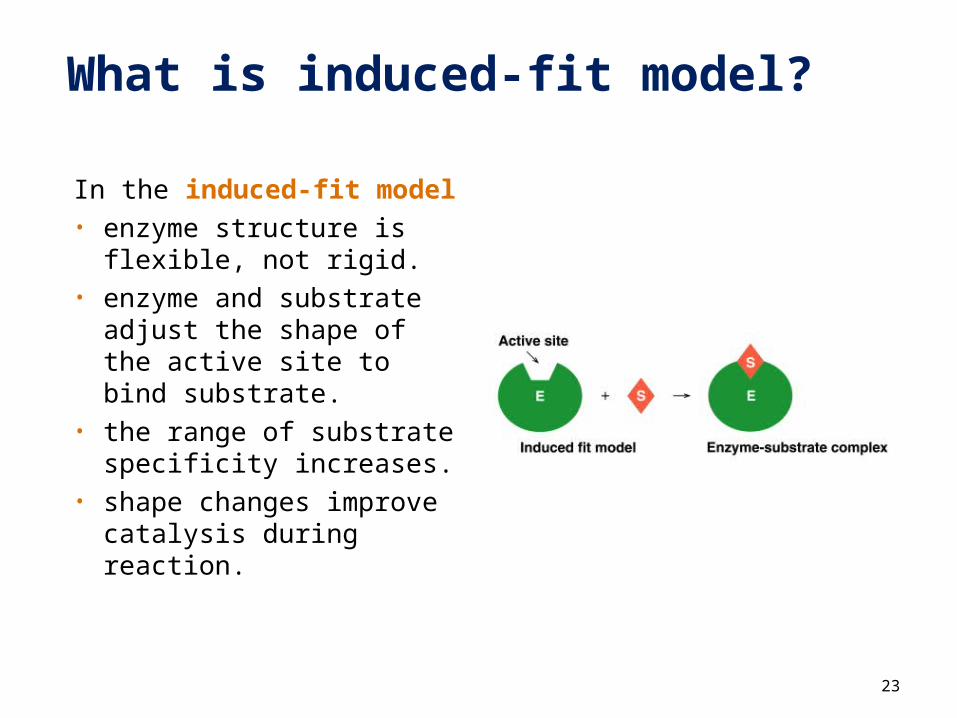
What is induced-fit model?
In the induced-fit model• enzyme structure is
flexible, not rigid.• enzyme and substrate
adjust the shape of the active site to bind substrate.
• the range of substrate specificity increases.
• shape changes improve catalysis during reaction.
23
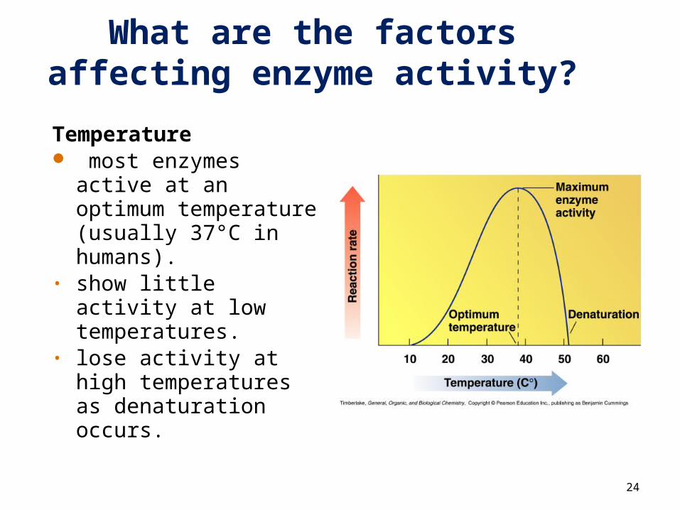
What are the factors affecting enzyme activity?
Temperature most enzymes active
at an optimum temperature (usually 37°C in humans).
• show little activity at low temperatures.
• lose activity at high temperatures as denaturation occurs.
24
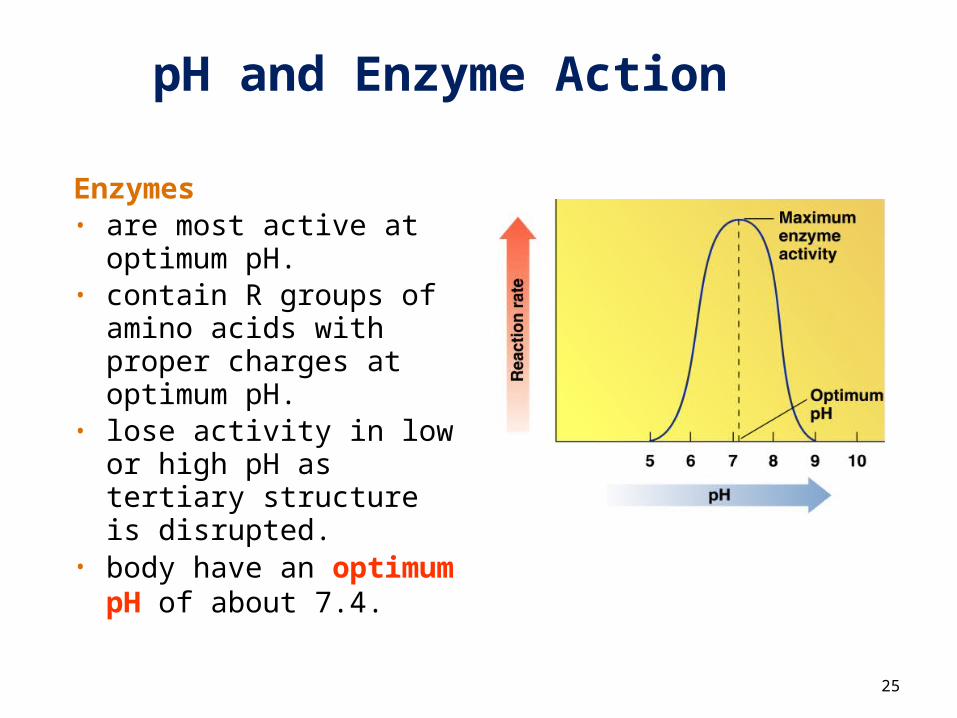
pH and Enzyme Action
Enzymes• are most active at
optimum pH.• contain R groups of
amino acids with proper charges at optimum pH.
• lose activity in low or high pH as tertiary structure is disrupted.
• body have an optimum pH of about 7.4.
25
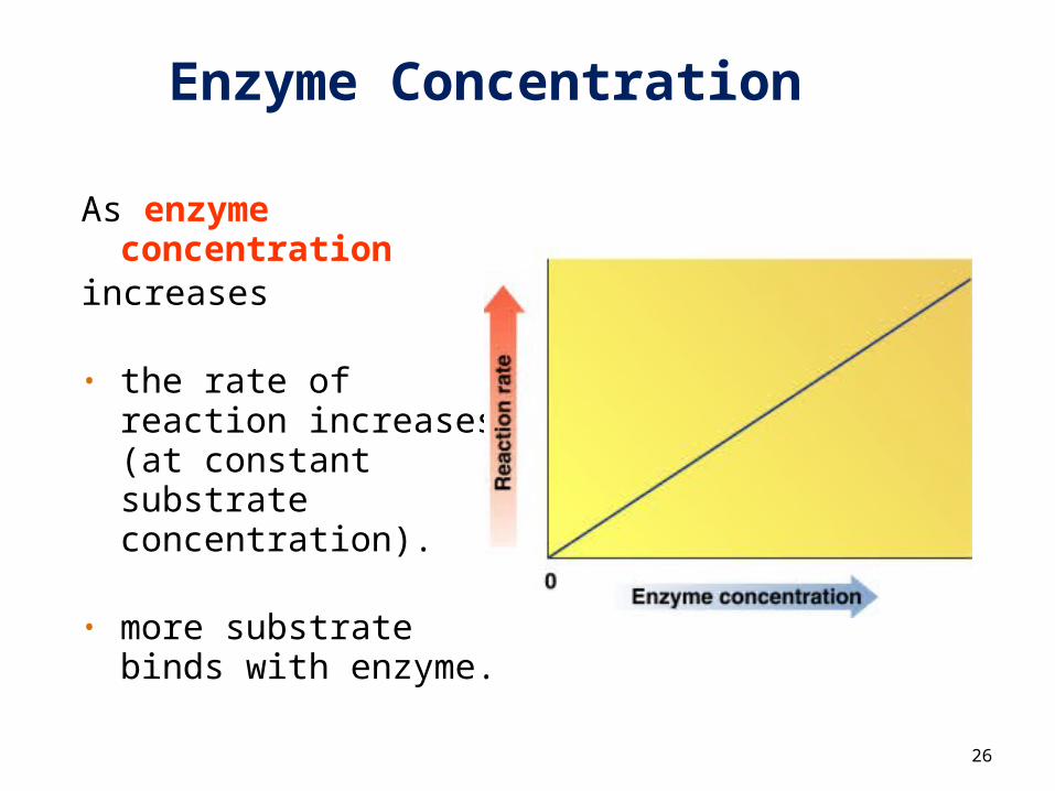
Enzyme Concentration
As enzyme concentration
increases
• the rate of reaction increases (at constant substrate concentration).
• more substrate binds
with enzyme.
26
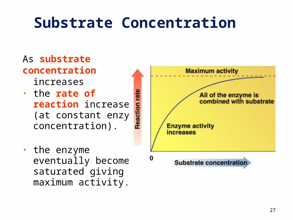
Substrate Concentration
As substrate concentration
increases • the rate of reaction
increases (at constant enzyme concentration).
• the enzyme eventually becomes saturated giving maximum activity.
27
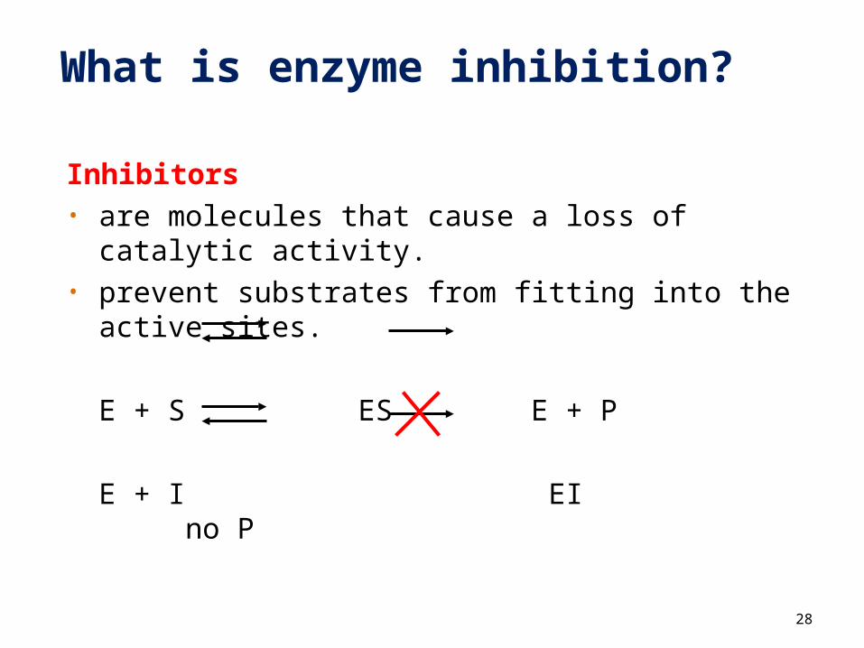
What is enzyme inhibition?
Inhibitors• are molecules that cause a loss of catalytic
activity.• prevent substrates from fitting into the active
sites.
E + S ES E + P
E + I EI no P
28
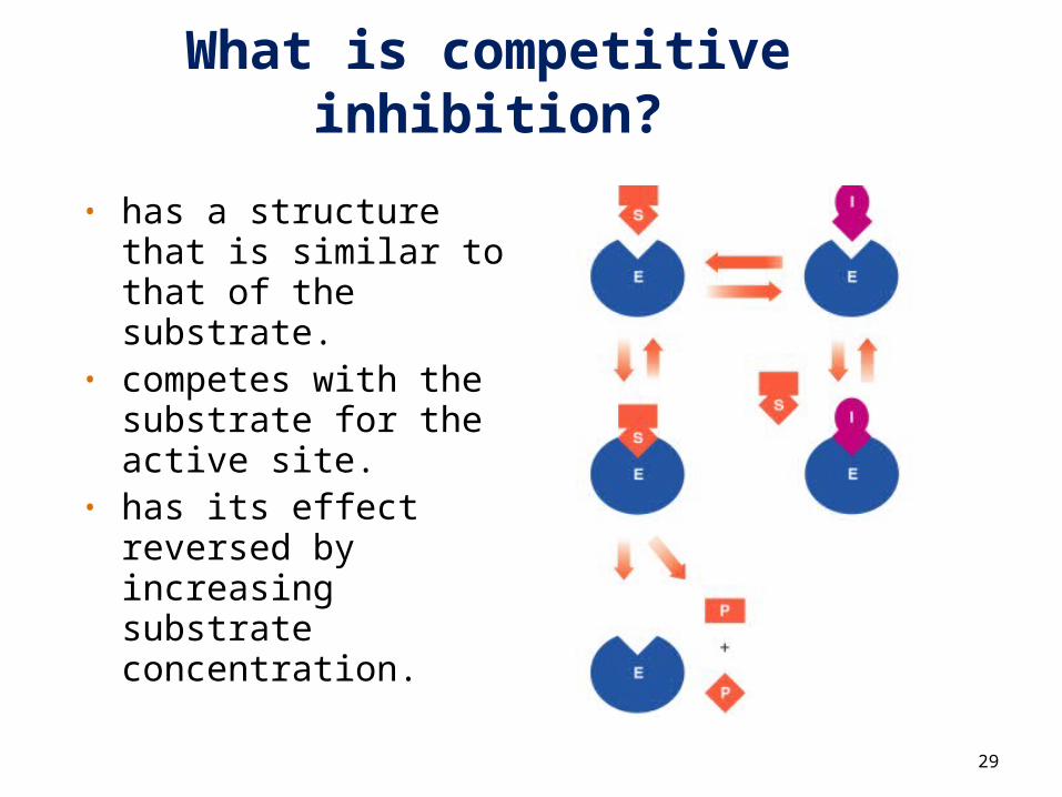
What is competitive inhibition?
• has a structure that is similar to that of the substrate.
• competes with the substrate for the active site.
• has its effect reversed by increasing substrate concentration.
29
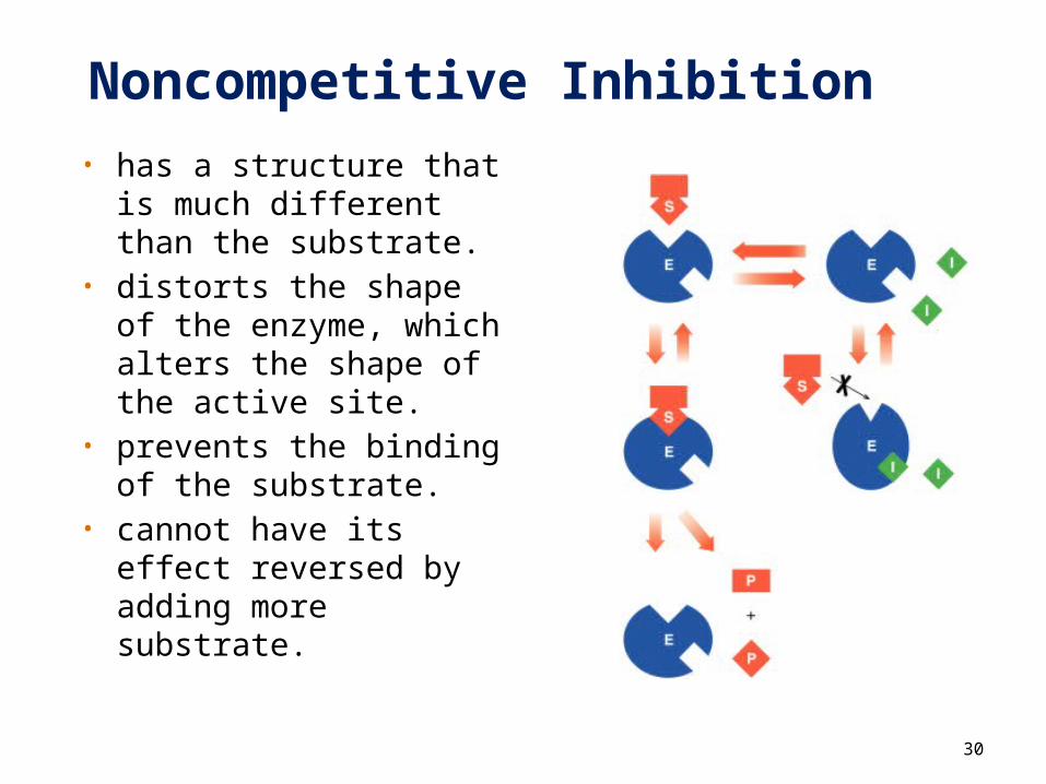
Noncompetitive Inhibition• has a structure that
is much different than the substrate.
• distorts the shape of the enzyme, which alters the shape of the active site.
• prevents the binding of the substrate.
• cannot have its effect reversed by adding more substrate.
30
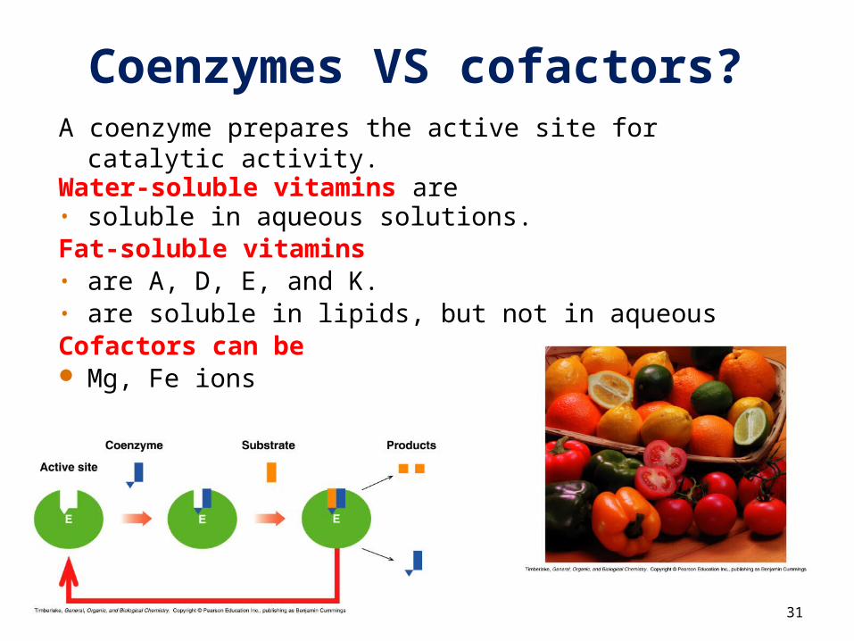
Coenzymes VS cofactors?A coenzyme prepares the active site for catalytic
activity.Water-soluble vitamins are• soluble in aqueous solutions.Fat-soluble vitamins• are A, D, E, and K. • are soluble in lipids, but not in aqueousCofactors can be Mg, Fe ions
31