Embed Size (px)
Citation preview
Name _____________________________________________
Honors Anatomy and Physiology Cardiovascular System Study Guide
5.1 Explain the relationship between the structure and function of arteries, capillaries, and veins.
1. Why do arteries have thicker walls than veins?
2. What would be the difference between blood leaving from an artery versus a vein when cut?
5.2 Label a diagram of the heart, including the chambers, blood vessels, valves, and the route of blood through the heart.
1. Label the external and internal structures of the heart. Word bank: Aorta, aortic valve, apex, Chordae tendineae, inferior vena cava, left atrium, left ventricle, mitral (bicuspid) valve, pulmonary artery, pulmonary trunk, pulmonary vein, pulmonic valve, right atrium, right ventricle, superior vena cava, tricuspid valve.
A: K:B: L:C: M:D: N:E: O:F: P:G: Q:H: R:I: S:J:
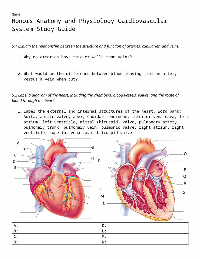
**On the internal diagram above, draw arrows with a colored pencil or crayon that show the path of the blood through the heart.**
Circulatory System Study Guide
5.3 Outline the mechanisms that control the heartbeat
1. What are systolic and diastolic blood pressure? Which one is higher, and why?
2. What activity or process causes heart sounds (the lub-dub you can hear through a stethoscope)?
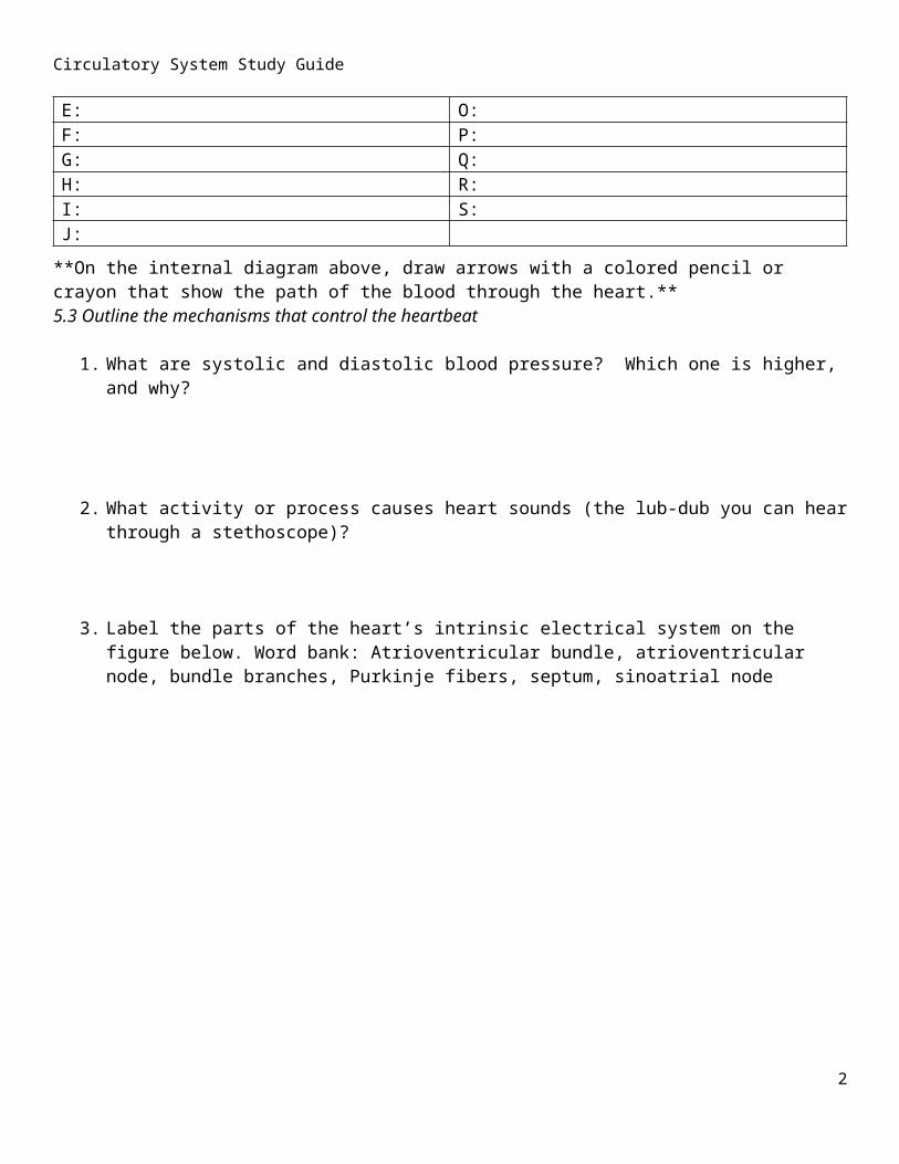
3. Label the parts of the heart’s intrinsic electrical system on the figure below. Word bank: Atrioventricular bundle, atrioventricular node, bundle branches, Purkinje fibers, septum, sinoatrial node
A EB FC GD
**With a crayon or colored pencil, draw arrows to show the path of electrical impulses through the heart**
4. Which of the above structures acts as the heart’s “pacemaker”? You can just write the name.
5. List 3 things that increase heart rate.
6. List 3 things that decrease heart rate. 2
Circulatory System Study Guide
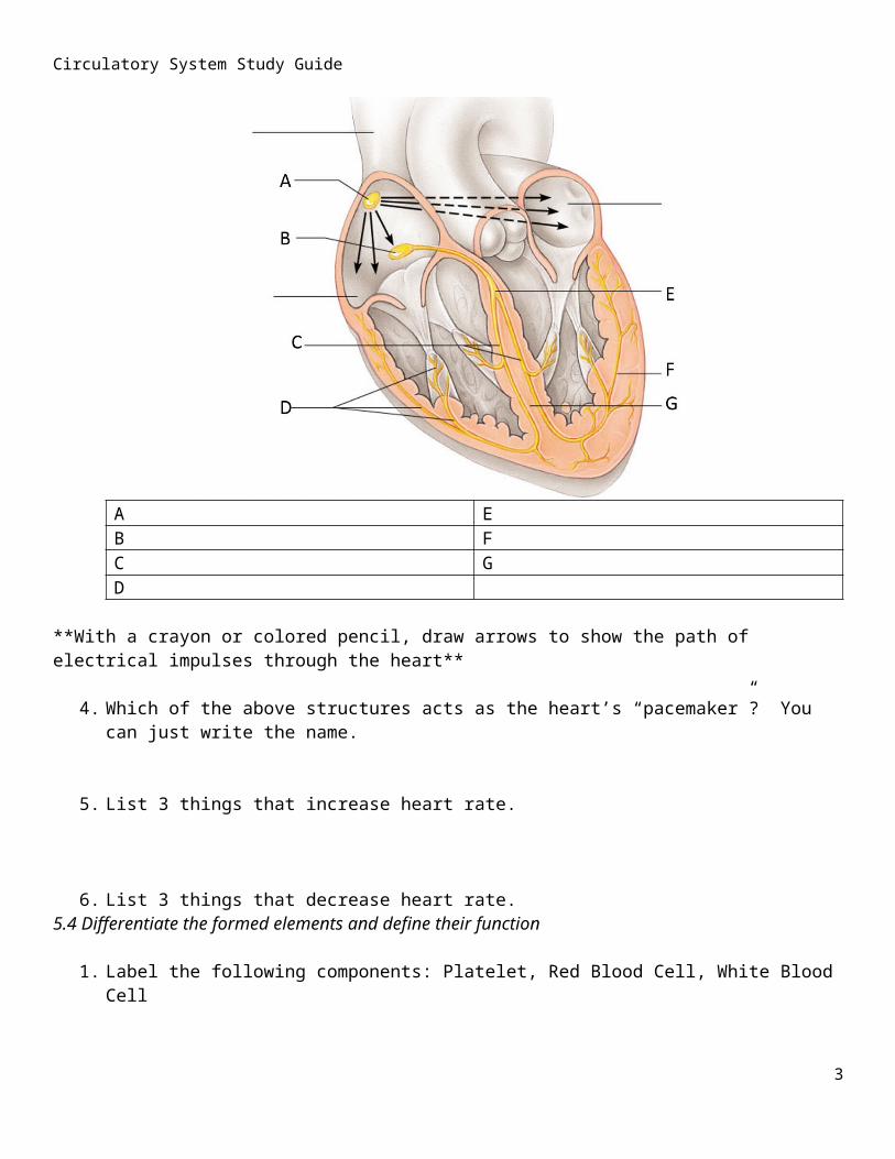
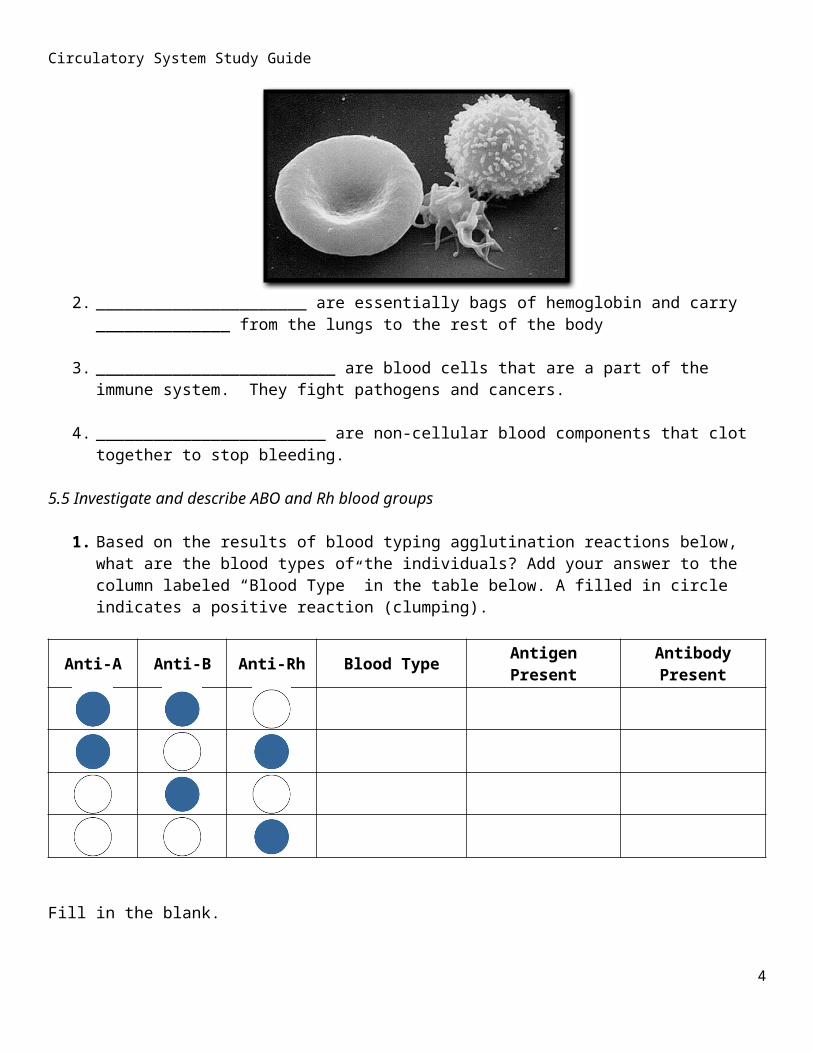
5.4 Differentiate the formed elements and define their function
1. Label the following components: Platelet, Red Blood Cell, White Blood Cell
2. ______________________ are essentially bags of hemoglobin and carry ______________ from the lungs to the rest of the body
3. _________________________ are blood cells that are a part of the immune system. They fight pathogens and cancers.
4. ________________________ are non-cellular blood components that clot together to stop bleeding.
5.5 Investigate and describe ABO and Rh blood groups
1. Based on the results of blood typing agglutination reactions below, what are the blood types of the individuals? Add your answer to the column labeled “Blood Type” in the table below. A filled in circle indicates a positive reaction (clumping).
Anti-A Anti-B Anti-Rh Blood Type Antigen Present Antibody Present
Fill in the blank.2. Blood types are determined by the presence of protein located on which blood cells? _______________
3. Blood type _________ is considered to be the universal recipient
4. If a person has type A– blood, then they have which proteins? ____________________________3
Circulatory System Study Guide
Complete the following:
1. Diagram how the blood flows through the body. Start with the oxygenated blood entering the left atrium and end with it returning. This can be done through words, pictures, or diagrams.
2. Describe each of the disease types: Blood Plasma disease, Red Blood Cell disease, Blood Clotting disease, and White Blood Cell disease.
4


![Review Article Treatment of Chagas Cardiomyopathy · complete atrioventricular block, and right bundle block [ , , , ]. Morphologically, hypertrophy, dilatation, and ... To reduce](https://img.dokumen.tips/doc/110x75/60f750f1c199d5733c62132f/review-article-treatment-of-chagas-cardiomyopathy-complete-atrioventricular-block.jpg)