Embed Size (px)
Citation preview

Brit. .j. Ophthal. (1973) 57, 773
Vitreous haemorrhage in tuberoussclerosis
Report of two cases
A. ATKINSON*, M. D. SANDERS, AND V. WONG**
Department of Ophthalmology, National Hospital, Queen Square, London
Tuberous sclerosis is a well-defined clinical entity characterized by a triad of epilepsy,mental retardation, and adenoma sebaceum, with excellent clinical descriptions elsewhere(Critchley and Earl, I932; Lagos and Gomez, I967). The principal ocular manifesta-tions comprise retinal tumours of two morphological types (van der Hoeve, 1920, 192I).Elevated mulberry-like tumours are found characteristically in the posterior pole wherethey may resemble drusen of the disc. Each tumour appears as a whitish-grey glisteningmass, studded with nodules which may become cystic. A second type occurs in the retinalperiphery which is flat, white or grey, circular or oval, and frequently multiple, rangingup to half the size of the optic disc (Koch and Walsh, I939).The condition is inherited as an autosomal dominant, Eo that early recognition is of
paramount importance for genetic counselling. A large proportion of new cases, however,occur as mutations (Bundey, I 971).The present report describes retinal phakomata in two cases of tuberous sclerosis.
Fluorescein angiographic studies in one patient demonstrated for the first time an associatedangiomatous anomaly in a disc tumour. Previous reports have neglected the vascularanomaly with its haemorrhagic potential.
Case reports
Case I, a 15-year-old schoolboy, was found, during a routine examination to have reduced visionin the right eye with a visual field defect. An ophthalmologist confirmed these findings, and notedin addition an angiomatous lesion of the right disc. The patient was admitted to hospital for in-vestigation. He had had infantile spasms at 4 months, and at 6 months developed a birthmark onthe left cheek. At I2 years a radiologist was consulted regarding x-ray therapy to the facial lesion;skull films showed areas of intracerebral calcification. There was no relevant family history.
Physical examination
He was a well-developed youth of average intelligence with no abnormality of the cardiovascular,respiratory, or nervous system. There was a pink elevated naevus on the left cheek, and numerouspink spots on both sides of the face in the distribution of the second and third divisions of the trige-minal nerve. A dermatologist confirmed adenoma sebaceum and a cavernous haemangioma of theleft side of the face.
Received for publication July 24, I972Address for reprints: M. D. Sanders, F.R.C.S., The National Hospital, Queeni Square, Lonidoln, WC IN 3BG* Moorfields Eye Hospital, London, E.C.i.** Clinical Center N.I.H., Bethesda, Md. 20014
copyright. on A
pril 18, 2020 by guest. Protected by
http://bjo.bmj.com
/B
r J Ophthalm
ol: first published as 10.1136/bjo.57.10.773 on 1 October 1973. D
ownloaded from

A. Atkinson, M. D. Sanders, and V. Jf4ong
Ophthalmological examination
The visual acuity was 6/6 in the left eye, and 6/6o in the right eye with an upper altitudinal defect.Slit-lamp examination of the right eye showed a small posteriorsubcapsular cataract with haemorrhageadherent to the posterior lens surface. The anterior segment was normal in the left eye and theintraocular pressure was normal in both eyes.The right fundus (Fig. I) showed a pre-retinal haemorihage extending into the vitreous, and
condensing below. A white mulberry-like tumour was present arising from the optic disc, withnumerous fine vascular channels throughout the tumour and on its surface. Scattered around thefundus were several small flat white lesions, with a smooth surface and apparently avascular. Themacula was detached.
FIG. I Case i. Right fundus,showing vitreous haemorrhage, andelevated mulberry tumour of the disc,and several flat peripheral retinalphakomata
The left fundus showed two small flat lesions in the upper nasal quadrant anterior to the equator,but was otherwise normal.
RadiologyThe skull showed three areas of intracerebral calcification. The two largest were defined by pneumo-encephalography forming papillary projections from the inferior surface of the right and left ventriclesriespectively, while the third lay in the brain substance. Bilateral carotid angiograms showed normalarterio-venous patterns.
Case 2, a 23-year-old man, was referred to Mr. K. Wybar at Moorfields Eye Hospital in May, I 96 I,with an abnormality of the right eye observed during a routine examination. The patient complainedof headaches for several years, but had no ocular symptoms. There was no relevant family history.
Ophthalmological examinationThe visual acuity was 6j6 in each eye. The anterior segments and media were normal. Projectinginto the vitreous from the lower part of the right optic disc (Fig. 2) was an extensive white mass withseedling-like nodules on its surface and an irregular border. The appearance of the left fundus wasnormal. A tentative diagnosis of drusen of the right disc was made.
774copyright.
on April 18, 2020 by guest. P
rotected byhttp://bjo.bm
j.com/
Br J O
phthalmol: first published as 10.1136/bjo.57.10.773 on 1 O
ctober 1973. Dow
nloaded from
Vitreous haemorrhage in tuberous sclerosis
FIG. 2 Case 2. Right fundus,showing nodular tumour arisingfrominfero-nasal part of optic disc
Course
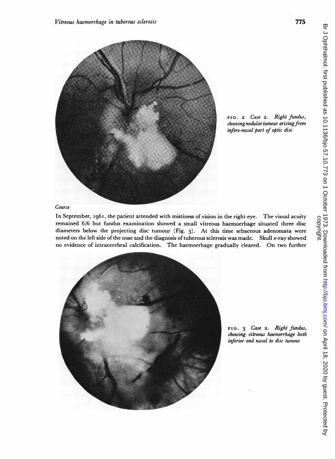
In September, I96I, the patient attended with mistiness of vision in the right eye. The visual acuityremained 6/6 but fundus examination showed a small vitreous haemorrhage situated three discdiameters below the projecting disc tumour (Fig. 3). At this time sebaceous adenomata werenoted on the left side of the nose and the diagnosis of tuberous sclerosis was made. Skull x-ray showedno evidence of intracerebral calcification. The haemorrhage gradually cleared. On two further
" j_ FIG. 3 Case 2. Right fundus,showing vitreous haemorrhage bothinferior and nasal to disc tumour
775copyright.
on April 18, 2020 by guest. P
rotected byhttp://bjo.bm
j.com/
Br J O
phthalmol: first published as 10.1136/bjo.57.10.773 on 1 O
ctober 1973. Dow
nloaded from

A. Atkinson, M. D. Sanders, and V. Wong
occasions during the next 3 years the patient presented with floaters in the right eye due to recurrentvitreous haemorrhages. In I966 the patient was seen at the National Hospital where skull filmsdemonstrated several areas of intracerebral calcification and an electroencephalogram showedchanges compatible with a diagnosis of tuberous sclerosis.The patient was last examined in March, I 971, when he was symptom free. The visual acuity was
6/6 in each eye, and fundus examination showed the mulberry tumoui of the right disc to be unchan-ged in size and configuration, and the vitreous to be clear. Central field examination showedenlargement of the blind spot, corresponding with the area of retina obscured by tumour. Theleft fundus was normal.
(I{ 'I).
(I fC!
FIG. 4 Case 2. Fluorescein angiograms of right fundus
(a) Pre-injection, showing auto-fluorescence of disc phakoma(b) Arterial phase: abnormal vessels on tumour surface are seen to be fillingfrom retinal arteries(c) Venous phase: numerous abnormal dilated capillary loops visible throughout tumour(d) Residual photograph, showing hyperfluorescence due to leakage of dye into retina around phakoma
776copyright.
on April 18, 2020 by guest. P
rotected byhttp://bjo.bm
j.com/
Br J O
phthalmol: first published as 10.1136/bjo.57.10.773 on 1 O
ctober 1973. Dow
nloaded from
Vitreous haemorrhage in tuberous sclerosis
A general examination showed the following positive features. Adenoma sebaceum of the faceappeared in a typical butterfly distribution, with involvement of the chin. A shagreen patch waspresent over the lower lumbar spine, and an "aspen leaf" area of hypopigmentation over the right6th intercostal space anteriorly. Subungual fibromata were noted on several digits of both hands.Fundus photography showed autofluorescence of the retinal tumour before the intravenous
injection of fluorescein (Fig. 4a). The arterial phase (Fig. 4b) showed normal flow with early fillingof the dilated capillaries on the surface of the tumour. Gross dilatation of the peripapillary retinalplexus was observed with filling from the retinal arteries. Abnormal dilated capillary loops werevisible in the venous phase (Fig. 4c) and some microaneurysms of the peripapillary plexus at the infer-ior and temporal disc margins. The residual photographs (Fig 4d) taken Io minutes after injectionshowed massive hyperfluorescence of the papillary region with leakage into the surrounding retina(Fig. 4e). Comparison with angiograms taken 4 years previously showed no change.
Discussion
Estimates of the incidence of phakomata in cases of tuberous sclerosis vary from 4 per cent.(Critchley and Earl, I932) to 53 per cent. (Lagos and Gomez, I967). When such lesionsare present they do not usually cause visual disturbance. Large tumours of the disc maycause subtle changes in the visual field (Case 2), though severe visual loss from vitreoushaemorrhage or macular detachment has not been reported.The first description of vitreous haemorrhage as a feature of tuberous sclerosis was by
van der Hoeve (I921). Cystic spaces in a mushroom-like tumour of the disc were notedto fill with blood on separate occasions, and on one occasion haemorrhage into thevitreous from a ruptured cyst was observed. Koch and Walsh (I939) reported a casewith vitreous haemorrhage occurring in association with papilloedema secondary to raisedintracranial pressure. A vascular retinopathy was described by Dyer, Hill, Rowan, andTaylor (I967) in a patient with tuberous sclerosis and megaloblastic anaemia, but thiscleared with treatment of the anaemia.The histological structure of ocular phakomata first received attention from van der
Hoeve (I923). A tumour arising from the disc was described, consisting of fibre-likeprocesses interspersed with large polymorphic cells having large nuclei and prominentnucleoli. The cytoplasm of adjacent cells fused in places to form a syncytium. Situatedbetween the fibres and cells were areas of calcification, and large spaces filled with bloodor serum. A disc tumour of similar structure has been described by Messinger and Clarke(I937).Peripheral retinal tumours consisting of pleomorphic cells within a framework of
fibre-like strands have also been described (Schob, I925; Feriz, 1930; Kuchenmeister,I934; Vogt, I934). Areas of calcification or cystic degeneration appear to be rare inperipheral lesions. These tumours usually lie in the nerve fibre or ganglion cell layer, butmay involve all layers of the retina. Fluorescein studies of the peripheral lesion demon-strated numerous abnormal vessels in association (Harley and Grover, I970).The fibrillar structure of phakomata and their apparent origin from the nerve fibre
layer led van der Hoeve to postulate that nerve fibres contributed to tumour formation.Subsequent reports have emphasized the glial-like nature of the fibre network (Schob,I925; Kuchenmeister, 1934), the tumours in cases of tuberous sclerosis described byMessinger and Clarke (I937) and McLean (I937) being classified as astrocytic in origin,probably hamartomata rather than true tumours.
Astrocytomas of the retina or optic disc occurring without clinical signs of neurofibro-matosis or tuberous sclerosis have been reported by McLean (I937), Foos, Straatsma, and
777copyright.
on April 18, 2020 by guest. P
rotected byhttp://bjo.bm
j.com/
Br J O
phthalmol: first published as 10.1136/bjo.57.10.773 on 1 O
ctober 1973. Dow
nloaded from

A. Atkinson, M. D. Sanders, and V. JWong
Allen (I965), and Ganley and Streeten (I 97i). The similarity in appearance and struc-ture to ocular phakomata and the rarity of such cases makes the search for other mani-festations of phakomatosis of great importance if reliable genetic counselling is to beachieved.
Vitreous haemorrhage occurring in drusen of the disc has been described by Gifford(I895), Reese (1940), and Gaynes and Towle (i967). Haemorrhages on or around thedisc have been reported by Bregeat (I 956), Sanders and ifytche (i 967), andWalsh and Hoyt(i969). Of particular interest is a recent report by Sanders, Gay, and Newman (I970)of seven cases of drusen of the disc with haemorrhagic complications. In two instances,the haemorrhage extended into the vitreous, while in the remainder the haemorrhage waseither within the substance of the nerve head or subretinal.The nature of the vascular supply of disc and retinal tumours has received little attention
in pathological studies. Numerous fine vessels on the surface of the disc tumour werenoted by van der Hoeve (I923), though Koch and Walsh (I939) emphasized the apparentavascularity of the flat peripheral tumours. The description of sebaceous adenomata byPringle (I890) emphasized the vascular nature of the skin tumours, the sebaceous lesionsshowing concomitant capillary dilatation and telangiectasis. Histological studies of skinbiopsies from 38 patients with tuberous sclerosis were presented by Nickel and Reed(i 962) . Dilatation of blood vessels and lymphatics wvas commonly observed, andthe prominent vascular changes led these authors to regard the lesions of adenoma seba-ceum as angiofibromata. The fluorescein angiograms presented here clearly demonstratenumerous dilated capillaries throughout the optic disc tumour, ws\hich are abnormal andhighly permeable to fluorescein.
These findings provide an explanation for the recurrent vitreous haemorrhages but alsosuggest that the ocular lesions resemble the cutaneous lesions in containing a significantangiomatous element. Pathological verification of this has been recently reported(Barsky and Wolter, 197 I).
Conclusion
Vitreous haemorrhages as a complication of tuberous sclerosis is reported in two caseswith large peripapillary tumours. Fluorescein angiograms show an extensive vascularplexus on the surface of the lesions and it is suggested that the vascular anomaly may bepart of the hamartomatous process.
WN'e should like to thank Mr. K. W\ybar for permission to publish case 2, Miss Sue Ford for the photographicprints, Miss Lace for secretarial assistance, and the Medical Research Council for financial support.
References
BARSKY, D., and WOLTER, J. R. (I97I) J. pediat. Ophthal., 8, 26IBREGEAT, P. (1956) "L'oedeme papillaire", p. 256. Masson, ParisBUNDEY, s. (I97I) Proc. roy. Soc. AMed., 64, i85CRITCHLEY, M., and EARL, c. J. c. (I932) Brain, 55, 31 I
DYER, A. M., HILL, R., ROWAN, R. M., and TAYLOR, W. D. E. (I967) Brit. nied. J., 4, 398FERIZ, H. (1930) Virchows Arch. Path. Anat., 278, 690FOOS, R. Y., STRAATSMA, B. R., and ALLEN, R. A. (i 965) A-ch. Ophthal. (Chicago), 74, 3 I 9
GANLEY, J. P., and STREETEN, B. W. (197I) Anmer. J. Ophthal., 71, 1099
GAYNES, P. M., and TOWLE, P. A. (I967) Ibid.. 63, I693GIFFORD, H. (I895) Arch. Ophthal., 24, 395
778copyright.
on April 18, 2020 by guest. P
rotected byhttp://bjo.bm
j.com/
Br J O
phthalmol: first published as 10.1136/bjo.57.10.773 on 1 O
ctober 1973. Dow
nloaded from

Vitreous haemorrhage in tuberous sclerosis 779
HIARLEY, R. D., and GROVER, W. D. (1970) Ann. Ophthal., I, 477KOCH, F. L. P., and WALSH, M. N. (I939) Arch. Ophthal., 21, 465KUCHENMEISTER, E. (I934) Derm. Wschr., 99, I333LAGOS, J. c., and GOMEZ, M. R. (i967) Mayo Clin. Proc., 42, 329MCLEAN, J. M. (1937) Arch. Ophthal., I8, 255MESSINGER, H. C., and CLARKE, B. E. (1937) Ibid., I8, I
NICKEL, W. R., and REED, W. B. (i962) Arch. Derm. (Chicago), 85, 89PRINGLE, J. J. (1890) Brit. J. Derml., 2, IREESE, A. B. (1940) Arch. Ophthal., 24, I87SANDERS, M. D., and FFYTCHE, T. J. (i967) Trans. ophthal. Soc. U.K., 87, 457SANDERS, T. E., GAY, A. J., and NEWMAN, M. (1970) Trans. Amer. ophthal. Soc., 68, i86SCHOB, F. (I925) Z. ges. N"eurol. Psychiat., 45, 731VAN DER IIOEVE, J. (1920) T7ans. ophthal. Soc. U.K., 40, 329
(I 92 I) v. Graefes Arch. Ophthal., 105, 88o(I923) Ibid., II I,
VOGT, A. (I934) Z. Augenheilk., 86, i8WALSH, F. B., and HOYT, W. F. (i969) "Clinical Neuro-Ophthalmology", 3rd ed., vol. 3, p.1957.
Williams and Wilkins, Baltimore
copyright. on A
pril 18, 2020 by guest. Protected by
http://bjo.bmj.com
/B
r J Ophthalm
ol: first published as 10.1136/bjo.57.10.773 on 1 October 1973. D
ownloaded from