Embed Size (px)
Citation preview
ORIGINAL CONTRIBUTIONS
Vitamin D3 Loading Is Superior to ConventionalSupplementation After Weight Loss Surgery in VitaminD-Deficient Morbidly Obese Patients: a Double-BlindRandomized Placebo-Controlled Trial
Maria Luger1,2,3 & Renate Kruschitz1,4 & Christian Kienbacher5 & Stefan Traussnigg5 &
Felix B. Langer6 & Gerhard Prager6 & Karin Schindler1 & Enikö Kallay7 &
Friedrich Hoppichler2,8 & Michael Trauner5 & Michael Krebs1 & Rodrig Marculescu9&
Bernhard Ludvik1,4
Published online: 12 November 2016# The Author(s) 2016. This article is published with open access at Springerlink.com
AbstractBackground Bariatric patients often suffer from vitaminD deficiency (VDD), and both, morbid obesity andVDD, are related to non-alcoholic fatty liver disease.However, limited data are available regarding best
strategies for treating VDD, particularly, in bariatric pa-tients undergoing omega-loop gastric bypass (OLGB).Therefore, we examined the efficacy and safety of aforced vitamin D dosing regimen and intervention ef-fects in liver fibrotic patients.
Clinical Trial Registry Number and WebsiteClinicaltrials.gov (NCT02092376) at https://clinicaltrials.gov/EudraCT (2013-003546-16) at https://eudract.ema.europa.eu/
Electronic supplementary material The online version of this article(doi:10.1007/s11695-016-2437-0) contains supplementary material,which is available to authorized users.
* Renate [email protected]
Maria [email protected]
Christian [email protected]
Stefan [email protected]
Felix B. [email protected]
Gerhard [email protected]
Karin [email protected]
Enikö [email protected]
Friedrich [email protected]
Michael [email protected]
Michael [email protected]
Rodrig [email protected]
Bernhard [email protected]
1 Division of Endocrinology and Metabolism, Department of InternalMedicine III, Medical University of Vienna, Vienna, Austria
2 Special Institute for Preventive Cardiology and Nutrition-SIPCANSave Your Life, Salzburg, Austria
3 Institute of Social Medicine, Centre for Public Health, MedicalUniversity of Vienna, Vienna, Austria
4 Department of Medicine 1 and Karl Landsteiner Institute for Obesityand Metabolic Diseases, Rudolfstiftung Hospital, Juchgasse 25,1030 Vienna, Austria
5 Division of Gastroenterology and Hepatology, Department ofInternal Medicine III, Medical University of Vienna, Vienna, Austria
OBES SURG (2017) 27:1196–1207DOI 10.1007/s11695-016-2437-0
Methods In this double-blind, randomized, placebo-controlled trial, 50 vitamin D-deficient patients undergoingOLGB were randomly assigned to receive, in the first monthpostoperatively, oral vitamin D3 (≤3 doses of 100,000 IU;intervention group) or placebo as loading dose (control group)with subsequent maintenance dose (3420 IU/day) in bothgroups until 6-month visit.Results Compared with control group, higher increase of25(OH)D (67.9 (21.1) vs. 55.7 nmol/L (21.1); p = 0.049) withlower prevalence of secondary hyperparathyroidism (10 vs.24 %; p = 0.045) was observed in intervention group. No(serious) adverse events related to study medication werefound. The loading dose regimen was more effective in in-creasing 25(OH)D in patients with significant liver fibrosiswhile this was not the case for conventional supplementation(placebo with maintenance dose) (71.5 (20.5) vs. 22.5 nmol/L(13.8); p = 0.022; n = 14).Conclusions Our findings indicate that a high vitamin D3
loading dose, in the first month postoperatively, with subse-quent maintenance dose is effective and safe in achievinghigher vitamin D concentrations in OLGB patients.Unexpectedly, it is more effective in patients with significantliver fibrosis which is of potentially high clinical relevanceand requires further investigation.
Keywords Vitamin D . Vitamin D supplementation .
Obesity . Gastric bypass . Omega-loop gastric bypass .
Weight loss . Secondary hyperparathyroidism . Liverfibrosis
Introduction
Vitamin D3 (cholecalciferol), derived both from dermalsynthesis and dietary sources (e.g., oily fish and supple-ments), is first modified mainly in the liver by 25-hydroxylation to generate the main circulating form, 25-hydroxy vitamin D (25(OH)D). This is followed by 1α-hydroxylation to produce the active hormone, 1,25-dihy-droxy vitamin D (1,25(OH)2D) mainly in the kidney andalso at numerous extra-renal sites [1]. The vitamin D re-ceptor (VDR) is expressed almost ubiquitously and regu-lates, upon ligand binding, the expression of over 200
genes [2]. In addition, non-VDR-mediated vitamin D ac-tions have been identified in various tissues and this fieldof research is rapidly expanding [3]. It is therefore notsurprising that the vitamin D endocrine system impactsthe function of virtually every organ system in the humanbody. Vitamin D deficiency has been connected by mech-anistic, epidemiologic, and clinical intervention studieswith the pathogenesis of numerous diseases [3]. Obesepatients are at higher risk of vitamin D deficiency com-pared with non-obese individuals [4], which seems to bebest explained by the dilution of vitamin D and 25(OH)Din the expanded adipose compartment [5, 6]. More sophis-ticated pharmacokinetic studies are needed to clarify de-tails such as the contribution of the adipose tissue itself tothe metabolism of vitamin D. Obesity is also associatedwith non-alcoholic fatty liver disease (NAFLD), whichcomprises a disease spectrum ranging from simplesteatosis to steatohepatitis, fibrosis, cirrhosis, and finallyhepatocellular carcinoma and is the most prevalent chron-ic liver disease in industrialized countries [7]. Vitamin Dinsufficiency (25(OH)D <75 nmol/L) is associated withNAFLD independently from diabetes, insulin resistance,and metabolic syndrome [8]. Increased stages of liver fi-brosis are associated with lower vitamin D concentrationsin morbidly obese patients [9], suggesting a causal role ofvitamin D deficiency in the pathogenesis of liver fibrosis.Vitamin D has even been proposed as a therapeutic agentfor this condition [10].
Bariatric surgery is an effective method to treat obesity,(pre)diabetes [11, 12], and NAFLD [13] in morbidly obesepatients and is associated with long-term weight loss and de-creased overall mortality [14]. Due to gastric restriction andmalabsorption following the gastric bypass procedure, 50–96 % of bariatric patients suffer from vitamin D deficiency,in addition to the obesity-related nutritional deficiencies[15–18].
Only limited data are available regarding the best strategiesfor treating vitamin D deficiency in bariatric patients. Theclinical practice guidelines differ among scientific societiesand often lack scientifically founded criteria. Therefore, thereis need for high quality randomized controlled trials to providereliable evidence for the recommendations on vitamin D sup-plementation in bariatric patients [19].
The primary aim of this study was to examine the efficacyand safety of a forced vitamin D dosing regimen (up to threeoral loading doses in the first month postoperatively, interven-tion group) vs. conventional supplementation (placebo withfollowing maintenance doses, control group) on parameters ofvitamin D metabolism in morbidly obese, vitamin D-deficientpatients undergoing omega-loop gastric bypass (OLGB)6 months after surgery. In addition, given the high prevalenceof liver fibrosis in these patients and the intricate role of vita-min D in this condition [9], we assessed markers of vitamin D
OBES SURG (2017) 27:1196–1207 1197
6 Division of General Surgery, Department of Surgery, MedicalUniversity of Vienna, Vienna, Austria
7 Department of Pathophysiology and Allergy Research, MedicalUniversity of Vienna, Vienna, Austria
8 Division of Internal Medicine, Krankenhaus der BarmherzigenBrüder Salzburg, Salzburg, Austria
9 Clinical Institute for Medical and Chemical Laboratory Diagnostics,Department of Laboratory Medicine, Medical University of Vienna,Vienna, Austria
metabolism in liver biopsies from the subgroup of patientsaffected by significant liver fibrosis.
Subjects and Methods
The BLink Between Obesity and Vitamin D^ (LOAD) studyconducted from April 2014 to October 2015 in Vienna(Austria), was a 6-month double-blind, placebo-controlled,randomized clinical trial with a parallel-group design in bar-iatric patients. The effects of up to three oral vitamin D3 load-ing doses in the first month postoperatively followed by amaintenance dose (intervention group) was compared withplacebo followed by a maintenance daily dose (control group)on 25(OH)D concentration and other vitamin D metabolismparameters. The details on design and the used materials andmethods of the study have been previously published [20].
Participant Recruitment and In- and Exclusion Criteria
Bariatric patients with following inclusion criteria were re-cruited: men and women aged 18–100 years with plannedOLGB surgery, serum 25(OH)D concentrations of<75 nmol/L, and body weight <140 kg (due to body weightlimitation of the dual energy X-ray absorptiometry (DEA)).Specific exclusion criteria included any other planned form ofbariatric surgery than OLGB, hypo- and hypercalcemia, renalinsufficiency (creatinine >133 μmol/L or glomerular filtrationrate <50mLmin−1 1.73m−2), or primary hyperparathyroidism[20]. In- and out-patients of the Obesity Clinics at theDepartment of Internal Medicine III or the Department ofSurgery in the General Hospital of Vienna were recruited be-tween April 2014 and April 2015. All study procedures werereviewed and approved by the local Ethics Committee of theMedical University of Vienna (No. 1899/2013) and by theAustrian Competent Authority (No. LCM-718280-0001) andcomply with the Declaration of Helsinki [21]. Moreover, theprotocol was registered at clinicaltrials.gov (identifier:NCT02092376) and EudraCT (identifier: 2013-003546-16).All study participants provided signed informed consent.The study methods are in accordance with the ConsolidatedStandards of Reporting Trials (CONSORT) guidelines forreporting randomized trials [22].
Sample size calculation was based on previously publisheddata from 50 bariatric patients who underwent OLGB [18]. Onbasis of an assumed 20 % dropout rate (including loss tofollow-up), we estimated a total sample size of 50 patients(25 in each group) by using 80 % statistical power and atwo-sided significance level of 0.05 to detect a 25(OH)D dif-ference of 30 nmol/L (standard deviation, 35) between inter-vention and control groups after 6 months.
Study Design and Randomization
After collecting baseline data, eligible patients were allocatedto receive either vitamin D3 (intervention group) or placebo(control group) with a computer-generated randomizationscheme in a 1:1 ratio, stratified by 25(OH)D, age, and genderlevel by a blinded study coordinator using the BRandomizerfor Clinical Trials 1.8.1^ [23]. Randomization was carried outduring the baseline assessment, after the patient had signed theinformed consent form. Everybody involved in the study wasblinded to the randomization status. Allocation was performedby consecutively numbered dark bottles with either vitaminD3 (cholecalciferol diluted in medium-chain triglycerides:Oleovit D3 drops, Fresenius) or placebo (carrier oil ofmedium-chain triglycerides) loading dose, labeled with therandomization number, which were created and bottled bythe in-house hospital pharmacy, so as to blind both studysubjects and investigators.
Vitamin D Dosing Regimen
In the field of pharmacology, a loading dose is an amount ofdrug designed to fill the central volume of distribution for adrug to a concentration that matches the final plateau concen-tration achieved with the maintenance dose. The purpose is toachieve this final plateau sooner than the four half-lives re-quired, if the drug is simply administered at the maintenancedose rate [24]. For vitamin D3, the functional half-life withinthe body is in the range of 2 to 3 months [25]. Accordingly, theloading dose for the intervention group was calculated as thecumulative maintenance dose that was planned to be giventhrough one functional half-life of vitamin D in the body(loading dose = (daily maintenance dose) × (60–90 days)).Moreover, it is safer not to take vitamin D at a dose beyond100,000 IU at one time to allow vitamin D to clear from thecirculation between each increment of the loading dose [1]. Inthat regard, the chosen loading dose of 300,000 IU vitamin D3
(cholecalciferol) was divided into three doses (each100,000 IU) and given on day 1 or 2, 2 weeks, and 4 weekspostoperatively with subsequent administration of the mainte-nance dose until the 6-month visit in the intervention group[20]. The first loading dose was given on day 1 or 2 aftersurgery, followed by the second (2 weeks) and third adminis-trations (4 weeks postoperatively) if 25(OH)D serum concen-tration remained below 75 nmol/L. The maximum loadingdose for the intervention group was 300,000 IU. After the lastloading dose, a maintenance dose of 3420 IU/day (approxi-mately translating to 24,000 IU/week) was given. The controlgroup received placebo as loading dose and subsequently themaintenance dose, the same way as the intervention group. Adetailed illustration of the dosing regimen is depicted in thestudy protocol [20]. At each study visit after surgery, partici-pants received a monthly supply of study medication (Oleovit
1198 OBES SURG (2017) 27:1196–1207
D3 drops, Fresenius, containing 12.5 mL of cholecalciferol inmedium-chain triglycerides carrier oil; 1 drop = 400 IU).Every week, the patients received instructions and further re-minders via text messages to take the supplement (mainte-nance dose). The patients were instructed to return emptybottles at the monthly study visit.
Assessment of Variables
At baseline (before randomization), age, sex, medical history(e.g., comorbidities, prescribed medication) were collected[20]. The following set of evaluations was obtained for eachparticipant before surgery and 0.5, 1, 2, 3, 4, 5, and 6 month(s)postoperatively: height and body weight (measured with thecalibrated scale seca mBCA 515), waist circumference mea-sured with an inelastic tape, supplement use, and parametersof vitamin D metabolism (25(OH)D (nmol/L), 1,25(OH)2D(pg/mL), intact parathyroid hormone PTHi (pg/mL), andalbumin-corrected calcium Ca (mmol/L)) [26]. Secondary hy-perparathyroidism (SHPT) was defined as PTHi >65 pg/mLwith simultaneous normal values for creatinine, calcium, andinorganic phosphate. Daily total energy (kcal), relative energyfrom protein (%), carbohydrate (%), fat (%), vitamin D (μg),and calcium (mg) intakes were calculated from 5-day foodrecords 1 week before surgery (baseline) and 1, 3, and6 month(s) after surgery. The computation of nutrient intakewas carried out with the nutritional software nut.s science(dato Denkwerkzeuge, v1.29.34, Austria). Additionally, habit-ual sun exposure was assessed by a series of questions forwhich categorical response options were provided [27] suchas Bhours spent outside between 9 a.m. and 3 p.m.^ during theweek (h/week), during the weekend (h/weekend), Bregulargoing to the tanning salon^ (yes, no), and Busing sunscreen^(yes, no). Furthermore, an average time spent outside wascalculated (h/week). Season was defined as spring (March–May), summer (June–August), autumn (September–Novembe r ) , and win t e r (December–Februa ry ) .Supplementation adherence was reviewed by medicationcounts.
Surgical Technique
All procedures were performed by the same surgical teamusing a laparoscopic approach. The OLGB is a simplifiedprocedure that consists of a unique gastrojejunal anastomosisbetween a 30- and 40-mL-sleeve gastric pouch and a jejunalomega-loop of approximately 200 cm [28].
Liver Biopsy and Histopathological Evaluation
Intraoperatively, fine-needle trucut biopsies were performedduring the laparoscopic OLGB. All tissues were fixed in10 % buffered formalin and embedded in paraffin. The three
histochemically stained biopsies (hematoxylin and eosin,Chromotrop Anilinblue and Prussian blue iron stains) wereanalyzed and interpreted by two independent, experiencedboard-certified pathologists of the General Hospital Vienna,Austria. The histological scoring system NAFLD activityscore (NAS; from 0 to 8) by Kleiner et al. [29] was used toevaluate the grade of steatosis (0–3), hepatocyte ballooning(0–2), lobular inflammation (0–3), and stage of fibrosis with a4-point scale.
Safety and Adverse Effects
The participants were interviewed after 2 weeks, 1, 2, 3, 4, 5,and 6 month(s) postoperatively for any signs or symptoms ofvitamin D toxicity or other adverse events, including seriousillness or hospitalizations.
Statistical Analysis
The results are expressed as mean (standard deviation) forcontinuous and as percentages for categorical variables. Inorder to test for normal distribution, a visual test (histogramsand box plots) was used and the Kolmogorov-Smirnov testwas applied in addition. Statistical significance tests such as ttest or Mann-Whitney U test, and Chi2 test were applied toassess differences between the intervention and control groupsat baseline. The primary outcome variable 25(OH)D was an-alyzed according to the intention-to-treat (ITT) principle (in-cluding all randomized participants). To handle missing data,the multiple imputation (MI) method was used for the mainanalysis [30]. All secondary outcome variables were assessedby per-protocol analysis (PP; all persons included in the studywithout major protocol deviation). The associations of25(OH)D and PTHi were assessed by multiple and singlelinear regression models with backward selection of variablesat a p value threshold of 0.20 or variables entered in the model.We used repeated measure analysis of covariance (ANCOVA)using random error (linear mixedmodel) to assess the effect oftime and the interaction for changes in parameters between thegroups, by using different covariance structure models as ap-propriate and were adjusted for age, sex, and baseline valuesto supply an unbiased estimate of the mean group difference[31].Moreover, a post hoc analysis with Bonferroni correctionwas used. Cohen’s d was used to quantify the effect of thetreatment (effect size) based on the mean pre-postchange inthe intervention group minus the mean pre-postchange in thecontrol group, divided by the pooled pretest standard devia-tion [32, 33]. Estimates of the prevalence of SHPT and vita-min D sufficiency between the intervention and controlgroups over time were calculated using generalized estimatingequation (GEE) with a logit link function for binary outcomesand unstructured covariance matrices. With this approach, weexamined effects with time as repeated factor and group as
OBES SURG (2017) 27:1196–1207 1199
between subject factor with prevalence of SHPT and vitaminD sufficiency (yes, no) as dependent variable, adjusted forage, sex, and baseline value. All statistical analyses were per-formed with IBM® SPSS® Statistics for Windows, v23 soft-ware (IBM Corporation, Armonk, New York, USA). p values<0.05 were considered statistically significant, and all testswere two sided.
Results
Study Recruitment and Follow-up
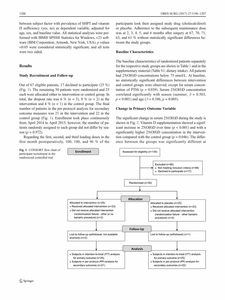
Out of 67 eligible patients, 17 declined to participate (25 %)(Fig. 1). The remaining 50 patients were randomized and 25each were allocated either to intervention or control group. Intotal, the dropout rate was 6 % (n = 3), 8 % (n = 2) in theintervention and 4 % (n = 1) in the control group. The finalnumber of patients in the per-protocol analysis for secondaryoutcome measures was 21 in the intervention and 22 in thecontrol group (Fig. 1). Enrollment took place continuouslyfrom April 2014 to April 2015, however, the number of pa-tients randomly assigned to each group did not differ by sea-son (p = 0.972).
Regarding the first, second, and third loading doses in thefirst month postoperatively, 100, 100, and 96 % of the
participants took their assigned study drug (cholecalciferol)or placebo. Adherence to the subsequent maintenance dosewas at 2, 3, 4, 5, and 6 months after surgery at 67, 70, 71,63, and 61 % without statistically significant differences be-tween the study groups.
Baseline Characteristics
The baseline characteristics of randomized patients separatelyfor the respective study groups are shown in Table 1 and in thesupplementary material (Table S1; dietary intake). All patientshad 25(OH)D concentrations below 75 nmol/L. At baseline,no statistically significant differences between interventionand control groups were observed, except for serum concen-tration of PTHi (p = 0.039). Serum 25(OH)D concentrationcorrelated significantly with season (summer; β = 0.503,p < 0.001) and age (β = 0.386, p = 0.005).
Change in Primary Outcome Variable
The significant change in serum 25(OH)D during the study isshown in Fig. 2. Vitamin D supplementation showed a signif-icant increase in 25(OH)D over time (p < 0.001) and with asignificantly higher 25(OH)D concentration in the interven-tion compared with the control group (p = 0.046). The differ-ence between the groups was significantly different at
Fig. 1 CONSORT flow chart ofparticipant recruitment in therandomized controlled trial
1200 OBES SURG (2017) 27:1196–1207
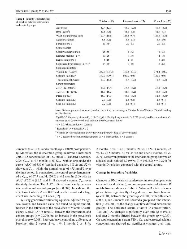
2 months (p = 0.031) and 6 months (p = 0.049) postoperative-ly. Moreover, the intervention group achieved a maximum25(OH)D concentration of 75.7 nmol/L (standard deviation,20.5; Cmax) at 4.7 months (1.6; Tmax) with an area under thecurve (AUC) of 339.6 (standard deviation, 103.2) and 52 %showed a Cmax within the normal range of >75 nmol/L overthe time period. In comparison, the control group demonstrat-ed a Cmax of 67.5 nmol/L (20.6) at 4.2 months (1.3) with anAUC of 261.6 (81.7) and 41 % showed a normal Cmax overthe study duration. The AUC differed significantly betweenintervention and control groups (p = 0.009). In addition, theeffect size Cohen’s d was 0.87 and can be considered a largeeffect size, according to Cohen [32].
By using generalized estimating equation, adjusted for age,sex, season, and baseline value, we found no significant dif-ference in the estimates of the prevalence of vitamin D suffi-ciency (25(OH)D ≥75 nmol/L) between the intervention andcontrol groups (p = 0.274), but an increase in the prevalenceover time (p = 0.008): intervention vs. control: no difference atbaseline; after 2 weeks, 2 vs. 1 %; 1 month, 5 vs. 3 %;
2 months, 6 vs. 3 %; 3 months, 24 vs. 15 %; 4 months, 23vs. 13 %; 5 months, 40 vs. 34 %; and after 6 months, 34 vs.22 %. Moreover, patients in the intervention group showed anadjusted odds ratio of 1.9 (95 % CI = 0.6, 5.9; p = 0.274) forvitamin D repletion compared with the control group.
Change in Secondary Variables
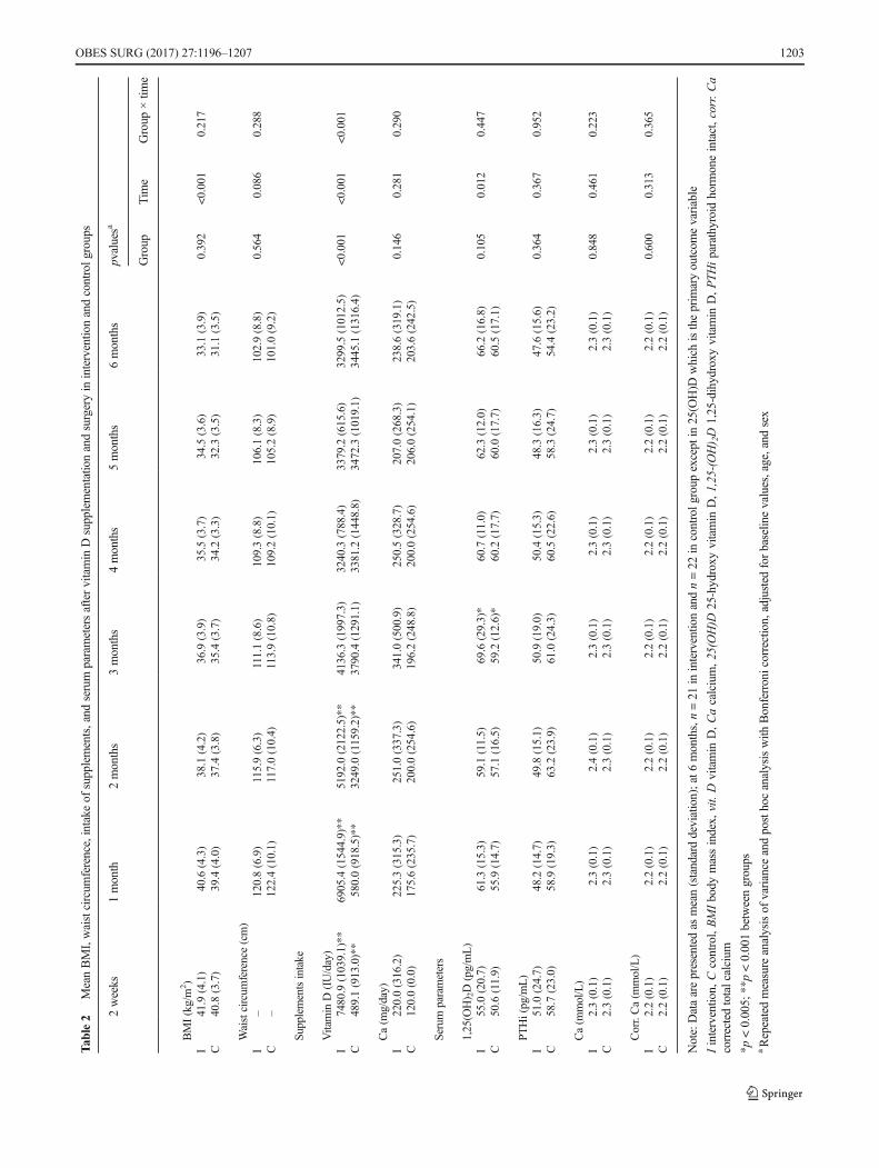
Changes in BMI, waist circumference, intake of supplements(vitamin D and calcium), and serum parameters of vitamin Dmetabolism are shown in Table 2. Vitamin D intake via sup-plementation significantly changed over time from baseline(p < 0.001) between the groups (p < 0.001) with differencesat 0.5, 1, and 2 months and showed a group and time interac-tion (p < 0.001), as the change over time differed between thegroups. The activated serum vitamin D concentration,1,25(OH)2D3, changed significantly over time (p = 0.012)and after 3 months differed between the groups (p = 0.050).Ca supplementation, serum PTHi, Ca, and corrected calciumconcentrations showed no significant changes over time,
Table 1 Patients’ characteristicsat baseline between interventionand control groups
Total (n = 50) Intervention (n = 25) Control (n = 25)
Age (years) 42.4 (12.7) 43.0 (12.6) 41.8 (13.0)
BMI (kg/m2) 43.8 (4.3) 44.6 (4.2) 42.9 (4.3)
Waist circumference (cm) 127.4 (10.6) 128.3 (9.7) 126.5 (11.5)
Number of drugs 5.8 (8.1) 5.8 (8.3) 5.8 (8.0)
Female (n (%)) 40 (80) 20 (80) 20 (80)
Comorbidities
Cardiovascular (n (%)) 28 (56) 13 (52) 15 (60)
Diabetes mellitus (n (%) 13 (26) 9 (36) 4 (18)
Depression (n (%)) 8 (16) 2 (8) 6 (24)
Significant liver fibrosis (n (%))a 14 (30) 9 (43) 5 (20)
Supplements intake
Vitamin D (IU/day)b 252.3 (675.2) 130.3 (407.3) 374.4 (856.7)
Calcium (mg/day)c 360.0 (339.4) 600.0 (0.0) 120.0 (0.0)
Time outside (h/week) 13.7 (11.1) 13.7 (10.0) 13.8 (12.3)
Serum parameters
25(OH)D (nmol/L) 39.0 (14.4) 38.8 (14.2) 39.3 (14.8)
1,25(OH)2D (pg/mL) 46.9 (16.2) 44.9 (14.2) 48.8 (17.9)
PTHi (pg/mL) 48.7 (14.3) 45.1 (14.7) 52.3 (13.3)*
Calcium (mmol/L) 2.3 (0.1) 2.3 (0.2) 2.3 (0.1)
Corr. Ca (mmol/L) 2.2 (0.1) 2.2 (0.1) 2.2 (0.1)
Note: Data are presented as mean (standard deviation) or percentages. T test or Mann-Whitney U test dependingon distribution
25(OH)D 25-hydroxy vitamin D, 1,25-(OH)2D 1,25-dihydroxy vitamin D, PTHi parathyroid hormone intact, Cacalcium, corr. Ca corrected total calcium, BMI body mass index
*p < 0.05 (intervention vs. control)a Significant liver fibrosis F ≥ 2bVitamin D via supplements before receiving the study drug of cholecalciferolc n = 2 received calcium supplementation (n = 1 intervention, n = 1 control)
OBES SURG (2017) 27:1196–1207 1201
between the study groups, and no group and time interactions.Dietary intake of energy, fat, carbohydrate, protein, calcium,and vitamin D is shown in Supplementary Material Table S2.The time spent outdoors (h/week; between 9 a.m. and 3 p.m. aday) was similar between the groups (intervention vs. control,0.7 h/week (95 % CI = −3.3, 4.7), p = 0.717), over the time(−1.5 h/week (95%CI = −6.6, 3.6), p = 0.345) and showed nogroup and time interactions (p = 0.877).
Impact of Vitamin D Supplementation in Patientswith Liver Fibrosis
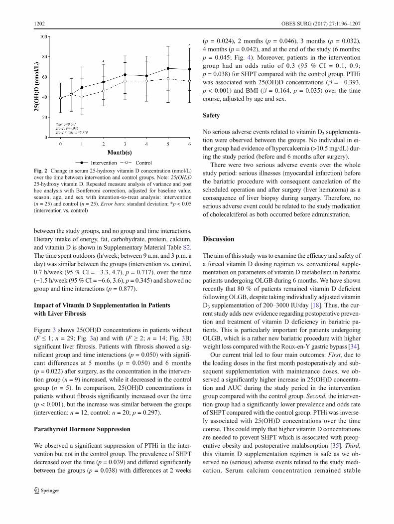
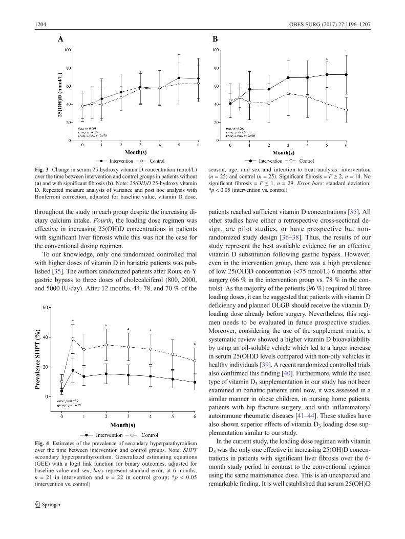
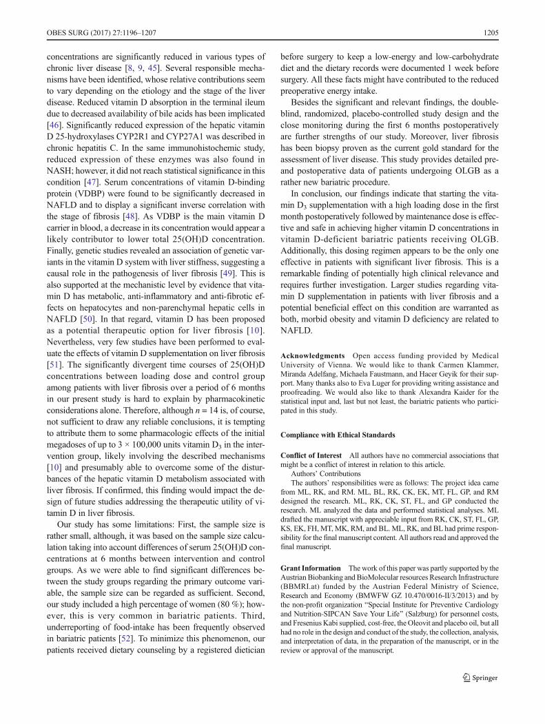
Figure 3 shows 25(OH)D concentrations in patients without(F ≤ 1; n = 29; Fig. 3a) and with (F ≥ 2; n = 14; Fig. 3B)significant liver fibrosis. Patients with fibrosis showed a sig-nificant group and time interactions (p = 0.050) with signifi-cant differences at 5 months (p = 0.050) and 6 months(p = 0.022) after surgery, as the concentration in the interven-tion group (n = 9) increased, while it decreased in the controlgroup (n = 5). In comparison, 25(OH)D concentrations inpatients without fibrosis significantly increased over the time(p < 0.001), but the increase was similar between the groups(intervention: n = 12, control: n = 20; p = 0.297).
Parathyroid Hormone Suppression
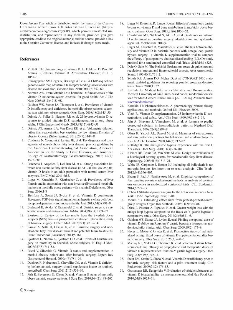
We observed a significant suppression of PTHi in the inter-vention but not in the control group. The prevalence of SHPTdecreased over the time (p = 0.039) and differed significantlybetween the groups (p = 0.038) with differences at 2 weeks
(p = 0.024), 2 months (p = 0.046), 3 months (p = 0.032),4 months (p = 0.042), and at the end of the study (6 months;p = 0.045; Fig. 4). Moreover, patients in the interventiongroup had an odds ratio of 0.3 (95 % CI = 0.1, 0.9;p = 0.038) for SHPT compared with the control group. PTHiwas associated with 25(OH)D concentrations (β = −0.393,p < 0.001) and BMI (β = 0.164, p = 0.035) over the timecourse, adjusted by age and sex.
Safety
No serious adverse events related to vitamin D3 supplementa-tion were observed between the groups. No individual in ei-ther group had evidence of hypercalcemia (>10.5 mg/dL) dur-ing the study period (before and 6 months after surgery).
There were two serious adverse events over the wholestudy period: serious illnesses (myocardial infarction) beforethe bariatric procedure with consequent cancelation of thescheduled operation and after surgery (liver hematoma) as aconsequence of liver biopsy during surgery. Therefore, noserious adverse event could be related to the study medicationof cholecalciferol as both occurred before administration.
Discussion
The aim of this study was to examine the efficacy and safety ofa forced vitamin D dosing regimen vs. conventional supple-mentation on parameters of vitamin D metabolism in bariatricpatients undergoing OLGB during 6 months. We have shownrecently that 80 % of patients remained vitamin D deficientfollowing OLGB, despite taking individually adjusted vitaminD3 supplementation of 200–3000 IU/day [18]. Thus, the cur-rent study adds new evidence regarding postoperative preven-tion and treatment of vitamin D deficiency in bariatric pa-tients. This is particularly important for patients undergoingOLGB, which is a rather new bariatric procedure with higherweight loss compared with the Roux-en-Y gastric bypass [34].
Our current trial led to four main outcomes: First, due tothe loading doses in the first month postoperatively and sub-sequent supplementation with maintenance doses, we ob-served a significantly higher increase in 25(OH)D concentra-tion and AUC during the study period in the interventiongroup compared with the control group. Second, the interven-tion group had a significantly lower prevalence and odds rateof SHPTcompared with the control group. PTHi was inverse-ly associated with 25(OH)D concentrations over the timecourse. This could imply that higher vitamin D concentrationsare needed to prevent SHPT which is associated with preop-erative obesity and postoperative malabsorption [35]. Third,this vitamin D supplementation regimen is safe as we ob-served no (serious) adverse events related to the study medi-cation. Serum calcium concentration remained stable
Fig. 2 Change in serum 25-hydroxy vitamin D concentration (nmol/L)over the time between intervention and control groups. Note: 25(OH)D25-hydroxy vitamin D. Repeated measure analysis of variance and posthoc analysis with Bonferroni correction, adjusted for baseline value,season, age, and sex with intention-to-treat analysis: intervention(n = 25) and control (n = 25). Error bars: standard deviation; *p < 0.05(intervention vs. control)
1202 OBES SURG (2017) 27:1196–1207
Tab
le2
MeanBMI,waistcircum
ference,intake
ofsupplements,and
serum
parametersaftervitamin
Dsupplementatio
nandsurgeryin
interventio
nandcontrolg
roups
2weeks
1month
2months
3months
4months
5months
6months
pvaluesa
Group
Tim
eGroup
×tim
e
BMI(kg/m
2)
I41.9(4.1)
40.6(4.3)
38.1(4.2)
36.9(3.9)
35.5(3.7)
34.5(3.6)
33.1(3.9)
0.392
<0.001
0.217
C40.8(3.7)
39.4(4.0)
37.4(3.8)
35.4(3.7)
34.2(3.3)
32.3(3.5)
31.1(3.5)
Waistcircum
ference(cm)
I–
120.8(6.9)
115.9(6.3)
111.1(8.6)
109.3(8.8)
106.1(8.3)
102.9(8.8)
0.564
0.086
0.288
C–
122.4(10.1)
117.0(10.4)
113.9(10.8)
109.2(10.1)
105.2(8.9)
101.0(9.2)
Supplementsintake
Vitamin
D(IU/day)
I7480.9(1039.1)**
6905.4(1544.9)**
5192.0(2122.5)**
4136.3(1997.3)
3240.3(788.4)
3379.2(615.6)
3299.5(1012.5)
<0.001
<0.001
<0.001
C489.1(913.0)**
580.0(918.5)**
3249.0(1159.2)**
3790.4(1291.1)
3381.2(1448.8)
3472.3(1019.1)
3445.1(1316.4)
Ca(m
g/day)
I220.0(316.2)
225.3(315.3)
251.0(337.3)
341.0(500.9)
250.5(328.7)
207.0(268.3)
238.6(319.1)
0.146
0.281
0.290
C120.0(0.0)
175.6(235.7)
200.0(254.6)
196.2(248.8)
200.0(254.6)
206.0(254.1)
203.6(242.5)
Serum
parameters
1,25(O
H) 2D(pg/mL)
I55.0(20.7)
61.3(15.3)
59.1(11.5)
69.6(29.3)*
60.7(11.0)
62.3(12.0)
66.2(16.8)
0.105
0.012
0.447
C50.6(11.9)
55.9(14.7)
57.1(16.5)
59.2(12.6)*
60.2(17.7)
60.0(17.7)
60.5(17.1)
PTHi(pg/m
L)
I51.0(24.7)
48.2(14.7)
49.8(15.1)
50.9(19.0)
50.4(15.3)
48.3(16.3)
47.6(15.6)
0.364
0.367
0.952
C58.7(23.0)
58.9(19.3)
63.2(23.9)
61.0(24.3)
60.5(22.6)
58.3(24.7)
54.4(23.2)
Ca(m
mol/L)
I2.3(0.1)
2.3(0.1)
2.4(0.1)
2.3(0.1)
2.3(0.1)
2.3(0.1)
2.3(0.1)
0.848
0.461
0.223
C2.3(0.1)
2.3(0.1)
2.3(0.1)
2.3(0.1)
2.3(0.1)
2.3(0.1)
2.3(0.1)
Corr.Ca(m
mol/L)
I2.2(0.1)
2.2(0.1)
2.2(0.1)
2.2(0.1)
2.2(0.1)
2.2(0.1)
2.2(0.1)
0.600
0.313
0.365
C2.2(0.1)
2.2(0.1)
2.2(0.1)
2.2(0.1)
2.2(0.1)
2.2(0.1)
2.2(0.1)
Note:Dataarepresentedas
mean(standarddeviation);at6
months,n=21
ininterventio
nandn=22
incontrolg
roup
except
in25(O
H)D
which
istheprim
aryoutcom
evariable
Iinterventio
n,Ccontrol,BMIbody
massindex,
vit.D
vitamin
D,Cacalcium,25(O
H)D
25-hydroxy
vitamin
D,1,25-(OH) 2D
1,25-dihydroxy
vitamin
D,PTH
iparathyroid
horm
oneintact,corr.C
acorrectedtotalcalcium
*p<0.005;
**p<0.001betweengroups
aRepeatedmeasure
analysisof
variance
andposthocanalysiswith
Bonferronicorrection,adjusted
forbaselin
evalues,age,and
sex
OBES SURG (2017) 27:1196–1207 1203
throughout the study in each group despite the increasing di-etary calcium intake. Fourth, the loading dose regimen waseffective in increasing 25(OH)D concentrations in patientswith significant liver fibrosis while this was not the case forthe conventional dosing regimen.
To our knowledge, only one randomized controlled trialwith higher doses of vitamin D in bariatric patients was pub-lished [35]. The authors randomized patients after Roux-en-Ygastric bypass to three doses of cholecalciferol (800, 2000,and 5000 IU/day). After 12 months, 44, 78, and 70 % of the
patients reached sufficient vitamin D concentrations [35]. Allother studies have either a retrospective cross-sectional de-sign, are pilot studies, or have prospective but non-randomized study design [36–38]. Thus, the results of ourstudy represent the best available evidence for an effectivevitamin D substitution following gastric bypass. However,even in the intervention group, there was a high prevalenceof low 25(OH)D concentration (<75 nmol/L) 6 months aftersurgery (66 % in the intervention group vs. 78 % in the con-trols). As the majority of the patients (96 %) required all threeloading doses, it can be suggested that patients with vitamin Ddeficiency and planned OLGB should receive the vitamin D3
loading dose already before surgery. Nevertheless, this regi-men needs to be evaluated in future prospective studies.Moreover, considering the use of the supplement matrix, asystematic review showed a higher vitamin D bioavailabilityby using an oil-soluble vehicle which led to a larger increasein serum 25(OH)D levels compared with non-oily vehicles inhealthy individuals [39]. A recent randomized controlled trialsalso confirmed this finding [40]. Furthermore, while the usedtype of vitamin D3 supplementation in our study has not beenexamined in bariatric patients until now, it was assessed in asimilar manner in obese children, in nursing home patients,patients with hip fracture surgery, and with inflammatory/autoimmune rheumatic diseases [41–44]. These studies havealso shown superior effects of vitamin D3 loading dose sup-plementation similar to our study.
In the current study, the loading dose regimen with vitaminD3 was the only one effective in increasing 25(OH)D concen-trations in patients with significant liver fibrosis over the 6-month study period in contrast to the conventional regimenusing the same maintenance dose. This is an unexpected andremarkable finding. It is well established that serum 25(OH)D
Fig. 3 Change in serum 25-hydroxy vitamin D concentration (nmol/L)over the time between intervention and control groups in patients without(a) and with significant fibrosis (b). Note: 25(OH)D 25-hydroxy vitaminD. Repeated measure analysis of variance and post hoc analysis withBonferroni correction, adjusted for baseline value, vitamin D dose,
season, age, and sex and intention-to-treat analysis: intervention(n = 25) and control (n = 25). Significant fibrosis = F ≥ 2, n = 14. Nosignificant fibrosis = F ≤ 1, n = 29. Error bars: standard deviation;*p < 0.05 (intervention vs. control)
Fig. 4 Estimates of the prevalence of secondary hyperparathyroidismover the time between intervention and control groups. Note: SHPTsecondary hyperparathyroidism. Generalized estimating equations(GEE) with a logit link function for binary outcomes, adjusted forbaseline value and sex; bars represent standard error; at 6 months,n = 21 in intervention and n = 22 in control group; *p < 0.05(intervention vs. control)
1204 OBES SURG (2017) 27:1196–1207
concentrations are significantly reduced in various types ofchronic liver disease [8, 9, 45]. Several responsible mecha-nisms have been identified, whose relative contributions seemto vary depending on the etiology and the stage of the liverdisease. Reduced vitamin D absorption in the terminal ileumdue to decreased availability of bile acids has been implicated[46]. Significantly reduced expression of the hepatic vitaminD 25-hydroxylases CYP2R1 and CYP27A1 was described inchronic hepatitis C. In the same immunohistochemic study,reduced expression of these enzymes was also found inNASH; however, it did not reach statistical significance in thiscondition [47]. Serum concentrations of vitamin D-bindingprotein (VDBP) were found to be significantly decreased inNAFLD and to display a significant inverse correlation withthe stage of fibrosis [48]. As VDBP is the main vitamin Dcarrier in blood, a decrease in its concentration would appear alikely contributor to lower total 25(OH)D concentration.Finally, genetic studies revealed an association of genetic var-iants in the vitamin D system with liver stiffness, suggesting acausal role in the pathogenesis of liver fibrosis [49]. This isalso supported at the mechanistic level by evidence that vita-min D has metabolic, anti-inflammatory and anti-fibrotic ef-fects on hepatocytes and non-parenchymal hepatic cells inNAFLD [50]. In that regard, vitamin D has been proposedas a potential therapeutic option for liver fibrosis [10].Nevertheless, very few studies have been performed to eval-uate the effects of vitamin D supplementation on liver fibrosis[51]. The significantly divergent time courses of 25(OH)Dconcentrations between loading dose and control groupamong patients with liver fibrosis over a period of 6 monthsin our present study is hard to explain by pharmacokineticconsiderations alone. Therefore, although n = 14 is, of course,not sufficient to draw any reliable conclusions, it is temptingto attribute them to some pharmacologic effects of the initialmegadoses of up to 3 × 100,000 units vitamin D3 in the inter-vention group, likely involving the described mechanisms[10] and presumably able to overcome some of the distur-bances of the hepatic vitamin D metabolism associated withliver fibrosis. If confirmed, this finding would impact the de-sign of future studies addressing the therapeutic utility of vi-tamin D in liver fibrosis.
Our study has some limitations: First, the sample size israther small, although, it was based on the sample size calcu-lation taking into account differences of serum 25(OH)D con-centrations at 6 months between intervention and controlgroups. As we were able to find significant differences be-tween the study groups regarding the primary outcome vari-able, the sample size can be regarded as sufficient. Second,our study included a high percentage of women (80 %); how-ever, this is very common in bariatric patients. Third,underreporting of food-intake has been frequently observedin bariatric patients [52]. To minimize this phenomenon, ourpatients received dietary counseling by a registered dietician
before surgery to keep a low-energy and low-carbohydratediet and the dietary records were documented 1 week beforesurgery. All these facts might have contributed to the reducedpreoperative energy intake.
Besides the significant and relevant findings, the double-blind, randomized, placebo-controlled study design and theclose monitoring during the first 6 months postoperativelyare further strengths of our study. Moreover, liver fibrosishas been biopsy proven as the current gold standard for theassessment of liver disease. This study provides detailed pre-and postoperative data of patients undergoing OLGB as arather new bariatric procedure.
In conclusion, our findings indicate that starting the vita-min D3 supplementation with a high loading dose in the firstmonth postoperatively followed bymaintenance dose is effec-tive and safe in achieving higher vitamin D concentrations invitamin D-deficient bariatric patients receiving OLGB.Additionally, this dosing regimen appears to be the only oneeffective in patients with significant liver fibrosis. This is aremarkable finding of potentially high clinical relevance andrequires further investigation. Larger studies regarding vita-min D supplementation in patients with liver fibrosis and apotential beneficial effect on this condition are warranted asboth, morbid obesity and vitamin D deficiency are related toNAFLD.
Acknowledgments Open access funding provided by MedicalUniversity of Vienna. We would like to thank Carmen Klammer,Miranda Adelfang, Michaela Faustmann, and Hacer Geyik for their sup-port. Many thanks also to Eva Luger for providing writing assistance andproofreading. We would also like to thank Alexandra Kaider for thestatistical input and, last but not least, the bariatric patients who partici-pated in this study.
Compliance with Ethical Standards
Conflict of Interest All authors have no commercial associations thatmight be a conflict of interest in relation to this article.
Authors’ ContributionsThe authors’ responsibilities were as follows: The project idea came
from ML, RK, and RM. ML, BL, RK, CK, EK, MT, FL, GP, and RMdesigned the research. ML, RK, CK, ST, FL, and GP conducted theresearch. ML analyzed the data and performed statistical analyses. MLdrafted the manuscript with appreciable input from RK, CK, ST, FL, GP,KS, EK, FH, MT, MK, RM, and BL. ML, RK, and BL had prime respon-sibility for the final manuscript content. All authors read and approved thefinal manuscript.
Grant Information The work of this paper was partly supported by theAustrian Biobanking and BioMolecular resources Research Infrastructure(BBMRI.at) funded by the Austrian Federal Ministry of Science,Research and Economy (BMWFW GZ 10.470/0016-II/3/2013) and bythe non-profit organization BSpecial Institute for Preventive Cardiologyand Nutrition-SIPCAN Save Your Life^ (Salzburg) for personnel costs,and Fresenius Kabi supplied, cost-free, the Oleovit and placebo oil, but allhad no role in the design and conduct of the study, the collection, analysis,and interpretation of data, in the preparation of the manuscript, or in thereview or approval of the manuscript.
OBES SURG (2017) 27:1196–1207 1205
Open Access This article is distributed under the terms of the CreativeCommons At t r ibut ion 4 .0 In te rna t ional License (h t tp : / /creativecommons.org/licenses/by/4.0/), which permits unrestricted use,distribution, and reproduction in any medium, provided you giveappropriate credit to the original author(s) and the source, provide a linkto the Creative Commons license, and indicate if changes were made.
References
1. Vieth R. The pharmacology of vitamin D. In: Feldman D, Pike JW,Adams JS, editors. Vitamin D. Amsterdam: Elsevier; 2011. p.1058–61.
2. Ramagopalan SV, Heger A, Berlanga AJ, et al. A ChIP-seq definedgenome-wide map of vitamin D receptor binding: associations withdisease and evolution. Genome Res. 2010;20(10):1352–60.
3. Norman AW. From vitamin D to hormone D: fundamentals of thevitamin D endocrine system essential for good health. Am J ClinNutr. 2008;88(2):491S–9S.
4. Goldner WS, Stoner JA, Thompson J, et al. Prevalence of vitaminD insufficiency and deficiency in morbidly obese patients: a com-parison with non-obese controls. Obes Surg. 2008;18(2):145–50.
5. Drincic A, Fuller E, Heaney RP, et al. 25-hydroxyvitamin D re-sponse to graded vitamin D(3) supplementation among obeseadults. J Clin Endocrinol Metab. 2013;98(12):4845–51.
6. Drincic AT, Armas LA, Van Diest EE, et al. Volumetric dilution,rather than sequestration best explains the low vitamin D status ofobesity. Obesity (Silver Spring). 2012;20(7):1444–8.
7. Chalasani N, Younossi Z, Lavine JE, et al. The diagnosis and man-agement of non-alcoholic fatty liver disease: practice guideline bythe American Gastroenterological Association, AmericanAssociation for the Study of Liver Diseases, and AmericanCollege of Gastroenterology. Gastroenterology. 2012;142(7):1592–609.
8. Barchetta I, Angelico F, Del Ben M, et al. Strong association be-tween non alcoholic fatty liver disease (NAFLD) and low 25(OH)vitamin D levels in an adult population with normal serum liverenzymes. BMC Med. 2011;9:85.
9. Luger M, Kruschitz R, Kienbacher C, et al. Prevalence of liverfibrosis and its association with non-invasive fibrosis andmetabolicmarkers in morbidly obese patients with vitaminD deficiency. ObesSurg. 2016;1-8
10. Beilfuss A, Sowa JP, Sydor S, et al. Vitamin D counteractsfibrogenic TGF-beta signalling in human hepatic stellate cells bothreceptor-dependently and independently. Gut. 2015;64(5):791–9.
11. Buchwald H, Avidor Y, Braunwald E, et al. Bariatric surgery: a sys-tematic review and meta-analysis. JAMA. 2004;292(14):1724–37.
12. Sjostrom L. Review of the key results from the Swedish obesesubjects (SOS) trial—a prospective controlled intervention studyof bariatric surgery. J Intern Med. 2013;273(3):219–34.
13. Sasaki A, Nitta H, Otsuka K, et al. Bariatric surgery and non-alcoholic fatty liver disease: current and potential future treatments.Front Endocrinol (Lausanne). 2014;5:164.
14. Sjostrom L, Narbro K, Sjostrom CD, et al. Effects of bariatric sur-gery on mortality in Swedish obese subjects. N Engl J Med.2007;357(8):741–52.
15. Bacci V, Silecchia G. Vitamin D status and supplementation inmorbid obesity before and after bariatric surgery. Expert RevGastroenterol Hepatol. 2010;4(6):781–94.
16. Ducloux R, Nobecourt E, Chevallier JM, et al. Vitamin D deficien-cy before bariatric surgery: should supplement intake be routinelyprescribed? Obes Surg. 2011;21(5):556–60.
17. Fish E, Beverstein G, Olson D, et al. Vitamin D status of morbidlyobese bariatric surgery patients. J Surg Res. 2010;164(2):198–202.
18. LugerM,Kruschitz R, Langer F, et al. Effects of omega-loop gastricbypass on vitamin D and bone metabolism in morbidly obese bar-iatric patients. Obes Surg. 2015;25(6):1056–62.
19. Chakhtoura MT, Nakhoul N, Akl EA, et al. Guidelines on vitaminD replacement in bariatric surgery: identification and systematicappraisal. Metabolism. 2016;4
20. Luger M, Kruschitz R, Marculescu R, et al. The link between obe-sity and vitamin D in bariatric patients with omega-loop gastricbypass surgery—a vitamin D supplementation trial to comparethe efficacy of postoperative cholecalciferol loading (LOAD): studyprotocol for a randomized controlled trial. Trials. 2015;16(1):328.
21. Dale O, Salo M. The Helsinki Declaration, research guidelines andregulations: present and future editorial aspects. Acta AnaesthesiolScand. 1996;40(7):771–2.
22. Schulz KF, Altman DG, Moher D, et al. CONSORT 2010 state-ment: updated guidelines for reporting parallel group randomisedtrials. Trials. 2010;11:32.
23. Institute for Medical Informatics Statistics and DocumentationMedical University of Graz. Web-based patient randomization ser-vice for Multi-Center Clinical Trials. [22.10.2014]. Available from:www.randomizer.at/.
24. Kenakin TP. Pharmacokinetics. A pharmacology primer: theory,applications, and methods. Oxford UK: Elsevier; 2009.
25. Vieth R. Vitamin D supplementation, 25-hydroxyvitamin D con-centrations, and safety. Am J Clin Nutr. 1999;69(5):842–56.
26. Jain A, Bhayana S, Vlasschaert M, et al. A formula to predictcorrected calcium in haemodialysis patients. Nephrol DialTransplant. 2008;23(9):2884–8.
27. Glanz K, Yaroch AL, Dancel M, et al. Measures of sun exposureand sun protection practices for behavioral and epidemiologic re-search. Arch Dermatol. 2008;144(2):217–22.
28. Rutledge R. The mini-gastric bypass: experience with the first 1,274 cases. Obes Surg. 2001;11(3):276–80.
29. Kleiner DE, Brunt EM, VanNatta M, et al. Design and validation ofa histological scoring system for nonalcoholic fatty liver disease.Hepatology. 2005;41(6):1313–21.
30. White IR, Carpenter J, Horton NJ. Including all individuals is notenough: lessons for intention-to-treat analysis. Clin Trials.2012;9(4):396–407.
31. Zhang S, Paul J, Nantha-Aree M, et al. Empirical comparison offour baseline covariate adjustment methods in analysis of continu-ous outcomes in randomized controlled trials. Clin Epidemiol.2014;6:227–35.
32. Cohen J. Statistical power analysis for the behavioral sciences. NewYork, USA: Psychology Press; 1988.
33. Morris SB. Estimating effect sizes from pretest-posttest-controlgroup designs. Organ Res Methods. 2008;11(2):364–86.
34. Disse E, Pasquer A, Espalieu P, et al. Greater weight loss with theomega loop bypass compared to the Roux-en-Y gastric bypass: acomparative study. Obes Surg. 2014;24(6):841–6.
35. GoldnerWS, Stoner JA, Lyden E, et al. Finding the optimal dose ofvitamin D following Roux-en-Y gastric bypass: a prospective, ran-domized pilot clinical trial. Obes Surg. 2009;19(2):173–9.
36. Flores L, Moize V, Ortega E, et al. Prospective study of individu-alized or high fixed doses of vitamin D supplementation after bar-iatric surgery. Obes Surg. 2015;25(3):470–6.
37. Mahlay NF, Verka LG, Thomsen K, et al. Vitamin D status beforeRoux-en-Y and efficacy of prophylactic and therapeutic doses ofvitamin D in patients after Roux-en-Y gastric bypass surgery. ObesSurg. 2009;19(5):590–4.
38. Stein EM, Strain G, Sinha N, et al. Vitamin D insufficiency prior tobariatric surgery: risk factors and a pilot treatment study. ClinEndocrinol. 2009;71(2):176–83.
39. Grossmann RE, Tangpricha V. Evaluation of vehicle substances onvitamin D bioavailability: a systematic review. Mol Nutr Food Res.2010;54(8):1055–61.
1206 OBES SURG (2017) 27:1196–1207
40. Wolf E, Utech M, Stehle P, et al. Oral high-dose vitamin D dis-solved in oil raised serum 25-hydroxy-vitamin D to physiologicallevels in obese patients after sleeve gastrectomy—a double-blind,randomized, and placebo-controlled trial. Obes Surg. 2016;26(8):1821–9.
41. Mak JC, Klein LA, Finnegan T, et al. An initial loading-dose vita-min D versus placebo after hip fracture surgery: baseline character-istics of a randomized controlled trial (REVITAHIP). BMCGeriatr.2014;14:101.
42. Radhakishun NN, van Vliet M, Poland DC, et al. Efficacy andtolerability of a high loading dose (25,000 IU weekly) vitamin D3supplementation in obese children with vitamin D insufficiency/deficiency. Horm Res Paediatr. 2014;82(2):103–6.
43. Sainaghi PP, Bellan M, Nerviani A, et al. Superiority of a highloading dose of cholecalciferol to correct hypovitaminosis d in pa-tients with inflammatory/autoimmune rheumatic diseases. JRheumatol. 2013;40(2):166–72.
44. Wijnen H, Salemink D, Roovers L, et al. Vitamin D supplementa-tion in nursing home patients: randomized controlled trial of stan-dard daily dose versus individualized loading dose regimen. DrugsAging. 2015;32(5):371–8.
45. Lim LY, Chalasani N. Vitamin d deficiency in patients with chronicliver disease and cirrhosis. Curr Gastroenterol Rep. 2012;14(1):67–73.
46. Miroliaee A, Nasiri-Toosi M, Khalilzadeh O, et al. Disturbances ofparathyroid hormone-vitamin D axis in non-cholestatic chronic liv-er disease: a cross-sectional study. Hepatol Int. 2010;4(3):634–40.
47. Barchetta I, Carotti S, Labbadia G, et al. Liver vitamin D receptor,CYP2R1, and CYP27A1 expression: relationship with liver histologyand vitamin D3 levels in patients with nonalcoholic steatohepatitis orhepatitis C virus. Hepatology. 2012;56(6):2180–7.
48. Miller MH, Walsh SV, Atrih A, et al. Serum proteome of nonalco-holic fatty liver disease: a multimodal approach to discovery ofbiomarkers of nonalcoholic steatohepatitis. J GastroenterolHepatol. 2014;29(10):1839–47.
49. Grunhage F, Hochrath K, Krawczyk M, et al. Common geneticvariation in vitamin D metabolism is associated with liver stiffness.Hepatology. 2012;56(5):1883–91.
50. Eliades M, Spyrou E. Vitamin D: a new player in non-alcoholicfatty liver disease? World J Gastroenterol. 2015;21(6):1718–27.
51. Song BJ, Rockey DC. Status of research on vitamin D supplemen-tation in treating or preventing liver fibrosis. Liver Int. 2013;33(5):653–5.
52. Quesada KR, Novais PF, Detregiachi CR, et al. Comparative anal-ysis of approaches for assessing energy intake underreporting byfemale bariatric surgery candidates. J Am Coll Nutr. 2014;33(2):155–62.
OBES SURG (2017) 27:1196–1207 1207