Embed Size (px)
Citation preview
Visual Psychophysics and Physiological Optics
Visual Acuity, Crowding, and Stereo-Vision Are Linked inChildren with and without Amblyopia
John A. Greenwood,1–4 Vijay K. Tailor,2,5 John J. Sloper,2,5 Anita J. Simmers,6 Peter J. Bex,7
and Steven C. Dakin1,2
PURPOSE. During development, the presence of strabismus andanisometropia frequently leads to amblyopia, a visual disordercharacterized by interocular acuity differences. Althoughadditional deficits in contrast sensitivity, crowding (theimpaired recognition of closely spaced objects), and stereo-acuity are common, the relationship between these abilities isunclear.
METHODS. We measured the covariation between these fourabilities in children 4 to 9 years of age (n¼72) with strabismus,anisometropia, or mixed strabismus/anisometropia, and unaf-fected controls. Children reported the orientation of a target (amodified ‘‘Pac-Man,’’ similar to Landolt-C stimuli) using four‘‘ghosts’’ as references. Using a modified staircase procedurewe measured threshold size (acuity), contrast detection, fovealcrowding (the minimum separation between target and ghost-flankers supporting accurate identification), and stereoacuity(with random-dot stereogram ghosts).
RESULTS. Group averages revealed significant interoculardifferences (IODs) in acuity for all three clinical groups (0.2–0.3 log minutes), and significant crowding IODs for thestrabismic and mixed groups (0.6 and 0.48, respectively).Nonetheless, crowding IODs were correlated with acuity IODsin all four groups (r values between 0.43 and 0.59 and P <0.05; P¼0.07 in the mixed group). Similarly, the occurrence ofstereo-blindness (most common in strabismic and mixedgroups) was associated with a significant increase in IODs forboth acuity and crowding (each P < 0.05). No correlationswere found with contrast detection.
CONCLUSIONS. Our results demonstrate an association betweenIODs in acuity and crowding and, furthermore, between theseIODs and the presence of stereo-vision. We suggest that thedeficits derived from strabismus and anisometropia lay along acontinuum with abilities observed during normal develop-ment. (Invest Ophthalmol Vis Sci. 2012;53:7655–7665) DOI:10.1167/iovs.12-10313
It is well known that abnormal visual experience can have aprofound effect on the development of the visual system. In
the case of children with anisometropia (unequal refractiveerror) and/or strabismus (ocular misalignment), a commonoutcome is unilateral amblyopia, a developmental visualdisorder whose defining feature is reduced acuity in one eyethat persists despite optical correction.1,2 The development ofamblyopia is also associated with a range of additional deficits,including anomalies in contrast detection,2–6 elevated visualcrowding,7–9 and reduced stereoacuity or complete stereo-blindness.2,10 Although each is becoming increasingly wellunderstood in isolation, little is known about the relationshipbetween these deficits, particularly during childhood whenstrabismus and anisometropia first develop.
Aside from the definitive acuity deficits, amblyopia is mostfrequently associated with deficits in stereo-vision. This canrange from an elevation in stereoacuity thresholds, themagnitude of which correlates with the interocular differencein acuity,10–12 to complete stereo-blindness, which is particu-larly common in strabismus2,13,14 and cases of severeanisometropia.2,10,12,15 The link between these deficits inacuity and stereo-vision has been attributed to altered neuralselectivities within cortical area V1: the abnormal visualexperience that causes amblyopia may shift both the selectivityof monocular neurons away from the amblyopic eye andreduce the number of binocular neurons.1,2,16–18 Improve-ments in visual acuity resulting from binocular therapy19,20
further suggest that there may be a causal link between acuityand stereo-vision, although a similar training regime focused onstereoacuity did not improve acuity thresholds.21 The relation-ship between these impairments in stereo-vision and otheramblyopic deficits is unknown.
A more complex pattern of deficits has been observed forachromatic contrast sensitivity. Although significant impair-ments in contrast sensitivity have been observed in theamblyopic eye of adults,4–6 amblyopic children may showmilder deficits.3 Additionally, although the amblyopic eye ofanisometropes typically supports higher contrast-detectionthresholds than does the fellow eye, in strabismus there havebeen reports of improved contrast-detection thresholds whenobservers use their amblyopic eye.2,6 It is possible that thesechanges reflect a redeployment of binocular cells as monocularcells,2 which may suggest a relationship between theseanomalies in contrast detection and the stereo-deficits de-scribed above. The variation in contrast-detection thresholds
From the 1UCL Institute of Ophthalmology, University CollegeLondon, London, United Kingdom; the 2National Institute for HealthResearch Biomedical Research Centre for Ophthalmology at Moor-fields Eye Hospital National Health Service Foundation Trust,London, United Kingdom; 3Laboratoire Psychologie de la Percep-tion, Universite Paris Descartes, Sorbonne Paris Cite, Paris, France;4Centre National de la Recherche Scientifique, UMR 8158, Paris,France; 5Strabismus and Paediatric Service, Moorfields Eye Hospital,London, United Kingdom; 6Vision Sciences, Department of LifeSciences, Glasgow Caledonian University, Glasgow, United King-dom; and the 7Schepens Eye Research Institute, Harvard MedicalSchool, Boston, Massachusetts.
Supported by the Special Trustees of Moorfields, the UKDepartment of Health through the award made by the NationalInstitute for Health Research to Moorfields Eye Hospital and UCLInstitute of Ophthalmology for a Biomedical Research Centre forOphthalmology, and a Marie Curie Intra-European Fellowship (JAG).
Submitted for publication June 1, 2012; revised August 14 andSeptember 29, 2012; accepted October 5, 2012.
Disclosure: J.A. Greenwood, None; V.K. Tailor, None; J.J.Sloper, None; A.J. Simmers, None; P.J. Bex, None; S.C. Dakin,None
Corresponding author: John A. Greenwood, Laboratoire Psy-chologie de la Perception, Universite Paris Descartes, Paris, France;[email protected].
Investigative Ophthalmology & Visual Science, November 2012, Vol. 53, No. 12
Copyright 2012 The Association for Research in Vision and Ophthalmology, Inc. 7655

also suggests that the nature of these effects depends on boththe progression and the type of amblyopia.
Finally, a particularly striking functional consequence ofamblyopia is an elevation in crowding, the impairment inobject recognition that occurs when an otherwise visible targetobject is flanked by similar elements.7,9,22 In ‘‘normal’’ adultvision, crowding is widely viewed as a ‘‘compulsory pooling’’of target and flanker signals23–25 that is strongest in theperipheral field and minimal in the fovea.9,26 In amblyopia,particularly when accompanied by strabismus, crowding isclearly evident in the fovea and can extend over spatial extentssimilar to crowding in the typical periphery.7–9 Elevated fovealcrowding has also been observed in typically developingchildren,27–29 which may persist as late as 11 years.29 Indeed,linear (so-called crowded) letter charts are generally consideredbetter detectors of amblyopia in children than isolatedletters.28,30,31 The relationship between these three instancesof crowding—in the ‘‘normal’’ periphery, the developing fovea,and the amblyopic fovea—is currently unclear. Many theorieshave been proposed to account for crowding,9 with one viewdescribing both peripheral and amblyopic forms as theconsequence of a neural undersampling of the visual field.8
As with the mechanisms underlying stereo-deficits, thissuggests a potential link with acuity loss. Accordingly, somerelation has been reported between acuity and crowding inadults,7,8,32 although others report that its magnitude exceedsthe scale of acuity losses.33,34 This relationship is yet to beexamined in children, and the relationship between crowdingand other amblyopic deficits is unknown.
These four deficits (acuity, contrast detection, crowding,and stereo-vision) are not the only functional consequences ofamblyopia, but the similarities in their proposed mechanismsled us to consider how they might covary across individuals. Inparticular, if each were indeed linked with alterations in eitherthe balance of monocular cells responding to each eye, or theproportion of binocular cells, then we may expect them tocovary in their magnitude. The aim of the present study wasthus to examine the relationship between acuity, contrastdetection, crowding, and stereoacuity in children with andwithout strabismus and/or anisometropia. We further sought toequate stimulus and task demands across these tasks andconsequently developed a novel paradigm, tailored to children,that involves simple characters in an easy-to-understand andengaging context.
METHODS
Participants
We examined 72 children, between 4 and 9 years of age (54–107
months; mean¼ 80.7), divided into four groups: those with strabismus
(n ¼ 18), anisometropia (n ¼ 16), or mixed strabismus/anisometropia
(n ¼ 19), and controls (n ¼ 19). All were tested at the Children’s Eye
Centre in Moorfields Eye Hospital (London, UK).
Children in the control group were selected to have acuity better
than 0.10 logMAR (logarithm of the minimum angle of resolution), in
the absence of any preexisting visual deficits. Inclusion in the
strabismic group was on the basis of manifest heterotropia and an
interocular refractive difference less than 1 diopter in any meridian.
Conversely, the anisometropic group had an interocular refractive
difference of 1 diopter or more in any meridian, without significant
heterotropia. Children with both a manifest heterotropia and
interocular refractive differences greater than 1 diopter were included
in the mixed group. There was no requirement for the presence of
amblyopia in the three ‘‘clinical’’ groups because we sought to assess a
range of visual abilities. We also sought a mix of stereo-vision abilities
for each group, and thus the observed frequencies of stereo-blindness
for these groups should not be taken as indicative of the overall
population. Children with paralytic or restrictive strabismus, as well as
those with additional visual deficits (e.g., cataracts or macular
dystrophies) or neurologic deficits (e.g., dyslexia or autism), were
excluded. The orthoptic details for all children are presented in
Supplemental Tables S1 to S4 (see Supplementary Material and
Supplementary Tables S1–S4, http://www.iovs.org/lookup/suppl/
doi:10.1167/iovs.12-10313/-/DCSupplemental).
Pretesting
All children underwent a full orthoptic assessment, including logMAR
acuity (with Thompson v2000 software; Thompson Software Solutions,
Herts, UK), TNO stereoacuity testing, ocular motility, binocular
function (prism fusion range), and cover tests for near and distance
fixation. The angle of any heterotropia or heterophoria was measured
by prism cover test. Where required, cycloplegic refraction was
performed within the 6 months prior to involvement in our study (but
never immediately prior to our experiments).
Apparatus
Experiments were run using a commercial software program (MATLAB;
The MathWorks, Ltd., Cambridge, UK) on a Dell PC (Dell, Round Rock,
TX) running PsychToolbox.35,36 Stimuli were presented on a liquid
crystal display monitor (SyncMaster 2233RZ LCD monitor; Samsung
Electronics, Seoul, South Korea), with 1680 3 1050 pixel resolution
and 120-Hz refresh rate. The monitor was calibrated using a
spectrophotometer (Konica Minolta Sensing Americas, Ramsey, NJ)
and linearized in software, giving a maximum luminance of 92.8 cd/m2.
Because these monitors allow only 6-bit contrast resolution, bit-
stealing37 was used to obtain 442 brightness levels (approximately 8.8
bits).
Children wore stereo-shutter glasses (nVidia Corp., Santa Clara,
CA), which alternated at 120 Hz (delivering images to each eye at 60
Hz) and were custom-mounted in a children’s ski-mask frame to give a
comfortable fit over spectacles (Fig. 1A). Children viewed the stimuli
from 3 m, wearing their full correction (as required), and made verbal
responses that were recorded by the experimenter using the computer
keyboard. To aid in the psychophysical tasks (e.g., for acuity: ‘‘which
color ghost was Vac-Man facing?’’), large reference pictures of the
ghosts were attached to the monitor edges. This allowed children to
report either the location of a ghost or, if preferred, its color.
Stimuli and Procedures
Overall, our tasks involved five video-game characters. The centrally
located target stimulus, known as Visual Acuity Man (Vac-Man; see Fig.
1A), was a circle with a horizontal bar missing from one half of the
center, similar to a ‘‘filled-in’’ Landolt C. The ‘‘mouth’’ width was equal
to one-fifth the stimulus diameter, as in the Sloan alphabet. Four ghost
characters were also present, acting either as color aids in the
identification of Vac-Man’s orientation (‘‘which color ghost was Vac-
Man facing?’’) or achromatic flanker stimuli in the crowding task. The
width of each ghost’s ‘‘leg’’ gaps was also one-fifth the stimulus
diameter.
Each child completed four tasks (acuity, contrast, crowding, and
stereoacuity) twice. The first three tasks involved monocular presen-
tation (once to each eye), whereas the stereoacuity task was performed
once with random-pixel arrays and once with additional monocular
contours. Children who did not complete all eight tasks were excluded
and are not included in the tallies above (three were excluded in this
way).
All children began with the acuity task. Here, Vac-Man was
presented in the center of the display and rendered in black at 50%
Weber contrast against a midgray background. Four ghosts moved
slowly along the monitor boundaries (approximately 2.2–4.48 from the
center), each with distinct colors (red to the right, green above, blue
7656 Greenwood et al. IOVS, November 2012, Vol. 53, No. 12
left, and orange below; see Fig. 1A). This combination of target-flanker
separation, flanker-color, and flanker-motion minimized the chance of
any crowding between target and ghosts.9,22,38,39
Children were required to indicate which of the four ghosts Vac-
Man was facing (four-alternative forced choice), because this was the
ghost ‘‘he wished to eat.’’ These responses (based on the color of
clearly individuated characters) were designed to avoid both left–right
response confusions40 and source confusions41,42 as to which element
in the multielement display was the target. Normal color-naming
abilities were checked using our stimuli, prior to running the
experiment. Feedback was given after each trial via brief animations
(Vac-Man smiled or frowned), with every third correct response giving
longer animations in which a color-rendered Vac-Man ‘‘ate’’ the correct
ghost (Fig. 1B; see also Supplementary Material and Supplementary
Movie S1, http://www.iovs.org/lookup/suppl/doi:10.1167/iovs.
12-10313/-/DCSupplemental).
Acuity thresholds were assessed by varying the size of Vac-Man (and
thus the visibility of the ‘‘mouth’’) using a QUEST algorithm staircase
procedure43 that converged on 62.5% correct performance. Incorrect
responses resulted in an increase in target size and correct responses in
a decrease. Three modifications were made to the QUEST routine to
increase its suitability for testing children. First, children were given
three practice trials with a target mouth-size of 4 minutes of arc to
begin each task (although this increased if these trials were failed).
Second, to minimize the frustration arising from adaptive procedures
placing many trials near threshold, we presented easier catch trials on
every fifth trial with stimuli scaled to double the current threshold
estimate. Third, to reduce the time required to estimate thresholds, we
added an exit criterion: if the SD of the estimated threshold for the past
eight trials was less than 0.03 log units (a value deemed reliable during
pilot tests), the experimenter was given the option of exiting the task.
Otherwise, the QUEST procedure terminated after 32 trials (plus
practice). The average number of trials required to reach threshold on
the acuity task was 14.7, not including practice.
Children completed the acuity task once for each eye, with
monocular presentation controlled using the stereo-shutter glasses. For
children in the control group, their left or right eye was randomly
selected to begin; those in clinical groups always began with their
fellow eye to ensure comprehension. Although acuity was always
completed first, subsequent tests were administered in random order.
For all tasks following the acuity measurement, Vac-Man and the ghost-
flankers were presented at sizes equal to 2.5-fold the size-threshold
(separately for each eye) to ensure their visibility.
For the contrast-detection task, Vac-Man was again presented in the
center of the display, surrounded by four distinctly colored ghosts, and
children indicated the ghost that Vac-Man was facing (see Supplemen-
tary Material and Supplementary Movie S2, http://www.iovs.org/
lookup/suppl/doi:10.1167/iovs.12-10313/-/DCSupplemental). QUEST
FIGURE 1. Apparatus and stimuli. (A) Stimuli were viewed through stereoscopic shutter glasses mounted in a children’s ski mask (inset) andpresented on a 3D-compatible monitor viewed at 3 m. An example trial of the acuity/contrast task is depicted. Children reported the color of theghost that Vac-Man was facing. Colored cards were present on the monitor edges for reference. (B) An example frame from the ‘‘reward animation’’(see Supplementary Material and Supplementary Movies S1–S4, http://www.iovs.org/lookup/suppl/doi:10.1167/iovs.12-10313/-/DCSupplemental).(C) Depiction of the crowding stimuli. Ghosts were rendered achromatically and presented at random orientations at a fixed separation from Vac-Man. The task was as in (A), with the reference cards assisting color memory. (D) An example frame from the stereo task with monocular ‘‘shadow’’contours, representing input to one of the eyes. Children were required to help Vac-Man find the ghost that ‘‘popped out’’ in depth. (E) An exampleghost from the stereo task with a monocular ‘‘shadow,’’ viewable with red–green anaglyphs (red filter to the right eye; actual stimuli wereachromatic). (F) An example ghost from the random-check stereo task, with a disparity offset defined purely by the random-check elements.
IOVS, November 2012, Vol. 53, No. 12 Acuity, Crowding, Stereo-Vision, and Amblyopia 7657
procedures were similar to the acuity task, albeit modulating Weber
contrast instead of size, beginning at 40% Weber contrast.
For the crowding task, Vac-Man and ghost elements were
achromatic and dark against the midgray background at 50% Weber
contrast (see Fig. 1C). The ghost-flankers were rendered without their
eyes and presented at random orientations. The target and flanker
stimuli were thus sufficiently similar to induce crowding9,38; indeed,
these stimuli produced strong crowding in the periphery of unaffected
adults during pilot testing. Ghost-flankers were positioned in each
cardinal direction at a given separation (determined by QUEST) from
the centrally located Vac-Man (see Supplementary Material and
Supplementary Movie S3, http://www.iovs.org/lookup/suppl/doi:10.
1167/iovs.12-10313/-/DCSupplemental). As before, children indicated
which ghost Vac-Man was facing, using the color of the ghosts that was
also available on reference cards at the monitor edges. As above, these
distinct character ‘‘identities’’ were intended to avoid poor perfor-
mance arising from source confusions,41,42 rather than an integrative
crowding process. QUEST operated as before, but now altered the
center-to-center separation between Vac-Man and ghost-flankers to
converge on the minimum/critical spacing leading to accurate
identification. Because crowding is largely insensitive to stimulus
size,26,44,45 critical spacing is a more reliable measure of crowding than
varying size at a fixed separation. Target-flanker separation began at six
times the Vac-Man radius. If identification was successful until target
and flanker elements were abutting, QUEST continued for three trials
and exited if all were correct, recording a zero value.
Note that these zero values reflect ceiling performance only for our
element sizes (scaled to 2.53 the acuity threshold for each child). It is
possible that a reduction in element size would reveal a small degree of
crowding in these cases by allowing smaller interelement separations.
However, because performance under such conditions is limited
largely by contrast masking,46 which operates at or near the resolution
limit, we did not attempt further testing with the very small stimuli
required to yield reliable estimates of performance. We also deemed
the demands of this task (e.g., very high levels of fixation stability) to
be beyond the children being tested. Instead, we interpret these zero
values as an indication of the functional absence of foveal crowding.
Finally, we measured stereoacuity both with and without the
presence of a monocular ‘‘shadow’’ contour. Ghost stimuli were
rendered as random-check stereograms within a circular region. The
target was now one of the four ghosts, presented with binocular
disparity, whereas the others lay in the zero-disparity plane. An
example screen frame is depicted in Figure 1D (also see Supplementary
Material and Supplementary Movie S4, http://www.iovs.org/lookup/
suppl/doi:10.1167/iovs.12-10313/-/DCSupplemental). Ghosts (without
eyes) were defined by a region of checks that were correlated in both
left and right eyes, with some displacement to give binocular disparity
(as rendered for red/green anaglyphs in Fig. 1E). Surrounding checks
were the same in each eye and random checks were used to fill the
region left by interocular displacements. The random checks were light
or dark squares with 50% Weber contrast.
In the ‘‘contour-based’’ stereograms, ghosts were additionally cued
by a 25% contrast decrement on top of the existing pixel modulations
(as in Fig. 1E), which followed the interocular correlation of the
constituent pixels. Prior studies have shown that contour information
may provide improvements to stereo-vision over purely random-pixel–
based stimuli,47–50 although it is not clear whether this reflects the use
of additional monocular ‘‘cues’’ to the task.51,52 In our task, this
monocular ‘‘shadow’’ was present in all four ghosts in the display.
However, to avoid this ‘‘monocular contour’’ being a cue to the
presence of disparity on its own, distractor ghost images were selected
randomly from either the right- or left-eye displacements of the target.
Thus, horizontal offsets of the ghosts within the circular random-check
array could not be used as a cue to the presence of the target ghost (as
in Fig. 1D). In the ‘‘random-check only’’ stereograms, monocular
shadows were absent and ghosts were defined solely by interocular
pixel correlations (depicted in Fig. 1F). Thus, with sufficient stereopsis,
the target ghost would be visible in one of the patches and no ghosts
would be visible in the distractor patches.
In each version of the stereo task, children were instructed to ‘‘help
Vac-Man find the ghost that pops off the screen.’’ During trials, Vac-Man
was centrally located at zero disparity and randomly faced each of the
ghosts for brief periods. As above, practice trials were given to begin,
with feedback indicating the target ghost to ensure task comprehen-
sion. Initially, each of the random checks corresponded to one monitor
pixel (19 arcseconds), but if children failed to identify the target these
checks were scaled with square dimensions of 2, 4, 8, or 16 monitor
pixels. Failure at one scale meant an increment to the next (which
typically required four to five trials), until either a satisfactory check
size was found or it was clear that the child had no demonstrable
stereo-vision. Once the correct scale had been selected, QUEST began
with a disparity of 160 arcseconds and ran as before, varying the
binocular disparity of the target ghost. Children completed the
monocular-contours task first before the random-checks only version.
All procedures were approved by local and National Health Service
ethics boards and conformed to the Declaration of Helsinki.
RESULTS
Thresholds for the Spatial Tasks
We first consider performance in the three spatial tasks (acuity,contrast-detection, and crowding). The distributions of acuityand contrast thresholds were both significantly skewed andwere consequently log-transformed. Crowding values were nottransformed due to the high number of zero values. Thresholdsfor the acuity, contrast-detection, and crowding tasks arepresented in Figure 2. Data from the clinical groups areattributed to either the ‘‘amblyopic’’ or ‘‘fellow eye’’; controlgroup data are kept as left/right eyes. Colored bars depictgroup means, whereas gray points show individual data.
Acuity thresholds (Fig. 2A) for the left and right eyes of thecontrol group averaged 0.03 and 0.01 log minutes of arc,respectively, both equivalent to a Snellen acuity of 6/6. Therewas no significant difference between these values (paireddifferences t-test: t[18]¼ 0.66, P¼ 0.5). Reduced acuity levelswere evident in the amblyopic eyes of all three clinical groups,with averages of 0.32, 0.23, and 0.32 log minutes for thestrabismic, anisometropic, and mixed groups, respectively(equivalent to Snellen acuities of 6/12 and 6/10), comparedwith average fellow-eye acuities of 0.10, �0.01, and 0.02 logminutes. These interocular differences were significant foreach group (paired differences for strabismic: t[17]¼ 4.6, P <0.001; anisometropic: t[15]¼ 5.9, P < 0.001; and mixed: t[18]¼ 9.4, P < 0.001). This is solely due to elevations in theamblyopic eye—acuity in the fellow eyes of these groups wasnot significantly different from that of controls (pooledunpaired t-test between all fellow eyes and both control eyes:t[89] ¼ �0.6, P ¼ 0.56). These acuity values also correlatehighly with logMAR values obtained during orthoptic exami-nation (see Supplementary Material and Supplementary Fig.S1A, http://www.iovs.org/lookup/suppl/doi:10.1167/iovs.12-10313/-/DCSupplemental) and show good agreement in aBland–Altman analysis (see Supplementary Material andSupplementary Fig. S1B, http://www.iovs.org/lookup/suppl/doi:10.1167/iovs.12-10313/-/DCSupplemental).
Contrast-detection thresholds (Fig. 2B) averaged 15.9% and14.8% Weber contrast for the left and right eyes of controls, anonsignificant difference (t[18]¼ 0.5, P¼ 0.5). Group averagesin the clinical groups showed slight but nonsignificantelevations in the amblyopic eye: strabismic children averaged19.2% and 17.7% for the amblyopic and fellow eyes (t[17] ¼0.5, P ¼ 0.49), anisometropes were 17.3% and 14.0% (t[15] ¼
7658 Greenwood et al. IOVS, November 2012, Vol. 53, No. 12
1.5, P ¼ 0.16), and the mixed group difference approachedsignificance with 20.0% and 15.8% (t[18] ¼ 1.97, P ¼ 0.06).
On average, foveal crowding was evident in both eyes ofcontrols (Fig. 2C), with threshold center-to-center separationsof 0.138 and 0.188 required for left and right eyes, respectively(a nonsignificant difference, t[18]¼�0.97, P¼ 0.34). These arelarge values relative to acuity: control children had an averageacuity of 1 arc minute, whereas the average crowding extent is69 arc minutes around this. Although the amblyopic eye ofanisometropes was slightly elevated, with 0.238 compared with0.178 in their fellow eyes, this difference was nonsignificant(t[18]¼ 3.03, P¼ 0.53). Large significant differences, however,were evident in the strabismic group, with an averageseparation of 0.668 required in the amblyopic eye and 0.108in the fellow eye (t[17] ¼ 3.7, P ¼ 0.002), and similarly in themixed group with 0.518 and 0.118, respectively (t[18]¼ 3.03, P
¼ 0.007).Note that in the fellow eye of the strabismic and mixed
groups, and both eyes of anisometropes, the level of crowdingresembles that of the control children rather than a zero mean.Indeed, a one-way t-test shows a significantly nonzero mean forthese eyes (pooled one-way analysis for all control and fellow-eye thresholds: t[90]¼ 5.63, P < 0.001). A pooled unpaired t-test between control eyes and fellow eyes was also nonsignif-icant (t[89] ¼ 0.63, P ¼ 0.53), demonstrating the similarity inthese values between the two groups. It would appear that thefellow eyes of these children follow a similar developmentaltrajectory to those of controls. There was, however, noinfluence of age across the approximately 50-month range ofour sample (see Supplementary Material and SupplementaryFig. S3, http://www.iovs.org/lookup/suppl/doi:10.1167/iovs.12-10313/-/DCSupplemental).
Correlations between the Spatial Tasks
To examine the relationship between the interocular differ-ences (IODs) shown above, we first converted monocularthresholds to IOD values by subtracting right-eye from left-eyethresholds in the control group and fellow-eye from amblyopic-eye thresholds in the clinical groups. Intertask comparisons ofthese IODs are plotted in Figure 3. Negative IOD valuesindicate better performance in the amblyopic eye (or right eyefor controls) than the fellow eye (or left eye), and vice versa forpositive values. The clustering of data points in Figure 3Ashows that there is no clear relationship between the IODs inacuity and contrast detection for any of the four groups(Control: r[17]¼�0.02, P¼ 0.94; strabismus: r[16]¼ 0.15, P¼0.55; anisometropia: r[14] ¼ 0.26, P ¼ 0.33; mixed: r[17] ¼0.21, P ¼ 0.38). Similarly, Figure 3B demonstrates the lack ofsignificant correlations between IODs for contrast detectionand crowding (Control: r[17] ¼�0.07, P ¼ 0.78; strabismus:r[16] ¼ 0.26, P ¼ 0.29; anisometropia: r[14] ¼ 0.15, P ¼ 0.59;mixed: r[17] ¼ 0.22, P ¼ 0.37).
Figure 3C shows the much stronger relationship betweenacuity and crowding. Positive correlations are evident in allgroups, with larger interocular differences in acuity associatedwith larger interocular differences in crowding. This wassignificant for the control (r[17] ¼ 0.54, P ¼ 0.02), strabismic(r[16]¼ 0.59, P¼ 0.01), and anisometropic (r[14]¼ 0.52, P¼0.04) groups, and approached significance in the mixed group(r[17] ¼ 0.43, P ¼ 0.07). The relationship is such that forstrabismic children, for instance, a 1-minute increase in theIOD for acuity (without log conversion) corresponds to a 0.248increase in the IOD for crowding. So, although the control andanisometropic groups do not show significant IODs incrowding at the group level (Fig. 2), the small IODs that dooccur are nonetheless well predicted by their interocularacuity differences. The larger crowding IODs of the strabismic
FIGURE 2. Thresholds for the three spatial tasks. (A) Acuity thresholds,in log minutes of visual arc (equivalent to logMAR). Data are separatedinto left- and right-eye thresholds for the controls and amblyopic/felloweye for the three clinical groups. Colored bars show group means; gray
points are individual data. Symbols at the top show the significance ofpaired-samples t-tests between the eyes of group members (as in thefigure legend). (B) Contrast-detection thresholds, in log units ofpercent Weber contrast, expressed as in (A). (C) Thresholds for thespatial extent of foveal crowding, expressed as the center-to-centerseparation between target and flankers in degrees of visual arc.
IOVS, November 2012, Vol. 53, No. 12 Acuity, Crowding, Stereo-Vision, and Amblyopia 7659
and mixed groups are similarly well predicted by interocular
acuity differences, although the nonsignificant correlation for
the mixed group appears to arise from those with the largest
acuity IODs, whose crowding IODs were disproportionately
large. Accordingly, Figure 3D plots the running mean for
crowding IOD values for a given child with their nearest
neighbors (61 child) sorted by the size of acuity IODs. For the
strabismic and mixed groups there is a sharp rise in crowding
IODs once acuity differences rise above 0.25 to 0.35 log
minutes. In contrast, control and anisometropic values show a
linear relationship throughout their ranges.
Note that the lack of correlation between contrast
sensitivity and either acuity or crowding, reported above, is
important since it demonstrates that children performing well
on one test did not necessarily mean they would perform well
on another. This rules out any explanation of the substantial
intertask correlations between acuity and crowding based on
general cognitive factors such as attention.
Stereoacuity Thresholds
We consider performance on the stereo tasks separately herebecause of the significant number of children who failed toachieve measurable stereoacuity values. Children were consid-ered to have ‘‘passed’’ these tasks if their thresholds werebelow 1000 arcseconds, a value well above the range ofstereoabilities expected for children of this age.10,14,51,53 Foreach group, the percentage of children who passed each stereotask is plotted in Figure 4A. In each case, more childrenachieved measurable stereoacuity thresholds in the monocular-contours–based task than in the random-checks–only version(control: 18/19 vs. 16/19; strabismic: 5/18 vs. 2/18; anisome-tropic: 13/16 vs. 10/16; and mixed: 5/19 vs. 2/19).
To examine the precise stereoacuity thresholds obtained,we next consider only the children with measurable stereo-acuity on both stereo tasks (i.e., the latter proportions for eachgroup above). Because this reduced the number of children inthe clinical groups significantly, we compare our controlchildren with a pooled ‘‘clinical’’ group consisting of children
FIGURE 3. Correlations between interocular differences on each of the three spatial tasks. (A) Correlations between interocular differences inacuity (log minutes difference) and contrast detection (log percent difference). Individual points show each observer and lines for each groupdisplay the best-fitting line to the data, with significance indicated on the rightward end. (B) Correlations between IODs in contrast detection andcrowding, plotted as in (A). (C) Correlations between IODs for acuity and crowding (degrees difference), plotted as in (A). (D) Acuity and crowdingIODs are replotted, with running means (61 child) for the crowding IODs shown as a function of the acuity IOD. Data points (as in C) are dimmedfor clarity.
7660 Greenwood et al. IOVS, November 2012, Vol. 53, No. 12
from each of the three categories. As with the acuity andcontrast-detection thresholds, stereoacuity thresholds weresignificantly skewed and were thus log-transformed, aspresented in Figure 4B. Control children achieved averagestereoacuities of 61 and 104 arcseconds on the contour andrandom-check tasks, respectively, whereas children in theclinical group achieved thresholds of 98 and 187 arcseconds onthe two tasks. A two-way ANOVA gave a significant main effectof group (F[1,56] ¼ 5.92, P ¼ 0.018), reflecting the higherthresholds in the clinical group, and a significant main effect ofstereo task (F[1,56] ¼ 7.25, P ¼ 0.009), with a nonsignificantinteraction (F[1,56] ¼ 0.06, P ¼ 0.80). That is, both groupsperformed better on the contours task than the random-checkstereo task, although clinical children were worse overall.These values correlate significantly with TNO stereoacuitythresholds obtained during orthoptic examination and showmodest agreement in Bland–Altman analyses (see Supplemen-tary Material and Supplementary Fig. S2, http://www.iovs.org/lookup/suppl/doi:10.1167/iovs.12-10313/-/DCSupplemental).
The Presence of Stereo-Vision
To examine the relationship between stereoacuity and thethree spatial tasks, we sought to include those withoutmeasurable stereo-thresholds in the analysis, but withoutassigning these children arbitrary stereo-thresholds (whichcould potentially bias correlation analyses). We thereforedivided children into two groups: ‘‘stereo,’’ for those withmeasurable thresholds (below 1000 arcseconds) on either ofour Vac-Man stereo tasks, and ‘‘nonstereo’’ for those who failedboth tasks. Children were grouped in this way regardless oftheir clinical condition. Note that we did not incorporate datafrom the control group simply to avoid them dominating the‘‘stereo’’ category. This gave a population of 23 stereo and 30nonstereo observers, whose thresholds are plotted in Figure 5.Individual points show observers grouped by their clinicalcondition, whereas gray bars show overall means.
Figure 5A plots the IODs in acuity, which are smaller in thestereo group (an average 0.2 log minute difference) than thenonstereo group (0.3 log minute), a significant difference(unpaired t-test: t[51] ¼ �2.07, P ¼ 0.04). Inspection of theindividual points reveals this pattern in each of the three
clinical groups. In contrast, interocular differences for contrastdetection (Fig. 5B) were similar for stereo and nonstereoindividuals, and did not differ significantly (t[51] ¼�0.87, P ¼0.39), although there was a tendency for nonstereo strabismicchildren to have improved contrast detection in theiramblyopic eye.
Crowding IODs show a large difference between the groups(Fig. 5C), with small IODs for stereo children (0.058 onaverage) and large IODs for nonstereo children (0.588 onaverage), a significant difference (t[51] ¼�3.78, P < 0.001).Although this could be due to the predominance of strabismicand mixed amblyopes in the nonstereo group, the samepattern is clearly evident within each of the three clinicalcategories. Interestingly, the sole control subject who failedboth our stereo tasks also showed elevated crowding with anIOD of �0.868, much larger than the average control IOD of�0.018. A similar pattern is evident within the ‘‘stereo’’category of children when precise stereoacuity thresholdsare plotted against these IOD values (see SupplementaryMaterial and Supplementary Fig. S4, http://www.iovs.org/lookup/suppl/doi:10.1167/iovs.12-10313/-/DCSupplemental),although the reduced range of these values and lower subjectnumbers considerably reduces the clarity of these effects.
DISCUSSION
We report four key findings. The first two document thethreshold abilities of children in the control and clinicalgroups, and replicate previous results, while the second twopresent novel insights into the correlation between thesethresholds.
Visual Abilities in Childhood and Amblyopia
First, our Vac-Man paradigm successfully detected theinterocular acuity differences of strabismic, anisometropic,and mixed amblyopia, as well as interocular differences infoveal crowding for the strabismic and mixed groups. Thispattern of crowding deficits is consistent with previousobservations in childhood28,30,31 and adult7–9 amblyopia, andrises above the already significant levels of crowding that we
FIGURE 4. Performance on the two stereo tasks. (A) The percentage of children from each group who achieved measurable stereoacuity thresholds(less than 1000 arcseconds) on either of the contour-based (light gray bars) or the random-check stereo task (RCS task; dark gray bars). (B)Thresholds on the two stereo tasks, plotted in log seconds of arc. Data are taken only from children with measurable stereo on both tasks, groupedinto either ‘‘control’’ or ‘‘clinical’’ categories. The latter includes children from the strabismic (blue triangles), anisometropic (green squares), andmixed (yellow inverted triangles) groups. Gray bars show the mean thresholds for each group on each task. The results of paired differences t-testsare shown for each group at the top of the figure, with significance levels shown in the figure legend.
IOVS, November 2012, Vol. 53, No. 12 Acuity, Crowding, Stereo-Vision, and Amblyopia 7661
observe in both eyes of all children (as observed previous-
ly27–29). Here we demonstrate crowding using a task with
clearly individuated elements (that are nonetheless sufficiently
similar to induce crowding), thus avoiding the possibility of
poor performance due to source confusions41,42 or miscom-
prehension of the task.
Our second key finding is that children had higher pass
rates and lower stereoacuity thresholds in a stereo task with
monocular contours than a random-checks–only version of the
same task. This is consistent with prior observations,47–50
although others have argued that these improvements reflect
the use of monocular cues.51,52 That is, the contours couldprovide either spatial displacement cues (e.g., if the elementoffset in depth was clearly displaced leftward in one eye) ormotion parallax cues during head movement. In our task, theuse of horizontal displacement cues was eliminated byrandomly sampling the zero-disparity distractors from eitherof the left- or right-eye stereo-images. Motion parallax cuesshould also have been minimal given the 3-m viewingdistance.54 We suggest instead that the contours may serve asa vergence cue to guide more efficient fusion of the twoimages.47,50 It is also possible that the larger contours aidstereopsis because of their additional low spatial frequencycontent, whereas random-check stimuli rely exclusively onhigh spatial frequencies. In anisometropic adults, stereoacuityindeed shows greater impairments in the high spatialfrequency range.55 This finding not only suggests thatamblyopic stereoacuities may be systematically underestimat-ed, but also that stereo-training programs may have greatersuccess if the initial stages use contour-rich stimuli.
Unlike previous studies, we did not observe any differencesin contrast-detection sensitivity between our groups. The lackof effect on contrast detection in children differs from adultamblyopia, where the amblyopic eye of anisometropes andmixed strabismic–anisometropes typically shows impairedcontrast detection, whereas that of strabismics often im-proves.2 We report a trend toward this pattern in our data,with the majority of our strabismic group showing bettercontrast detection in the amblyopic eye (67%) compared withmore even distributions in the anisometropic (50%) and mixed(42%) groups. The changes in the visual system that producethis pattern of improvements and deficits may simply takelonger to develop than the effects on acuity and crowdingobserved herein. Accordingly, although contrast deficits areevident in adults with broadband stimuli4,5 (albeit at differentmagnitudes depending on the spatiotemporal frequency andcolor content6), deficits have been observed in children onlywith high spatial-frequency stimuli.3 The use of narrowbandstimuli may more closely reveal the development of theseeffects.
Crowding and Its Relation to Acuity
Our third key finding is that interocular differences in acuityand crowding correlate for children in all groups (includingcontrol and anisometropic children, albeit trending in themixed group). This suggests that childhood deficits in acuityand crowding are linked, regardless of their magnitude.Importantly, this correlation is not simply a general covariationin psychophysical performance, as contrast-detection thresh-olds did not correlate with either acuity or crowding.
The correlation between acuity and crowding in strabismicchildren is consistent with that shown previously in strabismicadults.7,8,32 Others, however, have found that the scale offoveal crowding in strabismic adults can greatly exceed that oftheir acuity losses.33,34 In fact, our data show both features:although acuity and crowding IODs correlate for all children,crowding IODs in the strabismic and mixed groups increasesharply once acuity IODs exceed some critical value (Fig. 3D).This additional increase in crowding more closely resemblesthe excessive scale of crowding observed in cases of adultstrabismus33,34 and was greatest in the mixed group, resultingin the nonsignificant trend between acuity and crowding forthese children. Further, the few anisometropes in our samplewith large acuity IODs do not exhibit this sharp elevation incrowding, suggesting that this additive crowding may requirethe presence of strabismus. Were the strength of this additiveelement to increase over time, it could produce thedissociation between acuity and crowding observed in some
FIGURE 5. Interocular differences on the three spatial tasks for stereoand nonstereo observers in the three clinical groups. (A) IODs inacuity, plotted in log minutes difference. Points show individualobservers (colored according to their clinical classification), whereasgroup averages are shown as gray bars. The result of a two-way t-testbetween stereo and nonstereo observers is indicated by the centralbracket, with significance levels shown in the figure legend. (B) IODsin contrast-detection thresholds, in log percent difference units,plotted as in (A). (C) IODs in crowding, in degrees of difference,plotted as in (A).
7662 Greenwood et al. IOVS, November 2012, Vol. 53, No. 12
adult amblyopes.33,34 This differs from the ‘‘normal’’ adultfovea, where acuity and crowding continue to be correlated,7
consistent with our observations in unaffected children.Although the magnitude of foveal crowding decreases signif-icantly in adulthood,29 the correlation with acuity thus appearsto remain. It could be the case that acuity and crowding areinitially linked, before diverging in strabismic amblyopia (ifunsuccessfully treated) as the deficit builds over time.
A potential complication with the observed link betweenacuity and crowding arises from our variation in stimulus sizes,which were scaled relative to acuity thresholds. Althoughcrowding is largely size invariant in both the strabismic fovea34
and the normal periphery,44,56 in the normal adult fovea itvaries with size.46 Our observed covariations between acuityand the extent of foveal crowding could therefore simply resultfrom the larger stimulus sizes used to measure crowding.Although the size tuning of foveal crowding in children is notknown, dividing the spatial extent of crowding by acuitythresholds nonetheless reveals a persistent degree of crowdingin all groups (see Supplementary Material and SupplementaryFig. S5A, http://www.iovs.org/lookup/suppl/doi:10.1167/iovs.12-10313/-/DCSupplemental). The increase in crowding IODsfor nonstereo children over those with intact stereo-vision issimilarly resistant to this correction (see SupplementaryMaterial and Supplementary Fig. S5B, http://www.iovs.org/lookup/suppl/doi:10.1167/iovs.12-10313/-/DCSupplemental).Since stimulus size and acuity were directly proportional in ourstudy, it is not possible to determine whether size-correctedcrowding values continue to correlate with acuity, but giventhe above we suggest that these correlations are unlikely tosolely reflect variations in stimulus size.
Stereo-Vision and Its Relation to Acuity andCrowding
Our fourth key finding is that interocular differences in bothacuity and crowding were significantly larger for childrenwithout measurable stereoacuity than for those with measur-able stereoacuity. Correlations between these abilities werealso evident in children with measurable stereoacuity thresh-olds (see Supplementary Material and Supplementary Fig. S4,http://www.iovs.org/lookup/suppl/doi:10.1167/iovs.12-10313/-/DCSupplemental). Together, these results suggest alink between acuity, crowding, and stereo-vision both innormal development and in cases of strabismus and anisome-tropia. This link between large IODs in acuity and the loss ofstereo-vision has been observed previously in adults,2,10,12 butto our knowledge, the present study is the first demonstrationof a link between stereo-vision and crowding.
The correlation between these three deficits suggests thatthey may share a common cause. Accordingly, both interocularacuity differences and the loss of stereoacuity have previouslybeen attributed to a loss of binocular cells and a shift in theselectivity of monocular cells away from the amblyopiceye.1,2,16–18 The loss of binocular cells would obviously impairstereo-vision, while this in conjunction with the shift inmonocular cell selectivities could also impair acuity. That is,with monocular viewing, the amblyopic eye of a nonstereoobserver would have access only to its own monocular cells,whereas the fellow eye would also have access to the formerlybinocular cells. In addition, this redirection of cell selectivitiescould also lead to elevations in crowding; if crowding were theresult of a neural undersampling of the visual field,8 then thereduced sampling of the amblyopic eye may prompt anincrease in this pooling process. In the normally developingvisual system, the maturation of these cell selectivities may givesimilar linkages between these abilities.
Alternately, both strabismic and anisometropic amblyopiahave been linked with an increase in positional noise,57–59
which could certainly reduce the visibility of fine detail andhinder the detection of the interocular correlations in stereo-vision. Positional uncertainty has also been proposed toaccount for crowding,60,61 although weighted averagingprovides a closer approximation of the systematic errors thatarise in crowded conditions.23,24 It is possible that positionalnoise may contribute to these processes in the case ofamblyopia, however.62 Age-related improvements are also seenfor Vernier acuity in normally developing children,63,64
suggesting that the maturation of positional encoding process-es could similarly underlie the linked impairments in acuity,crowding, and stereo-vision. Of course, these two mechanisms(alterations in cell selectivity and increased positional noise)are not mutually exclusive and some combination of the two ispossible.
The Neural Loci of These Abilities
If acuity, crowding, and stereo-vision do share a common basis,as we suggest above, where could the neural changes that giverise to these covariations occur? An obvious candidate is V1,which changes substantially under amblyopic conditions.1,18
As above, shifts in the ocular dominance of V1 cells couldaccount for both the acuity losses and the disruption of stereo-vision observed herein. However, it has been noted that thescale of psychophysical deficits in amblyopia does not matchthe scale of physiologic changes in V1.65 Changes beyond V1are therefore likely, although precisely where these changesoccur is unclear.
The neural basis of crowding is even more elusive. It haslong been thought to occur at least at the level of V1 binocularcells,66 although many psychophysical effects have suggestedthat its primary operation may be at higher levels.26,67–70
Recent work suggests that crowding may in fact operate atmultiple levels in the visual hierarchy,71 increasingly modulat-ing activity throughout cortical regions V1 to V4.72 Themultilevel nature of crowding could mean that some of theelevated crowding we observe results from changes in earlyvisual cortex (e.g., V1). These early-stage deficits couldproduce the covariation with acuity and stereo-vision, althoughof course this could also arise if these alterations in V1selectivity were inherited by higher-level regions. In contrast,the apparent dissociation of crowding from acuity with largeIODs (in the presence of strabismus) could reflect distinctprocesses beyond V1. Similarly, although the developmentalforms of crowding we observe in our control group couldreflect the maturation of V1 function, the late maturation ofconnectivity and function in higher cortical regions73 suggeststhat later-stage processes could be what drive these elevations.The neural changes that give rise to elevated crowding maythus be widespread throughout the visual system. Regardlessof their origins, however, our results suggest that these changeshave consistent effects on acuity, crowding, and stereo-vision.
Acknowledgments
The authors thank Kate Naylor, Emma Jenkinson, Marko Nardini,Eliza Burton, and Jennifer Bales for help with recruitment, andElaine Anderson and Gary Rubin for helpful discussion.
References
1. Barrett BT, Bradley A, McGraw PV. Understanding the neuralbasis of amblyopia. Neuroscientist. 2004;10:106–117.
2. McKee SP, Levi DM, Movshon JA. The pattern of visual deficitsin amblyopia. J Vis. 2003;3:380–405.
IOVS, November 2012, Vol. 53, No. 12 Acuity, Crowding, Stereo-Vision, and Amblyopia 7663
3. Howell ER, Mitchell DE, Keith CG. Contrast thresholds for sinegratings of children with amblyopia. Invest Ophthalmol Vis
Sci. 1983;24:782–787.
4. Levi DM, Harwerth RS. Spatio-temporal interactions inanisometropic and strabismic amblyopia. Invest Ophthalmol
Vis Sci. 1977;16:90–95.
5. Hess RF, Howell ER. The threshold contrast sensitivity functionin strabismic amblyopia: evidence for a two type classification.Vision Res. 1977;17:1049–1055.
6. Davis AR, Sloper JJ, Neveu MM, Hogg CR, Morgan MJ, HolderGE. Differential changes of magnocellular and parvocellularvisual function in early- and late-onset strabismic amblyopia.Invest Ophthalmol Vis Sci. 2006;47:4836–4841.
7. Flom MC, Weymouth FW, Kahneman D. Visual resolution andcontour interaction. J Opt Soc Am. 1963;53:1026–1032.
8. Levi DM, Klein SA. Vernier acuity, crowding and amblyopia.Vision Res. 1985;25:979–991.
9. Levi DM. Crowding—an essential bottleneck for objectrecognition: a mini-review. Vision Res. 2008;48:635–654.
10. Weakley DR. The association between nonstrabismic aniso-metropia, amblyopia, and subnormal binocularity. Ophthal-
mology. 2001;108:163–171.
11. Goodwin RT, Romano PE. Stereoacuity degradation byexperimental and real monocular and binocular amblyopia.Invest Ophthalmol Vis Sci. 1985;26:917–923.
12. Levi DM, McKee SP, Movshon JA. Visual deficits in anisome-tropia. Vision Res. 2011;51:48–57.
13. Cooper J, Feldman J. Random-dot-stereogram performance bystrabismic, amblyopic, and ocular-pathology patients in anoperant-discrimination task. Am J Optom Physiol Opt. 1978;55:599–609.
14. Leske DA, Holmes JM. Maximum angle of horizontal strabis-mus consistent with true stereopsis. J Am Assoc Pediatr
Ophthalmol Strabismus. 2004;8:28–34.
15. Dobson V, Miller JM, Clifford-Donaldson CE, Harvey EM.Associations between anisometropia, amblyopia, and reducedstereoacuity in a school-aged population with a highprevalence of astigmatism. Invest Ophthalmol Vis Sci. 2008;49:4427–4436.
16. Wiesel TN, Hubel DH. Single-cell responses in striate cortex ofkittens deprived of vision in one eye. J Neurophysiol. 1963;26:1003–1017.
17. Marshman WE, Dawson E, Neveu MM, Morgan MJ, Sloper JJ.Increased binocular enhancement of contrast sensitivity andreduced stereoacuity in Duane syndrome. Invest Ophthalmol
Vis Sci. 2001;42:2821–2825.
18. Kiorpes L, McKee SP. Neural mechanisms underlying ambly-opia. Curr Opin Neurobiol. 1999;9:480–486.
19. Knox PJ, Simmers AJ, Gray LS, Cleary M. An exploratory study:prolonged periods of binocular stimulation can provide aneffective treatment for childhood amblyopia. Invest Ophthal-
mol Vis Sci. 2012;53:817–824.
20. Hess RF, Mansouri B, Thompson B. A binocular approach totreating amblyopia: antisuppression therapy. Optom Vis Sci.2010;87:697–704.
21. Ding J, Levi DM. Recovery of stereopsis through perceptuallearning in human adults with abnormal binocular vision. Proc
Natl Acad Sci USA. 2011;108:E733–E741.
22. Bouma H. Interaction effects in parafoveal letter recognition.Nature. 1970;226:177–178.
23. Parkes L, Lund J, Angelucci A, Solomon JA, Morgan M.Compulsory averaging of crowded orientation signals inhuman vision. Nat Neurosci. 2001;4:739–744.
24. Greenwood JA, Bex PJ, Dakin SC. Positional averaging explainscrowding with letter-like stimuli. Proc Natl Acad Sci USA.2009;106:13130–13135.
25. Greenwood JA, Bex PJ, Dakin SC. Crowding changesappearance. Curr Biol. 2010;20:496–501.
26. Pelli DG, Palomares M, Majaj NJ. Crowding is unlike ordinarymasking: distinguishing feature integration from detection. J
Vis. 2004;4:1136–1169.
27. Atkinson J, Braddick OJ. Assessment of visual acuity in infancyand early childhood. Acta Ophthalmol Scand Suppl. 1983;157(suppl):18–26.
28. Atkinson J, Anker S, Evans C, McIntyre A. The CambridgeCrowding Cards for preschool visual acuity testing. In:Transactions of the 6th International Orthoptic Congress,
Harrogate, UK. 1987:482–486.
29. Jeon ST, Hamid J, Maurer D, Lewis TL. Developmental changesduring childhood in single-letter acuity and its crowding bysurrounding contours. J Exp Child Psychol. 2010;107:423–437.
30. Hilton AF, Stanley JC. Pitfalls in testing children’s vision by theSheridan Gardiner single optotype method. Br J Ophthalmol.1972;56:135–139.
31. Simmers AJ, Gray LS, Spowart K. Screening for amblyopia: acomparison of paediatric letter tests. Br J Ophthalmol. 1997;81:465–469.
32. Bonneh YS, Sagi D, Polat U. Local and non-local deficits inamblyopia: acuity and spatial interactions. Vision Res. 2004;44:3099–3110.
33. Hess RF, Dakin SC, Tewfik M, Brown B. Contour interaction inamblyopia: scale selection. Vision Res. 2001;41:2285–2296.
34. Levi DM, Hariharan S, Klein SA. Suppressive and facilitatoryspatial interactions in amblyopic vision. Vision Res. 2002;42:1379–1394.
35. Brainard DH. The psychophysics toolbox. Spat Vis. 1997;10:433–436.
36. Pelli DG. The VideoToolbox software for visual psychophysics:transforming numbers into movies. Spat Vis. 1997;10:437–442.
37. Tyler CW, Chan H, Liu L, McBride B, Kontsevich LL. Bitstealing: how to get 1786 or more gray levels from an 8-bitcolor monitor. In: Human Vision, Visual Processing, and
Digital Display III. Bellingham, WA: SPIE Press;1992;1666:351–364.
38. Kooi FL, Toet A, Tripathy SP, Levi DM. The effect of similarityand duration on spatial interaction in peripheral vision. Spat
Vis. 1994;8:255–279.
39. Bex PJ, Dakin SC. Spatial interference among moving targets.Vision Res. 2005;45:1385–1398.
40. Corballis MC. The left-right problem in psychology. Can
Psychol. 1974;15:16–33.
41. Chastain G. Feature mislocalizations and misjudgments ofintercharacter distance. Psychol Res. 1982;44:51–65.
42. Goldberg MC, Maurer D, Lewis TL. Developmental changes inattention: the effects of endogenous cueing and of distractors.Dev Sci. 2001;4:209–219.
43. Watson AB, Pelli DG. QUEST: a Bayesian adaptive psychomet-ric method. Percept Psychophys. 1983;33:113–120.
44. Levi DM, Hariharan S, Klein SA. Suppressive and facilitatoryspatial interactions in peripheral vision: peripheral crowdingis neither size invariant nor simple contrast masking. J Vis.2002;2:167–177.
45. Strasburger H, Harvey LO, Rentschler I. Contrast thresholds foridentification of numeric characters in direct and eccentricview. Percept Psychophys. 1991;49:495–508.
46. Levi DM, Klein SA, Hariharan S. Suppressive and facilitatoryspatial interactions in foveal vision: foveal crowding is simplecontrast masking. J Vis. 2002;2:140–166.
47. Frisby JP, Mein J, Saye A, Stanworth A. Use of random-dotstereograms in the clinical assessment of strabismic patients.Br J Ophthalmol. 1975;59:545–552.
7664 Greenwood et al. IOVS, November 2012, Vol. 53, No. 12

48. Richards W. Stereopsis with and without monocular contours.Vision Res. 1977;17:967–969.
49. Garnham L, Sloper JJ. Effect of age on adult stereoacuity asmeasured by different types of stereotest. Br J Ophthalmol.2006;90:91–95.
50. Saye A, Frisby JP. The role of monocularly conspicuousfeatures in facilitating stereopsis from random-dot stereo-grams. Perception. 1975;4:159–171.
51. Fawcett SL, Birch EE. Validity of the Titmus and Randot circlestasks in children with known binocular vision disorders. J Am
Assoc Pediatr Ophthalmol Strabismus. 2003;7:333–338.
52. Simons K. A comparison of the Frisby, Random-Dot E, TNO,and Randot Circles stereotests in screening and office use.Arch Ophthalmol. 1981;99:446–452.
53. Simons K. Stereoacuity norms in young children. Arch
Ophthalmol. 1981;99:439–445.
54. Ono ME, Rivest J, Ono H. Depth perception as a function ofmotion parallax and absolute-distance information. J Exp
Psychol Hum Percept Perform. 1986;12:331–337.
55. Holopigian K, Blake R, Greenwald MJ. Selective losses inbinocular vision in anisometropic amblyopes. Vision Res.1986;26:621–630.
56. Tripathy SP, Cavanagh P. The extent of crowding in peripheralvision does not scale with target size. Vision Res. 2002;42:2357–2369.
57. Hess RF, Field DJ. Is the increased spatial uncertainty in thenormal periphery due to spatial undersampling or uncalibrat-ed disarray? Vision Res. 1993;33:2663–2670.
58. Neri P, Levi DM. Spatial resolution for feature binding isimpaired in peripheral and amblyopic vision. J Neurophysiol.2006;96:142–153.
59. Watt RJ, Hess RF. Spatial information and uncertainty inanisometropic amblyopia. Vision Res. 1987;27:661–674.
60. Wolford G. Perturbation model for letter identification.Psychol Rev. 1975;82:184–199.
61. Strasburger H. Unfocussed spatial attention underlies thecrowding effect in indirect form vision. J Vis. 2005;5:1024–1037.
62. Li RW, Levi DM. Characterizing the mechanisms of improve-ment for position discrimination in adult amblyopia. J Vis.2004;4:476–487.
63. Carkeet A, Levi DM, Manny RE. Development of Vernier acuityin childhood. Optom Vis Sci. 1997;74:741–750.
64. Zanker JM, Mohn G, Weber U, Zeitler-Driess K, Fahle M. Thedevelopment of Vernier acuity in human infants. Vision Res.1992;32:1557–1564.
65. Kiorpes L, Kiper DC, O’Keefe LP, Cavanaugh JR, Movshon JA.Neuronal correlates of amblyopia in the visual cortex ofmacaque monkeys with experimental strabismus and aniso-metropia. J Neurosci. 1998;18:6411–6424.
66. Flom MC, Heath GG, Takahashi E. Contour interaction andvisual resolution: contralateral effects. Science. 1963;142:979–980.
67. He S, Cavanagh P, Intriligator J. Attentional resolution and thelocus of visual awareness. Nature. 1996;383:334–337.
68. Dakin SC, Greenwood JA, Carlson TA, Bex PJ. Crowding istuned for perceived (not physical) location. J Vis. 2011;11:1–13.
69. Maus GW, Fischer J, Whitney D. Perceived positions determinecrowding. PLoS ONE. 2011;6:e19796.
70. Greenwood JA, Bex PJ, Dakin SC. Crowding follows thebinding of relative position and orientation. J Vis. 2012;12:1–20.
71. Whitney D, Levi DM. Visual crowding: a fundamental limit onconscious perception and object recognition. Trends Cogn
Sci. 2011;15:160–168.
72. Anderson EJ, Dakin SC, Schwarzkopf DS, Rees G, GreenwoodJA. The neural correlates of crowding-induced changes inappearance. Curr Biol. 2012;22:1199–1206.
73. Hensch TK. Critical period regulation. Annu Rev Neurosci.2004;27:549–579.
IOVS, November 2012, Vol. 53, No. 12 Acuity, Crowding, Stereo-Vision, and Amblyopia 7665
1
Supplementary materials for: Visual acuity, crowding and stereo-vision are linked in children with and without amblyopia John A. Greenwood1,2,3,4, Vijay K. Tailor2,5, John J. Sloper2,5, Anita J. Simmers6, Peter J. Bex7, and Steven C. Dakin1,2
1 UCL Institute of Ophthalmology, University College London, London, UK 2 NIHR Biomedical Research Centre for Ophthalmology at Moorfields Eye Hospital NHS
Foundation Trust, London, UK 3 Laboratoire Psychologie de la Perception, Université Paris Descartes, Sorbonne Paris Cité,
Paris, France 4 Centre National de la Recherche Scientifique, UMR 8158, Paris, France 5 Strabismus and Paediatric Service, Moorfields Eye Hospital, London, UK
6 Vision Sciences, Department of Life Sciences, Glasgow Caledonian University, Glasgow, UK 7 Schepens Eye Research Institute, Harvard Medical School, Boston, MA, USA
2
Supplementary movie files
We include four supplementary movie files to demonstrate the operation of each task in
the VacMan battery. For demonstration, screen dimensions have been reduced to
640×480 pixels, though this was considerably larger in the actual task (see main text). A
description of each movie follows:
Supplementary Movie 1. Two example trials of the acuity task. The first response is
incorrect, causing an increase in stimulus size on the second trial. The second response
is correct and the reward animation follows (though in the actual task, three correct
responses were required for the animation).
Supplementary Movie 2. Two example trials of the contrast-detection task. The first
response is correct and VacMan becomes dimmer on the second trial as a result.
Supplementary Movie 3. Two example trials of the crowding task. The first response is
incorrect, and the centre-to-centre spacing between VacMan and the achromatic “ghost”
flankers increases on the subsequent trial.
Supplementary Movie 4. Two example trials of the stereo-acuity task, with monocular
“shadows” present. The presence of binocular disparity is demonstrated here by
showing the signal to each eye on subsequent frames of the movie (causing the “ghost”
with disparity to move back-and-forth, which was not present in the actual task). The
first trial elicits a correct response, causing the binocular disparity to decrease on the
subsequent trial.
3
Clinical details of the children
All children underwent a full orthoptic examination prior to participating in the
experiments. The four tables on the following pages display the details of each child and
their results in the orthoptic exam. The selection criteria for each group are described in-
text, but note that there was no requirement for the presence of amblyopia in the three
“clinical” groups, as we wished to assess a range of visual abilities. Though the majority
do meet this criterion (either acuity worse than 0.1 logMAR or an interocular acuity
difference greater than 0.1 logMAR), one of the anisometropic children (MF) and four of
the strabismic children (SS, AI, JM, and HA) do not. Nonetheless, removing these
children does not qualitatively alter the analyses described in-text. We also sought a mix
of stereo-vision abilities for each group, and thus the observed frequencies of stereo-
blindness for these groups should not be taken as indicative of the overall population
(e.g. Richards, 1970, 1971).
4
Initials Age
(months) Sex Refractive error logMAR
acuity TNO stereo.
MW 67 F R: Plano L: Plano
R: 0.00 L: 0.00
60”
KK 70 F R: +2.00/-1.25×180° L: +2.00/-1.25×180°
R: 0.075 L: 0.075
240”
OC 64 F R: Plano L: Plano
R: 0.06 L: 0.02
60”
IB 85 M R: Plano L: Plano
R: 0.00 L: 0.00
120”
BM 94 F R: Plano L: Plano
R: 0.00 L: 0.00
60”
BR 83 F R: Plano L: Plano
R: 0.05 L: 0.00
60”
AH 86 M R: Plano L: Plano
R: 0.025 L: 0.025
60”
FC 75 M R: Plano L: Plano
R: 0.04 L: 0.04
60”
KA 63 M R: Plano L: Plano
R: 0.00 L: 0.00
120”
BD 83 F R: Plano L: Plano
R: 0.00 L: 0.00
60”
SA 90 M R: Plano L: Plano
R: -0.10 L: 0.00
60”
SL 75 M R: Plano L: Plano
R: 0.025 L: 0.075
120”
JB 70 M R: Plano L: Plano
R: 0.00 L: 0.00
60”
SA 92 F R: Plano L: Plano
R: 0.06 L: 0.00
60”
JT 100 F R: +2.75 DS L: +2.75/-0.50×165°
R: 0.00 L: -0.04
60”
IK 86 F R: Plano L: Plano
R: 0.00 L: 0.00
60”
GJ 60 M R: Plano L: Plano
R: -0.10 L: -0.10
60”
EG 60 F R: Plano L: Plano
R: 0.00 L: 0.04
60”
GF 81 F R: Plano L: Plano
R: 0.00 L: 0.00
60”
Supplementary Table 1. Clinical details of the 19 control children. Age is reported in months. Optical correction includes cylindrical and spherical values with appropriate axes for each eye, where R = right eye and L = left eye. logMAR acuity is similarly reported for each eye, and results of the TNO stereo-acuity test are reported in seconds of arc.
5
Initials
Age (months)
Sex Refractive error logMAR acuity
TNO stereo.
GF 102 M R: +7.00/-1.00×180° L: +6.00/-1.00×180°
R: 0.10 L: 0.10
360”
AK 83 F R: +1.00/-0.5×5° L: +3.50/-0.5×180°
R: 0.00 L: 0.125
60”
SA 85 F R: -5.75/-0.5×20° L: +1.75/-1.25×20°
R: 0.25 L: 0.05
Nil
RH 101 M R: +4.50 DS L: +5.50/-2.25×175
R: 0.025 L: 0.125
120”
AV 98 F R: +1.25 DS L: +7.00/-1.00×180°
R: -0.22 L: 0.10
240”
AA 80 M R: -0.25/-1×25° L: 0/-2.00×165°
R: 0.00 L: 0.10
60”
BH 72 M R: Plano L: +4.50 DS
R: -0.10 L: 0.10
120”
CT 95 M R: +6.50/-0.50×60° L:+1.75 DS
R: 0.10 L: -0.10
240”
JK 80 F R:+2.50/-0.25×180° L:+6.00/-1.75×170°
R: 0.00 L: 0.10
120”
JB 86 M R:+1.00 DS L:+3.00 DS
R: 0.00 L: 0.28
120”
NR 104 F R:+5.50/-1.25×180° L:+1.00/-0.50×5°
R: 0.60 L: 0.00
Nil
PS 90 F R:+5.75/-1.75×10° L:+2.75/-0.50×170°
R: 0.14 L: 0.00
120”
MF 83 F R: -0.50/-0.50×180° L:+1.25/-0.25×180°
R: 0.00 L: 0.00
60”
CB 62 M R:+3.25/-1.5×10° L:+2.00/-0.75×180°
R: 0.80 L: 0.10
Nil
LC 108 M R:+5.50 DS L:+3.75 DS
R: 0.12 L: -0.10
60”
SS 56 F R:+3.25/-3.50×95° L:+1.75/-1.00×95°
R: 0.20 L: 0.00
160”
Supplementary Table 2. Clinical details of the 16 anisometropic children. Values reported are in the same format as Supplementary Table 1.
6
Initials Age
(months) Sex Ocular alignment
(with Rx) Refractive error logMAR
acuity TNO stereo.
MO 80 F n: L Alt. SOT 16∆ d: L Alt. SOT 6∆
R: +5.50/-3.00×180° L: +5.75/-2.50×165°
R: 0.275 L: 0.45
Nil
SS 92 F n: straight d: R XOT 18∆
R: Plano L: Plano
R: 0.00 L: 0.00
60”
NJ 94 M n: L XOT 10∆ d: L XOT 8∆ L/R 10∆
R: +3.75/-0.25×10° L: +4.50/-0.25×70°
R: 0.10 L: 0.15
Nil
EG 60 F n: straight d: R XOT 20∆
R: +1.00/-0.50×90° L: +1.50 DS
R: 0.15 L: 0.00
240”
GD 92 F n: L SOT 45∆ d: L SOT 40∆
R: +5.75/-1.25×175° L: +6.50/-1.25×180°
R: 0.15 L: 0.75
Nil
DS 94 M n: R SOT 8∆ R/L 6∆ d: R SOT 6∆ R/L 11∆
R: Plano L: Plano
R: 0.20 L: 0.00
Nil
HD 83 M n: R SOT 10∆ d: R SOT 6∆
R: +8.50/-0.50×155° L: +8.25/-2.00×45°
R: 0.36 L: 0.10
Nil
JB 64 M L SOT 20∆ R: +4.00/+0.50×180° L: +4.75/-0.75×175°
R: 0.06 L: 0.46
Nil
NO 96 M L SOT <10∆ R: +1.50 DS L: +2.25 DS
R: -0.14 L: 0.06
60”
OC 105 M R SOT 12∆ R: +7.00/-1.00×20° L: +7.50/-1.50×170°
R: 0.54 L: 0.10
Nil
RS 75 M L SOT 40∆ R: +8.25/-1.00×30° L: +8.25/-1.50×160°
R: 0.10 L: 0.76
Nil
LH 60 F L SOT <10∆ R: +6.25/-0.75×180° L: +6.50 DS
R: 0.00 L: 0.18
Nil
AI 53 F L SOT <10∆ R: +5.50/-0.50×180° L: +5.75/-0.25×30°
R: 0.00 L: 0.06
Nil
TP 95 F R SOT 10∆ R: +6.50/-2.50×140° L: +6.50/-2.75×170°
R: 0.40 L: 0.10
Nil
CA 97 F n: L SOT 45∆
d: L SOT 25∆ R: +2.50 DS L: +2.25 DS
R: 0.10 L: 0.15
Nil
JM 58 M Alt. R SOT 15∆ R: +1.50 DS L: +1.25 DS
R: 0.06 L: 0.02
Nil
HA 61 F n: Alt. R SOT 20∆
d: Alt. R SOT 10∆ R: +4.50 DS L: +5.00 DS
R: 0.02 L: 0.04
60”
HD 61 F R SOT 20∆ R: +6.00 DS L: +6.00 DS
R: 0.26 L: 0.00
Nil
Supplementary Table 3. Clinical details of the 18 strabismic children. The “ocular alignment” column reports the outcome of both near (n) and distance (d) prism cover tests. Here, SOT = esotropia, XOT = exotropia, Alt. = alternating, L/R = left over right, and R/L = right over left. The degree of deviation is shown in prism dioptres and the amblyopic eye is denoted. All other values are in the same format as the preceding tables.
7
Initials Age
(months) Sex Ocular alignment
(with Rx) Refractive error logMAR
acuity TNO stereo.
JP 86 M R SOT 10∆ R: -9.75/-1.50×75° L -3.25/-1.75×125°
R: 0.75 L: 0.05
Nil
SH 95 M L SOT <10∆ R: +3.25 DS L: +6.75 DS
R: -0.10 L: 0.05
120”
AW 65 M n: R SOT 25∆
d: R SOT 20∆ R: +7.25/-1.50×20° L: +6.25/-1.50×155°
R: 0.25 L: 0.05
Nil
RM 70 M L SOT 25∆ L/R 12∆ R: +2.50/-0.50×180° L: +4.00/-1.75×65°
R: 0.05 L: 0.125
Nil
GE 75 F n: R SOT 16∆
d: R SOT 12∆ R: +4.50/-0.5×160° L: +0.75 DS
R: 0.20 L: 0.00
Nil
BW 82 M L SOT 30∆ R: +3.00/-1.00×100° L: +6.00/-1.50×80°
R: -0.20 L: 0.14
Nil
AJ 96 M L SOT <10∆ R: 0.00/-0.50×5° L: +4.00/-1.25×175°
R: 0.00 L: 0.16
240”
MM 59 F R SOT 30∆ R: +6.25/-1.25×115° L: +5.25/-0.75×50°
R: 0.30 L: 0.10
Nil
AW 66 M R SOT 35∆ R: +7.00/-0.75×180° L: +6.00/-1.00×180°
R: 0.38 L: 0.10
Nil
EO 62 F L SOT 10∆ R: +4.5 DS L: +6.5 DS
R: 0.10 L: 0.36
Nil
SG 104 F R SOT <10∆ R: +5.50/-4.00×10° L: +3.00/-1.50×160°
R: 0.14 L: -0.10
60”
DB 84 F L SOT <10∆ R: +2.75 DS L: +4.25 DS
R: 0.00 L: 0.30
360”
EP 80 F L SOT <10∆ R: +1.5/-0.5×180° L: +6.25/-2.25×10°
R: 0.00 L: 0.40
Nil
TH 107 M L SOT 12∆ L/R 9∆ R: +1.75/-0.5×20° L: +3.00/-1.5×170°
R: 0.00 L: 0.54
Nil
HB 70 F L SOT <10∆ R: +4.25/-0.50×150° L: +5.25/-0.75×170°
R: 0.14 L: 0.46
Nil
NO 77 F R SOT 18∆ R: +4.5/-0.25×180° L: +3.5/-0.50×180°
R: 0.14 L: 0.03
Nil
NT 73 F L SOT <10∆ R: +4.25 DS L: +6.25 DS
R: 0.04 L: 0.14
450”
DB 93 M n: R SOT 25∆
d: R SOT 14∆ R: +4.00 DS L: +2.75/+0.75×180°
R: 0.24 L: 0.00
Nil
JD 77 F SOT 30∆ R: +3.50/-0.75×5° L: +5.00/+3.50×175°
R: 0.14 L: 0.14
Nil
Supplementary Table 4. Clinical details of the 19 mixed anisometropic and strabismic children. All values are reported in the same format as Supplementary Table 3.
8
Inter-task correlation and agreement
Because all children underwent a full orthoptic examination prior to their inclusion in
the study, we can compare values obtained using our Vac-Man procedures and those
obtained using more established methods. In the clinic, logMAR acuity was measured
with the Thompson V2000. When these values are compared with our acuity thresholds
(in equivalent units of log minutes of arc), a strong correlation is observed: r(142) = 0.68,
p<.001 (Supplementary Figure 1A). A Bland-Altman analysis (Bland & Altman, 1986)
was conducted to examine the level of agreement between these values. This involves a
comparison between the mean of acuity values on each task as a function of the
difference between these values, as plotted in Supplementary Figure 1b. This shows an
acceptable level of agreement between the two tasks: 95% of our acuity values were
between ±0.31 log min. (dashed lines), around a mean difference of 0.02 log minutes.
A. Inter-task correlation
-0.25 0 0.25 0.5 0.75 1-0.25
0
0.25
0.5
0.75
1
Orthoptic logMAR
log
VacM
an A
cuity
B. Bland-Altman analysis
-0.2 0 0.2 0.4 0.6 0.8-0.8-0.6-0.4-0.2
00.20.40.60.8
Acuity task mean
Acui
ty ta
sk d
iffer
ence
Supplementary Figure 1. A. Correlation between our VacMan acuity thresholds and logMAR values obtained during orthoptic examination. Our values (in log minutes of arc) correlate strongly with orthoptic values. B. Bland-Altman test between VacMan acuity thresholds and the orthoptic logMAR values. The x-axis plots the average of the orthoptic logMAR values and log VacMan Acuity values, while the y-axis plots the difference between the two values. Dashed lines indicate the 95% confidence limits on the differences between values, which indicate that the differences largely fall within a range of ±0.31 log minutes around a mean difference (solid line) of 0.02 log minutes.
Stereo-acuity was also assessed during the orthoptic examinations, using the TNO
circles test. For comparison with our Vac-Man measures, we converted these values into
log seconds of arc. If we examine only those with measurable stereo-thresholds on the
contour-based Vac-Man stereo task, there is a strong correlation between these two

9
measures (r(39)=0.63 p<0.001), as plotted in Supplementary Figure 2A. When submitted
to a Bland-Altman analysis, as in Supplementary Figure 2B, these values show modest
agreement, with 95% of values falling within ±0.64 log seconds around a mean of -0.01
log seconds. Similarly, for the random-check stereogram (RCS) version of the task, there
was a strong correlation with TNO performance (r(28)=0.49 p=0.01), as plotted in
Supplementary Figure 2C, despite the lower number of children with measurable
thresholds in this task. The Bland-Altman analysis for these tasks, plotted in
Supplementary Figure 2D, reveals some degree of bias – Vac-Man RCS thresholds were
consistently higher than those obtained in the TNO, with a mean difference of 0.25 log
seconds. The spread of error is nonetheless similar, with 95% of values contained within
±0.72 log seconds. The discrepancy between these stereo-measures is likely due to our
Vac-Man task being conducted at a distance of 3m, compared to the arm’s length
distance of the TNO test. Although this greater distance has the advantage of
minimising potential motion parallax cues (Ono, Rivest, & Ono, 1986), it also increases
the likelihood that individual checks in the display will fall below acuity limits and fail
to be resolved. The same is not true for the contour-based task where lower spatial
frequency information is also available. Future experiments using the Vac-Man stereo-
task may nonetheless benefit from closer viewing distances.
10
A. Contour-TNO correlation B. Contour-TNO Bland-Altman test
1 2 31
2
3
TNO stereo-acuity (log sec.)
Vac-
Man
ster
eo-a
cuity
(log
sec.)
1 2 3-0.8-0.6-0.4-0.2
00.20.40.60.8
Stereo-task mean
Ster
eo-ta
sk d
iffer
ence
C. RCS-TNO correlation D. RCS-TNO Bland-Altman test
1 2 31
2
3
TNO stereo-acuity (log sec.)
Vac-
Man
ster
eo-a
cuity
(log
sec.)
1 2 3-0.8-0.6-0.4-0.2
00.20.40.60.8
11.2
Stereo-task mean
Ster
eo-ta
sk d
iffer
ence
Supplementary Figure 2. A. Correlation between our Vac-Man stereo-acuity thresholds, obtained with monocular “shadows” present, and values obtained from the TNO circles test during orthoptic examination. Both values (in log seconds of arc) correlate strongly. B. Bland-Altman test between our contour-based stereo-acuity thresholds and the TNO values. The x-axis plots the average of the orthoptic two values, while the y-axis plots the difference. Dashed lines indicate the 95% confidence limits on the differences between values. C. Correlation between our Vac-Man stereo-acuity thresholds, obtained in the random-check stereogram (RCS) version of the task, and TNO values, plotted as in panel A. D. Bland-Altman test between our random-check stereogram Vac-Man test and the TNO circles test, plotted as in panel B.

11
The effect of age
We include in our sample of children an age range from 54-107 months, with a mean of
80.7 months. To examine whether this age range contains variation in abilities, we
examined the effect of age on acuity and crowding. Prior studies have shown that acuity
is typically adult-like by approximately 6 years of age (Atkinson, Anker, Evans, &
McIntyre, 1987; Ellemberg, Lewis, Hong Liu, & Maurer, 1999; Pan et al., 2009; Jeon,
Hamid, Maurer, & Lewis, 2010), approximately midway through our age-range. The
improvement over time for amblyopic children will obviously be confounded by the age
at which treatment regimes begin, and so we separate amblyopic eyes from the fellow
eyes and those of controls. Supplementary Figure 3A plots the relationship between
acuity and age (in months) for these two groups of values. The “non-amblyopic” group
includes averaged values for each control subject, and the fellow eye of all our
amblyopic children. Computed in this way, there is no relationship between acuity and
age over this range, r(70)=-.15, p=.22. Similarly, there is no improvement in acuity for the
amblyopic eyes of children in the three clinical groups, r(51)=-.12, p=.38.
A. Acuity with age B. Crowding with age
0.5
50 70 90 110-0.25
0
0.25
0.75
1
Age (months)
Acui
ty (l
og m
in.)
Non-clinicalClinical
1.5
50 70 90 1100
0.5
1
2
Age (months)
Crow
ding
(deg
.)
Low crowdingHigh crowding
Supplementary Figure 3. Age effects on acuity and crowding. A. The effect of age (in months) on acuity values (in log minutes of arc). Here, “non-amblyopic” values (blue circles) represent the average of both eyes (for control subjects) or the fellow eye of children in the three clinical groups. The “amblyopic” values (red triangles) represent the amblyopic eye of children in all three clinical groups. There is no relationship with age for either group. B. The effect of age on crowding (in degrees). Here the “low crowding” values (green squares) represent the average of both eyes (for control subjects) or the fellow eye of children in the three clinical groups, while the “high crowding” values are the amblyopic eyes of children in the strabismic and mixed groups. There is no relationship for either dataset.
In contrast with the early development of acuity, elevated foveal crowding has been
observed in “normally” developing children as late as 11 years (Atkinson & Braddick,
12
1983; Atkinson et al., 1987; Jeon et al., 2010). However, it is possible that age may
improve crowding in the amblyopic group due to the progression of treatment in this
time. To examine this, data was separated into those with “high crowding”, i.e. the
amblyopic eye of children in the strabismic and mixed groups, with comparison to those
with “low crowding” – an average of foveal values in control children, and the fellow
eye of all amblyopes. When computed this way, as plotted in Supplementary Figure 3B,
there is no relationship between age and crowding (in degrees) for either of the “low
crowding” (r(70)=-0.10, p=.41) or “high crowding” (r(35)=-0.06, p=.72) datasets.
Correlations between stereo-acuity thresholds and the spatial tasks
In the main-text, Figure 4 displays the stereo-acuity thresholds for those with
measurable stereo-abilities. To examine the relationship between stereo-vision and
interocular differences (IODs) in each of the three spatial tasks (acuity, contrast-
detection, and crowding), we divided children into “stereo” and “non-stereo” categories
based on their ability to achieve measureable stereo-acuity thresholds (i.e. below 1000”
of arc) on either of the two stereo-tests. As outlined in-text, we considered this to be the
conservative method of comparison rather than assigning arbitrary values to the “non-
stereo” children in each group. In this section we examine the correlation between the
stereo-thresholds achieved by children in the “stereo” category and their IODs in the
three spatial tasks.
Because it is the existence of any interocular difference that may disrupt stereo-acuity
(Goodwin & Romano, 1985), the direction of interocular differences is unimportant. We
thus took the absolute interocular difference in the three spatial tasks. This does not
affect data in the three clinical groups considerably, but does alter the distribution of
control values. The relationship between contour-based stereo-thresholds and absolute
interocular differences in acuity are plotted in Supplementary Figure 4A. Overall, this
relationship is significant, with a correlation of r(39)=0.41, p=0.008. Because the selection
of only those children with measureable stereo-thresholds reduces group numbers
substantially (particularly for the strabismic and mixed groups), here we combine the
three clinical groups into a single category. When combined in this way, there was a
marginally non-significant correlation with interocular acuity differences in the control
13
group (r(16)=0.44, p=0.068), and a non-significant relationship for the clinical group
(r(21)=0.15, p=0.51). Both groups can however be seen to follow roughly the same
pattern, albeit noisily. Similarly, Supplementary Figure 4B plots the relationship
between the random-check stereogram version of our task and IODs in acuity. Overall,
this relationship was highly significant, with r(28)=0.57, p=0.001. When divided by
category, there was a non-significant correlation for the control group (r(14)=0.34,
p=0.20), though this data can be seen to follow the same trend as the clinical group for
whom the relationship was significant (r(12)=0.63, p=0.016).
Supplementary Figure 4C plots the relationship between contrast-detection thresholds
and contour-based stereo. Overall there is no significant correlation between the two
(r(39)=-0.03, p=0.84), which is true for the individual groups as well (control: r(16)=0.17,
p=0.49; clinical: r(21)=-0.09, p=0.70). The same is true for random-check stereo-acuities,
as plotted in Supplementary Figure 4D. There is no significant correlation either overall
(r(28)=0.01, p=0.94), or for the separate groups (control: r(14)=0.14, p=0.60; clinical:
r(12)=0.05, p=0.87).
For contour-based stereo, the relationship with crowding is similar to that of acuity. The
two are plotted in Supplementary Figure 4E, and overall this relationship is highly
significant (r(39)=0.39, p=0.011). When broken down, this relationship is significant for
the control group (r(16)=0.49, p=0.038) but not for the clinical group (r(21)=0.34,
p=0.109). In contrast, there was no significant relationship between random-check
stereo-acuities and crowding for the data as a whole (r(28)=0.23, p=0.22), nor for the
clinical group separately (r(12)=0.10, p=0.73), though the control children show a
marginally non-significant correlation (r(14)=0.49, p=0.06).
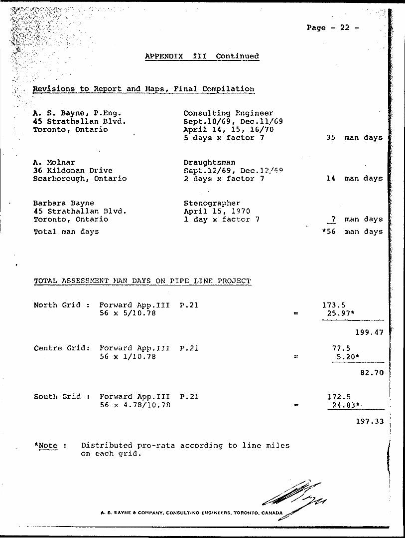
14
0 0.25 0.51
1.5
2
2.5
3
Acuity IOD (log min.)
Ster
eo-a
cuity
(log
sec.)
n.s.
n.s.
n.s.*
0 0.25 0.51
1.5
2
2.5
3
Acuity IOD (log min.)
Ster
eo-a
cuity
(log
sec.)
0 0.4 0.8 1.21
1.5
2
2.5
3
Crowding IOD (deg.)
Ster
eo-a
cuity
(log
sec.)
* n.s.
0 0.4 0.8 1.21
1.5
2
2.5
3
Crowding IOD (deg.)
Ster
eo-a
cuity
(log
sec.)
n.s.
n.s.
A. Acuity vs. contour stereo B. Acuity vs. random-check stereo
E. Crowding vs contour stereo F. Crowding vs. random-check stereo
* p < .05n.s. Non-sign.
Data
Statistics
ControlStrab.Aniso.Mixed
0 0.2 0.4 0.61
1.5
2
2.5
3
Contrast IOD (log %)
Ster
eo-a
cuity
(log
sec.)
n.s.n.s.
0 0.2 0.4 0.61
1.5
2
2.5
3
Contrast IOD (log %)
Ster
eo-a
cuity
(log
sec.)
n.s.n.s.
C. Contrast vs. contour stereo D. Contrast vs. random-check stereo
Supplementary Figure 4. Correlations between the three spatial tasks and stereo-acuity thresholds, for observers with measurable stereo-acuity. A. The relationship between contour-based stereo-acuity thresholds (in log seconds of arc) and interocular differences (IODs) in acuity (in log minutes). Children are divided into “control” and “clinical” categories, though separate symbols show whether clinical children are derived from the strabismic (upright triangles), anisometropic (squares), or mixed (inverted triangles) groups. The correlation for each group is shown at the right-hand side of the line of best fit. B. Random-check stereo-acuity thresholds, plotted against IODs for acuity, as in panel A. C. Contour-based stereo and IODs for contrast detection, the latter plotted in log Weber percentage units. D. Random-check stereo-acuity thresholds and contrast-detection IODs. E. IODs for crowding (in degrees of visual arc), plotted against contour-based stereo-acuity thresholds. F. Crowding IODs and random-check stereo-acuity thresholds.
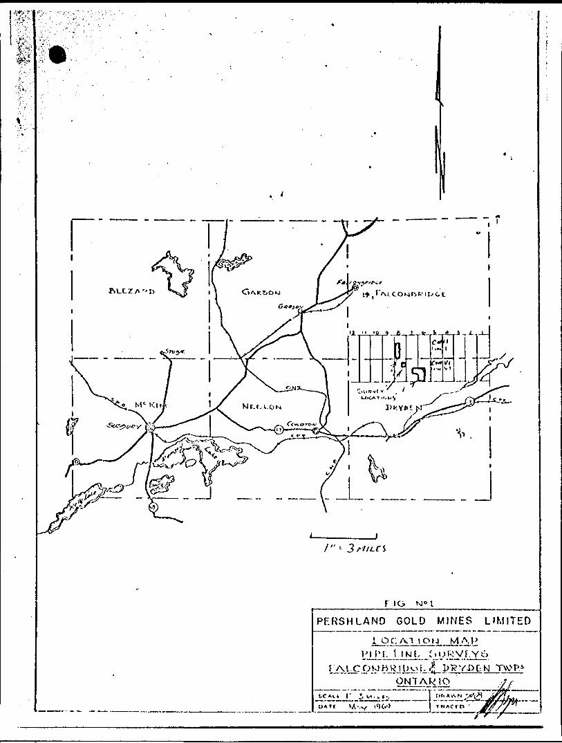
15
Together, we see a similar pattern in these results as when children are divided into
“stereo” and “non-stereo” categories (as in Figure 5 in-text). Namely, those with poor
stereo-acuity have large interocular differences in both acuity and crowding. The
correlation between IODs in acuity and stereo-thresholds has been observed previously
in adults (Weakley, 2001; McKee, Levi, & Movshon, 2003; Levi, McKee, & Movshon,
2011); here we demonstrate that this is true for both contour-based and random-check
stereo stimuli. Differences emerge with crowding however, where we demonstrate a
strong relationship between contour-based stereo-acuity and crowding, while the
relationship between random-check stereo-acuity and crowding is non-significant. This
could be due to the stronger demands placed on acuity by the random-check
stereograms – the individual checks in these stimuli must be resolved in order to detect
the interocular correlations. The relationship with acuity may dominate these thresholds
as a result, whereas the additional low spatial frequency information in the contour-
based stereograms may allow the relationship with crowding to emerge.
Size-corrected crowding values
In the “normal” visual system, the extent of foveal crowding in adults has been found to
follow the size of the stimuli used in testing (Levi, Klein, & Hariharan, 2002), while in
the periphery (Levi, Hariharan, & Klein, 2002b; Tripathy & Cavanagh, 2002) and the
amblyopic fovea (Levi, Hariharan, & Klein, 2002a), crowding is largely size-invariant. In
our study, the large variation in acuity within our population necessitated that stimuli in
the remaining tasks was scaled to clearly visible sizes to ensure visibility. However, this
variation in stimulus size raises the possibility that the elevations in foveal crowding
seen in some groups could simply reflect the variation in stimulus size (and the
corresponding variation in crowding). To examine this possibility, our measured extents
of crowding (in degrees of visual angle) were divided by the size of stimuli used in this
task (also in degrees, which corresponds to values 2.5× acuity thresholds). The resulting
values represent the spatial extent of foveal crowding expressed in multiples of stimulus
size. Note that this is equivalent to expressing the extent of crowding in units of “letter
widths”, as is sometimes performed for crowding experiments.
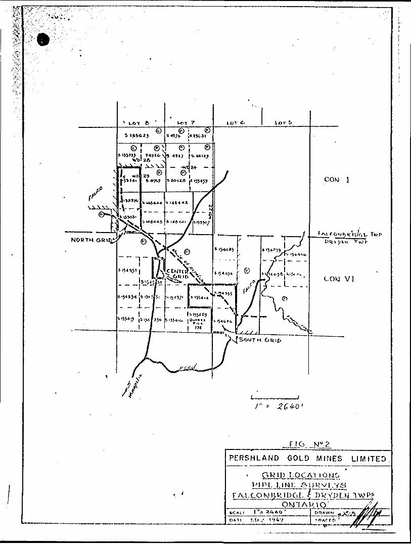
16
Supplementary Figure 5A plots the spatial extent of this size-corrected foveal crowding
in each eye for all children in our study. The same pattern of results as in Figure 2C is
clearly evident. There is no interocular difference between the degree of crowding in
either of the control (t(18)=-1.16, p=.26) or anisometropic (t(15)=-0.02, p=.99) groups,
while a clear interocular difference is evident in the strabismic group (t(17)=3.38, p=.004)
and a marginally significant difference exists for the mixed group (t(18)=2.10, p=.049).
On top of this, there is still a clear degree of crowding in both eyes of controls and the
fellow eyes of all clinical groups (slightly below two times the stimulus size in each
case), with a combined analysis showing a significant difference (t(90)=5.63, p<.001).
This is consistent with prior studies (Jeon et al., 2010).
A. Size-corrected crowding thresholds B. Size-corrected crowding IODs
Control Strab. Aniso. Mixed02468
10
1214
Size
!cor
rect
ed cr
owdi
ng ("
)
n.s. n.s.** *Amblyopic /left eye meanFellow / right eye meanIndividuals
******
p < .001p < .01p < .05
n.s. Non-sig.
Data
Statistics
Stereo Non-stereo-5
0
5
10
15Si
ze!c
orr.
crow
ding
IOD
*
******
p < .001p < .01p < .05
n.s. Non-sig.
Data
Statistics
Strab.Aniso.MixedMeans
Supplementary Figure 5. Size-corrected crowding values. A. When crowding values are expressed in multiples of acuity thresholds, the degree of foveal crowding remains significant in all eyes, as do interocular differences for the strabismic and mixed groups (data as in Figure 2 in-text). B. Interocular crowding differences are also larger for “non-stereo” children than for those with measurable stereo-vision (data plotted as in Figure 5 in-text).
It is also possible that the higher levels of crowding observed in our non-stereo subjects
(Figure 5C of the main text) reflects the potential size-tuning of foveal crowding. To
examine whether the larger IODs persisted after size correction, we split our observers
into “stereo” and “non-stereo” categories, as in the main text, and plot size-corrected
crowding extents in Supplementary Figure 5B. Both the pattern of individual observers,
and the large group difference on average are maintained, which remains significant
(t(51)=-2.39, p=.021). The relationship between stereo-vision and crowding is thus
independent of stimulus size.
17
Supplementary References
Atkinson, J., Anker, S., Evans, C., & McIntyre, A. (1987). The Cambridge Crowding Cards for preschool visual acuity testing. Transactions of the Sixth International Orthoptic Congress, 482-486.
Atkinson, J., & Braddick, O. J. (1983). Assessment of visual acuity in infancy and early childhood. Acta Ophthalmologica, 157 Suppl, 18-26.
Bland, M. J., & Altman, D. G. (1986). Statistical methods for assessing agreement between two methods of clinical measurement. The Lancet, 327(8476), 307-310.
Ellemberg, D., Lewis, T. L., Hong Liu, C., & Maurer, D. (1999). Development of spatial and temporal vision during childhood. Vision Research, 39(14), 2325-2333.
Goodwin, R. T., & Romano, P. E. (1985). Stereoacuity degradation by experimental and real monocular and binocular amblyopia. Investigative Ophthalmology & Visual Science, 26(7), 917-923.
Jeon, S. T., Hamid, J., Maurer, D., & Lewis, T. L. (2010). Developmental changes during childhood in single-letter acuity and its crowding by surrounding contours. Journal of Experimental Child Psychology, 107(4), 423-437.
Levi, D. M., Hariharan, S., & Klein, S. A. (2002a). Suppressive and facilitatory spatial interactions in amblyopic vision. Vision Research, 42(11), 1379-1394.
Levi, D. M., Hariharan, S., & Klein, S. A. (2002b). Suppressive and facilitatory spatial interactions in peripheral vision: Peripheral crowding is neither size invariant nor simple contrast masking. Journal of Vision, 2, 167-177.
Levi, D. M., Klein, S. A., & Hariharan, S. (2002). Suppressive and facilitatory spatial interactions in foveal vision: Foveal crowding is simple contrast masking. Journal of Vision, 2(2), 140-166.
Levi, D. M., McKee, S. P., & Movshon, J. A. (2011). Visual deficits in anisometropia. Vision Research, 51(1), 48-57.
McKee, S. P., Levi, D. M., & Movshon, J. A. (2003). The pattern of visual deficits in amblyopia. Journal of Vision, 3(5), 380-405.
Ono, M. E., Rivest, J., & Ono, H. (1986). Depth perception as a function of motion parallax and absolute-distance information. Journal of Experimental Psychology: Human Perception and Performance, 12(3), 331-337.
Pan, Y., Tarczy-Hornoch, K., Cotter, S. A., Wen, G., Borchert, M. S., Azen, S. P., et al. (2009). Visual acuity norms in pre-school children: The multi-ethnic pediatric eye disease study. Optometry & Vision Science, 86(6), 607-612.
Richards, W. (1970). Stereopsis and stereoblindness. Experimental Brain Research, 10(4), 380-388.

18
Richards, W. (1971). Anomalous stereoscopic depth perception. Journal of the Optical Society of America, 61(3), 410-414.
Tripathy, S. P., & Cavanagh, P. (2002). The extent of crowding in peripheral vision does not scale with target size. Vision Research, 42(20), 2357-2369.
Weakley, D. R. (2001). The association between nonstrabismic anisometropia, amblyopia, and subnormal binocularity. Ophthalmology, 108(1), 163-171.