Embed Size (px)
Citation preview

VIRAL-CYCLES: Four Basic Shapes
COLORADO STATE UNIVERSITY EXTENSION
4-H PROGRAMS ARE AVAILABLE TO ALL WITHOUT DISCRIMINATION
VOLUME 10, ISSUE 3, May 12, 2020
There are more viruses than stars in the universe. It is estimated that there are 10 nonillion individuals. Nonillion is a really big number. It is 100,000,000,000,000,000,000,000,000,000,000. (I counted the zeros twice to make sure I had all of them). There should be 32 zeros after the 1. That is a big number! Viruses are everywhere. They are in the soil, water, and air. They can infect all living cells, bacteria and archaea (simple cells, like bacteria, but the DNA is more similar our DNA), protists, fungi, plants, and animals. If there are that many viruses, and they are found everywhere, why are we not always sick? All viruses are extremely simple. They are: • genetic material (single
strand DNA, double strand DNA, or RNA, which is single strand)
• genetic material is protected by a capsid protein coat
(depicted as the grey sphere in the image below of the coronavirus)
• glycoproteins that are embedded on the outside of the capsid (the nail shaped red and orange imaged below) that help the virus recognize a cell to infect
The glycoproteins only recognize certain cells. Maybe they recognize a bat cell, or a mouse cell. They do not recognize a person cell. This novel virus is thought to have been a bat virus. It infected a human. Because humans have not had this virus before, our immune system has to figure out how to fight it. Antibodies mutate to recognize new pathogens. They signal our WBC to fight the invader. It takes time, but they will win!
THIS ISSUE • Shapes page 2 • Nets page 3-6
POWER WORDS • antibody: blood protein
produced in response to and counteracting a specific invading virus, bacterium, or other foreign body
• archaea: microorganisms similar to bacteria in size and simplicity of structure but radically different in molecular organization
• morphology: the study of the forms of things
• novel virus: a virus not previously recorded
• pathogen: a bacterium, virus, or other microorganism that can cause disease.
• protist: a single-celled organism of the kingdom Protista
Morphology of Viruses

Four Basic Shapes 2
Viruses have four basic shapes: • helical (rabiesvirus)
• icosahedral (poliovirus)
• spherical (coronavirus)
• complex (bacteriophage)
The first three shapes have viruses that can cause human disease. The complex form, the bacteriophage, only attacks bacterial cells. In fact, doctors are looking at bacteriophages as the next generation of medicine protecting us from bacterial illnesses. For more information, watch this cartoon video: https://www.youtube.com/watch?v=YI3tsmFsrOg&t=2s. Very cool!
POWER WORDS • biconvex: having
an outline or surface curved like the exterior of a circle or sphere on both sides
• complex: consisting of many different and connected parts
• helical: having the shape or form of a spiral
• helix: a spiral • icosahedral : a
polyhedron have 20 faces
• net: A pattern that you can cut and fold to make a model of a solid shape
• polyhedral: of, relating to, or having the shape of a polyhedron
• polyhedron: a solid figure having many faces
• spherical: shaped like a sphere
• triangle: a plane figure with three straight sides and three angles
MATERIALS • print pages 3-6 single-sided • color pencils or markers • scissors • tape • string • 3 small paperclips • optional needle point pliers
In this activity, you will make a paper model of each basic shape of the viruses. The following pages give directions and the template to make the morphology of these four basic shapes.
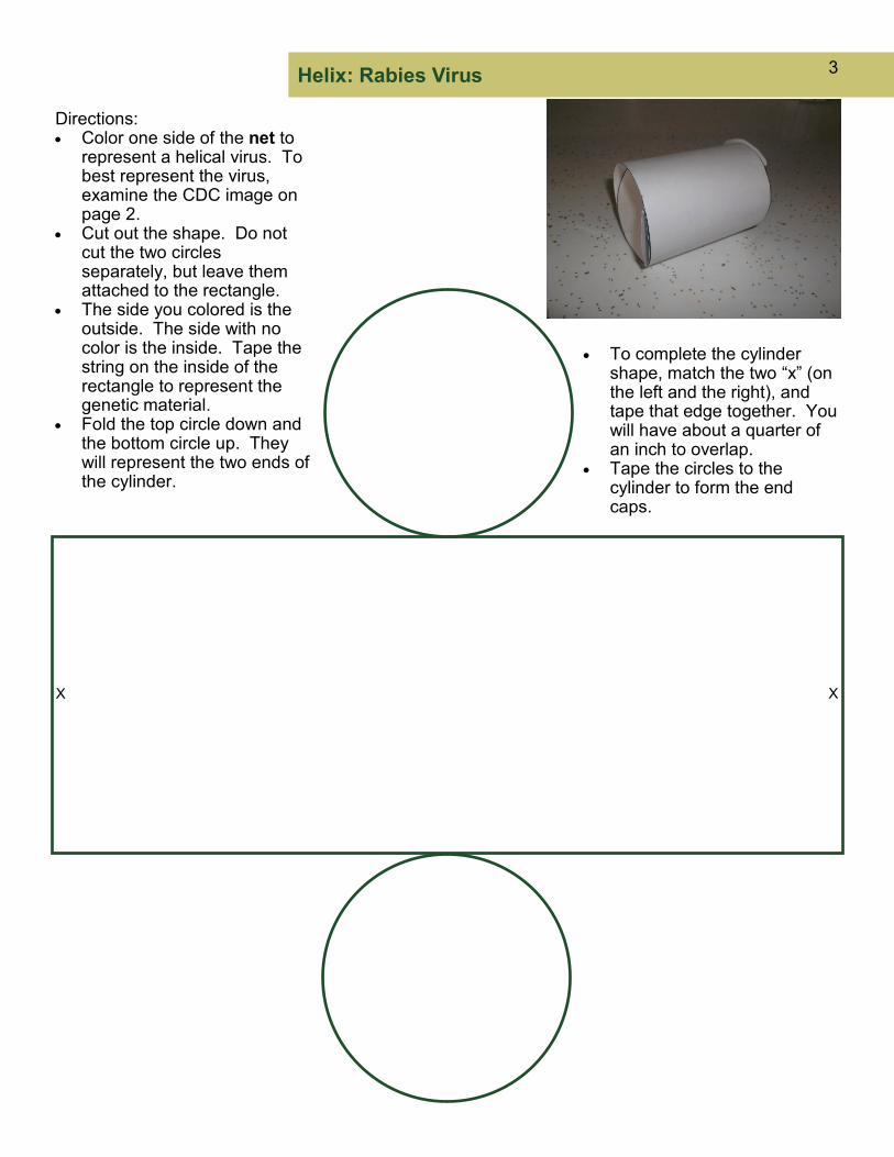
Helix: Rabies Virus 3
Directions: • Color one side of the net to
represent a helical virus. To best represent the virus, examine the CDC image on page 2.
• Cut out the shape. Do not cut the two circles separately, but leave them attached to the rectangle.
• The side you colored is the outside. The side with no color is the inside. Tape the string on the inside of the rectangle to represent the genetic material.
• Fold the top circle down and the bottom circle up. They will represent the two ends of the cylinder.
• To complete the cylinder shape, match the two “x” (on the left and the right), and tape that edge together. You will have about a quarter of an inch to overlap.
• Tape the circles to the cylinder to form the end caps.
X X

Icosahedral — Poliovirus 4
Directions: • Color one side of the net to
represent a icosahedralvirus. To best represent the virus, examine the CDC image on page 2.
• Cut out the shape on the outer black lines. Do not cut the individual triangle shapes out separately. This should be in one piece.
• The side you colored is the outside. The side with no color is the inside.
• Fold along each line. When you have folded all the lines, you may need to refold the lines so that the shape curls inward, towards the side you did not color.
• Tape the edges except one together to form your 3-D icosahedral.
• Cut about 2’ of string, and place inside the shape. The string represents the genetic material of this virion.
• Tape the last triangle.

Sphere — Coronavirus 5
Directions: • Color one side of the net to
represent the spherical coronavirus. To best represent the virus, examine the CDC image on page 2.
• Cut out the shape on the outer black lines. This should be in one piece.
• The side you colored is the outside. The side with no color is the inside.
• Fold along each side that connects to the next biconvex shape. When you have folded all the lines, you may need to refold the lines so that the shape curls inward, towards the side you did not color.
• Tape together the edges except the last two to form your 3-D sphere.
• Cut about 2’ of string, and place inside the shape. The string represents the genetic material of this virion.
• Tape the last two biconvex shapes to complete the sphere.
Hint: use a small ball (size of a tennis ball), orange, or apple when taping your sphere. It is easier to manage this step.

Complex Virus: Bacteriophage 6
Directions: • Color one side of the net of the complex
bacteriophage. There isn’t a CDC model to use. Be creative when you color your bacteriophage.
• This model is five pieces: capsid, tail with pins (triangles), tail fibers (paperclips), collar, and base plate.
• Cut out the four shapes on the outer dark green lines. Cut out the two gold hatch-mark inner circles on the collar and base plate.
• The side you colored is the outside. The side with no color is the inside.
• Fold along each line. When you have folded all the lines, you may need to refold the lines so that the shape curls inward, towards the side you did not color.
• Tape the edges except one together to form your polyhedron.
• Cut about 2’ of string and place inside the shape. The string represents the genetic material.
• Tape the last triangle. • Fold the top circle down and the bottom
circle up on the tail. They are the two ends of the cylinder.
• To complete the cylinder shape, match the two “x” (on the left and the right), and tape that edge together. You will have about a quarter of an inch to overlap.
• Tape the top circle to the cylinder. • Carefully insert the collar over the top and
the base plate on the bottom of the cylinder. Tape in place.
• Unbend your two paperclips. You can use needlepoint pliers to help you with this step. Shape the paperclip, with the center 3/4”, and the two legs about equal:
• Insert one end of the paperclip on the back side, poking through the “x” on the opposite side. Poke the other side of the paperclip through the second “x.” Repeat with the second paperclip, inserting both ends in the “y” on the bottom circle. The paperclip ends need to stick out like legs at the bottom when inserted correctly. Tape the circle to the cylinder.
• Assemble the bacteriophage. Tape the capsid to the top of the tail.
capsid
tail
collar
base plate
x y
x y
pins (also called spikes) Image on page 7
of bacteriophage model

ACKNOWLEDGMENTS AND CITATIONS 7
AUTHORS • Dr. Barbara J. Shaw, Colorado State University Extension Western Region Youth Development
4-H STEM K/12 Specialist • Tom Lindsay, retired Portland State University instructor (geology and paleontology); HS science
teacher (AP and IB Chemistry, Physics, Biology, and Calculus)
ACKNOWLEDGMENTS • Funding for this project provided by Colorado State University System Venture Capital Fund • CJ Mucklow, Colorado State University Extension Western Regional Director • Stephanie Lamm, Colorado State University Extension Tri-River Area STEM Agent • Andrew Reed, Colorado State University Extension, Tri-River Area, Montrose County Video
Editor • Dr. Joe Cannon and Marketing Strategies students Berlyn Anderson, Jenna Balsley, Rachel
Kassirer, Rachel Richman, Colorado State University, College of Business, for marketing strategies and ST[EMpower] graphics
• Doug Garcia, Colorado State University Creative Services Communication Coordinator/ Designer
CITATIONS Information: • https://www.exploratorium.edu/snacks/life-size; https://www.exploratorium.edu/snacks/viral-
packaging; https://ellenjmchenry.com/; National Geographic https://www.nationalgeographic.com/science/2020/04/factors-allow-viruses-infect-humans-coronavirus/#close;
Images: • Shapes of Viruses: Medical Microbiology. 4th edition.
Baron S, editor. Galveston (TX): University of Texas Medical Branch at Galveston; 1996.; Center for Disease Control and Prevention images of viral shapes: Coronavirus: https://www.cdc.gov/coronavirus/2019-nCoV/index.html; Poliovirus: https://www.cdc.gov/dotw/polio/index.html; Rabiesvirus: https://www.cdc.gov/rabies/about.html; Bacteriophage: Doss, Janis, Culbertson, Kayla, Hanh, Delila, Camacho, Joanna, Barekzi, Nazir (2017) A review of phage therapy against Bacterial Pahtogens of Aquatic and Terrestrial Organisms. Multidisciplinary Digital Publishing Institute 9:3, https://www.mdpi.com/1999-4915/9/3/50/htm
Paper Model of the Bacteriophage