Embed Size (px)
Citation preview
Tumor and Stem Cell Biology
Vimentin–ERK Signaling Uncouples Slug GeneRegulatory FunctionReettaVirtakoivu1,2, AnjaMai1, ElinaMattila1,2, NicolaDe Franceschi1,2, SusumuY. Imanishi1,Garry Corthals1, Riina Kaukonen1,2, Markku Saari1,2, Fang Cheng1,3, Elin Torvaldson1,3,Veli-Matti Kosma4, Arto Mannermaa4, Ghaffar Muharram1,2, Christine Gilles5,John Eriksson3, Ylermi Soini4, James B. Lorens6, and Johanna Ivaska1,2,7
Abstract
Epithelial–mesenchymal transition (EMT) in cells is a develop-mental process adopted during tumorigenesis that promotes met-astatic capacity. In this study, we advance understanding of EMTcontrol in cancer cells with the description of a novel vimentin–ERK axis that regulates the transcriptional activity of Slug (SNAI2).Vimentin, ERK, and Slug exhibited overlapping subcellular local-ization in clinical specimens of triple-negative breast carcinoma.RNAi-mediated ablation of these gene products inhibited cancercell migration and cell invasion through a laminin-rich matrix.
Biochemical analyses demonstrated direct interaction of vimentinand ERK, which promoted ERK activation and enhanced vimentintranscription. Consistent with its role as an intermediate filament,vimentin acted as a scaffold to recruit Slug to ERK and promoteSlug phosphorylation at serine-87. Site-directed mutagenesisestablisheda requirement for ERK-mediatedSlugphosphorylationin EMT initiation. Together, these findings identified a pivotal stepin controlling the ability of Slug to organize hallmarks of EMT.Cancer Res; 75(11); 2349–62. �2015 AACR.
IntroductionCancer cell dissemination and metastasis is the underlying
cause of mortality in cancer patients and is closely linked to adevelopmental process called epithelial–mesenchymal transition(EMT). During EMT, epithelial cells lose apical–basal polarity,disassemble cell–cell contacts, and adopt a more mesenchymaland motile phenotype (1, 2). In the course of malignant progres-sion, cancer cells acquire the ability to break through the basallamina, invade surrounding tissues and capillaries, extravasateand colonize distant organs (3). The acquisition of EMT char-acteristics is believed to be one important mechanism, wherebycancer cells achieve increased motility and invasiveness to pro-mote metastasis (4, 5). In breast cancer, EMT characteristics areenriched in the aggressive and metastatic triple-negative breastcancer subtype (6, 7), suggesting a role for EMT in breast cancermetastasis. Importantly, vimentin, the mesenchymal intermedi-ate filament (IF) and a hallmark of EMT, is overexpressed inmalignant epithelial cancers, including breast cancer, and corre-lates with poor prognosis (8). Vimentin is also present in mam-
mary and breast cancer stem cells, providing a further linkbetween EMT and malignancy (9, 10). However, the functionalrole of vimentin in EMT and/or stem cells remains incompletelyunderstood. Strong evidence indicates that vimentin regulatesmesenchymal cell shape and mammary epithelial cell migration(11–13), and plays a role in regulating signal transduction,necessary for EMT induction, downstream of mutant H-Ras andTGFb via a yet unknown mechanism (13).
Activated ERK kinases participate in the regulation of severalcellular processes such as cell proliferation, survival, andmotility.Recent evidence has highlighted that ERK2, and not ERK1, isinvolved in EMT induction (14, 15) and cancer cell migration andinvasion (16–18). Vimentin has been shown to function as asignaling scaffold in many different cell types and biologic pro-cesses (19). In neurons, vimentin fragments interact with ERK andsupport the spatial translocation of active ERK along axons inresponse to injury (20).However, it is currently unknownwhethera similar mechanism functions outside of the nervous system orwhether vimentin upregulation during cancer progression plays arole in regulating ERK signaling.
Slug belongs to the Snail family of EMT-inducing transcrip-tion factors (Snail, Slug, and Smuc; refs. 2, 21). Posttransla-tional modifications of Snail, and as recently described for Slug,have emerged as critical levels of control for their abundanceand subcellular localization. In the nucleus, Lats2 interacts withand directly phosphorylates Snail on residue T203, supportingEMT by influencing the stability and nuclear localization ofSnail (22). Glycogen synthase kinase 3b (GSK3b) phosphor-ylates both Slug and Snail and negatively regulates their sta-bility thus maintaining epithelial morphology (23–27). Theactivation of oncogenic pathways, including PI3K–Akt andRas–MAPK signaling, suppresses GSK3b activity and GSK3b-dependent reduction in Snail and Slug phosphorylation result-ing in increased transcription factor stability and accumulationand EMT induction.
1Turku Centre for Biotechnology, University of Turku, Turku, Finland.2Medical Biotechnology, VTT Technical Research Centre of Finland,Turku, Finland. 3ÅboAkademiUniversity,Turku, Finland. 4UniversityofEastern Finland, Cancer Center of Eastern Finland, Kuopio, Finland.5Universit�e de Li�ege, GIGA-Cancer, Liege, Belgium. 6Department ofBiomedicine, University of Bergen, Bergen, Norway. 7Department ofBiochemistry and FoodChemistry, University of Turku,Turku, Finland.
Note: Supplementary data for this article are available at Cancer ResearchOnline (http://cancerres.aacrjournals.org/).
Corresponding Author: Johanna Ivaska, University of Turku, Turku Centre forBiotechnology, Tykist€okatu 6, FI-20520, Turku, Finland. Phone: 358-405020812;Fax: 358-24788601; E-mail: [email protected]
doi: 10.1158/0008-5472.CAN-14-2842
�2015 American Association for Cancer Research.
CancerResearch
www.aacrjournals.org 2349
on July 11, 2018. © 2015 American Association for Cancer Research. cancerres.aacrjournals.org Downloaded from
Published OnlineFirst April 8, 2015; DOI: 10.1158/0008-5472.CAN-14-2842
In addition to ERK2 activity-dependent inhibition of GSK3bactivity, other direct mechanisms have also been implicated inEMT onset, including Fra1-mediated induction of ZEB1/2 tran-scription factors (15, 28) and stromal collagen-induced Snailphosphorylation, leading to increased stability and nuclear accu-mulation of Snail (14). Altogether, a deeper appreciation of themechanisms regulating the Snail family of transcription factors,including posttranslational modifications, will be vital in ourefforts to prevent EMT and EMT-related processes during cancerprogression. Here, we identify a pathway linking vimentin expres-sion, ERK activity, and Slug phosphorylation to EMT inductionand positive regulation of cancer cell invasion,migration, and thegene regulatory function of Slug.
Materials and MethodsAntibodies and reagents
For antibodies and reagents, see Supplementary ExperimentalProcedures.
Cell culture and stable cell linesFor cell culture and stable cell lines, see Supplementary Exper-
imental Procedures.
Histologic material and immunohistochemistryThe histologic breast cancer material of triple-negative (n ¼
118) and estrogen receptor (ER) and/or progesterone receptor(PR), and/or HER2-positive tumors (n ¼ 356) consisted offormalin-fixed, paraffin-embedded or frozen samples retrievedfrom the files of the Department of Pathology, Kuopio UniversityHospital (Kuopio, Finland). Sampleswere sectioned, prepared forimmunohistochemistry, and stained with appropriate primaryand secondary antibodies. For more information on tissue prep-aration and immunohistochemistry, see Supplementary Experi-mental Procedures.
Tumor xenografts on chick embryo chorioallantoicmembranesFor tumor xenografts on chick embryos, see Supplementary
Experimental Procedures.
In vitro kinase assay and alkaline phosphatase protection assayIn in vitro kinase assays purified recombinant active Akt1,
ERK1, or ERK2 (ProQinase GmbH; BSA as the control) wereincubated for 20 minutes at 30�C with recombinant Snail orSlug and ATP-g-P32, in buffer containing 20 mmol/L HEPESpH 7.4, 10 mmol/L MgCl2, and serine–threonine phosphataseinhibitor cocktail 1. Reactions were stopped with Laemmlisample buffer and heating at 100�C for 10 minutes. Sampleswere resolved by SDS-PAGE, and the gel was Coomassie-stained and dried before detection by autoradiography. Forthe alkaline phosphatase protection assays, recombinant activeERK2 (ProQinase) was incubated with either vimentin (Cyto-skeleton Inc.) or actin (non-muscle human platelets; Cytoskel-eton Inc.) in reaction buffer (125 mmol/L HEPES–NaOH pH7.5, 150 mmol/L NaCl, 7.5 mmol/L MgCl2, 10 mmol/L CaCl2,and 2.5 mmol/L DTT) for 30 minutes at 37�C. Alkaline phos-phatase (calf intestinal; Promega) was then added to the pre-formed protein complexes or to ERK2 alone and incubated for afurther 2 or 20 minutes at 37�C. The reaction was stopped bythe addition of 8� Laemmli sample buffer followed by SDS-PAGE and Western blotting with the indicated antibodies.
Pulldowns, cosedimentation, and immunoprecipitationDirect protein binding was assessed by GST and antibody
pulldowns using recombinant proteins. For GST pulldowns,recombinant proteins [phosphorylated or nonphosphorylatedERK2 (ProQinase) and GST-vimentin (Spring Bio) or GST as acontrol] were incubated for 1 hour at room temperature inreaction buffer (20 mmol/L Tris–HCl, 150 mmol/L NaCl, 1mmol/L MgCl2, and 10% glycerol) and complexes isolated usingpreblocked glutathione beads for 1 hour. For antibody pull-downs, recombinant proteins [ERK2 and either vimentin or actinor keratin-8 (Sigma)] were allowed to react for 30minutes at 37�Cand complexes isolated using anti-ERK antibody for 30 minutesfollowed by the addition of preblocked Protein G Sepharosebeads (GE Healthcare) for 1 hour.
For cosedimentation assays, polymerization of vimentin [0.5mg/mL in 50 mL of assembly buffer: 20 mmol/L Tris, pH 7.4, 3mmol/L KCl, 0.2% 2-mercaptoethanol, 0.2% phenylmethylsul-fonylfluoride (PMSF)] was triggered by the addition of 3 mL of 5mol/L NaCl at 37�C for 1 hour. Vimentin IFs were then incubatedwith the indicated recombinant proteins (1–2 mg each) for anadditional hour. Proteins were collected by ultracentrifugation at100,000 � g for 30 minutes and the total soluble (supernatant)and nonsoluble (pellet) fractions were analyzed by SDS-PAGEfollowed by Western blotting with the indicated antibodies.
In immunoprecipitation assays, cells were treatedwith EGF (50ng/mL) or with DMSO as control for 20 minutes, washed withPBS, and detached with HyQTase (Thermo Scientific). Cells werelysed with RIPA lysis buffer (50 mmol/L Tris–HCl, 150 mmol/LNaCl, 1 mmol/L EDTA, 1%NP-40, 0.5% sodium deoxycholate, 1mmol/L EGTA, and 0.1% SDS) for 30minutes at 4�C, centrifugedat 13,000 rpm for 10minutes at 4�C. Lysates were incubated withanti-FLAG antibody (2.5 mg/reaction) for 1 hour, and washedthree times. Where indicated immunoprecipitations were per-formed in cells preincubatedwith 32P-ATP–supplementedmedia.
Luciferase reporter assays and DNA-binding ELISAFor luciferase reporter assays and DNA-binding ELISA, see
Supplementary Experimental Procedures.
qRT-PCRFor qRT-PCR methods, see Supplementary Experimental
Procedures.
Immunofluorescenc, fluorescence recovery afterphotobleaching, and flow cytometry
For immunofluorescence, fluorescence recovery after photo-bleaching (FRAP), and flow cytometry, see Supplementary Exper-imental Procedures.
Stimulated emission depletion microscopyLeica TCS SP5 stimulated emission depletion (STED) laser
scanning microscope was used to image in super-resolution level(Leica Microsystems GmbH), where approximately 65-nm reso-lution in x- and y-axis was achieved. Mega-520-labeled vimentinwas excited with 532-nm pulse laser (PicoQuant), and respec-tively Star-635 (for slug or pERK) at 635-nm wavelength (Pico-Quant). The channels were scanned sequentially and emissionwas detected by avalanche photodiode detectors at emissionrange of 685/40 (Leica Microsystems GmbH). Leica LAS software(Leica Microsystems GmbH) was used do perform background
Virtakoivu et al.
Cancer Res; 75(11) June 1, 2015 Cancer Research2350
on July 11, 2018. © 2015 American Association for Cancer Research. cancerres.aacrjournals.org Downloaded from
Published OnlineFirst April 8, 2015; DOI: 10.1158/0008-5472.CAN-14-2842
subtraction and then deconvolution of all images. In the decon-volution process, a Lorentzian PSF was generated by using mea-sured PSF value of 62 nm, which was exploited to signal energybased deconvolution algorithm.
Proliferation, migration, and invasionFor proliferation, migration, and invasion assays, see Supple-
mentary Experimental Procedures.
In situ proximity ligation assayFor proximity ligation assay (PLA), see Supplementary Exper-
imental Procedures.
Mass spectrometryFor mass spectrometry, see Supplementary Experimental
Procedures.
Generation of phospho-Slug antibodyRabbit polyclonal antibody against phosphorylated Slug was
generated following immunization with a synthetic peptide con-jugated to OVA (NH2-CYSSSLGRV(Sp)-COOH for Slug(P)87site. The rabbits were immunized according to the standardprocedure of the service provider (Primm srl) with five injectionsand the antibody was affinity purified from immune serum usingCNBr-Sepharose–conjugated phospho-peptides.
Statistical analysisAll statistical analyseswere done using the Student t test. For the
clinical samples, cytoplasmic and nuclear Slug and vimentinexpression were analyzed on a per patient basis and the statisticalsignificance and associations with the parameters (triple-negativeor other samples) was analyzed with the x2 test.
ResultsVimentin, ERK, and Slug expression correlate with triple-negative status of breast cancer and regulate breast cancer cellmigration and invasion
Vimentin, Slug, and ERK kinases, especially ERK2, have beenlinked to EMT and breast cancer cell invasion. In a cohort of 356breast cancer tissue samples, we found that vimentin and Slugexpression were, as described earlier (29, 30), significantly higherin triple-negative tissues (Fig. 1A, 164 samples). In these samples,61% of cells were vimentin-positive, 73% showed positive cyto-plasmic and 42% positive nuclear Slug staining, 71% were ERK-positive and 49% displayed phosphorylated ERK-1/2. Moreover,among these triple-negative samples, Slug, ERK, and phosphor-ylated ERK (pERK) expressionwasmore pronounced at the tumorperiphery in line with a recent report (Fig. 1B; Slug in 44% andpERK in 39% of the tumors; ref. 31), whereas vimentin wasexpressed throughout the tumor, including at the periphery. Inthese triple-negative breast carcinomas, pERK did not correlatewith slug (P ¼ 0.12) or vimentin (P ¼ 0.23) expression asdetermined by the x2 test. As these EMT-linked proteins werecoexpressed at the tumor invasive front, we investigated theirfunctional impact on migration and invasion of triple-negativebreast cancer cells using the MDA-MB-231 cell line. We reportedrecently that vimentin is a critical regulator of migration in thesecells (13). Here, we further demonstrate that silencing of Slug,ERK1, or ERK2, using either smart pool or individual siRNAoligos, also significantly inhibits MDA-MB-231 cell migration
(Fig. 1C and Supplementary Fig. S1A). ERK2 appeared to be thepredominant ERK isoform regulating cell migration as recentlydescribed (16, 17). Moreover, these effects were motility-specificas suppressed cellmotility ((13) and Supplementary Fig. S1A)wasnot accompanied by defects in cell proliferation (SupplementaryFig. S1B). In addition to roles in cell migration, ERK2, vimentin,and Slug were all required for efficient directional cell invasionthrough a laminin-rich extracellular matrix (Matrigel; Fig. 1D andSupplementary Fig. S1A). As ERK1 silencing did not appear tohave any impact on cell invasion (Fig. 1D), we focused our futureefforts on delineating the role of the ERK2 isoform in EMTinduction.
Remarkably, in an orthotopic xenograft model, the populationof MDA-MB-231 cells that spontaneously metastasized to thecontralateral lymphnodes exhibited significantly higher vimentinlevels than the cell population remaining in the primary tumor(Fig. 1E), whereas DAPI staining intensity was even between theprimary tumor and lymph node metastases (Supplementary Fig.S1C). These data suggest that increased vimentin expressioncorrelates with enhanced tumor metastasis in vivo even in cellsthat already displays all the hallmarks of EMT. Together, theseresults demonstrate that vimentin, Slug, and ERK2 are coex-pressed in the majority of triple-negative breast cancers and areimportant regulators of breast cancer cellmigration, invasion, andmetastasis.
Vimentin and ERK form a reciprocal regulatory complexWe previously identified a functional role for vimentin in
supporting EMT downstream of TGFb and active Ras signaling(13); however, the molecular mechanisms underlying this obser-vation were unclear. Here, we show that H-Ras–transformedMCF10A cellswith elevated vimentin expression exhibit increasedERK phosphorylation relative to the nontransformed low vimen-tin-expressing cells (Fig. 2A). Moreover, TGFb-induced vimentinexpression in MCF10A breast epithelial cells results in a concom-itant increase in ERK phosphorylation (Fig. 2B), indicating acorrelation between vimentin expression and ERK activity. Inter-estingly, silencing of vimentin in TGFb-induced MCF10A and invimentin-highMDA-MB-231 cells significantly reduced the phos-phorylation and total levels of ERK kinases (Fig. 2B and Supple-mentary Fig. S2A), suggesting that vimentin expression may benecessary to stabilize activated ERK in cells. To validate thesefindings, we generated immortalizedwild-type (WT) or vimentin-null (Vim KO) mouse embryonic fibroblasts (MEF) and investi-gated total and active ERK levels in these cells. Congruent with thesiRNA data, the levels of pERK and, to a lesser extent, total ERKwere significantly decreased in Vim KOMEFs compared with WTMEFs (Fig. 2C). Importantly, ERK levels were rescued by reexpres-sion of vimentin in the null cells (Fig. 2D).
In order to elucidate the mechanism of vimentin-dependentERK regulation, we first investigated ERK and vimentin localiza-tion in cells. Using super-resolution STED microscopy, wedetected a subset of pERK coincident with vimentin filaments.Importantly, treatment of cells with ERK inhibitor U0216 notice-ably reduced the pERK signal along vimentin filaments (Fig. 2E).Moreover, in situ PLAs demonstrated colocalization betweenendogenous pERK and vimentin in the cytoplasm of MDA-MB-231 cells (Fig. 2F). Notably, the PLA signal was lost upon inhi-bition of MEK1/2 with U0126, indicating a specific interactionbetween ERK and vimentin (Fig. 2F). These data suggest thatvimentin and active ERK could form a complex in cells. To clarify
Slug Phosphorylation Induces Vimentin
www.aacrjournals.org Cancer Res; 75(11) June 1, 2015 2351
on July 11, 2018. © 2015 American Association for Cancer Research. cancerres.aacrjournals.org Downloaded from
Published OnlineFirst April 8, 2015; DOI: 10.1158/0008-5472.CAN-14-2842
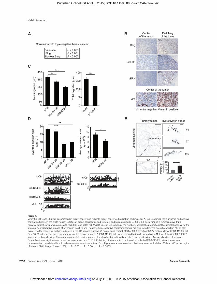
Figure 1.Vimentin, ERK, and Slug are coexpressed in breast cancer and regulate breast cancer cell migration and invasion. A, table outlining the significant and positivecorrelation between the triple-negative status of breast carcinomas and vimentin and Slug staining (n ¼ 356). B, IHC staining of a representative triple-negative patient carcinoma sample with Slug, ERK, and pERK T202/Y204 (n¼ 30–40 samples). The numbers indicate the proportion (%) of samples positive for thestaining. Representative images of a vimentin-positive and -negative triple-negative carcinoma sample are also included. The overall proportion (%) of cellsexpressing the respective proteins indicated in the IHC images is shown. C, migration of control, ERK1 or ERK2 smart pool (SP), or Slug-silenced MDA-MB-231 cells(n ¼ 38–56 cells; shown are representatives of three experiments). D, MDA-MB-231 cells were allowed to invade for 4 days in Matrigel following ERK1, ERK2,vimentin, or Slug silencing. Shown are representative micrographs of phalloidin-stained invading cells (z-stack, side-view). Arrows, direction of invasion(quantification of eight invasion areas per experiment; n ¼ 3). E, IHC staining of vimentin in orthotopically implanted MDA-MB-231 primary tumors andrepresentative contralateral lymph node metastasis from three animals (n¼ 7 lymph node lesions and n¼ 5 primary tumors). Scale bar, 300 and 100 mm for regionof interest (ROI) images (mean � SEM; � , P < 0.05; �� , P < 0.001; ��� , P < 0.0001).
Virtakoivu et al.
Cancer Res; 75(11) June 1, 2015 Cancer Research2352
on July 11, 2018. © 2015 American Association for Cancer Research. cancerres.aacrjournals.org Downloaded from
Published OnlineFirst April 8, 2015; DOI: 10.1158/0008-5472.CAN-14-2842
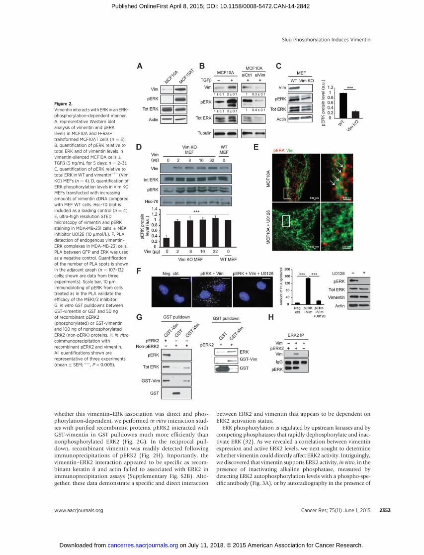
whether this vimentin–ERK association was direct and phos-phorylation-dependent, we performed in vitro interaction stud-ies with purified recombinant proteins. pERK2 interacted withGST-vimentin in GST pulldowns much more efficiently thannonphosphorylated ERK2 (Fig. 2G). In the reciprocal pull-down, recombinant vimentin was readily detected followingimmunoprecipitations of pERK2 (Fig. 2H). Importantly, thevimentin–ERK2 interaction appeared to be specific as recom-binant keratin 8 and actin failed to associated with ERK2 inimmunoprecipitation assays (Supplementary Fig. S2B). Alto-gether, these data demonstrate a specific and direct interaction
between ERK2 and vimentin that appears to be dependent onERK2 activation status.
ERK phosphorylation is regulated by upstream kinases and bycompeting phosphatases that rapidly dephosphorylate and inac-tivate ERK (32). As we revealed a correlation between vimentinexpression and active ERK2 levels, we next sought to determinewhether vimentin could directly affect ERK2 activity. Intriguingly,we discovered that vimentin supports ERK2 activity, in vitro, in thepresence of inactivating alkaline phosphatase, measured bydetecting ERK2 autophosphorylation levels with a phospho-spe-cific antibody (Fig. 3A), or by autoradiography in the presence of
Figure 2.Vimentin interactswith ERK in an ERK-phosphorylation-dependent manner.A, representative Western blotanalysis of vimentin and pERKlevels in MCF10A and H-Ras–transformed MCF10AT cells (n ¼ 3).B, quantification of pERK relative tototal ERK and of vimentin levels invimentin-silenced MCF10A cells �TGFb (5 ng/mL for 5 days; n ¼ 2–3).C, quantification of pERK relative tototal ERK in WT and vimentin�/� (VimKO) MEFs (n ¼ 4). D, quantification ofERK phosphorylation levels in Vim KOMEFs transfected with increasingamounts of vimentin cDNA comparedwith MEF WT cells. Hsc-70 blot isincluded as a loading control (n ¼ 4).E, ultra-high resolution STEDmicroscopy of vimentin and pERKstaining in MDA-MB-231 cells � MEKinhibitor U0126 (10 mmol/L). F, PLAdetection of endogenous vimentin–ERK complexes in MDA-MB-231 cells.PLA between GFP and ERK was usedas a negative control. Quantificationof the number of PLA spots is shownin the adjacent graph (n ¼ 107–132cells; shown are data from threeexperiments). Scale bar, 10 mm.Immunobloting of pERK from cellstreated as in the PLA validate theefficacy of the MEK1/2 inhibitor.G, in vitro GST pulldowns betweenGST-vimentin or GST and 50 ngof recombinant pERK2(phosphorylated) or GST-vimentinand 100 ng of nonphosphorylatedERK2 (non-pERK) proteins. H, in vitrocoimmunoprecipitation withrecombinant pERK2 and vimentin.All quantifications shown arerepresentative of three experiments(mean � SEM; ��� , P < 0.005).
Slug Phosphorylation Induces Vimentin
www.aacrjournals.org Cancer Res; 75(11) June 1, 2015 2353
on July 11, 2018. © 2015 American Association for Cancer Research. cancerres.aacrjournals.org Downloaded from
Published OnlineFirst April 8, 2015; DOI: 10.1158/0008-5472.CAN-14-2842
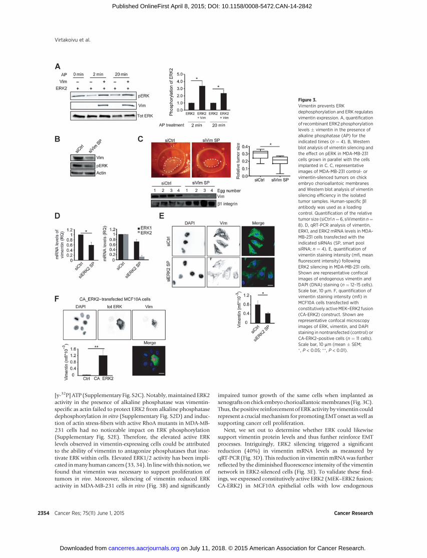
[y-32P] ATP (Supplementary Fig. S2C).Notably,maintainedERK2activity in the presence of alkaline phosphatase was vimentin-specific as actin failed to protect ERK2 from alkaline phosphatasedephosphorylation in vitro (Supplementary Fig. S2D) and induc-tion of actin stress-fibers with active RhoA mutants in MDA-MB-231 cells had no noticeable impact on ERK phosphorylation(Supplementary Fig. S2E). Therefore, the elevated active ERKlevels observed in vimentin-expressing cells could be attributedto the ability of vimentin to antagonize phosphatases that inac-tivate ERK within cells. Elevated ERK1/2 activity has been impli-cated inmanyhuman cancers (33, 34). In linewith this notion,wefound that vimentin was necessary to support proliferation oftumors in vivo. Moreover, silencing of vimentin reduced ERKactivity in MDA-MB-231 cells in vitro (Fig. 3B) and significantly
impaired tumor growth of the same cells when implanted asxenografts on chick embryo chorioallantoicmembranes (Fig. 3C).Thus, thepositive reinforcement of ERKactivity by vimentin couldrepresent a crucialmechanism for promoting EMTonset as well assupporting cancer cell proliferation.
Next, we set out to determine whether ERK could likewisesupport vimentin protein levels and thus further reinforce EMTprocesses. Intriguingly, ERK2 silencing triggered a significantreduction (40%) in vimentin mRNA levels as measured byqRT-PCR (Fig. 3D). This reduction in vimentinmRNAwas furtherreflected by the diminished fluorescence intensity of the vimentinnetwork in ERK2-silenced cells (Fig. 3E). To validate these find-ings, we expressed constitutively active ERK2 (MEK–ERK2 fusion;CA-ERK2) in MCF10A epithelial cells with low endogenous
Figure 3.Vimentin prevents ERKdephosphorylation and ERK regulatesvimentin expression. A, quantificationof recombinant ERK2 phosphorylationlevels � vimentin in the presence ofalkaline phosphatase (AP) for theindicated times (n ¼ 4). B, Westernblot analysis of vimentin silencing andthe effect on pERK in MDA-MB-231cells grown in parallel with the cellsimplanted in C. C, representativeimages of MDA-MB-231 control- orvimentin-silenced tumors on chickembryo chorioallantoic membranesand Western blot analysis of vimentinsilencing efficiency in the isolatedtumor samples. Human-specific b1antibody was used as a loadingcontrol. Quantification of the relativetumor size (siCtrl n¼ 6, siVimentin n¼8). D, qRT-PCR analysis of vimentin,ERK1, and ERK2 mRNA levels in MDA-MB-231 cells transfected with theindicated siRNAs (SP, smart poolsiRNA; n ¼ 4). E, quantification ofvimentin staining intensity (mfi, meanfluorescent intensity) followingERK2 silencing in MDA-MB-231 cells.Shown are representative confocalimages of endogenous vimentin andDAPI (DNA) staining (n ¼ 12–15 cells).Scale bar, 10 mm. F, quantification ofvimentin staining intensity (mfi) inMCF10A cells transfected withconstitutively activeMEK–ERK2 fusion(CA-ERK2) construct. Shown arerepresentative confocal microscopyimages of ERK, vimentin, and DAPIstaining in nontransfected (control) orCA-ERK2–positive cells (n ¼ 11 cells).Scale bar, 10 mm (mean � SEM;� , P < 0.05; �� , P < 0.01).
Virtakoivu et al.
Cancer Res; 75(11) June 1, 2015 Cancer Research2354
on July 11, 2018. © 2015 American Association for Cancer Research. cancerres.aacrjournals.org Downloaded from
Published OnlineFirst April 8, 2015; DOI: 10.1158/0008-5472.CAN-14-2842
vimentin expression. Strong vimentin staining was detectedexclusively in CA-ERK2 expressing cells compared with thenontransfected cells (Fig. 3F) indicative of an ERK2-dependentrole in inducing vimentin expression. These data together withthe observed direct interaction between vimentin and ERK andthe vimentin-mediated stabilization of ERK activity stronglysupport the existence of a reciprocal vimentin–ERK regulatorycomplex.
Both Slug and ERK can interact at vimentin filamentsIn addition to ERK and vimentin, we found the EMT transcrip-
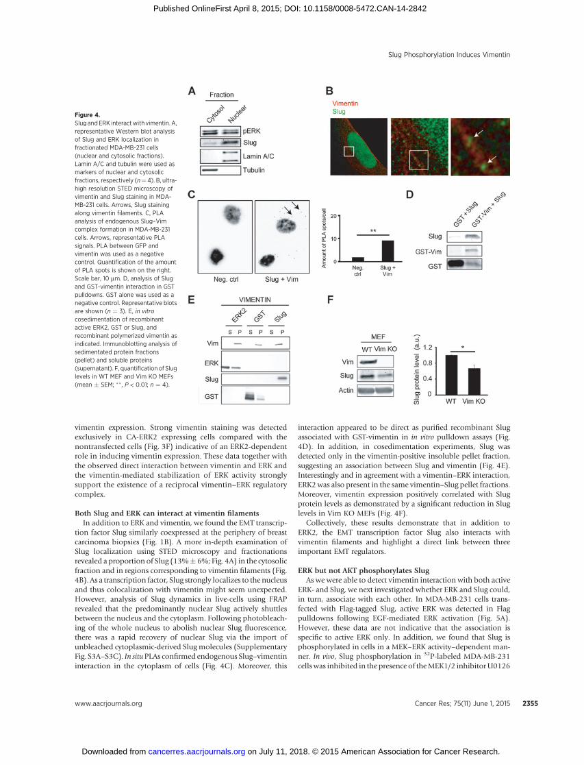
tion factor Slug similarly coexpressed at the periphery of breastcarcinoma biopsies (Fig. 1B). A more in-depth examination ofSlug localization using STED microscopy and fractionationsrevealed a proportion of Slug (13%� 6%; Fig. 4A) in the cytosolicfraction and in regions corresponding to vimentin filaments (Fig.4B). As a transcription factor, Slug strongly localizes to the nucleusand thus colocalization with vimentin might seem unexpected.However, analysis of Slug dynamics in live-cells using FRAPrevealed that the predominantly nuclear Slug actively shuttlesbetween the nucleus and the cytoplasm. Following photobleach-ing of the whole nucleus to abolish nuclear Slug fluorescence,there was a rapid recovery of nuclear Slug via the import ofunbleached cytoplasmic-derived Slug molecules (SupplementaryFig. S3A–S3C). In situPLAs confirmed endogenous Slug–vimentininteraction in the cytoplasm of cells (Fig. 4C). Moreover, this
interaction appeared to be direct as purified recombinant Slugassociated with GST-vimentin in in vitro pulldown assays (Fig.4D). In addition, in cosedimentation experiments, Slug wasdetected only in the vimentin-positive insoluble pellet fraction,suggesting an association between Slug and vimentin (Fig. 4E).Interestingly and in agreement with a vimentin–ERK interaction,ERK2was also present in the same vimentin–Slug pellet fractions.Moreover, vimentin expression positively correlated with Slugprotein levels as demonstrated by a significant reduction in Sluglevels in Vim KO MEFs (Fig. 4F).
Collectively, these results demonstrate that in addition toERK2, the EMT transcription factor Slug also interacts withvimentin filaments and highlight a direct link between threeimportant EMT regulators.
ERK but not AKT phosphorylates SlugAs we were able to detect vimentin interaction with both active
ERK- and Slug, we next investigated whether ERK and Slug could,in turn, associate with each other. In MDA-MB-231 cells trans-fected with Flag-tagged Slug, active ERK was detected in Flagpulldowns following EGF-mediated ERK activation (Fig. 5A).However, these data are not indicative that the association isspecific to active ERK only. In addition, we found that Slug isphosphorylated in cells in a MEK–ERK activity–dependent man-ner. In vivo, Slug phosphorylation in 32P-labeled MDA-MB-231cells was inhibited in the presence of theMEK1/2 inhibitorU0126
Figure 4.Slug andERK interactwith vimentin. A,representative Western blot analysisof Slug and ERK localization infractionated MDA-MB-231 cells(nuclear and cytosolic fractions).Lamin A/C and tubulin were used asmarkers of nuclear and cytosolicfractions, respectively (n¼ 4). B, ultra-high resolution STED microscopy ofvimentin and Slug staining in MDA-MB-231 cells. Arrows, Slug stainingalong vimentin filaments. C, PLAanalysis of endogenous Slug–Vimcomplex formation in MDA-MB-231cells. Arrows, representative PLAsignals. PLA between GFP andvimentin was used as a negativecontrol. Quantification of the amountof PLA spots is shown on the right.Scale bar, 10 mm. D, analysis of Slugand GST-vimentin interaction in GSTpulldowns. GST alone was used as anegative control. Representative blotsare shown (n ¼ 3). E, in vitrocosedimentation of recombinantactive ERK2, GST or Slug, andrecombinant polymerized vimentin asindicated. Immunoblotting analysis ofsedimentated protein fractions(pellet) and soluble proteins(supernatant). F, quantification of Sluglevels in WT MEF and Vim KO MEFs(mean � SEM; �� , P < 0.01; n ¼ 4).
Slug Phosphorylation Induces Vimentin
www.aacrjournals.org Cancer Res; 75(11) June 1, 2015 2355
on July 11, 2018. © 2015 American Association for Cancer Research. cancerres.aacrjournals.org Downloaded from
Published OnlineFirst April 8, 2015; DOI: 10.1158/0008-5472.CAN-14-2842
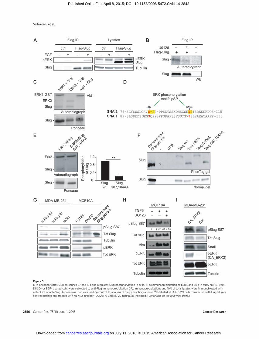
Figure 5.ERK phosphorylates Slug on serines 87 and 104 and regulates Slug phosphorylation in cells. A, coimmunoprecipitation of pERK and Slug in MDA-MB-231 cells.DMSO- or EGF- treated cells were subjected to anti-Flag immunoprecipitation (IP). Immunoprecipitations and 10% of total lysates were immunoblotted withanti-pERK or anti-Slug. Tubulin was used as a loading control. B, analysis of Slug phosphorylation in 32P-labeled MDA-MB-231 cells transfected with Flag-Slug orcontrol plasmid and treated with MEK1/2 inhibitor (U0126; 10 mmol/L, 20 hours), as indicated. (Continued on the following page.)
Virtakoivu et al.
Cancer Res; 75(11) June 1, 2015 Cancer Research2356
on July 11, 2018. © 2015 American Association for Cancer Research. cancerres.aacrjournals.org Downloaded from
Published OnlineFirst April 8, 2015; DOI: 10.1158/0008-5472.CAN-14-2842
(Fig. 5B). To clarify whether ERK phosphorylates Slug directly, weperformed in vitro kinase assays using purified recombinant GST-Slug and recombinant ERK1 and ERK2 kinases. Both ERK1 andERK2 directly phosphorylated Slug (Fig. 5C). Interestingly, phos-phorylation of Slug by ERK isoforms appeared to be specific asanother kinase, Akt1, was not able to induce Slug phosphoryla-tion (Fig. 5C). The possible ERK phosphorylation sites in Slugwere determined by liquid chromatography-tandem mass spec-trometry (LC/MS-MS) analysis (Supplementary Fig. S4A). Inter-estingly, two major serine phosphosites (S87 and S104) wereidentified that matched the minimal consensus ERK phosphor-ylation motif (pSP) and ERK2 appeared to be more efficient,than ERK1, in phosphorylating these sites (Supplementary Fig.S4B). Unlike the four conserved central GSK3b phosphoryla-tion sites in Slug and Snail (25, 26) that are implicated inregulation of protein stability, the two ERK consensus motifsidentified in Slug are absent in Snail (Fig. 5D), suggesting thatERK may regulate Slug distinctly from Snail (14). To confirmthe ERK phosphorylation sites within Slug, we generated arecombinant Slug mutant with alanine substitutions at thespecific phosphoserine residues (Slug S87,104AA), identifiedby LC/MS-MS, and used this as a substrate for in vitro kinaseassays. ERK-dependent phosphorylation of Slug S87,104AAwas clearly reduced compared with the WT Slug protein (Fig.5E). The relevant contribution of the two ERK-sites to Slugphosphorylation was further studied in cells. Phos-Tag resolu-tion of cell lysates, expressing exogenous Slug constructs, fol-lowed by immunolabeling with anti-Slug antibody revealed asingle slow-migrating phosphorylated Slug band in cellsexpressing Slug WT and Slug S104A only. In contrast, SlugS87A and Slug S87,104AA double mutant migrated at a sizecorresponding to the control recombinant nonphosphorylatedSlug on Phos-Tag gels (Fig. 5F). Together, these data indicatethat S87 is the principal ERK phosphorylation site in Slug asdisrupting the S104 site does not influence the phosphoryla-tion status of Slug in MDA-MB-231 cells, albeit this residue canbe phosphorylated by ERK in vitro.
To examine whether endogenous Slug is phosphorylated onserine 87, we generated a recombinant antibody against phos-phorylated S87. The antibody detected Slug from cell lysates,but not the nonphosphorylated recombinant Slug and anti-body reactivity, was lost upon Slug silencing (Fig. 5G), dem-onstrating antibody specificity toward phosphorylated Slug.Importantly, treatment of MCF10A and H-Ras–transformedMCF10A cells with MEK1/2 inhibitor, U0126, clearly reducedphosphorylation of Slug in cells, providing further evidencethat Slug is phosphorylated on serine 87 in a MEK/ERK1/2-dependent manner (Fig. 5G and Supplementary Fig. S4C).Antibody specificity was further validated by transfecting cellswith Flag-Slug WT/S87A/S87,104AA constructs. S87-P anti-
body clearly showed reduced immunoreactivity toward over-expressed Slug mutants as compared with WT Slug (Supple-mentary Fig. S4D). Importantly, TGFb induced the levels ofS87-P in MCF10A cells and this was sensitive to MEK1/2inhibition (Fig. 5H). Finally, overexpression of a constitutivelyactive MEK–ERK2 fusion plasmid in MDA-MB-231 cellsincreased the phosphorylation levels of S87. In line with arecent report demonstrating ERK2-mediated phosphorylationand stabilization of Snail (14), cells expressing active ERK2possessed higher levels of total Snail protein (Fig. 5I).
ERK phosphorylation of Slug regulates the expression ofvimentin
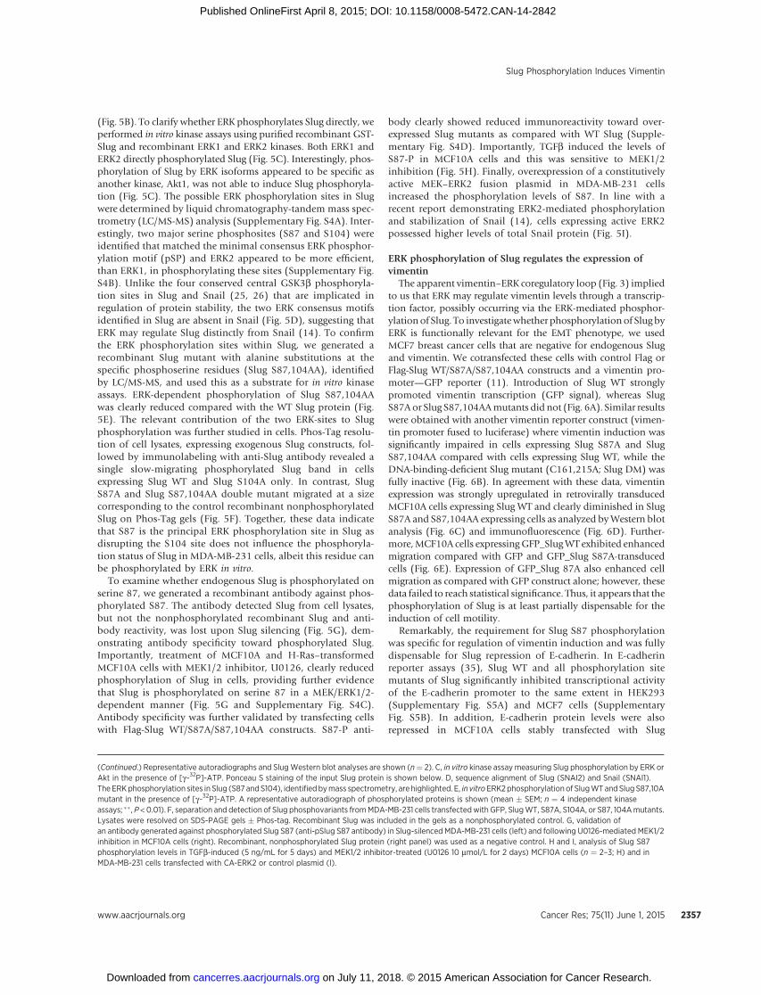
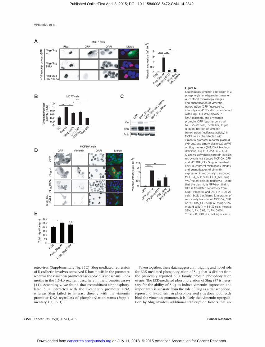
The apparent vimentin–ERK coregulatory loop (Fig. 3) impliedto us that ERK may regulate vimentin levels through a transcrip-tion factor, possibly occurring via the ERK-mediated phosphor-ylation of Slug. To investigatewhether phosphorylation of Slug byERK is functionally relevant for the EMT phenotype, we usedMCF7 breast cancer cells that are negative for endogenous Slugand vimentin. We cotransfected these cells with control Flag orFlag-Slug WT/S87A/S87,104AA constructs and a vimentin pro-moter—GFP reporter (11). Introduction of Slug WT stronglypromoted vimentin transcription (GFP signal), whereas SlugS87A or Slug S87,104AAmutants did not (Fig. 6A). Similar resultswere obtained with another vimentin reporter construct (vimen-tin promoter fused to luciferase) where vimentin induction wassignificantly impaired in cells expressing Slug S87A and SlugS87,104AA compared with cells expressing Slug WT, while theDNA-binding-deficient Slug mutant (C161,215A; Slug DM) wasfully inactive (Fig. 6B). In agreement with these data, vimentinexpression was strongly upregulated in retrovirally transducedMCF10A cells expressing Slug WT and clearly diminished in SlugS87A and S87,104AA expressing cells as analyzed byWestern blotanalysis (Fig. 6C) and immunofluorescence (Fig. 6D). Further-more,MCF10A cells expressingGFP_SlugWT exhibited enhancedmigration compared with GFP and GFP_Slug S87A-transducedcells (Fig. 6E). Expression of GFP_Slug 87A also enhanced cellmigration as compared with GFP construct alone; however, thesedata failed to reach statistical significance. Thus, it appears that thephosphorylation of Slug is at least partially dispensable for theinduction of cell motility.
Remarkably, the requirement for Slug S87 phosphorylationwas specific for regulation of vimentin induction and was fullydispensable for Slug repression of E-cadherin. In E-cadherinreporter assays (35), Slug WT and all phosphorylation sitemutants of Slug significantly inhibited transcriptional activityof the E-cadherin promoter to the same extent in HEK293(Supplementary Fig. S5A) and MCF7 cells (SupplementaryFig. S5B). In addition, E-cadherin protein levels were alsorepressed in MCF10A cells stably transfected with Slug
(Continued.) Representative autoradiographs and SlugWestern blot analyses are shown (n¼ 2). C, in vitro kinase assaymeasuring Slug phosphorylation by ERK orAkt in the presence of [g-32P]-ATP. Ponceau S staining of the input Slug protein is shown below. D, sequence alignment of Slug (SNAI2) and Snail (SNAI1).TheERKphosphorylation sites in Slug (S87 andS104), identifiedbymass spectrometry, are highlighted. E, in vitroERK2phosphorylation of SlugWTandSlug S87,10Amutant in the presence of [g-32P]-ATP. A representative autoradiograph of phosphorylated proteins is shown (mean � SEM; n ¼ 4 independent kinaseassays; ��, P <0.01). F, separation and detection of Slug phosphovariants fromMDA-MB-231 cells transfectedwith GFP, SlugWT, S87A, S104A, or S87, 104Amutants.Lysates were resolved on SDS-PAGE gels � Phos-tag. Recombinant Slug was included in the gels as a nonphosphorylated control. G, validation ofan antibody generated against phosphorylated Slug S87 (anti-pSlug S87 antibody) in Slug-silenced MDA-MB-231 cells (left) and following U0126-mediated MEK1/2inhibition in MCF10A cells (right). Recombinant, nonphosphorylated Slug protein (right panel) was used as a negative control. H and I, analysis of Slug S87phosphorylation levels in TGFb-induced (5 ng/mL for 5 days) and MEK1/2 inhibitor-treated (U0126 10 mmol/L for 2 days) MCF10A cells (n ¼ 2–3; H) and inMDA-MB-231 cells transfected with CA-ERK2 or control plasmid (I).
Slug Phosphorylation Induces Vimentin
www.aacrjournals.org Cancer Res; 75(11) June 1, 2015 2357
on July 11, 2018. © 2015 American Association for Cancer Research. cancerres.aacrjournals.org Downloaded from
Published OnlineFirst April 8, 2015; DOI: 10.1158/0008-5472.CAN-14-2842
retrovirus (Supplementary Fig. S5C). Slug-mediated repressionof E-cadherin involves conserved E-box motifs in the promoter,whereas the vimentin promoter lacks obvious consensus E-boxmotifs in the 1.5-kb segment used here in the promoter assays(11). Accordingly, we found that recombinant unphosphory-lated Slug interacted with the E-cadherin promoter DNA,whereas Slug failed to interact directly with the vimentinpromoter DNA regardless of phosphorylation status (Supple-mentary Fig. S5D).
Taken together, these data suggest an intriguing and novel rolefor ERK-mediated phosphorylation of Slug that is distinct fromthe previously reported Slug family protein phosphorylationevents. The ERK-mediated phosphorylation of Slug S87 is neces-sary for the ability of Slug to induce vimentin expression andimportantly is separate from the role of Slug as a transcriptionalrepressor of E-cadherin. As phosphorylated Slug does not directlybind the vimentin promoter, it is likely that vimentin upregula-tion by Slug involves additional transcription factors that are
Figure 6.Slug induces vimentin expression in aphosphorylation-dependent manner.A, confocal microscopy imagesand quantification of vimentintranscription (GFP fluorescenceintensity) in MCF7 cells cotransfectedwith Flag-Slug WT/S87A/S87,104A plasmids, and a vimentinpromoter-GFP reporter construct(n ¼ 25–28 cells). Scale bar, 10 mm.B, quantification of vimentintranscription (luciferase activity) inMCF7 cells cotransfected withvimentin promoter reporter plasmid(VP-Luc) and empty plasmid, SlugWTor Slug mutants (DM, DNA binding-deficient Slug C161,215A; n ¼ 3–5).C, analysis of vimentin protein levels inretrovirally transduced MCF10A_GFPand MCF10A_GFP Slug WT/mutantcells. D, confocal microscopy imagesand quantification of vimentinexpression in retrovirally transducedMCF10A_GFP or MCF10A_GFP SlugWT/mutant cells stained for GFP (notethat the plasmid is GFP-ires, that is,GFP is translated separately fromSlug), vimentin, and DAPI (n ¼ 21–25cells). Scale bar, 10 mm. E, migration ofretrovirally transduced MCF10A_GFPor MCF10A_GFP Slug WT/Slug S87Amutant cells (n ¼ 34–39 cells; mean�SEM; � , P < 0.05; �� , P < 0.001;��� , P < 0.0001; n.s., not significant).
Virtakoivu et al.
Cancer Res; 75(11) June 1, 2015 Cancer Research2358
on July 11, 2018. © 2015 American Association for Cancer Research. cancerres.aacrjournals.org Downloaded from
Published OnlineFirst April 8, 2015; DOI: 10.1158/0008-5472.CAN-14-2842
recruited to Slug in a Slug-phosphorylation–dependent manner.However, this remains to be investigated.
ERKphosphorylationdoes not regulate Slug stability or nuclearaccumulation
Recently, there has been intense focus on the link betweenphosphorylation of Slug family transcription factors and theirstability and nuclear localization. We found that all GFP-taggedSlug mutants localized predominantly to the nucleus in MCF10Acells with no significant difference detected between the serinephosphositemutants and SlugWT (Supplementary Fig. S5E). Thisnuclear staining was not a consequence of the GFP tag as resultswere reproducible in cells expressing Flag-tagged Slug mutants(not shown). Furthermore, silencing of vimentin had no effect onthe relative abundance of endogenous Slug in the nuclear andcytoplasmic fractions in MDA-MB-231 cells (Supplementary Fig.S5F). This suggests that ERK does not regulate Slug nuclearlocalization and would be in agreement with the fact that Slug
lacks the phosphorylation-masked nuclear export motif found inSnail (36). During the course of these studies, Slug appeared to berather stable in different cell lines regardless of GSK3b activity andcorrespondingly we did not detect any significant differences inSlugWT or Slug S87,104AA protein stability in cells (Supplemen-tary Fig. S5G).
Slug-S87 phosphorylation is necessary for Axl inductionWe have shown earlier that vimentin regulates EMT induction
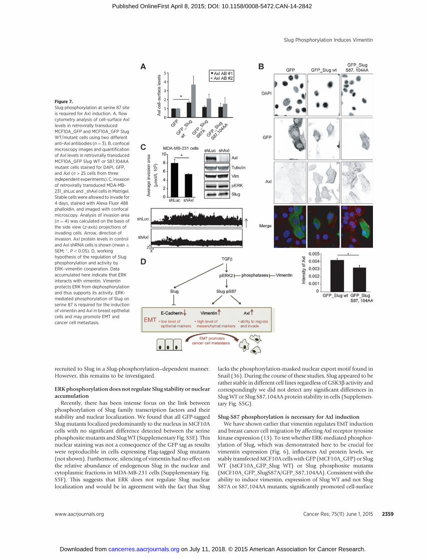
and breast cancer cell migration by affecting Axl receptor tyrosinekinase expression (13). To test whether ERK-mediated phosphor-ylation of Slug, which was demonstrated here to be crucial forvimentin expression (Fig. 6), influences Axl protein levels, westably transfectedMCF10A cells withGFP (MCF10A_GFP) or SlugWT (MCF10A_GFP_Slug WT) or Slug phosphosite mutants(MCF10A_GFP_SlugS87A/GFP_S87,104AA). Consistent with theability to induce vimentin, expression of Slug WT and not SlugS87A or S87,104AA mutants, significantly promoted cell-surface
Figure 7.Slug phosphorylation at serine 87 siteis required for Axl induction. A, flowcytometry analysis of cell-surface Axllevels in retrovirally transducedMCF10A_GFP and MCF10A_GFP SlugWT/mutant cells using two differentanti-Axl antibodies (n¼ 3). B, confocalmicroscopy images and quantificationof Axl levels in retrovirally transducedMCF10A_GFP Slug WT or S87,104AAmutant cells stained for DAPI, GFP,and Axl (n > 25 cells from threeindependent experiments). C, invasionof retrovirally transduced MDA-MB-231_shLuc and _shAxl cells in Matrigel.Stable cells were allowed to invade for4 days, stained with Alexa Fluor 488phalloidin, and imaged with confocalmicroscopy. Analysis of invasion area(n ¼ 4) was calculated on the basis ofthe side view (z-axis) projections ofinvading cells. Arrow, direction ofinvasion. Axl protein levels in controland Axl shRNA cells is shown (mean�SEM; �, P < 0.05). D, workinghypothesis of the regulation of Slugphosphorylation and activity byERK–vimentin cooperation. Dataaccumulated here indicate that ERKinteracts with vimentin. Vimentinprotects ERK from dephosphorylationand thus supports its activity. ERK-mediated phosphorylation of Slug onserine 87 is required for the inductionof vimentin and Axl in breast epithelialcells and may promote EMT andcancer cell metastasis.
Slug Phosphorylation Induces Vimentin
www.aacrjournals.org Cancer Res; 75(11) June 1, 2015 2359
on July 11, 2018. © 2015 American Association for Cancer Research. cancerres.aacrjournals.org Downloaded from
Published OnlineFirst April 8, 2015; DOI: 10.1158/0008-5472.CAN-14-2842
levels of Axl, based on FACS analysis (Fig. 7A) and total Axlexpression levels detected by immunofluorescence staining (Fig.7B). Because vimentin, ERK, and Slug were important for MDA-MB-231 cell invasion and ERK-mediated phosphorylation of Slugis required for Axl induction in MCF10A cells, we tested whetherAxl inhibition could also impede cell invasion. Stable silencing ofAxl in MDA-MB-231 cells did not influence levels of vimentin,pERK, or Slug but significantly reduced MDA-MB-231 cell inva-sion comparedwith control knockdown cells (Fig. 7C). These datasuggest that Axl contributes to EMT-linked invasion downstreamof the EMT-inducing factors critical for Axl expression. In conclu-sion, our data show that phosphorylation of Slug at serine 87 isessential for Slug-mediated vimentin induction and Axl expres-sion. Importantly, direct inhibition of Axl expression or of thepathways that impinge on Axl expression hinders MDA-MB-231cell invasion.
DiscussionHere, we found that a reciprocal regulatory vimentin–ERK
interaction facilitates ERK phosphorylation of Slug at a novel site(S87) that determines the ability of Slug to induce vimentin andAxl expression. Intriguingly, ERK-mediated Slug S87 phosphor-ylation uncouples the activating and repressing functions of Slugso that ERK-mediated S87 phosphorylation is fully dispensablefor E-cadherin repression by Slug. Our workingmodel, posits thatthe vimentin–ERK axis regulates EMT via phosphorylation of SlugS87 and induces expression of vimentin and Axl (Fig. 7D). Inaddition, ERK-dependent inhibition of GSK3b activity canenforce this EMT induction indirectly by stabilizing Slug to repressE-cadherin, as implied by data linking GSK3b activity to desta-bilization of Slug (25). Hence, vimentin functions as an impor-tant and central EMT signaling scaffold supporting ERK activityand possibly bringing together Slug and ERK in the cytoplasm(though this remains to be formally shown; Fig. 7D).
As discussed in detail below, several studies have investigatedthe role of phosphorylation on the subcellular localization andprotein stability of Snail family members. In contrast, the linkbetween phosphorylation and the ability of these transcriptionfactors to regulate gene expression remains poorly investigatedand the limited data available focuses on transcriptional repres-sion. Phosphorylation of serine 11 and 92 is independentlyrequired for Snail repression of E-cadherin expression and forefficient recruitment of the corepressor mSin3A (37). In addition,Slug phosphorylation at serine 4 has been functionally implicatedin E-cadherin repression (24); however, the detailed mechanismremains to be investigated. We show here that ERK2-mediatedphosphorylation of Slug on S87 specifically influences Slug-dependent transcriptional activation of vimentin, and to the bestof our knowledge, this is the first example of how repressionversus activation of genes is achieved by an EMT-regulatingtranscription factor. Our data suggest that in contrast to Slug-mediated E-cadherin repression, the ability of Slug to inducevimentin transcription does not involve direct Slug-recruitmentto the vimentin promoter DNA. In the future, it will be importantto identify the additional transcription factors necessary for Slugphosphorylation–dependent vimentin induction and to fullydissect the repressor and activator functions of Slug in EMTregulation.
Vimentin is considered a canonicalmarker of EMT; however, itsfunctional role remains elusive (2, 8). We have recently shown
that vimentin is required for EMT induction by H-RasV12, Slug,and TGFb (13). Accruing evidence highlights the multifacetedrole of vimentin in determining cell shape, regulating cellmotility, and integrin turnover and as a signaling scaffold thatcan bind several different proteins (12, 13, 19, 38). Therefore, afunctional role for vimentin in cellular processes, includingEMT, is evident, albeit incompletely understood at present. Inthe majority of carcinomas, vimentin is overexpressed andseveral studies have linked vimentin expression to tumoraggressiveness and metastasis (8). We find here that vimentinexpression is critical for maintaining high ERK activity in cells.The ability of vimentin to bind to and to protect ERK fromdephosphorylation is most likely a contributing factor toincreased ERK activity. However, we also expect other mechan-isms to be involved in the regulation of ERK activity as theproportion of ERK sequestered by vimentin in cells in low.Conversely, ERK activity regulates vimentin expression andSlug phosphorylation in breast cancer cells.
Our identification of Slug, active ERK, and vimentin coexpres-sion at the invasive edges of triple-negative breast carcinomas andthe role of these proteins in regulating breast cancer cell motilityand invasion in vitro suggest that this pathway would be animportant clinical target. Furthermore, the fact that vimentin-positive MDA-MB-231 cell metastases, from orthotopic xeno-grafts, express higher level of vimentin than in the primary tumors(Fig. 1E) and that vimentin contributes to xenograft growth in vivo(Fig. 3C), underlines the apparent role for vimentin in cancerprogression and metastatic dissemination. Therefore, under-standing the role and regulation of vimentin in EMTmay unravelnew strategies to target this important pathway in cancer.
GSK3b has been suggested to regulate the stability of Slugand Snail in epithelial cells via posttranslational modification.Phosphorylation-deficient alanine substitutions of four keyphosphorylation sites, which appear to be conserved betweenSlug and Snail, results in a more stable protein with increasedefficiency in EMT induction (14, 23, 25, 26). One of thesephosphorylation sites (S104 in Slug), identified here as an ERKphosphorylation site in vitro, is also phosphorylated by GSK3b.According to our data, this site may not be prominentlyphosphorylated at least in MDA-MB-231 cells where mutagen-esis of another serine (S87) clearly reduces overall Slug phos-phorylation. This further emphasizes the seemingly dominantrole of ERK in the regulation of Slug function because inaddition to controlling GSK3b activity ERK-mediated phos-phorylation of Slug at S87 controls the overall phosphorylationof Slug in cells.
It is possible that these findings are also relevant for ourunderstanding of the regulatory networks that determine themammary stem cell state and regulate human breast cancer stemcells (9). Vimentin is abundantly expressed in stem cells and isinduced alongside Slug during conversion of luminal mammaryepithelial cells into MaSC (9). The vimentin–ERK axis is criticallyimportant for Axl tyrosine kinase expression in breast cancer cells(13), and Axl expression is also associated with the expression ofstem cell genes in addition to metastasis-linked genes (39).Therefore, it is likely that vimentin–ERK coregulation and con-tribution to Slug phosphorylation is also functional in the reg-ulationof stemness.However,more studies are required to furtherexamine the activity of this pathway and its potential implicationsin tumorigenesis, metastasis, and the maintenance of humanbreast cancer stem cells.
Cancer Res; 75(11) June 1, 2015 Cancer Research2360
Virtakoivu et al.
on July 11, 2018. © 2015 American Association for Cancer Research. cancerres.aacrjournals.org Downloaded from
Published OnlineFirst April 8, 2015; DOI: 10.1158/0008-5472.CAN-14-2842
Disclosure of Potential Conflicts of InterestJ.B. Lorens is a CSO at BerGenBio and has ownership interest (including
patents) in the same. No potential conflicts of interest were disclosed by theother authors.
Authors' ContributionsConception and design: R. Virtakoivu, E. Mattila, J.B. Lorens, J. IvaskaDevelopment of methodology: E. Mattila, G. CorthalsAcquisition of data (provided animals, acquired and managed patients,provided facilities, etc.): R. Virtakoivu, A. Mai, E. Mattila, N. De Franceschi,S.Y. Imanishi, G. Corthals, M. Saari, F. Cheng, E. Torvaldson, A. Mannermaa,G. Muharram, Y. Soini, J. IvaskaAnalysis and interpretation of data (e.g., statistical analysis, biostatistics,computational analysis): R. Virtakoivu, E. Mattila, S.Y. Imanishi, G. Corthals,F. Cheng, Y. SoiniWriting, review, and/or revisionof themanuscript:R.Virtakoivu, A.Mai,N.DeFranceschi, G. Corthals, R. Kaukonen, V.-M. Kosma, A. Mannermaa, C. Gilles,J. Eriksson, Y. Soini, J.B. Lorens, J. IvaskaAdministrative, technical, or material support (i.e., reporting or organizingdata, constructing databases): R. Virtakoivu, A. Mai, R. Kaukonen, M. Saari,A. Mannermaa, C. Gilles, Y. SoiniStudy supervision: J. IvaskaOther (expertise and model systems related to vimentin): J. Eriksson
AcknowledgmentsThe authors thank L. Lahtinen, J. Siivonen, S. Kaustara, K. Vuoriluoto, andH.
Marttila for excellent technical assistance and H. Hamidi for scientific writingand editing of the article. The authors are grateful to Dr. J. Westermarck forsharing his reagents and critically reading the article.
Grant SupportThis study has been supported by the Academy of Finland, ERC
Starting Grant (#202809), ERC Consolidator Grant (#615258), theSigrid Juselius Foundation, and the Finnish Cancer Organization. R.Virtakoivu has been supported by the K. Albin Johansson Foundation,Lounais-Suomen sy€op€ayhdistys, Instrumentariumin tiedes€a€ati€o, OrionResearch Foundation and by the Turku Doctoral Program of BiomedicalSciences. N. De Franceschi has been supported by the Drug ResearchDoctoral Program. E. Mattila has been supported by the Academy ofFinland postdoc grant.
The costs of publication of this articlewere defrayed inpart by the payment ofpage charges. This article must therefore be hereby marked advertisement inaccordance with 18 U.S.C. Section 1734 solely to indicate this fact.
Received September 24, 2014; revised February 9, 2015; accepted February 22,2015; published OnlineFirst April 8, 2015.
References1. Moustakas A, Heldin CH. Induction of epithelial–mesenchymal tran-
sition by transforming growth factor beta. Semin Cancer Biol 2012;22:446–54.
2. Thiery JP, Acloque H, Huang RY, Nieto MA. Epithelial–mesenchymaltransitions in development and disease. Cell 2009;139:871–90.
3. Hanahan D, Weinberg RA. Hallmarks of cancer: the next generation. Cell2011;144:646–74.
4. De Craene B, Berx G. Regulatory networks defining EMT during cancerinitiation and progression. Nat Rev Cancer 2013;13:97–110.
5. Puisieux A, Brabletz T, Caramel J. Oncogenic roles of EMT-inducingtranscription factors. Nat Cell Biol 2014;16:488–94.
6. Perou CM. Molecular stratification of triple-negative breast cancers.Oncologist 2010;15(Suppl 5):39–48.
7. Shah SP, Roth A, Goya R,, Oloumi A, Ha G, Zhao Y, et al. The clonal andmutational evolution spectrum of primary triple-negative breast cancers.Nature 2012;486:395–9.
8. Satelli A, Li S. Vimentin in cancer and its potential as a molecular target forcancer therapy. Cell Mol Life Sci 2011;68:3033–46.
9. Guo W, Keckesova Z, Donaher JL, Shibue T, Tischler V, Reinhardt F, et al.Slug and Sox9 cooperatively determine the mammary stem cell state. Cell2012;148:1015–28.
10. Mani SA, Guo W, Liao MJ,, Eaton EN, Ayyanan A, Zhou AY, et al. Theepithelial–mesenchymal transition generates cells with properties of stemcells. Cell 2008;133:704–15.
11. Gilles C, Polette M, Zahm JM,, Tournier JM, Volders L, Foidart JM, et al.Vimentin contributes to human mammary epithelial cell migration. J CellSci 1999;112:4615–25.
12. Mendez MG, Kojima S, Goldman RD. Vimentin induces changes in cellshape, motility, and adhesion during the epithelial to mesenchymaltransition. FASEB J 2010;24:1838–51.
13. Vuoriluoto K, HaugenH, Kiviluoto S, Mpindi JP, Nevo J, GjerdrumC, et al.Vimentin regulates EMT induction by slug and oncogenic H-ras andmigration by governing axl expression in breast cancer. Oncogene 2011;30:1436–48.
14. Zhang K, Corsa CA, Ponik SM,, Prior JL, Piwnica-Worms D, Eliceiri KW,et al. The collagen receptor discoidin domain receptor 2 stabilizes SNAIL1to facilitate breast cancer metastasis. Nat Cell Biol 2013;15:677–87.
15. Shin S, Dimitri CA, Yoon SO, Dowdle W, Blenis J. ERK2 but not ERK1induces epithelial-to-mesenchymal transformation via DEF motif-depen-dent signaling events. Mol Cell 2010;38:114–27.
16. Radtke S,MilanovicM, Rosse C,De RyckerM, Lachmann S,Hibbert A, et al.ERK2 but not ERK1mediates HGF-inducedmotility in non–small cell lungcarcinoma cell lines. J Cell Sci 2013;126:2381–91.
17. von Thun A, Birtwistle M, Kalna G, Grindlay J, Strachan D, Kolch W, et al.ERK2 drives tumour cell migration in three-dimensional microenviron-ments by suppressing expression of Rab17 and liprin-beta2. J Cell Sci2012;125:1465–77.
18. Botta GP, Reginato MJ, Reichert M, Rustgi AK, Lelkes PI. Constitutive K-RasG12D activation of ERK2 specifically regulates 3D invasion of humanpancreatic cancer cells via MMP-1. Mol Cancer Res 2012;10:183–96.
19. Ivaska J, Pallari HM,Nevo J, Eriksson JE.Novel functions of vimentin in celladhesion, migration, and signaling. Exp Cell Res 2007;313:2050–62.
20. Perlson E,Michaelevski I, KowalsmanN, Ben-Yaakov K, ShakedM, Seger R,et al. Vimentin binding to phosphorylated ERK sterically hinders enzy-matic dephosphorylation of the kinase. J Mol Biol 2006;364:938–44.
21. Nieto MA, Cano A. The epithelial–mesenchymal transition under control:global programs to regulate epithelial plasticity. Semin Cancer Biol 2012;22:361–8.
22. Zhang K, Rodriguez-Aznar E, Yabuta N, Owen RJ, Mingot JM, Nojima H,et al. Lats2 kinase potentiates Snail1 activity by promoting nuclear reten-tion upon phosphorylation. EMBO J 2012;31:29–43.
23. Zhou BP, Deng J, Xia W, Xu J, Li YM, Gunduz M, et al. Dual regulation ofsnail by GSK-3beta-mediated phosphorylation in control of epithelial–mesenchymal transition. Nat Cell Biol 2004;6:931–40.
24. Molina-Ortiz P, Villarejo A, MacPherson M, Santos V, Montes A, Souchel-nytskyi S, et al. Characterization of the SNAG and SLUG domains of Snail2in the repression of E-cadherin and EMT induction:modulation by serine 4phosphorylation. PLoS One 2012;7:e36132.
25. Wu ZQ, Li XY, Hu CY, FordM, Kleer CG,Weiss SJ. Canonical wnt signalingregulates slug activity and links epithelial–mesenchymal transition withepigenetic breast cancer 1, early onset (BRCA1) repression. Proc Natl AcadSci U S A 2012;109:16654–9.
26. Kao SH, Wang WL, Chen CY, Chang YL, Wu YY, Wang YT, et al. GSK3betacontrols epithelial–mesenchymal transition and tumor metastasis byCHIP-mediated degradation of slug. Oncogene 2014;33:3172–82.
27. Kim JY, Kim YM, Yang CH, Cho SK, Lee JW, Cho M. Functional regulationof slug/Snail2 is dependent on GSK-3beta–mediated phosphorylation.FEBS J 2012;279:2929–39.
28. Chen H, Zhu G, Li Y, Padia RN, Dong Z, Pan ZK, et al. Extracellular signal-regulated kinase signaling pathway regulates breast cancer cell migrationby maintaining slug expression. Cancer Res 2009;69:9228–35.
29. Geradts J, de Herreros AG, Su Z, Burchette J, Broadwater G, Bachelder RE.Nuclear Snail1 and nuclear ZEB1 protein expression in invasive andintraductal human breast carcinomas. Hum Pathol 2011;42:1125–31.
30. Karihtala P, Auvinen P, Kauppila S, Haapasaari KM, Jukkola-Vuorinen A,Soini Y. Vimentin, zeb1 and Sip1 are up-regulated in triple-negative and
www.aacrjournals.org Cancer Res; 75(11) June 1, 2015 2361
Slug Phosphorylation Induces Vimentin
on July 11, 2018. © 2015 American Association for Cancer Research. cancerres.aacrjournals.org Downloaded from
Published OnlineFirst April 8, 2015; DOI: 10.1158/0008-5472.CAN-14-2842
basal-like breast cancers: associationwith an aggressive tumour phenotype.Breast Cancer Res Treat 2013;138:81–90.
31. Bartholomeusz C, Gonzalez-Angulo AM, Liu P, Hayashi N, Lluch A, Ferrer-Lozano J, et al. High ERK protein expression levels correlate with shortersurvival in triple-negative breast cancer patients. Oncologist 2012;17:766–74.
32. Junttila MR, Li SP, Westermarck J. Phosphatase-mediated crosstalkbetweenMAPK signaling pathways in the regulation of cell survival. FASEBJ 2008;22:954–65.
33. Downward J. Targeting RAS signalling pathways in cancer therapy. Nat RevCancer 2003;3:11–22.
34. Matallanas D, Crespo P. New druggable targets in the ras pathway? CurrOpin Mol Ther 2010;12:674–83.
35. Slorach EM, Chou J, Werb Z. Zeppo1 is a novel metastasis promoter thatrepresses E-cadherin expression and regulates p120-catenin isoformexpression and localization. Genes Dev 2011;25:471–84.
36. Dominguez D, Montserrat-Sentis B, Virgos-Soler A, Guaita S, Grueso J,Porta M, et al. Phosphorylation regulates the subcellular location andactivity of the snail transcriptional repressor. Mol Cell Biol 2003;23:5078–89.
37. MacPhersonMR, Molina P, Souchelnytskyi S, Wernstedt C, Martin-P�erez J,Portillo F, et al. Phosphorylation of serine 11 and serine 92 as new positiveregulators of human Snail1 function: potential involvement of caseinkinase-2 and the cAMP-activated kinase protein kinase A. Mol Biol Cell2010;21:244–53.
38. Ivaska J, Vuoriluoto K, Huovinen T, Izawa I, Inagaki M, Parker PJ. PKCep-silon-mediated phosphorylation of vimentin controls integrin recyclingand motility. EMBO J 2005;24:3834–45.
39. Byers LA, Diao L, Wang J, Saintigny P, Girard L, Peyton M, et al. Anepithelial–mesenchymal transition gene signature predicts resistance toEGFR and PI3K inhibitors and identifies axl as a therapeutic target forovercoming EGFR inhibitor resistance. Clin Cancer Res 2013;19:279–90.
Cancer Res; 75(11) June 1, 2015 Cancer Research2362
Virtakoivu et al.
on July 11, 2018. © 2015 American Association for Cancer Research. cancerres.aacrjournals.org Downloaded from
Published OnlineFirst April 8, 2015; DOI: 10.1158/0008-5472.CAN-14-2842
2015;75:2349-2362. Published OnlineFirst April 8, 2015.Cancer Res Reetta Virtakoivu, Anja Mai, Elina Mattila, et al.
ERK Signaling Uncouples Slug Gene Regulatory Function−Vimentin
Updated version
10.1158/0008-5472.CAN-14-2842doi:
Access the most recent version of this article at:
Material
Supplementary
http://cancerres.aacrjournals.org/content/suppl/2015/05/15/0008-5472.CAN-14-2842.DC2
http://cancerres.aacrjournals.org/content/suppl/2015/04/10/0008-5472.CAN-14-2842.DC1Access the most recent supplemental material at:
Cited articles
http://cancerres.aacrjournals.org/content/75/11/2349.full#ref-list-1
This article cites 39 articles, 16 of which you can access for free at:
Citing articles
http://cancerres.aacrjournals.org/content/75/11/2349.full#related-urls
This article has been cited by 12 HighWire-hosted articles. Access the articles at:
E-mail alerts related to this article or journal.Sign up to receive free email-alerts
Subscriptions
Reprints and
To order reprints of this article or to subscribe to the journal, contact the AACR Publications Department at
Permissions
Rightslink site. Click on "Request Permissions" which will take you to the Copyright Clearance Center's (CCC)
.http://cancerres.aacrjournals.org/content/75/11/2349To request permission to re-use all or part of this article, use this link
on July 11, 2018. © 2015 American Association for Cancer Research. cancerres.aacrjournals.org Downloaded from
Published OnlineFirst April 8, 2015; DOI: 10.1158/0008-5472.CAN-14-2842