Embed Size (px)
Citation preview

NOTES OF ABDOMINAL WALL & INGUINAL REGION:
-planes that divide the abdomin into 9 regions are :
-2 vertical planes : R&L mid clavicular planes -2 transverse planes:( transpyloric & subcostal planes)
muscles of the anterior abdominal wall are 2 kinds-:
1/flat muscles, which include:(internal oblique, external oblique, transverse abdominis) 2/vertical muscles (found inside the rectus sheath):which include: rectus abdominis & pyramidalis ( small triangular m absent in most human beings but very important in dogs
-note: there is no deep fascia in the layers of the anterior abdominal wall, only superficial fascia is present which is composed of 1 layer above the umbilicus(scarpa) & 2 layers below the umbilicus (camper & scarpa)
the fibrous scarpa fascia(below the umbilicus) will continue to descend downward & laterally to reach the inguinal ligament & fuse with the fascia lata . & descends medially in the direction of pubic symphysis around the root of penis & fuse
with colle's fascis
-blood supply of anterior abdominal wall:
1-superior epigastric & musculophrenic As ------internal thoracic------subclavian
2-inferior epigastric (runs upward within the rectus sheath)-------anastomosing between branches of external iliac
1
تشريح مالحظات /4 / 18ثانيطب /
vessels of femoral (continue the B.supply on slide 10)
-linea alba: fibrous structure runs between the xyphoid processs superiorly & pubic symphysis inferiorly, represent a common insertion for 3 abdominal muscles: (ext oblique ,int oblique, transversus abdominis)
linea alba is devoid of blood supply therefore a surgical incision is made through the linea alba during operation
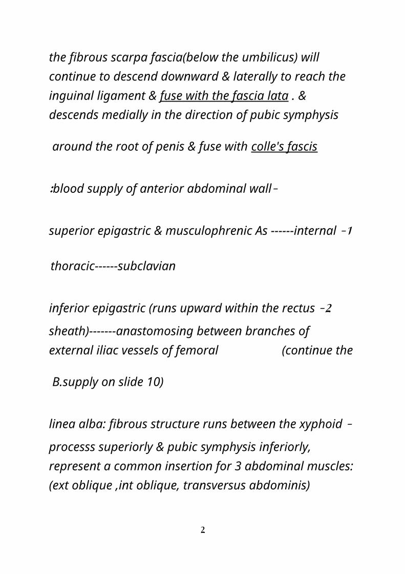
RECTUS SHEATH:- 2 muscles lies on the rectus sheath ( rectus abdominis & pyramidalis)
boundaries of rectus sheath: medially----linea alba laterally----linea semilunaris
formation of rectus sheath is by aponeurosis of:1/ext oblique 2/int oblique 3/transverse abdominis
content of rectus sheath : 1/ muscles (rectus abdominis & pyramidalis) 2/lower 5 intercostal nerves 3/superior & inferior epigastric As rectus sheath include 3 areas: 1/first part: above the ribs 2/second part: below the costal cartilages to the arcuate line 3/third part: from arcuate line downward including the first part : rectus abdominis will rest on the lower ribs & intercostal
spaces & only ext oblique aponeurosis is present
2

second part(above the arcuate line): the aponeurosis of int oblique will split & envelope the rectus abdomins m the superficial aponeurosis of int oblique will meet the aponeurosis of ext oblique forming the anterior layer of rectus sheath ,while the inner(deep) aponeurosis of int oblique meet hte aponeurosis of transverse abdominis forming the posterior layer of rectus sheath
note: ant layer of R.sheat is adherant 2 rectus abdominis while post layer of R sheath is NOT adherant to rectus
abdominis
third part(below the arcuate line): the 3 aponeurosis will meet above the rectus abdominis muscle forming ant layer of
3

R sheath
*femoral ring is wider in female than male , therfore the common type of hernia in females is femoral hernia while the common type of hernia in male is inguinal hernia
NOTES
-hessselbach triangle: is the site where direct hernia occurs specifically occus in the superficial inguinal ring while the indirect(oblique) hernia occurs in the inguinal canal and this type of hernia can descend into the scrotum
-the inferior epigastic artery is related lateral to direct hernia & medial to indirect hernia
4
-floor of hessselbach tiangle is corresponding to medial inguinal fossa & lateral border of hesselbach triangle is related to lateral inguinal fossa
-superficial inguinal ring is a defect in the external oblique aponeurosis & communicate with the scrotum while deep inguinal ring is a defect in transversalis fascia & communicate with the abdominal cavity . between them there is an oblique canal called inguinal canal
-some fibers of internal oblique m will form cremastric muscle while other fibers will form conjoint tendon along with transversus abdominis
-vas deferens open in prostatic urethra
-there 3 layers of fascia covering the spermatic cord : 1/external spermatic fascia ------- from external oblique aponeurosis
5

2/cremastric fascia ------from internal oblique fibers
3/internal spermatic fascia ------from fascia transversalis
NOTES INCLUDING THE PELVIS & PERINEUM
-the boney pelvis consist of 2 hip bones meets anteriorly at the pubic symphysis , each hip bone consist of : pubis , ilium, ischium ) , the 3 hip bones meet at floor of acetabulum
-the joint between the ilium & sacrum is called sacro-iliac joint , infront of this joint there is anteroir sacr-oiliac ligament while behind it there is posterior sacro-iliac ligament
6

- pelvic brim starts at pectineal line above the pelvic brim ------ greater pelvis or false pelvis the false pelvis is continuous with the lower part of abdominal cavity sigmoid colon , uterus & uterine tubes are seen in the false pelvis region below the level of pelvic brim-------- there is the true pelvis of lesser pelvis
-at the 38 weak of gestation the head of the fetus will pass below the pelvic brim and arrest on the true pelvis in a process called engagement
-types of female pelvis according to the shape of pelvic brim
7

-pelvic brim --------inlet of lesser pelvis perineum --------outlet or birth canal
outlet of pelvis is bounded : 1/posteriorly ----tip of coccyx , 2/anteriorly -----pubic arch+ pubic symphysis + inferior pubic ramus
3/on both sides -------ischial tuberosities , 4/sacro-spinal & sacro-tuberous ligaments
-pelvic floor = pelvic diaphragm urogenital diaphragm = deep perineal pouch
pelvic diaphragm------mainly levator ani m + coccygeus m
-external anal sphincter (skeletal muscle) supplied by : inferior rectal branch of perineal nerve
internal anal sphincter (smooth muscle) supplied by autonomic from inferior hypogastric plexus
-in the male ischiocavernousus muscle will arrest on the crura of penis while in the female it will arrest on the root of clitoris
8