Embed Size (px)
DESCRIPTION
Citation preview

disc pathology and non-surgical decompression
reuben henderson, , d.od.o..
• 1993 - michigan state university college of osteopathic medicine
• internship: flint osteopathic and st. lawrence hospitals 1994 & 1995
• pm&r residency: university of michigan 2001
• private practice 2004: pm&r
low back pain world wide
• common complaint among adults
• lifetime prevalence in working population up to 80%
• 60% experience functional limitation or disability
• second most common reason for work disability
• despite advances in imaging and surgical techniques
LBP prevalence and its cost are relatively unchanged
back pain causes
• de-conditioning• sprain/strain• spondylolithesis• spondylosis• facet syndrome• disc herniation
• disc bulge• spinal stenosis• biomechanical• inflammatory• infection• cancer
recent research on DDD
1. heredity may be largely responsible for degeneration/herniation of intervertebral disc.
2. genetic influences have been confirmed by the identification of several gene forms associated with disc degeneration.
• 1. Kenneth M C Cheung, “How has genetics research altered understanding of degenerative disc disease: implications for intervertebral disc regeneration.” European Cells and Materials Vol. 16 Suppl. 4, 2008 (page 8)
• 2. Yin’gang Zang, “Advances in susceptibility genetics of intervertebral degenerative disc disease.” Int J Biol Sci 2008 4:283-290
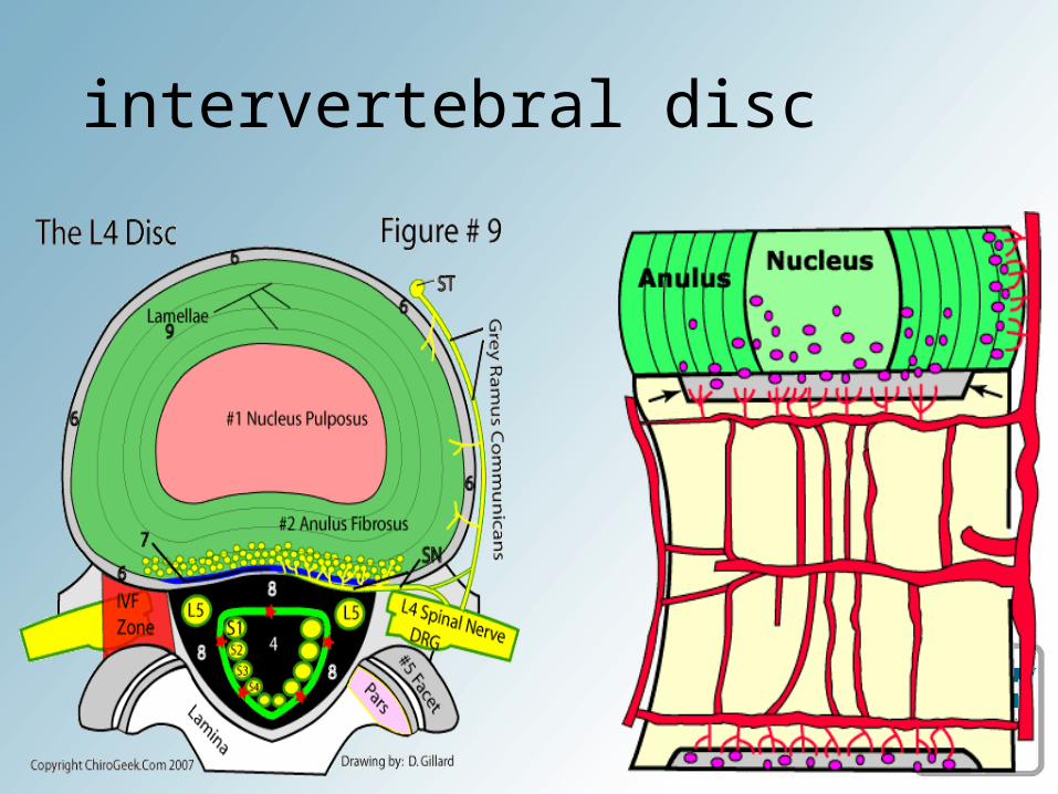
intervertebral disc
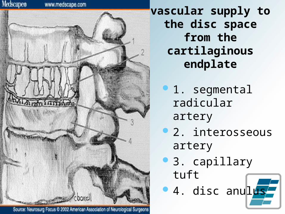
vascular supply to the disc space from
the cartilaginous endplate
1. segmental radicular artery
2. interosseous artery
3. capillary tuft 4. disc anulus

neurological innervation of
posterior spinal column
1. ascending branch of the sinuvertebral nerve
2. dorsal root ganglion3. descending branch of
the sinuvertebral nerve
4. disc anulus5. posterior
longitudinal ligament

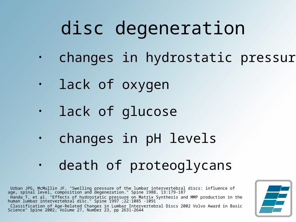
disc degeneration• changes in hydrostatic pressure
• lack of oxygen
• lack of glucose
• changes in pH levels
• death of proteoglycans
Urban JPG, McMullin JF, "Swelling pressure of the lumbar intervertebral discs: influence of age, spinal level, composition and degeneration." Spine 1988, 13:179-187 Handa T, et al. "Effects of hydrostatic pressure on Matrix Synthesis and MMP production in the human lumbar intervertebral disc." Spine 1997 ;22:1085 -1091 Classification of Age-Related Changes in Lumbar Intervertebral Discs 2002 Volvo Award in Basic Science" Spine 2002; Volume 27, Number 23, pp 2631-2644
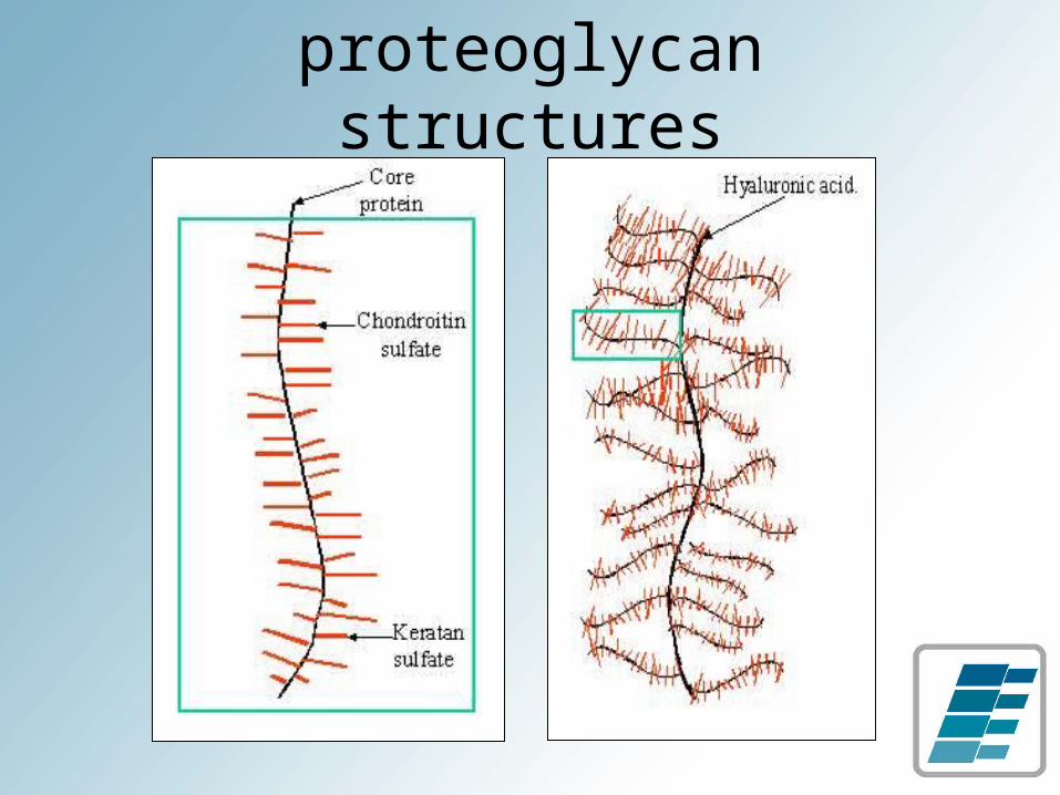
proteoglycan structures
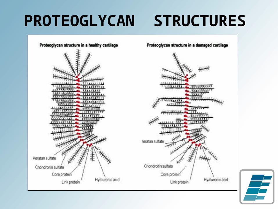
PROTEOGLYCAN STRUCTURES

annular tears
rim lesion
concentric tear
radial tear
Osti OL, Vernon-Roberts B, et al. “Annular Tears & Disc Degeneration” J Bone Joint Surg. [Br] 1992; 74-B:678-82 Gordon SJ, Yang KH, Mayer PJ, et al: Mechanism of disc rupture. A preliminary report. Spine 16:450-456, 1991
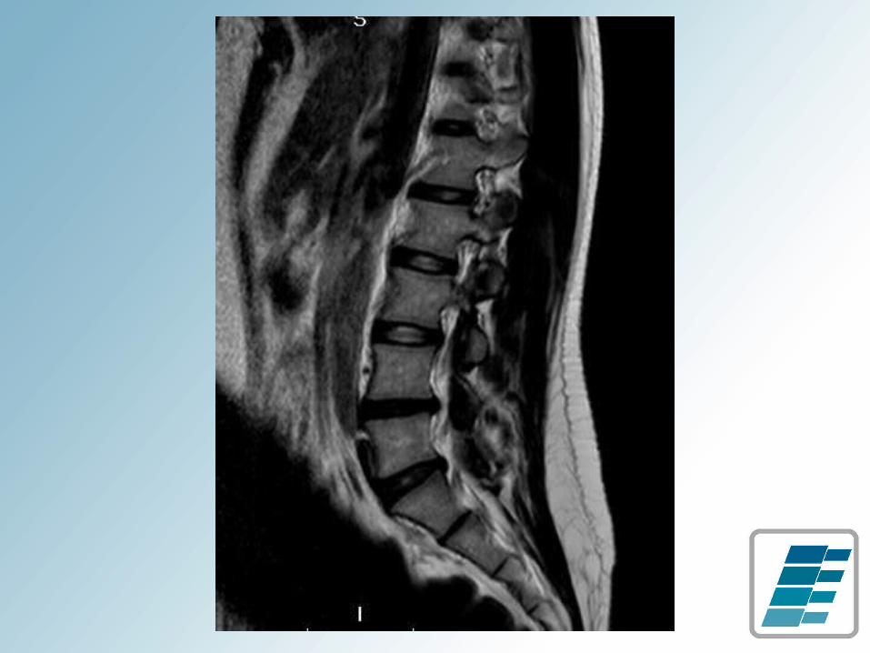
disc pathology vs pain
• degree of disc injury (size of tear / herniation), nor the degree of nerve root compression correlate with subjective pain or functional disability
Karppinen J. et al. “Severity of Symptoms and Signs in Relation to MRI Findings Among Sciatica Patients.” Spine 2001; 26(7):E149-E154

internal disruption
Crock HV, Internal disc disruption. A challenge to disc prolapse fifty years on. Spine 1986 ;11:650-3

current therapies for discogenic pain or disc pathology
• medication and limited activity
• spinal rehabilitation
• interventional pain management
• spinal surgery

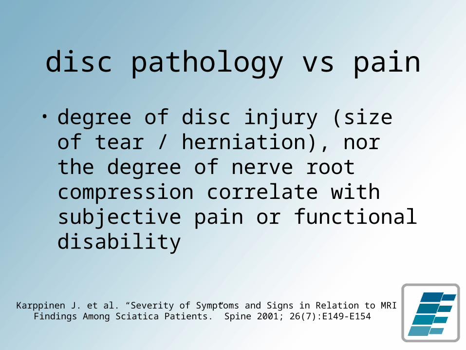
non-surgical decompression
• non-invasive procedure designed to target underlying disc pathology
• improve nutrient exchange
• create environment for healing
non-surgical decompression

goals of treatment
• actively distract and passively retract the spine in order to affect intervertebral disc space
• reduce intradiscal pressures
• increase fluid and nutrient exchange
• promote disc regeneration
• retract nucleic material of bulging or herniated disc
guarding reflex
• traction causes natural guarding reflex
• muscles contract or spasm to prevent distraction in order to protect the spine
• traction devices are rarely able to bypass or overpower reflex contractions and achieve distraction of the disc space
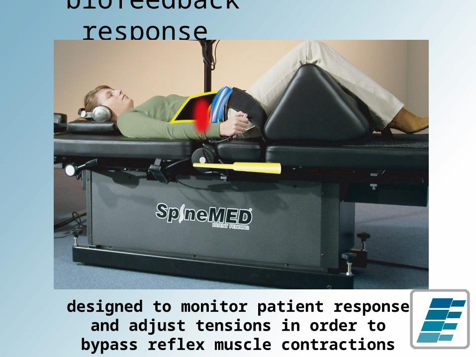
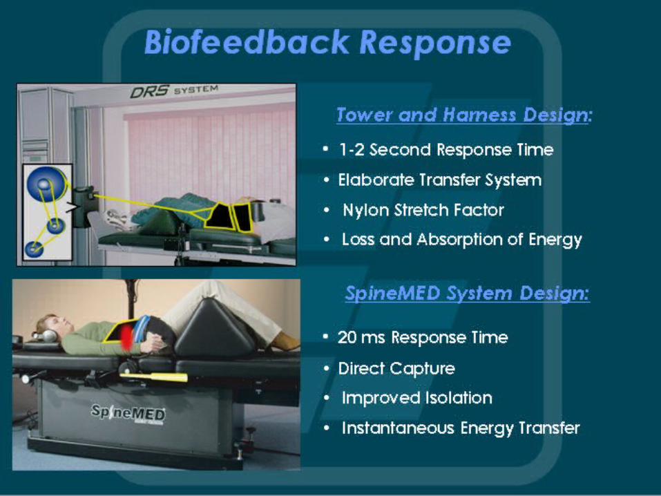
biofeedback response
designed to monitor patient response and adjust tensions in order to bypass
reflex muscle contractions
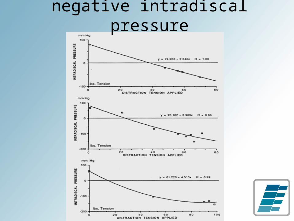
negative intradiscal pressure
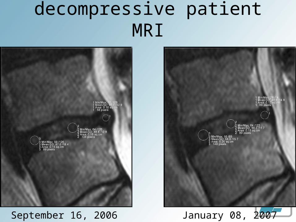
decompressive patient MRI
September 16, 2006 January 08, 2007
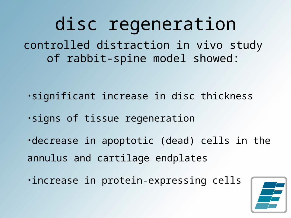
disc regenerationcontrolled distraction in vivo study of
rabbit-spine model showed:
•significant increase in disc thickness
•signs of tissue regeneration
•decrease in apoptotic (dead) cells in the annulus
and cartilage endplates
•increase in protein-expressing cells
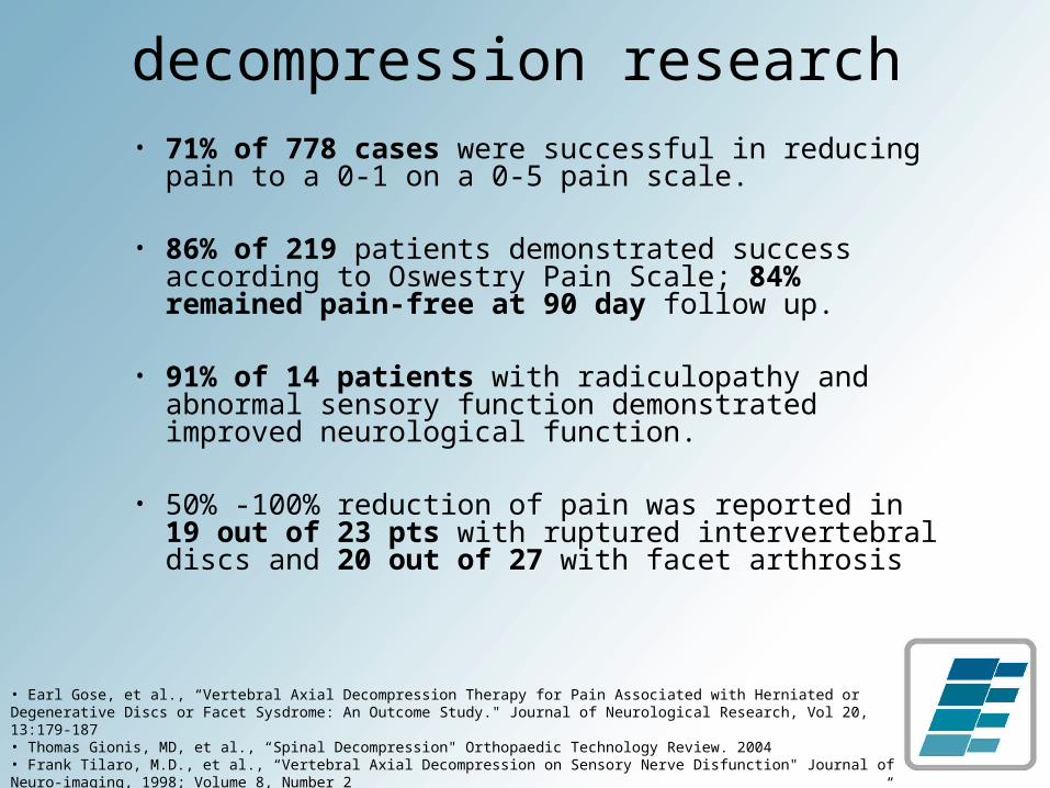
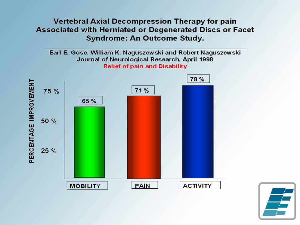
decompression research• 71% of 778 cases were successful in reducing
pain to a 0-1 on a 0-5 pain scale.
• 86% of 219 patients demonstrated success according to Oswestry Pain Scale; 84% remained pain-free at 90 day follow up.
• 91% of 14 patients with radiculopathy and abnormal sensory function demonstrated improved neurological function.
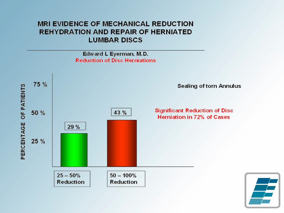
• 50% -100% reduction of pain was reported in 19 out of 23 pts with ruptured intervertebral discs and 20 out of 27 with facet arthrosis
• Earl Gose, et al., “Vertebral Axial Decompression Therapy for Pain Associated with Herniated or Degenerative Discs or Facet Sysdrome: An Outcome Study." Journal of Neurological Research, Vol 20, 13:179-187• Thomas Gionis, MD, et al., “Spinal Decompression" Orthopaedic Technology Review. 2004 • Frank Tilaro, M.D., et al., “Vertebral Axial Decompression on Sensory Nerve Disfunction" Journal of Neuro-imaging, 1998; Volume 8, Number 2• Shealy, et al., “New Concepts in Back Pain Management. Decompression, Reduction and Stabilization”. Pain Management. 1998 239-257
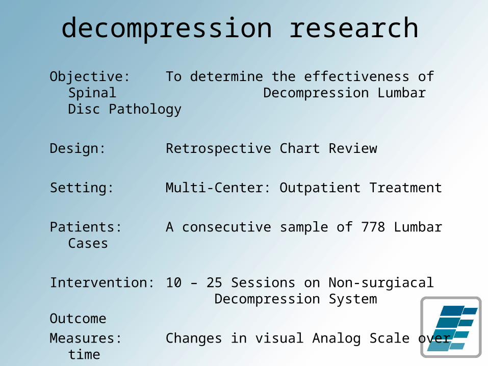
Objective: To determine the effectiveness of Spinal Decompression Lumbar Disc Pathology
Design: Retrospective Chart Review
Setting: Multi-Center: Outpatient Treatment
Patients: A consecutive sample of 778 Lumbar Cases
Intervention: 10 – 25 Sessions on Non-surgiacal Decompression System
Outcome Measures: Changes in visual Analog Scale over time
Improvements in MobilityImprovements in Functioning
decompression research
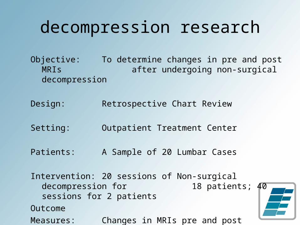
Objective: To determine changes in pre and post MRIs after undergoing non-surgical decompression
Design: Retrospective Chart Review
Setting: Outpatient Treatment Center
Patients: A Sample of 20 Lumbar Cases
Intervention: 20 sessions of Non-surgical decompression for 18 patients; 40 sessions for 2 patients
Outcome Measures: Changes in MRIs pre and post treatment
decompression research
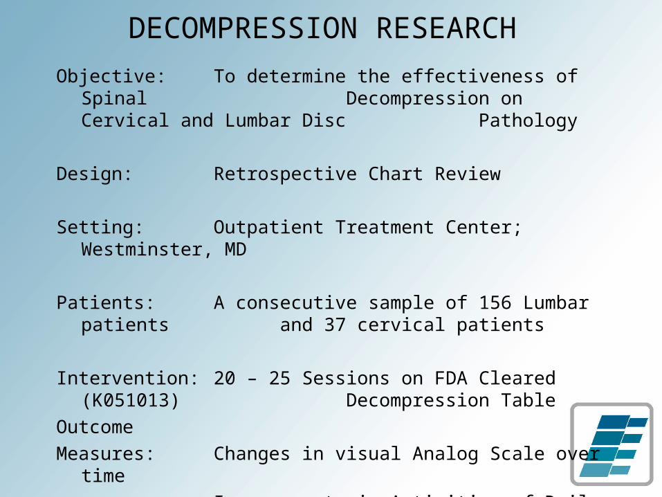
Objective: To determine the effectiveness of Spinal Decompression on Cervical and Lumbar
Disc Pathology
Design: Retrospective Chart Review
Setting: Outpatient Treatment Center; Westminster, MD
Patients: A consecutive sample of 156 Lumbar patients and 37 cervical patients
Intervention: 20 – 25 Sessions on FDA Cleared (K051013) Decompression Table
Outcome Measures: Changes in visual Analog Scale over time
Improvements in Activities of Daily LivingImprovements in Functioning
DECOMPRESSION RESEARCH

lumbar improvement in V.A.S.
• Reduction in Mean from 5.8 to 0.8 V.A.S.• Same improvement noted for both post
surgical and non-post surgical patients
Reported Pain at Each Session
0123456789
10
0 5 10 15 20 25
Treatment Day
No Pain Severe Pain
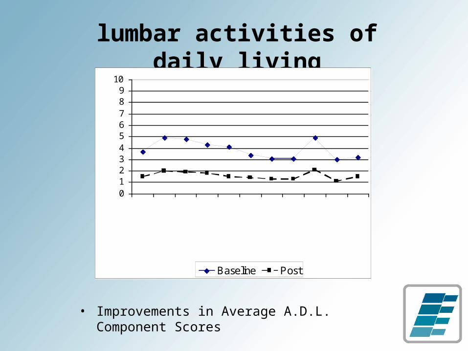
lumbar activities of daily living
• Improvements in Average A.D.L. Component Scores
0123456789
10
SittingStandingWalking
Stair ClimbingIn/Out of Car
DrivingDressingBathing
House/Year Work
WorkSleeping
No Pain Severe Pain
Baseline Post
decompressive research
• SUNY research foundation
• randomized, double-blind, controlled trial
• subjective VAS pain and oswestry measurements
• objective pre and post MRIs
decompressive research
• Greater Baltimore Medical Center
• Randomized Controlled Trials: SpineMED vs conventional traction
• Subjective VAS pain and Oswestry Measurements
case study - annie
• 30 y.o. female presents with low back pain
• pain radiating down right leg
• initial onset approximately 1 year
• referred by orthopedic surgeon
• on motrin, previously darvocet, flexeril and valium
• previous treatments: chiropractic and physical therapy
findings • ROM: decreased in the lumbar spine to flexion,
rotation and side bending
• Strength: right side 4/5 for L4-L5 innervated muscles
• FABER test: positive on the right
• Reflexes: ¼ and symmetrical
• SLR: negative
diagnostic studies
• A-P / lateral Plain Film:– degenerative disc height loss at L4-5 level
• MRI:– L4-L5: large central disc herniation (9mm in AP
X 10mm Broad) effacing the ventral thecal sac and impressing upon the central canal.
• This produces moderate canal stenosis.
– L5-S1: broad disc bulge with radial tear.• mild effacement upon the ventral thecal sac.
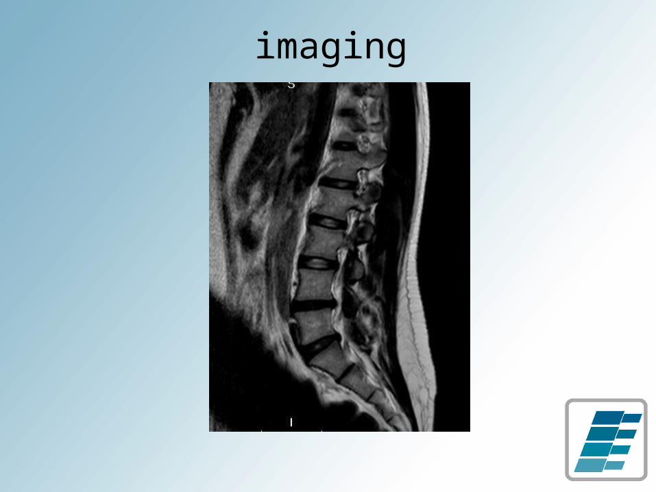
imaging

case 1 outcomecase 1 outcome
VISIT #6> pain reduced:4 to 1
VISIT #10> pain reduced: 1 to 0> core exercises initiated
VISIT # 20> pain stabilized: 1
case 1 outcomecase 1 outcome
VISIT #24> Pain stable at 1> Released to home exercise program> Inversion table recommended
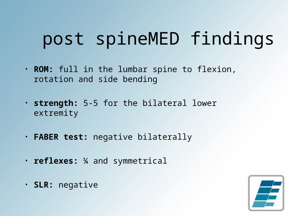
post spineMED findings
• ROM: full in the lumbar spine to flexion, rotation and side bending
• strength: 5-5 for the bilateral lower extremity
• FABER test: negative bilaterally
• reflexes: ¼ and symmetrical
• SLR: negative
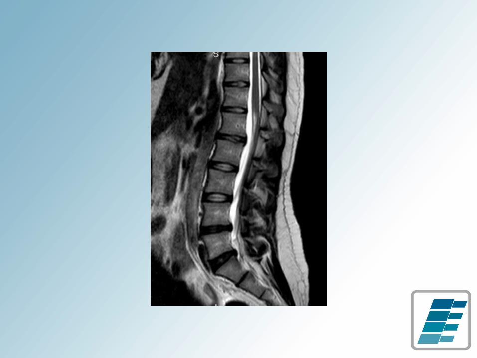
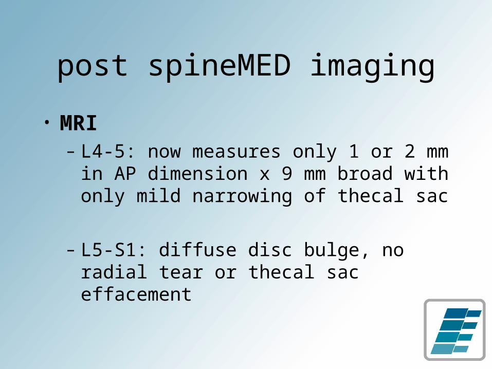
post spineMED imaging
• MRI– L4-5: now measures only 1 or 2 mm
in AP dimension x 9 mm broad with only mild narrowing of thecal sac
– L5-S1: diffuse disc bulge, no radial tear or thecal sac effacement
conclusion
• non-surgical decompression can significantly improved the clincal outcome of patients with discogenic pain
• in treating over 300 patients
• no incidence of injury, some incidence of residual pain
• many successful outcomes
• mostly lasting results & healing

intervertebral disc