Embed Size (px)
Citation preview
Ann OWl RhiTlOILaryngol 101: 1992
VIDEOSTROBOSCOPY OF HUMAN VOCAL FOLD PARALYSIS
JOEL A. SERCARZ, MD
GERALD S. BERKE, MD
BRUCE R. GERRATI, PHD
Los ANGELES, CALIFORNIA
YEMING,MD
MANUEL NATIVIDAD
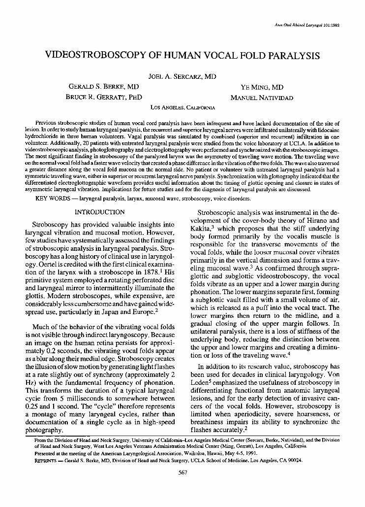
Previous stroboscopic studies of human vocal cord paralysis have been infrequent and have lacked documentation of the site oflesion. In order to study human laryngeal paralysis, the recurrent and superior laryngeal nerves were infiltrated unilaterally with lidocainehydrochloride in three human volunteers. Vagal paralysis was simulated by combined (superior and recurrent) infiltration in onevolunteer. Additionally, 20 patients with untreated laryngeal paralysis were studied from the voice laboratory at UCLA. In addition tovideostroboscopic analysis, photoglottography and electroglottography were performed and synchronized with the stroboscopic images.The most significant finding in stroboscopy of the paralyzed larynx was the asymmetry of traveling wave motion. The traveling waveon the normal vocal fold had a faster wave velocity that created aphase difference in the vibration of the two folds. The wave also traverseda greater distance along the vocal fold mucosa on the normal side. No patient or volunteer with untreated laryngeal paralysis had asymmetric traveling wave, either in superior or recurrent laryngeal nerve paralysis. Synchronization with glottography indicated that thedifferentiated electroglottographic waveform provides useful information about the timing of glottic opening and closure in states ofasymmetric laryngeal vibration. Implications for future studies and for the diagnosis of laryngeal paralysis are discussed.
KEY WORDS - laryngeal paralysis, larynx, mucosal wave, stroboscopy, voice disorders.
INTRODUCTION
Stroboscopy has provided valuable insights intolaryngeal vibration and mucosal motion. However,few studies have systematically assessed the findingsof stroboscopic analysis in laryngeal paralysis. Stroboscopy has a long history ofclinical use in laryngology. Oertel is credited with the first clinical examination of the larynx with a stroboscope in 1878. 1 Hisprimitive system employed a rotating perforated discand laryngeal mirror to intermittently illuminate theglottis. Modern stroboscopes, while expensive, areconsiderably less cumbersome and have gained widespread use, particularly in Japan and Europe.s
Much of the behavior of the vibrating vocal foldsis not visible through indirect laryngoscopy. Becausean image on the human retina persists for approximately 0.2 seconds, the vibrating vocal folds appearas a blur along their medial edge. Stroboscopy createsthe illusion ofslow motion by generating light flashesat a rate slightly out of synchrony (approximately 2Hz) with the fundamental frequency of phonation.This transforms the duration of a typical laryngealcycle from 5 milliseconds to somewhere between0.25 and 1 second. The "cycle" therefore representsa montage of many laryngeal cycles, rather thandocumentation of a single cycle as in high-speedphotography.
Stroboscopic analysis was instrumental in the development of the cover-body theory of Hirano andKakita, 3 which proposes that the stiff underlyingbody formed primarily by the vocalis muscle isresponsible for the transverse movements of thevocal folds, while the looser mucosal cover vibratesprimarily in the vertical dimension and forms a traveling mucosal wave.I As confirmed through supraglottic and subglottic videostroboscopy, the vocalfolds vibrate as an upper and a lower margin duringphonation. The lower margins separate first, forminga subglottic vault filled with a small volume of air,which is released as a puff into the vocal tract. Thelower margins then return to the midline, and agradual closing of the upper margin follows. Inunilateral paralysis, there is a loss of stiffness of theunderlying body, reducing the distinction betweenthe upper and lower margins and creating a diminution or loss of the traveling wave.t
In addition to its research value, stroboscopy hasbeen used for decades in clinical laryngology. VonLeden'' emphasized the usefulness of stroboscopy indifferentiating functional from anatomic laryngeallesions, and for the early detection of invasive cancers of the vocal folds. However, stroboscopy islimited when aperiodicity, severe hoarseness, orbreathiness impairs its ability to synchronize theflashes accurately.s
From the Division of Head and Neck Surgery, University of California-Los Angeles Medical Center (Sercarz, Berke, Natividad), and the Divisionof Head and Neck Surgery, West Los Angeles Veterans Administration Medical Center (Ming, Gerratt), Los Angeles, California.
Presented at the meeting of the American Laryngological Association, Waikoloa, Hawaii, May 4-5,1991.REPRINTS - Gerald S. Berke, MD, Division of Head and Neck Surgery, UCLA School of Medicine, Los Angeles, CA 90024.
567

568 Sercarz et al, Stroboscopy of Vocal Fold Paralysis
Previous research supported the widespread viewthat laryngeal paralysis is accompanied by a loss ofthe mucosal traveling wave on the paralyzed fold.2
Schoenharls studied laryngeal paralysis with stroboscopy and found that in 55 of62 cases the travelingwave was absent. The presence of the mucosal wavein patients with a paralyzed fold was interpreted as asign of some degree of reinnervation, suggesting animproved prognosis for eventual complete recovery.2,7 Fex7 published a study in 1970 on the stroboscopy of laryngeal paralysis. He agreed with previous authors that in acute recurrent laryngeal nerve(RLN) paralysis the glottic wave is absent. The mucosal traveling wave was found to be an accuratereflection of thyroarytenoid muscle tonus, and itsreturn indicated recovery ofRLN function even whenvocal fold abduction could not be detected'? Thepossibility that the return of the traveling wave waspromoted by vocal cord fibrosis was not discussed.
Isshiki et al8 published a detailed account of theeffects of asymmetric vocal fold extension, usingexcised larynges and a computer model to assess theeffect of unilateral changes of cricothyroid (CT)contraction and subsequent vocal fold stiffness. According to Isshiki et al, the vibratory findings produced by asymmetric laryngeal tension depend onthe degree of glottic closure. They found that insimulated superior laryngeal nerve (SLN) paralysisthe true vocal folds vibrated at the same frequency butout of phase, with the more tense fold (active CT)preceding the less tense one. This finding was laterconfirmed by Isshiki' s group in a study performed onlive dogs.?
Another study of canine laryngeal paralysis wasperformed by Moore et a14 employing videostroboscopy. The authors found that canine RLN paralysisproduces a difference in the timing of the onset ofvocal fold lateral displacement and loss ofthe normaltwo-mass laryngeal vibration. The mucosal wavewas markedly diminished but not lost in RLN paralysis."
The morphological findings in laryngeal paralysis,including the Wagner-Grossman theory ofvocal foldposition in RLN and vagal paralysis, can be appreciated through indirect laryngoscopy alone and will notbe the focus of this research.
The purpose of this study is to update earlieranalyses of asymmetric laryngeal vibration by usingvideostroboscopy. First, it will describe the stroboscopic appearance ofthe larynx in paralysis. Previousreports of vibrational findings in laryngeal paralysishave been limited by the difficulty in defining theactual type of paralysis. Temporary induced paraly-
sis was used in this study and provides a known siteof lesion and the ability to compare laryngeal vibration before and after paralysis. Second, videostroboscopic images were synchronized with glottographicwaveforms to more accurately assess the timing ofglottic opening and closure on the glottographicwaveforms. Third, an analysis of a clinical series ofpatients with various types of vocal fold paralysiswas performed to verify that the findings noted ininduced paralyses are similar to those in a typicallaryngology practice. Finally, asymmetric vibrationof the laryngeal mucosa provides an opportunity toutilize recently developed techniques for the objective analysis of videostroboscopic images for thedegree of vocal fold symmetry.
MATERIALS AND METHODS
Method of Paralysis Induction. Three adult malevolunteers with a mean age of 36 years were studiedin this experiment. The volunteers appeared normalon laryngeal examination and had no history oflaryngeal disorders. Vocal fold paralysis was induced by infiltrating 2% lidocaine hydrochloridewith a 25-gauge needle into the expected location ofthe nerve. The left RLN was infiltrated in the tracheoesophageal groove approximately 2 em inferior to thecricoid cartilage. The left SLN was infiltrated asdescribed by Abelson and Tucker. 10 Ten cubic centimeters were necessary, with injection both at theposterior edge ofthe thyroid ala and inferiorly near itspassage posterior to the thyrohyoid muscle. To createa combined ("vagal") paralysis, an SLN injectionwas performed, verified by stroboscopy, and immediately followed by an RLN injection. Subject 1 hadSLN, RLN, and combined paralysis induced withlidocaine. Subject 2 had the RLN only injected, andsubject 3 had the SLN only injected.
Videostroboscopic Analysis. The method ofvideostroboscopic analysis was modified from previousreports by Bless et all and Kitzing.I Emphasis wasplaced in the analysis on the characteristics of thetraveling mucosal wave. The onset, extent, and velocity ofthe traveling wave were analyzed and estimatedby advancing the videotape frame by frame. Phasedifferences were noted in cases ofasymmetric vibration. Other characteristics included the degree ofglottic closure, the lateral excursion of the fold duringvibration (termed amplitude by Bless et all), and theregularity ofglottic cycles, reflecting the presence orabsence of severe frequency perturbation.
Stroboscopy. The experimental equipment requiredfor these studies is depicted in Fig 1.Stroboscopy wasperformed with a Bruel & Kjaer 49l4A (Orange,Calif) stroboscope. A microphone (Sennheiser MD

Sercar; et al, Stroboscopy of Vocal Fold Paralysis
ENOOSCOPE
EYE PIECE ADAPlDA.. OKJPTER LENS/X ~ 50rrm MACROLENS
',/,/'.I"'N;"~~'VIDEO, CAMERA.,. I-(l~ /--->~
'~J.j t/«~I f
569
Fig 1. Experimental setup forrecording of videostroboscopicimages and concurrent glottographic waveforms, Computerincludes Frame Grabber hardware, which allows digitizationof videostroboscopic images,and Image Pro software foranalysis of images.
4026) transduced the speech signal to allow synchronization with the video images. A Toshiba ChargeCoupled Device (CCD) color video camera (modelIK-C30A, Buffalo Grove, Ill) imaged the glottis. Theimages were recorded onto a 3/4-in professional videocassette recorder (Sony YO-9850, Teaneck, NJ)equipped with a time coder. Still images from thevideotape were made with a color video printer (SonyMavigraph, Teaneck, NJ).
Glottography. Videostroboscopy and glottographywere performed simultaneously during phonation ofthe vowel IiI produced at a constant pitch and loudness.Transduction ofphotoglottography (PGG) waveforms required constant illumination of the larynxwith a miniature light source (Karl Storz 481-C,Culver City, Calif) in addition to the stroboscopiclight source. A 90° telescope (Wolfe, Rosemont, Ill)with two light inputs was used to visualize the glottis.Light transmission for PGG waveforms was transduced with a photosensor (Centronics OSD 50-2,Mountainside, NJ) held in position on the skin overlying the CT membrane. Electroglottography (EGG)recording electrodes were strapped into position ontothe subject's neck, overlying the thyroid lamina at thelevel of the true vocal folds. A Synchrovoice electroglottograph (Harrison, NJ) recorded the EGG waveforms.
Synchronization. Videostroboscopic images werecorrelated with glottographic signals to provide pre-
liminary information about the timing of events inlaryngeal paralysis. Further details about the synchronization method are being published in a separate report from this laboratory.l! Briefly, a 5-millisecond square wave pulse was digitized and simultaneously recorded on the audio channel by the videotape recorder. By correlating the 5-millisecondsquarewave pulse with the vertical synchronization trace ofthe video signal, the position on the glottographicwaveform could be precisely correlated with individual video images. A Hitachi oscilloscope (modelY-1050 F, Torrance, Calif) was used to extract thevertical trace of the video signal for recording.
Electroglottography, photoglottography, synchronizing pulse and vertical trace of the video signalwere digitized via a Labmaster 12-bit analog-todigital board housed in an IBM compatible computer.The EGG and PGG signals were verified on a Tektronix 5116 (Beaverton, Ore) storage oscilloscopeprior to recording. The waveforms were analyzed byusing a commercially available software package forthe PC system (C-Speech, Paul Milenkovic, University of Wisconsin, Madison, Wis).
Videostroboscopic Image Evaluation. Images wereanalyzed by using an image processing softwarepackage previously described12(Image Pro II, MediaCybernetics, Silver Spring, Md). The hardware necessary for image analysis included a Frame Grabberto digitize and analyze video images (Data Transla-

570 Sercarz et al, Stroboscopy of Vocal Fold Paralysis
TABLE 1. SUMMARY OF STROBOSCOPIC EVALUATION OF LIDOCAINE-INDUCED LARYNGEAL PARALYSIS
Subject Site Regularity* ClosureSymmetry of Vibration and Characteristics
ofMucosal Wave
Variable
Complete
Complete
Complete
Large chink
Small posteriorglottic chink
Regular
Regular
Regular
Regular
Variableaperiodicity
Variableaperiodicity
RLN
SLN
SLN
RLN
RLN paresis
SLN +RLN
2
Moderate asymmetry of TW; greater velocity and excursion ofTW on normal side, particularly at low frequency of vibration;decreased tension of TVF on injected side; occasional shifting ofglottis from side to side during vibration
TVFs vibrated at different level during portions of cycle (Fig 3C);profound asymmetry of TW (Fig 3); normal TVF crossed midlineduring vibration; TW Velocity and excursion greater on normalside; loss of TVF tension; TVF higher on normal side
Marked diminution of TW excursion and velocity on side ofinjection; greater TVF excursion on normal side (see symmetryratio, Fig 4A)
Asymmetry of vibration with greater excursion and velocity ofTW on normal side; normal TVF adduction and abduction
Marked asymmetry of TW (Fig 2B); greater velocity of TW onnormal side; vibration out of phase; normal TVF precedesparalyzed TVF
Diminished mucosal wave on side of paralysis; flaccidity of TVFon injected side
SLN - superior laryngeal nerve, RLN - recurrent laryngeal nerve, TW - traveling wave, TVF - true vocal fold.
*Regularity indicates ability of subject to maintain constant frequency and therefore facilitate stroboscopy. High jitter (frequency perturbation)results in irregular vibration and difficulty in analyzing glottic wave.
3
tion, DT-2853 60SQ, Marlboro, Mass). The desiredportion of the video image can be outlined by use ofa pointing device, and then the area of the trace or thelength of a line is calculated by and expressed in pixelunits by the software.
Images were digitized from the most closed andmost open portions of the vocal cycle, and the widthof the vocal folds was measured during these intervals. A measure was then computed of the symmetryratio, which expresses the excursion of one vocal foldin proportion to that of the other fold. Symmetricvibratory movement is marked by an equal excursionof both vocal folds from the midline during phonation. When the lateral motion of one fold is markedlyreduced, as in unilateral RLN paralysis, the symmetry ratio approaches zero. Further details regardingthe objective analysis of videostroboscopic imageshave been recently published by Sercarz et al.12
Chronic Paralysis Study. We reviewed the videotapes and clinical files ofall patients with a diagnosisof vocal fold paralysis evaluated in the UCLA VoiceLaboratory during the years 1988 to 1991. Patientswere excluded if the paralyses had been previouslytreated or if inadequate videotape or clinical informationwas available. All patients underwent stroboscopyand glottographic (PGG and EGG) recordings. Ineach case, medical records were analyzed to determine the probable site of the paralysis. Three cases ofvagal paralysis had documented sites of lesion. Twopatients had laryngeal paralysis following penetrating trauma to the skull base and subsequent highvagal paralysis. The third had undergone a tumorwith sacrifice of cranial nerve X at the skull base.
Two clinical cases were diagnosed as SLN paralysis. One patient developed a dysphonia followingthyroid surgery marked by a reduced ability to modulate pitch and vocal fatigue. Later exploration revealed a lack of function of the SLN on direct nervestimulation. Another patient suffered neck traumaand developed a dysphonia marked by abnormallaryngeal vibration and normal vocal fold abductionand adduction. In the latter patient, there was arotation of the posterior commissure in the directionof the paralysis side.
Fifteen additional patients received diagnoses ofunilateral RLN paralysis based on history and indirect laryngeal examination.
RESULTS
Induced Paralysis. The three subjects underwentstroboscopy prior to induction of paralysis. In eachcase, vibration was normal with equivalent velocityand excursion of the traveling wave and symmetrictension of the vocal folds bilaterally. Closure wascomplete in each case.
In each case of SLN paralysis, the volunteer reported anesthesia of the larynx and an inability to singat a high pitch in addition to the findings described.
Table 1 summarizes the videostroboscopic findings in induced vocal fold paralysis. Following RLNinjection, the vocal process on the paralyzed sideremained in the paramedian position. There was norotation of the posterior glottis that was appreciablein any of the induced paralysis states.

Sercarz: et al, Stroboscopy of Yocal Fold Paralysis 571
Fig 2. (Subject 2) A) Normal phonation, showing symmetric traveling mucosal wave. B) Following left recurrent laryngeal nerveinjection. Right mucosal wave has completed its vibration. Wave of lesser excursion is now traveling along left (paralyzed) vocalfold.
The most consistent finding in each case of induced paralysis was a diminution of traveling wavevelocity and less lateral excursion on the side of theinjection, whether SLN, RLN, or combined. Although the traveling wave was attenuated in eachcase on the side of the paralysis, the wave wasobserved during each trial. For subject 1, who under-
went all three types of paralysis, the traveling waveasymmetry was greatest in the vagal paralysis, followed by RLN and SLN in that order.
Figure 2A is normal phonation from subject 2 witha symmetric traveling wave. Figure 2B, followingleft RLN injection, demonstrates asymmetric vibra-
Fig 3. (Subject 1) Portions of glottic cycle following injection ofsuperior and recurrent laryngeal nerves. A) Midcycle, with brisklateral motion of right (normal) vocal fold, without visiblevibration of paralyzed fold. B) Later in cycle, markedly attenuated wave traverses left, paralyzed fold. C) Normal fold, havingcompleted its vibration, returns to position just past midline, atlevel higher than paralyzed left fold.

572 Sercarz et al, Stroboscopy of Yocal Fold Paralysis
Fig 4. Symmetry ratio for two volunteers with induced recurrent laryngeal nerve paralysis (see text). A) Subject 1. B) Subject 2.
tion. The normal (right) vocal fold has completed itsvibration, and the paralyzed (left) fold is shown withan attenuated traveling wave. Figure 3 is a series ofstill video frames from subject I with a left combinedRLN and SLN paralysis. Figure 3A is early in theglottic cycle, with a vigorous excursion of theuninjected (right) vocal fold. Later in the glotticcycle, a minimal mucosal wave is seen on the leftvocal fold (Fig 3B). Finally, the right vocal fold,positioned at a higher level than the paralyzed leftvocal fold, crosses the midline, and the glottis closes(Fig 3C).
Figure 4 depicts measurement of the symmetryratio for subjects 1 and 2 following RLN injection.The lengths a and b (most closed portion of the glottalcycle) and c and d (most open) represent the width ofthe right and left vocal folds. The calculated symmetry ratio for subject 1 was 0.139 (Fig 4A) and forsubject 2 was 0.151 (Fig 4B). In a perfectly symmetric case, the ratio is 1.0, and in a case with no lateralvocal fold movement in one of the folds the ratio is o.
Figure 5 demonstrates the results of synchronizingthe glottographic signal to the videostroboscopicimage from subject 1 following RLN injection. Figure 5A is a stroboscopic image taken immediatelyfollowing opening of the glottis. Figure 5B is thesynchronized glottography. There is an upward deflection in the differentiatedEGG (dEGG) waveformthat correlates well with the onset of vocal foldopening. Figure 5C,D is synchronized from a point atmidcycle. Figure 5E,F is from an image immediatelypreceding the point ofglottic closure. The downwarddeflection in the dEGG waveform closely correlatedwith the instant oflaryngeal closure. Similar findingsare documented in Fig 6, which shows the results of
synchronizing strobe images and glottography insubject 1 with SLN paralysis at the moment of opening (Fig 6A,B) and closure (Fig 6C,D). Again, thetiming of opening and closure of the vocal folds ispredicted by the dEGG waveform.
Clinical Cases. The results of a series of 20 consecutive patients analyzed at the UCLA Voice Laboratory will now be described. Ofthe 15RLN patients,3 could not be included because aperiodic vibrationor a large glottic gap prevented analysis of the mucosal traveling wave. The results of the 12 remainingpatients with unilateral RLN paralysis and adequatestroboscopy are summarized in Table 2. Ten of 12patients had a mucosal wave present on the paralysisside. The wave asymmetry was similar to that observed in the induced paralysis: the normal wave hada greater velocity and traveled farther along the vocalfold mucosa. There was a phase shift, with the normalside vibrating sooner than the side of the paralysis.The asymmetry was marked in the majority of patients with unilateral RLN paralysis.
The findings in the two patients with SLN paralysis parallel those in the group with induced paralysis.There was normal abduction and adduction of thevocal folds in both cases. Glottic closure and regularity (lack of noticeable frequency perturbation) werenormal. Analysis of the mucosal wave revealed mildto moderate asymmetry of the traveling wave. On thenormal (nonparalysis) side, the wave appeared earlier, had a greater velocity, and traveled farther alongthe surface of the vocal cord mucosa.
The mucosal wave findings in vagal paralysis aresimilar to those in RLN paralysis. Only one of thethree patients had the frequently cited finding ofrotation of the posterior glottis in the direction of the

Sercarz et al, Stroboscopy of Vocal Fold Paralysis 573
strobe flash --i II
strobe flash -----.
EGG
PGG
dEGG
F
EGG
PGG
dEGG
B
EGG
PGG
dEGGo
Fig 5. (Subject 1) Correlation of glottographic information with stroboscopic images in recurrent laryngeal nerve paralysis.EGG - electroglottography, PGG - photoglottography, dEGG - differentiated EGG. A) Immediately following vocal foldopening. B) Corresponding glottographic waveforms, with strobe flash occurring just after positive deflection in dEGG waveform. C) Later in cycle, at rnid-opening. D) Glottographic waveforms, with strobe occurring nearpeakofPGG waveform. E) Imageof glottis at point of closure. F) Corresponding waveforms; nadir of dEGG waveform corresponds to strobe flash.
paralysis. In one of the patients with vagal paralysis,the glottic gap was wide throughout the cycle andthere was very irregular vibration of the mucosalwave bilaterally. The other two patients had markedasymmetry of the traveling wave, vibration out ofphase, and differences in the velocity and excursionof the traveling wave similar to those found withinduced vagal paralysis in this study.
chemical paralysis allows comparison with normalstroboscopy in the same individual. It also providesan opportunity to study recovering RLN weaknessand observe the gradual recovery of symmetry, as inthe study of vocal fold paresis in subject 1. Beforediscussing the stroboscopic findings in particularparalytic states, we will review the overall relationship between tension of the vocal fold and vibratorycharacteristics.
DISCUSSION
There are several advantages of studying inducedvocal fold paralysis stroboscopically. Temporary
Unlike previous studies2,6,7,13 of stroboscopy invocal cord paralysis, the data in the present reportindicate that the mucosal wave is always affected but

574 Sercar: et al, Stroboscopy of Vocal Fold Paralysis
strobe flash
EGG
PGGdEGG
B
EGG
PGGdEGG
oFig 6. (Subject 1) Correlation of glottographic information with stroboscopic images in superior laryngeal nerve paralysis. Abbreviations as in Fig 5. A) Immediately following vocal fold opening. B) Corresponding glottographic waveforms, with strobeflash occurring just after positive deflection in dEGG waveform. C) At point of closure of glottis. D) Corresponding glottographicwaveforms, with strobe again occurring at nadir of dEGG waveform.
not invariably absent in RLN and vagal paralysis. Allpatients with laryngeal paralysis of any type, experimental or clinical, had an unambiguous asymmetryoflaryngeal vibration demonstrated stroboscopically.Furthermore, the data presented indicate that theasymmetry follows a pattern: the normal vocal foldtraveling wave has a greater velocity than that of theparalyzed fold, is observed earlier in the glottic cycle,and traverses farther over the surface of the vocal foldmucosa. The symmetry ratio data were presented todocument the greater excursion of the vocal foldmargin on the nonparalyzed side during the mostopen portion of the cycle in unilateral RLN paralysis.
The findings described here are similar to thosereported by Tanabe et al? in a report on asymmetricglottic vibration studied in canine larynges. It is notsurprising that there was a visible difference in thevelocity of the traveling waves on two vocal folds.Basic research on the propagation of waves in elasticmedia have indicated that there is a direct relationshipbetween the stiffness of the material or reactance todeformation and the velocity of a harmonic travelingwave.!" The lack of thyroarytenoid stiffness and/orCT contraction is probably the central cause of the
vibratory differences in laryngeal paralysis. In amorphologic study of the vocal fold as a vibrator,Hirano'> stated that the CT and thyroarytenoid muscleshad the greatest effect on the stiffness relationshipbetween the body and the cover of the vocal fold; thisfinding may explain why either muscle may elicitsimilar changes in the traveling wave velocity andexcursion.
For the two patients with absent glottic waves,each had poor glottic closure that reduced the degreeof vocal fold contact. This tended to decrease theability of stroboscopy to detect the very subtle mucosal wave that generally occurs on the paralyzed vocalfold.
Because of its sensitivity in demonstrating slightdifferences in vocal fold vibration, stroboscopy isparticularly useful in the study oflaryngeal paralysis.In subtle cases, such as RLN paresis or isolated SLNparalysis, an abnormality of traveling wave motionmay be the easiest finding to elicit. The results of thisstudy suggest that traveling wave asymmetry may beone of the most salient observations to make in suspected paralysis, because traveling wave asymmetry

Sercarz et al, Stroboscopy of Vocal Fold Paralysis 575
TABLE 2. RESULTS OF PATIENTS WITH RECURRENTLARYNGEAL NERVE PARALYSIS
No. ofPatients
Glottic closureComplete 2Mild to moderate incomplete closure 5Severely incomplete closure 5
Glottic wave: extent of wave excursion alongvocal fold mucosa
Symmetric 0Mild to moderate asymmetry 2Marked asymmetry 8Absent on paralysis side 2
Glottic wave: estimated speed of glottic waveSymmetric 0Greater velocity (earlier wave) on normalside 10Greater velocity on paralyzed side 0Absent wave on paralyzed side 2
Vocal fold lateral displacement during vibration"Equivalent 0Mild to moderate asymmetry 4Marked asymmetry 8
Of 15 patients with clinical diagnosis of recurrent laryngeal nerveparalysis, 12had adequate regularity ofvibration for suitable stroboscopy. Their results are presented here.
*Lateral motion during vibration was invariably greater on side opposite paralysis (normal side). See Fig 4, showing symmetry ratio ininduced paralysis.
is a consistent finding in untreated laryngeal paralysis. In subject 1, traveling wave asymmetry was alsoidentified in RLN paresis.
Despite this consistency, there is a significantvariability of findings among different patients andeven within the same individual phonating at twodifferent fundamental frequencies. For example, inRLN paralysis, CT muscle contraction at high pitchadds vocal fold stiffness and tends to reduce the degree of asymmetry. One patient in the study developed RLN paralysis following thyroid surgery 20years before the examination; the paralysis is stilllargely uncompensated. He apparently compensateswith CT tension, producing speech at a high pitchlevel (paralytic falsetto) that provides glottic closureand only moderately asymmetric traveling wavemotion.
Both RLN paralysis and combined SLN and RLNparalysis produced the above changes in the travelingwave. Although the combined paralysis was characterized by a greater degree of asymmetry and a moreflaccid or "wavy" paralyzed cord, it is doubtful fromour data whether stroboscopy alone can be of significant assistance in differentiating vagal from RLNparalysis.
Isolated SLN paralysis is a rarely noted clinical
entity that probably often goes undetected. IO,16,17
The SLN provides sensation to the supraglottic larynx through its internal branch and motor fibers to theCT muscle via its external branch. The motor branchis in close proximity to the superior thyroid vesselsand is therefore vulnerable to injury during thyroidsurgery. According to Ward et al!? and other authors.l" paralysis of the CT muscle causes 1) lack oflongitudinal tension of the true vocal folds, 2) a tilt ofthe larynx because of the downward motion of thecontralateral intact CT muscle, and 3) a rotation of theposterior glottis toward the side of the paralysisbecause of the unopposed CT muscle's pulling theanterior thyroid cartilage toward the intact side.
Unfortunately for the clinician, as stated by Dedoin his study on experimental and clinical vocal foldparalysis, the expected findings in SLN paralysis" ...are not consistent or obvious enough to be usefulin clinically diagnosing superior laryngeal nerveparalysis."I6(pI503) Neither ofthe two volunteers undergoing SLN injection for this study had an unequivocal rotation of the posterior glottis toward theparalyzed side, as described in the literature. Theasymmetry of the traveling wave, however, was easyto observe stroboscopically.
Although the SLN patients demonstrated that theirfolds were vibrating at the same frequency, there wasa difference in velocity and a phase lag, with thenonparalyzed fold completing the vibratory cyclebefore the paralyzed fold. This finding has beenpreviously reported in both live dogs and excisedlarynges.v? Similar asymmetry occurred, although toa greater degree, in the present study in RLN paralysis.
It is possible that the expected rotation of theposterior glottis was too subtle to be appreciated inthese patients. In a canine study, the glottic rotationfollowing unilateral CT paralysis was only 3.50 to170
•9 At the lower end of this range, the rotation
probably is imperceptible.
Following SLN injection, the asymmetric vibration and morphology, supraglottic anesthesia, andinability to sing at a high pitch produced a constellation of findings consistent only with SLN paralysis,in the opinion of the authors. It is difficult to provecomplete SLN paralysis, however, even if a loss ofCT activity is documented on electromyography. It ispossible, but unlikely, that there is another explanation for the stroboscopic findings.
Clinical correlation is required in the diagnosis ofan isolated SLN paralysis with stroboscopy. Otherpossible causes of asymmetric laryngeal vibrationshould be considered, including scarring or a submu-

576 Sercarz et al, Stroboscopy of Vocal Fold Paralysis
cosal mass, which could rarely produce a similarstroboscopic picture.2,18 In the setting of suspectedSLN injury - for example, a postthyroidectomy patient with a loss ofthe ability to sing or modulate pitchin continuous speech - an asymmetric mucosal traveling wave or asymmetric vocal fold excursion suggests SLN paralysis, even if other characteristicssuch as posterior glottic rotation or tension asymmetry are not observed on indirect laryngoscopy alone.In such a situation, laryngeal electromyography provides a sensitive method to verify the presence of aparalysis.19
A method of synchronizing glottographic waveforms with videostroboscopic images has recentlybeen developed in our laboratory.11 Figures 5 and 6indicate that in studies of patients with asymmetriclaryngeal vibration, the first derivative of the EGGwaveform provides information about the timing oflaryngeal opening and closure, provided that adequate vocal fold contact occurs during phonation.The upward deflection in the EGG first-derivativewaveform is a good indicator of the moment ofopening, whether in normal patients or in those withasymmetric vibration. The nadir of the dEGG waveform has previously been shown to closely correspond to the moment of laryngeal closure.20,21 Thelargest positive peak in the dEGG waveform has beenshown to correlate with opening.j? Our data fromsynchronizing the videostroboscopic images withglottographic waveforms (Figs 5 and 6) indicate thata similar relationship exists in both RLN and SLNparalyses. Childers et al20 noted that there was asignificant variation in dEGG waveforms dependenton the experimental conditions, such as frequency,which somewhat limits the accuracy of estimatingglottic events based on dEGG.
Computerized digital analysis of stroboscopicimages holds the promise for quantifying travelingwave abnormalities associated with laryngeal disorders. Figure 4 demonstrates the symmetry ratio applied to laryngeal paralysis. The ratios can provide anobjective measure, allowing interpatient and intrapatient comparisons of asymmetry.
Although this study did not report data describingthe results of reinnervation or treatment on videostroboscopic findings in RLN paralysis, stroboscopycan document a return of normal symmetry of laryngeal vibration, particularly when the thyroarytenoidmuscle is reinnervated. Crumley-s recently updatedhis experience with ansa hypoglossi nerve transferfor the treatment ofunilateral vocal fold paralysis. Heused stroboscopic analysis to document reinnervation,observing return to symmetric vibration in four offive patients assessed following reinnervation.P
In conclusion, stroboscopy is a useful tool in theevaluation of patients with suspected laryngeal paralysis. Paralysis of the SLN, RLN, or both results inasymmetric laryngeal vibration that is easily identified even by inexperienced observers. The mucosalwave has a greater velocity and travels further alongthe mucosa on the normal fold. The probable cause ofthese vibratory findings is the reduced stiffness in theparalyzed cord, which reduces the velocity and extent of the traveling mucosal wave. Stroboscopy canidentify abnormal vibration in patients with otherwise normal findings on indirect examination. Ourearly experience suggests that stroboscopy cannotreliably distinguish RLN paralysis from vagal paralysis. Studies are being planned in the canine model to better quantify the traveling wave findings inlaryngeal paralysis.
REFERENCES1. Bless DM, Hirano M, Feder RJ. Videostroboscopic evalu
ation of the larynx. Ear Nose Throat J 1987;66:48-58.
2. Kitzing P. Stroboscopy - a pertinent laryngeal examination. J OtolaryngoI1985;14:151-7.
3. Hirano M, Kakita Y. Cover-body theory of vocal foldvibration. In: DaniloffRO, ed. Speech science. San Diego, Calif:College-Hill Press, 1985:1-46.
4. Moore DM, Berke OS, Hanson DO, Ward PH. Videostroboscopy of the canine larynx: the effects of asymmetric laryngeal tension. Laryngoscope 1987;97:543-53.
5. Von Leden H. The electronic synchron-stroboscope. Itsvalue for the practicing laryngologist. Ann Otol Rhinol Laryngol1961;70:881-93.
6. Schoenharl E. Die Stroboskopie in der PraktischenLaryngologie. Stuttgart, Germany: Georg Thieme Verlag, 1960.
7. Fex S. Judging the movements of vocal cords in larynxparalysis. Acta Otolaryngol [Suppl] (Stockh) 1970(suppI263):82-3.
8. Isshiki N, Tanabe M, Ishizaka K, Broad D. Clinicalsignificanceof asymmetrical vocal cord tension. Ann Otol RhinolLaryngoI1977;86:58-66.
9. Tanabe M, Isshiki N, Kitajima N. Vibratory pattern of thevocal cord in unilateral paralysis of the cricothyroid muscle. Anexperimental study. Acta Otolaryngol (Stockh) 1972;74:339-45.
10. Abelson Tl, Tucker HM. Laryngeal findings in superiorlaryngeal nerve paralysis: a controversy. Otolaryngol Head NeckSurg 1981;89:463-70.
11. Sercarz JA, Berke OS, Ming Y, Gerratt BR, Kreiman J,Natividad M. Synchronizing videostroboscopic images of human laryngeal vibration with physiologic signals. AmJ Otolaryngol (in press).
12. Sercarz JA, Berke OS, Arnstein D, Gerratt B, NatividadM. A new technique for quantitative measurement of laryngealvideostroboscopic images. Arch Otolaryngol Head Neck Surg1991;117:871-5.
13. Fex S, Elmqvist D. Endemic recurrent laryngeal nerve
Sercar; et al, Stroboscopy of Yocal Fold Paralysis 577
paresis. Correlation between EMG and stroboscopic findings.Acta Otolaryngol (Stockh) 1973;75:368-9.
14. AchenbachID. Wave propagation inelastic solids. Amsterdam, the Netherlands: Elsevier Science Publications, 1987.
15. Hirano M. Morphological structure of the vocal cord as avibrator and its variations. Folia Phoniatr (Basel) 1974;26:89-94.
16. Dedo HH. The paralyzed larynx: an electromyographicstudy in dogs and humans. Laryngoscope 197D;80:1455-517.
17. Ward PH, Berci G, Calcaterra TC. Superior laryngealnerve paralysis: an often overlooked entity. Trans Am AcadOphthalmolOtolaryngoI1977;84:78-89.
18. Hirano M. Clinical examination ofvoice. New York, NY:Springer-Verlag, 1981.
19. Blair RL, Berry H, Briant TDR. Laryngeal electromyography: techniques and application. Otolaryngol Clin NorthAm 1978;11:325-46.
20. Childers 00, Hicks DM, Moore GP, Eskenazi L, LalwaniAL. Electroglottography and vocal fold physiology. J SpeechHear Res 1990;33:245-54.
21. Gerratt BR, Hanson 00, Berke GS. Glottographic measures of laryngeal function in individuals with abnormal motorcontrol. In: Baer T, ed. Proceedings of the 4th InternationalConference on Vocal Fold Physiology. San Diego, Calif: College-Hill Press, 1986:521-32.
22. Crumley R. Update: ansa cervicalis to recurrent laryngealnerveanastomosis for unilateral laryngealparalysis.Laryngoscope1991;101:384-8.
7m WORLD CONGRESS FOR BRONCHOLOGY & BRONCHOESOPHAGOLOGY
The 7th World Congresses for Bronchology & Bronchoesophagology will be held Sept 28-0ct 2, 1992, at the Mayo Clinic and MayoMedical Center in Rochester, Minnesota. For further information, contact Udaya B. S. Prakash, MD, Secretary-General & Director, 7thWCB & WCBE, East-18, Mayo Clinic, Rochester, MN 55905.
FIRST EUROPEAN SYMPOSIUM ON PEDIATRIC COCHLEAR IMPLANTATION
The First European Symposium on Pediatric Cochlear Implantation will be held September 24-27, 1992, at University Hospital,Nottingham. For further information, contact the University of Nottingham, Office for Professional and Industrial Training, UniversityPark, Nottingham NG7 2RD, England; telephone (0602) 792841, fax (0602) 501718.