Embed Size (px)
Citation preview
August 1996 • Volume 110 • Number 2
Ortho Bytes Video cephalometric diagnosis (VCD): A new concept in treatment planning?
David M. Sarver, DMD, MS Birmingham, Ala.
Sections
• Treatment Planning of Adult and Orthognathic Cases Video Cephalometrics • Conclusion • References • Publishing and Reprint Information
Let's take a minute and think back, historically, how the orthodontic treatment planning process has evolved to what we have today. The original diagnostic records consisted of a set of study models and the patient's orthodontic problems were categorized by dental classification, Angle I, II, and III. Treatment was then prescribed to reach the norm or Class I aligned dental relationships. With the arrival of cephalometric technology in the 1930s and its increase in popularity, the clarification of the anatomic basis for malocclusion became possible. The recognition of the interaction of jaw and tooth positions began a refinement of orthodontic diagnosis and treatment beyond just dental terms. Cephalometric statistical analyses were evolved and more quantitative decision making was the result. With the integration of computers and cephalometric technology in the 1970s, complex statistical analyses of growth patterns and dentoskeletal relations were established. Computers moved into the practice environment, initially in the business office, where computer number crunching was very useful in managing the business end of the practice. Recently, the speed of computerized cephalometric programs has helped streamline the laborious manual measurement of patient cephalograms and has also sped the creation of the visualization treatment objective (VTO). In the VTO of an orthognathic surgery case, the clinician classically has used acetate templates of the teeth and jaws to predict orthodontic and surgical movements to attain their esthetic and functional goals, and the final profile is determined by the reaction of the soft tissue to the hard tissue movements. In an effort to predict the final esthetic profile configuration, the orthodontic and surgical publications are replete with studies of the final soft tissue reaction to these hard tissue movements. Cephalometric digitizing programs are useful in automating these predictions and, in both cases, single line profile renderings serve as the profile outline of the final treatment goal.
Where does computerized video image modification fit in this time-proven treatment planning scenario? Video imaging technology allows the orthodontist to gather frontal and profile images and modify them to project overall esthetic treatment goals. In the case of surgical treatment, patients are very motivated to know what they will look like after surgery. Profile line renderings may represent a reasonable feedback system for the orthodontist, but has little cognitive value to the patient. It is possible to cut photographs and move the sections in a way that somewhat simulates the surgical outcome,1 but does not allow the planner to visualize limiting factors such as the dental relationships (overjet) or differential soft tissue reaction to hard tissue movement. Gaps in the manipulated photographs are unavoidable. The use of video imaging technology allows us to modify facial images to project treatment goals and then discuss them with the patient. The video image is much more realistic than photograph simulation and it is much easier for the patient to comprehend than just the soft tissue profile of a cephalometric tracing. Video imaging, then, appears to be the next step in the natural progression of the application of technology to orthodontic treatment planning . In recent studies, Kiyak2 found that 53% of female patients and 41% of male patients listed esthetics as a major factor in their decision to proceed with orthognathic surgery. Other studies reported patients who rate esthetics as a moderate to major factor in the decision to pursue treatment range from 76% to 89%. Definition of esthetic parameters would then be quite important in maximizing the chances for patient satisfaction in surgical cases, and it would be reasonable to make the same assumption in adolescent orthodontics. In the past several years, there has been an emphasis on facial esthetics and its relation to orthodontic treatment. This has been particularly true in orthognathic surgery where the dramatic changes we have come to expect in our surgery cases have become more predictable. The trend toward more emphasis on the facial outcome of our orthodontic treatment plans has been a result of several factors:
1. The natural evolution of orthodontics from a tooth oriented specialty to a more comprehensively oriented specialty. (Thus the change in the journal name from Orthodontics to Orthodontics and Dentofacial Orthopedics.)
2. Criticism in the past two decades of occlusal treatment schemes designed without regard to their effect on facial outcome.
3. The facial principles learned by orthodontists through the dramatic increase in surgical treatment in the past two decades has drawn attention to the desirability of combining functional treatment goals with facial esthetic goals.
For example, recognition and treatment of mandibular deficiency in the adult patient results in the two options of nonsurgical dental compensation or orthodontic decompensation and surgical mandibular advancement. In an effort to provide facial improvement while correcting Class II malocclusions in children, growth modification is now the treatment of choice rather than dentoalveolar compensation, which was often the recommended treatment 20 years ago.
Treatment planning of adult and orthognathic cases video cephalometrics TOP
Stressing the importance of soft tissue analysis in orthodontic treatment planning, Holdaway3 thought “we should determine beforehand that the proposed orthodontic treatment will not result in adverse facial change.” The advantage that video cephalometric planning often offers is that (1) it allows facial visualization for better comprehension of the facial response to the dental and/or soft tissue manipulation involved in a particular treatment plan; (2) it allows quantification of the planned dental and/or osseous movements to reduce the guesswork as to the facial response to our orthodontic treatment plan; and (3) it allows the clinician to test various treatment
plans before deciding on the final plan. This is the essence of the VCD concept because it allows us, at least in adult or surgical cases, to determine beforehand the facial result of proposed treatment. We still have a lot of research and understanding of the application of this technology that needs to be accomplished. The experience of the planner is still, without question, the single most important factor in treatment planning success. Clinicians who begin to use this technology should quickly recognize its usefulness, but they will also realize that it is they who must make decisions as to what, in reality, can be accomplished. In other words, the clinician must design attainable treatment plans and provide “cerebral override” of the computer when needed. It will be most useful to illustrate these concepts of treatment planning by using adult and orthognathic cases for illustration. As in our treatment plan presentations to patients, verbal descriptions simply do not communicate the issues as well as images. We are going to present a nonsurgical case and a surgical case. A case with an adolescent patient is not presented because of the complexity of growth prediction of both hard and soft tissues in the patient greatly complicates the predictability of VCD outcome. Video cephalometric planning is certainly not useless in the case of the adolescent patient because it has many valuable applications in this type of case, but the orthodontic/orthognathic case with the adult patient is much more amenable to any sort of predictable planning because of the static nature of the dental and soft tissue relationships. The traditional treatment planning flowchart for orthognathic patients follows this general outline:
1. Patient and record analysis a. Facial analysis: frontal, 45°, and profile examinations reveal hard and soft tissue
relationships, such as incisor to lip relationships at rest and on smile, gingival display on smile and on profile, maxillomandibular and chin relationships. There are a number of published soft tissue analyses available.
b. Dental analysis: This analysis most often includes Angle classifications, but is also related to cephalometric measurements that quantify protrusion, proclinations (compensation), and procumbency.
c. Cephalometric analysis: Hard tissue cephalometric analysis tend to be presented in “normative values” by which the ideal or normal skeletal relationships are compared with each individual patient. Templates based on the Bolton's standards have even been advocated as a method of guiding clinicians as to where to direct surgery.
2. Prediction of outcome
In orthognathic surgery, the primary decision from which all other decisions must flow concerns which operation will produce the most stable correction of the existing malocclusion. Whatever movement is required to correct the malocclusion obviously has an effect on the facial structure. In some cases this effect may be an improvement and in some cases actually quite deleterious to the esthetic outcome. For example, posterior maxillary impaction may be necessary to close an anterior open bite. In the long-face patient, the esthetic changes with shortening of the face go hand-in-hand with the correction of the malocclusion. However, in a short-face patient, impaction of the posterior maxilla to close an open bite might result in an unduly short face, and adjunctive genioplasty to increase the facial height by lengthening the chin may be needed to maintain facial esthetics. Traditionally, acetate tracings have been used for prediction of the movements required to correct the existing malocclusion. Acetate tracings are very useful to determine placement of hard tissue and the response of soft tissue to those movements. However, they are of lesser value for visualization of the profile outcome and obviously of virtually no value in frontal prediction. Cephalometric normative values are often used as a guideline for the placement of the skeletal units. The final esthetic outcome is heavily dependent on the experience and/or artistic
skill of the treatment planner. Therefore photograph modification was advocated in the 1980s for improved communication and planning, and with the evolution of computer technology, visualization of coordinated facial and dental plans has been greatly facilitated. The use of computerized video cephalometrics to serve as an interactive tool in designing and measuring treatment plans may be best illustrated through case presentation.
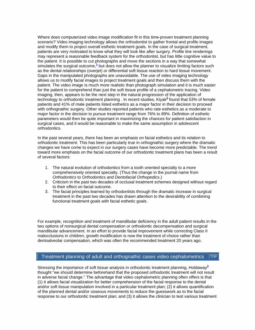
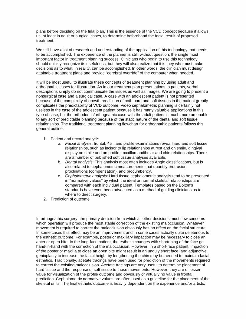
Case 1 This patient presented for treatment after previously having undergone orthodontic treatment (Figs. 1 and 2).
Fig. 1. This patient's profile would be considered by orthodontic standards to be “full” or bidentally protrusive. Facial analysis with plastic surgery in mind would indicate chin deficiency and lack of nasal tip projection.
Click on Image to view full size
Fig. 2. Her occlusal relationships were Class I.
Click on Image to view full size
Her abbreviated problem list was as follows:
1. Profile: Characterized by lip fullness, an acute nasolabial angle, and mild chin deficiency. 2. Dental relations: Nicely treated to a Class I occlusion, however, her maxillary and
mandibular incisors were proclined and protruded. 3. Cephalometric analysis: The patient measured to be not only bidentally protrusive, but
also had only 1 mm of chin projection (NB-Pg).
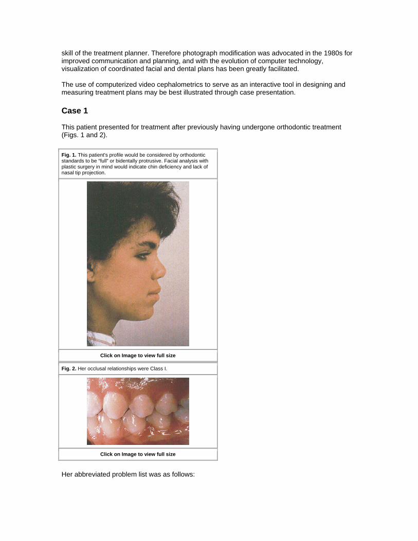
Cephalometric measurements help as an initial guide, but in dentofacial planning with computerized video cephalometrics, the application of norms as the treatment goal itself is not advocated. In VCD, the final dental and esthetic goals are designed and the proposed movements quantified with the computer through “retroengineering.” In other words, the end in mind is planned and the technology helps measure how we get there. The essence of this patient's problem revolved around her chief complaint. Although orthodontic treatment had successfully aligned the teeth and adequately spaced them, the patient was not pleased with her profile. Further advice was sought as to what other avenues of treatment might be provided to attain the desired outcome. With conventional profile outline tracing, orthodontic retreatment shows the lip response to four premolar extraction and a 5 mm posterior movement of both upper and lower incisors (Fig. 3). Fig. 3. Conventional acetate tracing to outline proposed profile changes expected with orthodontic treatment with four premolar extraction and maximum retraction of anterior teeth.
Click on Image to view full size
After the tracing is completed, several questions still remain:
1. While orthodontic retreatment reduces the lip projection, is a genioplasty desirable to improve the mildly deficient chin? Obviously, visualization of those changes and the desirability of a genioplasty depends on the skill of the clinician in being able to visualize from the acetate tracing what the outcome might be, and the patient's ability to help the clinician decide as to whether a genioplasty would be desirable. This is a valuable aspect of video imaging as far as bioethics and informed consent is concerned, as described by Ackerman and Proffit.4 Of course, a genioplasty may be considered as a staged procedure after the orthodontic retreatment.
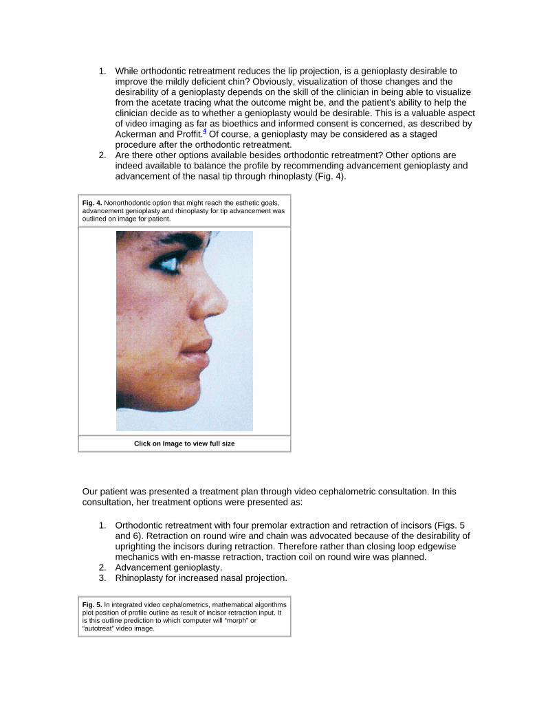
2. Are there other options available besides orthodontic retreatment? Other options are indeed available to balance the profile by recommending advancement genioplasty and advancement of the nasal tip through rhinoplasty (Fig. 4).
Fig. 4. Nonorthodontic option that might reach the esthetic goals, advancement genioplasty and rhinoplasty for tip advancement was outlined on image for patient.
Click on Image to view full size
Our patient was presented a treatment plan through video cephalometric consultation. In this consultation, her treatment options were presented as:
1. Orthodontic retreatment with four premolar extraction and retraction of incisors (Figs. 5 and 6). Retraction on round wire and chain was advocated because of the desirability of uprighting the incisors during retraction. Therefore rather than closing loop edgewise mechanics with en-masse retraction, traction coil on round wire was planned.
2. Advancement genioplasty. 3. Rhinoplasty for increased nasal projection.
Fig. 5. In integrated video cephalometrics, mathematical algorithms plot position of profile outline as result of incisor retraction input. It is this outline prediction to which computer will “morph” or “autotreat” video image.
Click on Image to view full size
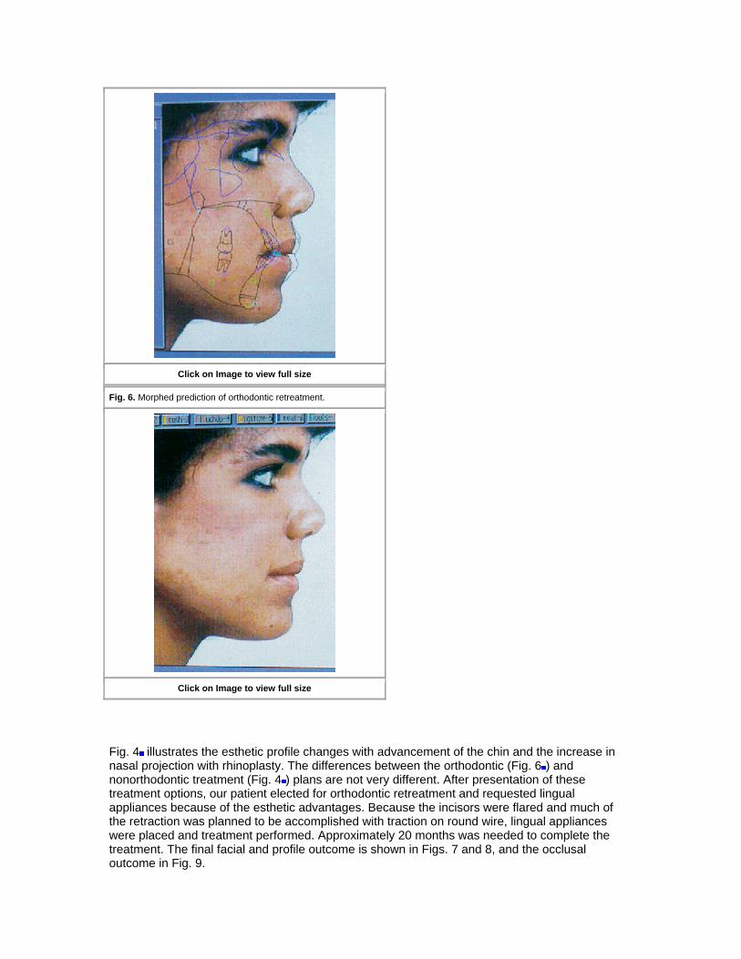
Fig. 6. Morphed prediction of orthodontic retreatment.
Click on Image to view full size
Fig. 4 illustrates the esthetic profile changes with advancement of the chin and the increase in nasal projection with rhinoplasty. The differences between the orthodontic (Fig. 6 ) and nonorthodontic treatment (Fig. 4 ) plans are not very different. After presentation of these treatment options, our patient elected for orthodontic retreatment and requested lingual appliances because of the esthetic advantages. Because the incisors were flared and much of the retraction was planned to be accomplished with traction on round wire, lingual appliances were placed and treatment performed. Approximately 20 months was needed to complete the treatment. The final facial and profile outcome is shown in Figs. 7 and 8, and the occlusal outcome in Fig. 9.
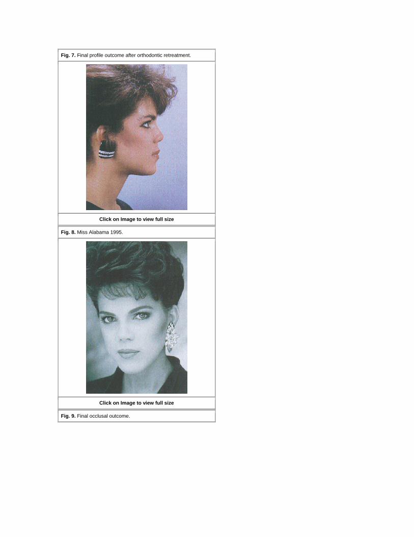
Fig. 7. Final profile outcome after orthodontic retreatment.
Click on Image to view full size
Fig. 8. Miss Alabama 1995.
Click on Image to view full size
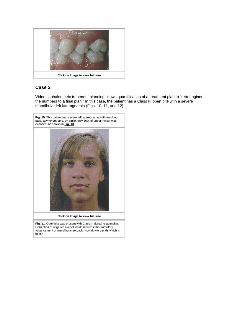
Fig. 9. Final occlusal outcome.
Click on Image to view full size
Case 2 Video cephalometric treatment planning allows quantification of a treatment plan to “retroengineer the numbers to a final plan.” In this case, the patient has a Class III open bite with a severe mandibular left laterognathia (Figs. 10, 11, and 12).
Fig. 10. This patient had severe left lateregnathia with resulting facial asymmetry and, on smile, only 50% of upper incisor was exposed, as shown in Fig. 13.
Click on Image to view full size
Fig. 11. Open bite was present with Class III dental relationship. Correction of negative overjet would require either maxillary advancement or mandibular setback. How do we decide which is best?
Click on Image to view full size
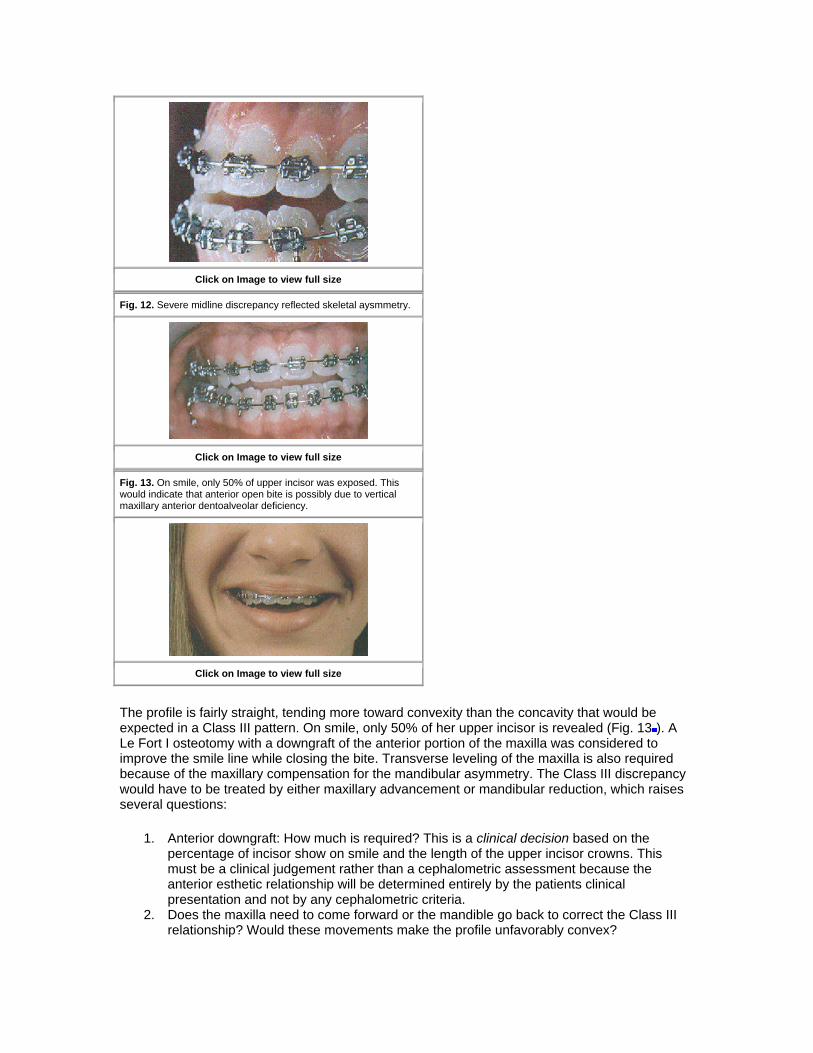
Fig. 12. Severe midline discrepancy reflected skeletal aysmmetry.
Click on Image to view full size
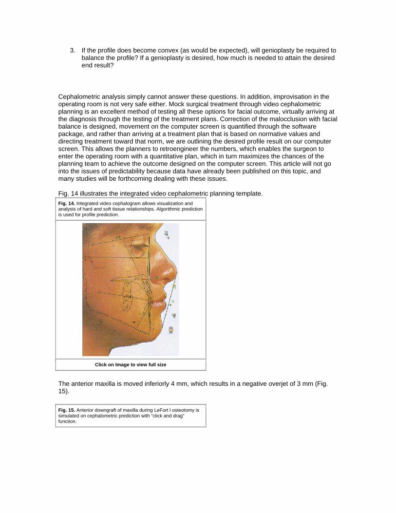
Fig. 13. On smile, only 50% of upper incisor was exposed. This would indicate that anterior open bite is possibly due to vertical maxillary anterior dentoalveolar deficiency.
Click on Image to view full size
The profile is fairly straight, tending more toward convexity than the concavity that would be expected in a Class III pattern. On smile, only 50% of her upper incisor is revealed (Fig. 13 ). A Le Fort I osteotomy with a downgraft of the anterior portion of the maxilla was considered to improve the smile line while closing the bite. Transverse leveling of the maxilla is also required because of the maxillary compensation for the mandibular asymmetry. The Class III discrepancy would have to be treated by either maxillary advancement or mandibular reduction, which raises several questions:
1. Anterior downgraft: How much is required? This is a clinical decision based on the percentage of incisor show on smile and the length of the upper incisor crowns. This must be a clinical judgement rather than a cephalometric assessment because the anterior esthetic relationship will be determined entirely by the patients clinical presentation and not by any cephalometric criteria.
2. Does the maxilla need to come forward or the mandible go back to correct the Class III relationship? Would these movements make the profile unfavorably convex?
3. If the profile does become convex (as would be expected), will genioplasty be required to balance the profile? If a genioplasty is desired, how much is needed to attain the desired end result?
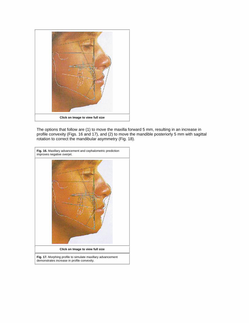
Cephalometric analysis simply cannot answer these questions. In addition, improvisation in the operating room is not very safe either. Mock surgical treatment through video cephalometric planning is an excellent method of testing all these options for facial outcome, virtually arriving at the diagnosis through the testing of the treatment plans. Correction of the malocclusion with facial balance is designed, movement on the computer screen is quantified through the software package, and rather than arriving at a treatment plan that is based on normative values and directing treatment toward that norm, we are outlining the desired profile result on our computer screen. This allows the planners to retroengineer the numbers, which enables the surgeon to enter the operating room with a quantitative plan, which in turn maximizes the chances of the planning team to achieve the outcome designed on the computer screen. This article will not go into the issues of predictability because data have already been published on this topic, and many studies will be forthcoming dealing with these issues. Fig. 14 illustrates the integrated video cephalometric planning template. Fig. 14. Integrated video cephalogram allows visualization and analysis of hard and soft tissue relationships. Algorithmic prediction is used for profile prediction.
Click on Image to view full size
The anterior maxilla is moved inferiorly 4 mm, which results in a negative overjet of 3 mm (Fig. 15).
Fig. 15. Anterior downgraft of maxilla during LeFort l osteotomy is simulated on cephalometric prediction with “click and drag” function.
Click on Image to view full size
The options that follow are (1) to move the maxilla forward 5 mm, resulting in an increase in profile convexity (Figs. 16 and 17), and (2) to move the mandible posteriorly 5 mm with sagittal rotation to correct the mandibular asymmetry (Fig. 18).
Fig. 16. Maxillary advancement and cephalometric prediction improves negative overjet.
Click on Image to view full size
Fig. 17. Morphing profile to simulate maxillary advancement demonstrates increase in profile convexity.
Click on Image to view full size
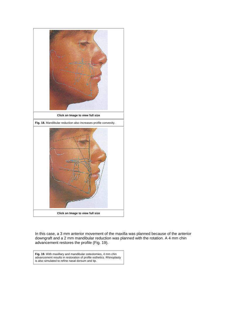
Fig. 18. Mandibular reduction also increases profile convexity.
Click on Image to view full size
In this case, a 3 mm anterior movement of the maxilla was planned because of the anterior downgraft and a 2 mm mandibular reduction was planned with the rotation. A 4 mm chin advancement restores the profile (Fig. 19).
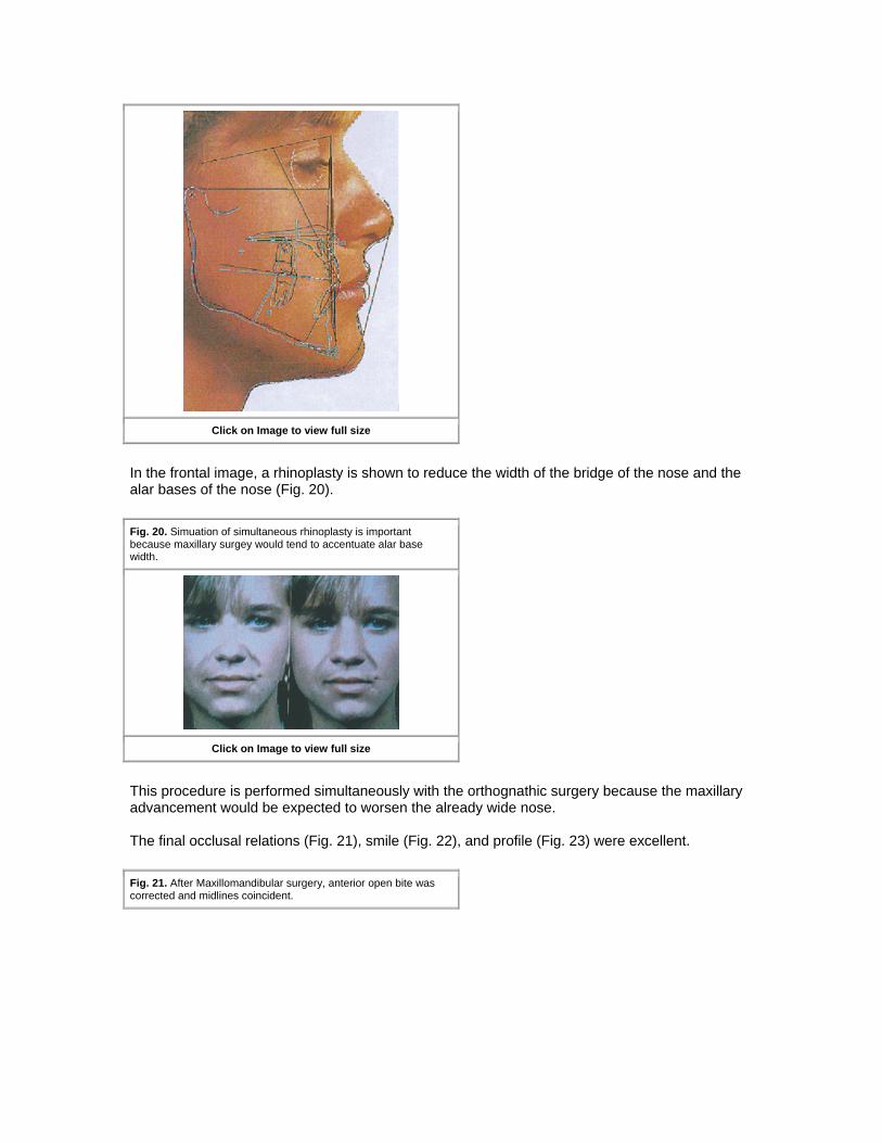
Fig. 19. With maxillary and mandibular osteotomies, 4 mm chin advancement results in restoration of profile esthetics. Rhinoplasty is also simulated to refine nasal dorsum and tip.
Click on Image to view full size
In the frontal image, a rhinoplasty is shown to reduce the width of the bridge of the nose and the alar bases of the nose (Fig. 20).
Fig. 20. Simuation of simultaneous rhinoplasty is important because maxillary surgey would tend to accentuate alar base width.
Click on Image to view full size
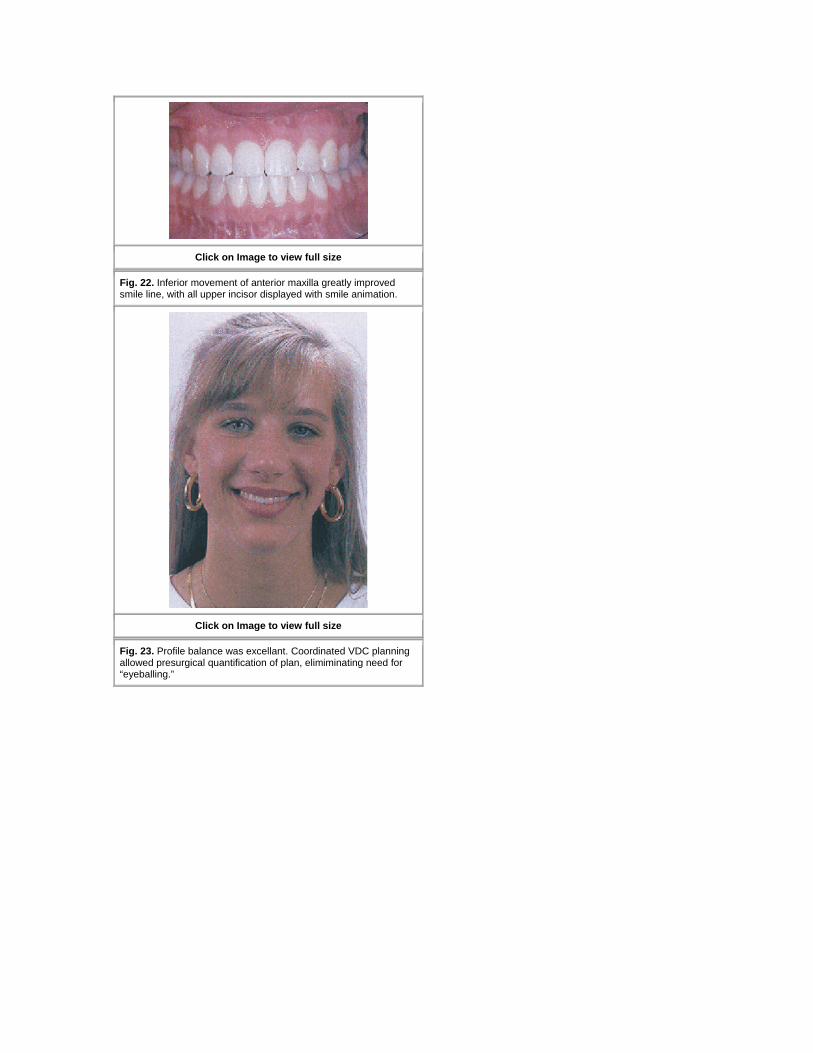
This procedure is performed simultaneously with the orthognathic surgery because the maxillary advancement would be expected to worsen the already wide nose. The final occlusal relations (Fig. 21), smile (Fig. 22), and profile (Fig. 23) were excellent.
Fig. 21. After Maxillomandibular surgery, anterior open bite was corrected and midlines coincident.
Click on Image to view full size
Fig. 22. Inferior movement of anterior maxilla greatly improved smile line, with all upper incisor displayed with smile animation.
Click on Image to view full size
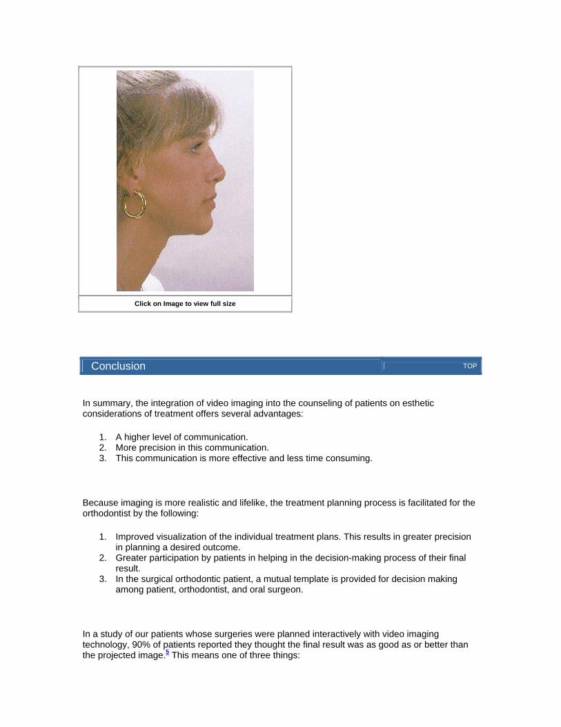
Fig. 23. Profile balance was excellant. Coordinated VDC planning allowed presurgical quantification of plan, elimiminating need for “eyeballing.”
Click on Image to view full size
Conclusion TOP
In summary, the integration of video imaging into the counseling of patients on esthetic considerations of treatment offers several advantages:
1. A higher level of communication. 2. More precision in this communication. 3. This communication is more effective and less time consuming.
Because imaging is more realistic and lifelike, the treatment planning process is facilitated for the orthodontist by the following:
1. Improved visualization of the individual treatment plans. This results in greater precision in planning a desired outcome.
2. Greater participation by patients in helping in the decision-making process of their final result.
3. In the surgical orthodontic patient, a mutual template is provided for decision making among patient, orthodontist, and oral surgeon.
In a study of our patients whose surgeries were planned interactively with video imaging technology, 90% of patients reported they thought the final result was as good as or better than the projected image.5 This means one of three things:
1. We are very accurate and honest with our projected treatment goals as far as their attainability.
2. In surgical cases, our surgeons are very good at placing the osteotomies where they are planned.
3. The use of imaging more clearly describes to the patient what to expect from their procedures, and therefore their expectations may be more reasonable.
Patient unhappiness would tend to occur then, when (1) the planners outline treatment that is clinically unattainable, and (2) the orthodontist or surgeon is clinically unable to “deliver the goods” as outlined. Video cephalometric treatment planning (1) quantitates movements in adult surgical cases; (2) allows interaction of the patient with the clinicians to help direct the treatment plan to the desired end result; and (3) allows facial planning and provides quantitative feedback as to what is required to attain a particular treatment plan.
References TOP
1. Kinnebrew MC. Hoffman DR. Carlton DM. Projecting the soft tissue outcome of surgical and orthodontic manipulation of the maxillofacial skeleton. Am J Orthod Dentofac Orthop 1983;84:508-19. 2. Kiyak HA Hohl T, West RA McNeill RW. Psychological change in orthognathic surgery patients: a 24 month followup. J Oral Maxillofac Surg 1984;42:506-12.
3. Holdaway RA. A soft tissue cephalometric analysis and its u e in orthodontic treatment planning. Am J Orthod 1983;84:1-28 4. Ackerman JL. Proffit WR. Communication in orthodontic treatment planning: bioet-hical and informed consent issues. Angle Orthod 1995;65:253-62 5. Sarver DM. Johnston MW. Matukas VJ. Video imaging in orthognathic surgery. J Oral Maxillofac Surg 1988;46:939-45.
Publishing and Reprint Information TOP
• Copyrights © 1997 by the American Association of Orthodontists • doi:10.1067/mod.1996.1100128
![Research Article Correlation Assessment between Three ...craniofacial diagnosis and orthodontic treatment planning []. Hard tissue is routinely evaluated by means of lateral cephalometric](https://img.dokumen.tips/doc/110x75/611e36bbf65f44201c695ce8/research-article-correlation-assessment-between-three-craniofacial-diagnosis.jpg)