Embed Size (px)
Citation preview
59
VI nerve palsy (abducens palsy)Paralisia do VI nervo (abducente)
Renato Luiz Nahoum Curi¹, Ian Curi Bonotto de Oliveira Costa², Tábatta Graciolli Moreira Barroso3
1Fluminense Federal University – UFF – Rio de Janeiro (RJ), Brazil. Niterói Institute of Ophthalmology – ION – Rio de Janeiro (RJ), Brazil.2Medical Student; Intern at the Ophthalmology Service, Antônio Pedro University Hospital (HUAP), Fluminense Federal University — UFF – Rio deJaneiro (RJ), Brazil.
Study conducted at the Ophthalmology Department, Fluminense Federal University Medical School – UFF - Rio de Janeiro (RJ), and the ION —Niterói Institute of Ophthalmology - Rio de Janeiro (RJ), Brazil
ABSTRACT
The authors review the basic aspects, etiology, clinical signs, diagnosis and treatment of the VI nerve palsy. Review the possible causesof abducent paralysis and location of determinant lesions. The clinical signs and clinical follow up are also observed in order to guide theetiology and therapeutic. The authors describe the clinical, pharmacological and surgical treatment. The authors emphasizes theirproposal of VI nerve palsy correction using the isolated Carlson-Jampolsky transposition.
Keywords: Abducens nerve; Abducens nerve diseases/etiology; Abducens nerve diseases/surgery; Palsy/surgery; Paresis;Ophthalmologic surgery procedures/methods
RESUMO
Neste trabalho foi realizada uma revisão da literatura com o objetivo de integrar e compilar artigos disponíveis sobre a paralisia doVI nervo (abducente) para rever suas características clínicas, etiologias possíveis e os procedimentos clínicos, farmacológicos ecirúrgicos para seu tratamento. Primeiramente, descreve-se sua ação, localização, trajeto e possíveis lesões, depois seus principaisfatores etiológicos para em seguida abordar-se o diagnóstico e o tratamento. Proposta de transposição de Carlson-Jampolsky isoladano tratamento cirúrgico da paralisia do VI nervo é também apresentada.
Descritores: Nervo abducente; Doenças do nervo abducente/etiologia; Doenças do nervo abducente/cirurgia; Paralisia/cirur-gia; Paresia; Transposições musculares; Procedimentos cirúrgicos oftalmológicos/métodos
REVIEW ARTICLE
Rev Bras Oftalmol. 2013; 72 (1): 59-69
The authors declare no conflicts of interest
Received for publication: 26/10/2011 - Accepted for publication: 18/5/2012
60
Figure 1: Left VI nerve (abducens) paresis or paralysis. Left esotropiawith major limitation of abduction, increasing on left gaze
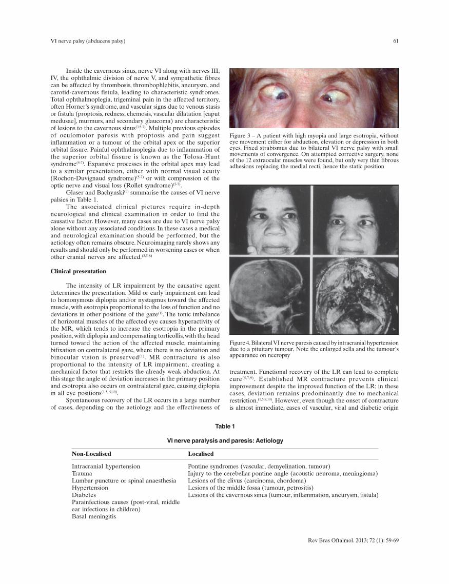
Figure 2: Bilateral Duane syndrome, pre- and postoperatively
Rev Bras Oftalmol. 2013; 72 (1): 59-69
Curi RLN, Costa ICBO, Barroso TGM
INTRODUCTION
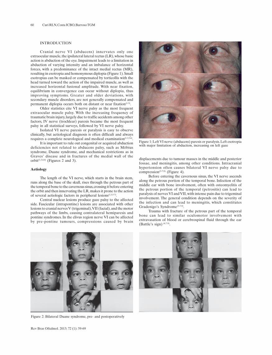
Cranial nerve VI (abducens) innervates only oneextraocular muscle, the ipsilateral lateral rectus (LR), whose basicaction is abduction of the eye. Impairment leads to a limitation inabduction of varying intensity and an imbalance of horizontalforces, with a predominance of the intact medial rectus (MR),resulting in esotropia and homonymous diplopia (Figure 1). Smallesotropias can be masked or compensated by torticollis with thehead turned toward the action of the impaired muscle, as well asincreased horizontal fusional amplitude. With near fixation,equilibrium in convergence can occur without diplopia, thusimproving symptoms. Greater and older deviations, withsecondary muscle disorders, are not generally compensated andpermanent diplopia occurs both on distant or near fixation(1-5).
Older statistics cite VI nerve palsy as the most frequentextraocular muscle palsy. With the increasing frequency oftraumatic brain injury, largely due to traffic accidents among otherfactors, IV nerve (trochlear) paresis became the most frequentpalsy in all statistical surveys, followed by VI nerve palsy.
Isolated VI nerve paresis or paralysis is easy to observeclinically, but aetiological diagnosis is often difficult and alwaysrequires a complete neurological and medical examination(1,3,5,6).
It is important to rule out congenital or acquired abductiondeficiencies not related to abducens palsy, such as Möbiussyndrome, Duane syndrome, and mechanical restrictions as inGraves’ disease and in fractures of the medial wall of theorbit(1,3,4,6) (Figures 2 and 3).
Aetiology
The length of the VI nerve, which starts in the brain stem,runs along the base of the skull, rises through the petrous part ofthe temporal bone to the cavernous sinus, crossing it before enteringthe orbit and then innervating the LR, makes it prone to the actionof several aetiologic factors in peripheral lesions(1,3,5-7).
Central nuclear lesions produce gaze palsy to the affectedside. Fascicular (intrapontine) lesions are associated with otherlesions to cranial nerves V (trigeminal), VII (facial), and the motorpathways of the limbs, causing contralateral hemiparesis andpontine syndromes. In the clivus region nerve VI can be affectedby pre-pontine tumours, compressions caused by brain
displacements due to tumour masses in the middle and posteriorfossae, and meningitis, among other conditions. Intracranialhypertension often causes bilateral VI nerve palsy due tocompression(5-7,9) (Figure 4).
Before entering the cavernous sinus, the VI nerve ascendsalong the petrous portion of the temporal bone. Infection of themiddle ear with bone involvement, often with osteomyelitis ofthe petrous portion of the temporal (petrositis) can lead toparalysis of nerves VI and VII, with intense pain due to trigeminalinvolvement. The general condition depends on the severity ofthe infection and can lead to meningitis, which constitutesGradenigo’s Syndrome(5-7,9).
Trauma with fracture of the petrous part of the temporalbone can lead to similar oculomotor involvement withextravasation of blood or cerebrospinal fluid through the ear(Battle’s sign) (6,7,9).
61
Non-Localised Localised
Intracranial hypertension Pontine syndromes (vascular, demyelination, tumour)Trauma Injury to the cerebellar-pontine angle (acoustic neuroma, meningioma)Lumbar puncture or spinal anaesthesia Lesions of the clivus (carcinoma, chordoma)Hypertension Lesions of the middle fossa (tumour, petrositis)Diabetes Lesions of the cavernous sinus (tumour, inflammation, aneurysm, fistula)Parainfectious causes (post-viral, middleear infections in children)Basal meningitis
Table 1
VI nerve paralysis and paresis: Aetiology
Figure 3 – A patient with high myopia and large esotropia, withouteye movement either for abduction, elevation or depression in botheyes. Fixed strabismus due to bilateral VI nerve palsy with smallmovements of convergence. On attempted corrective surgery, noneof the 12 extraocular muscles were found, but only very thin fibrousadhesions replacing the medial recti, hence the static position
Figure 4. Bilateral VI nerve paresis caused by intracranial hypertensiondue to a pituitary tumour. Note the enlarged sella and the tumour’sappearance on necropsy
Rev Bras Oftalmol. 2013; 72 (1): 59-69
VI nerve palsy (abducens palsy)
Inside the cavernous sinus, nerve VI along with nerves III,IV, the ophthalmic division of nerve V, and sympathetic fibrescan be affected by thrombosis, thrombophlebitis, aneurysm, andcarotid-cavernous fistula, leading to characteristic syndromes.Total ophthalmoplegia, trigeminal pain in the affected territory,often Horner’s syndrome, and vascular signs due to venous stasisor fistula (proptosis, redness, chemosis, vascular dilatation [caputmedusae], murmurs, and secondary glaucoma) are characteristicof lesions to the cavernous sinus(3,5-7). Multiple previous episodesof oculomotor paresis with proptosis and pain suggestinflammation or a tumour of the orbital apex or the superiororbital fissure. Painful ophthalmoplegia due to inflammation ofthe superior orbital fissure is known as the Tolosa-Huntsyndrome(5-7). Expansive processes in the orbital apex may leadto a similar presentation, either with normal visual acuity(Rochon-Duvignaud syndrome)(5-7) or with compression of theoptic nerve and visual loss (Rollet syndrome)(5-7).
Glaser and Bachynski(5) summarise the causes of VI nervepalsies in Table 1.
The associated clinical pictures require in-depthneurological and clinical examination in order to find thecausative factor. However, many cases are due to VI nerve palsyalone without any associated conditions. In these cases a medicaland neurological examination should be performed, but theaetiology often remains obscure. Neuroimaging rarely shows anyresults and should only be performed in worsening cases or whenother cranial nerves are affected.(3,5-8)
Clinical presentation
The intensity of LR impairment by the causative agentdetermines the presentation. Mild or early impairment can leadto homonymous diplopia and/or nystagmus toward the affectedmuscle, with esotropia proportional to the loss of function and nodeviations in other positions of the gaze(1). The tonic imbalanceof horizontal muscles of the affected eye causes hyperactivity ofthe MR, which tends to increase the esotropia in the primaryposition, with diplopia and compensating torticollis, with the headturned toward the action of the affected muscle, maintainingbifixation on contralateral gaze, where there is no deviation andbinocular vision is preserved(1). MR contracture is alsoproportional to the intensity of LR impairment, creating amechanical factor that restricts the already weak abduction. Atthis stage the angle of deviation increases in the primary positionand esotropia also occurs on contralateral gaze, causing diplopiain all eye positions(1,5, 9,10).
Spontaneous recovery of the LR occurs in a large numberof cases, depending on the aetiology and the effectiveness of
treatment. Functional recovery of the LR can lead to completecure(5,7,9). Established MR contracture prevents clinicalimprovement despite the improved function of the LR; in thesecases, deviation remains predominantly due to mechanicalrestriction.(1,5,9,10). However, even though the onset of contractureis almost immediate, cases of vascular, viral and diabetic origin
62
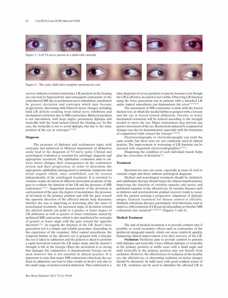
Figure 5 - Left VI nerve paresis in a child with varicella
Figure 6 – The same child after complete spontaneous cure
Rev Bras Oftalmol. 2013; 72 (1): 59-69
Curi RLN, Costa ICBO, Barroso TGM
recover without eventual contracture. LR paralysis in the fixatingeye can lead to hyperactivity and consequent contracture of thecontralateral MR due to permanent nerve stimulation, manifestedby greater deviation and esotropia which may becomeprogressively alternating with bilateral motor changes, includingmild LR deficits resulting from initial nerve inhibition andmechanical restriction due to MR contracture. Bilateral paralysisis not uncommon, with large angles, permanent diplopia andtorticollis with the head turned toward the fixating eye. In thiscase, the torticollis is not to avoid diplopia, but due to the staticposition of the eye in esotropia(1,5,9-12).
Diagnosis
The presence of diplopia and oculomotor signs, withesotropia and unilateral or bilateral impairment of abductioneasily lead to the diagnosis of VI nerve palsy. Clinical andneurological evaluation is essential for aetiologic diagnosis andappropriate treatment. The ophthalmic evaluation aims to ob-serve motor changes, their consequences on the oculomotorsystem and their progression, in order to determine theappropriate ophthalmic management to minimise symptoms andavoid sequels which, once established, can be treatedindependently of the aetiological treatment. It is essential toexamine ocular deviation in different horizontal positions of thegaze to evaluate the function of the LR and the presence of MRcontracture(1,3-5,7). Sequential measurements of the deviation ateach position of the gaze, the degree of incomitance, the presenceof deviation in the primary position and with the gaze towardthe opposite direction of the affected muscle help determinewhether the case is improving or worsening after the onset ofneurological treatment. An increased angle of deviation towardthe affected muscle can point to a greater or lesser degree ofLR deficiency as well as greater or lesser restriction caused byipsilateral MR contracture, which is also manifested by esotropiaof greater or lesser angle with the gaze toward the oppositedirection(1,5,9). As regards the function of the LR, Scott’s forcegeneration test is a simple and reliable procedure, depending onthe experience of the examiner. After topical anaesthesia, thetemporal limbus of the affected eye is grasped with a forceps;the eye is taken to adduction and the patient is asked to performa rapid movement toward the LR under study, and the muscle’sstrength is felt at the forceps. Often the movement is so strongthat damages the conjunctiva. A dynamometric forceps can beused, however this is not essential in clinical practice. It isimportant to note that major MR contractures that keep the eyefixed in adduction can lead to false results in Scott’s test due tothe small range of motion toward abduction. This could lead to a
false diagnosis of severe paralysis or paresis, because even thoughthe LR is effective, its action is not visible. Observing LR functionusing the force generation test in patients with a detached LRunder topical anaesthesia can demonstrate the error(1,5,9,10,13).
The assessment of MR contracture is done with the forcedduction test, in which the medial limbus is grasped with a forcepsand the eye is forced toward abduction. Greater or lessermechanical restriction will be noticed according to the strengthneeded to move the eye. Major contractures may prevent anypassive movement of the eye. Restrictions induced by conjunctivalchanges can also be demonstrated, especially with the formationof conjunctival folds toward the forceps(1,3,5,9,10).
Electrooculography or electromyography can yield thesame results, but these tests are not commonly used in clinicalpractice. The improvement or worsening of LR function can beassessed with sequential electrooculographies(9,10,13).
Diagnosing the condition of each individual muscle helpsplan the correction of deviation(1,3).
Treatment
Spontaneous cure can occur, especially in cases of viral orvascular origin and those without aetiological diagnosis.
Medical and neurological treatment should be instituted,and ophthalmic therapy should target the clinical manifestations,improving the function of extrinsic muscles and motor andpositional sequelae in the affected eye. In vascular diseases suchas diabetes and arteriosclerosis gradual recovery tends to occur,with the patient noticing a progressive alignment of diplopicimages. General treatment for disease control is effective.Similarly, infectious diseases, particularly viral infections, tend toimprove, with remission of LR paresis depending on whether MRcontracture has occurred(1,3,6,8,10,12) (Figures 5 and 6) .
Medical Treatment
The aim of medical treatment is to provide comfort and, ifpossible, to avoid secondary effects such as contracture of theipsilateral antagonist muscle, which can occur relatively quickly,hampering clinical improvement even after recovery of the LR.
Occlusion: Occlusion aims to provide comfort to patientswith diplopia and torticollis. Cases without diplopia or torticollisin the primary position or stable cases with a small angle andmild torticollis in the primary position may not benefit fromocclusion. However, the effectiveness of occlusion of the healthyeye, the affected eye, or alternating occlusion on motor changesshould be discussed. In mild cases with good residual action ofthe LR, occlusion can be used to stimulate the affected LR in
63
Rev Bras Oftalmol. 2013; 72 (1): 59-69
VI nerve palsy (abducens palsy)
order to fixate it in abduction, thus over-stimulating the LR andinducing increased neural inhibition of the MR in the same eye.This constant relaxation of the MR prevents contracture whileallowing the LR to be activated. In more severe cases, withseverely impaired abduction and torticollis due to MRcontracture, this approach can be difficult. Occlusion of thehealthy eye can lead to maintenance of torticollis depending onthe position of static equilibrium of the eye, leading to difficultiesin spatial orientation and rejection by the patient despite theabsence of diplopia, but without any prospect of influencingmuscle contraction in itself(1,5,9,10).
In cases with larger angles, longer progression or torticolliswith large deviations of the head, in which a more importantimbalance occurs in the PPG, or even with permanent diplopia inall horizontal positions of the gaze and consequent MRcontracture, occlusion of the affected eye is satisfactory, as itprevents diplopia and the spatial disorientation induced by thestatic position of the affected eye in adduction, which may be thenorm in all unilateral cases(1,3,10,12).
Alternating occlusion can be considered in cases where,due to muscle rebalancing, bilateral motor abnormalities occurleading to paralytic alternating esotropia, which is not uncommonand suggests a longer progression or bilateral VI nerveimpairment. It is important to note that in alternating cases, botheyes can fixate in the PPG, thus improving the acceptance ofocclusion(1,5,10).
Prism therapy: Temporal prisms can be used in cases withsmaller angles or stable, residual esotropia after surgicaltreatment. Satisfactory correction of diplopia is achieved in thePPG, though it should be present in other positions of the gaze.Torticollis also improves, usually with good patient satisfaction(1,3,8,10).
At greater angles acceptance is more difficult, either withunilateral therapy, due to the reduced vision and colourdissociation produced by the prism, or with bilateral therapy, dueto the spatial disorientation caused by the prism in front of thefixating eye.
Assessment of prism therapy by the physician and thepatient is critical, and the prism can be assembled in a test frameto allow the patient to get used to it.
Prism therapy can be associated with occlusion to controldiplopia while acting on secondary effects (Guibor prismtherapy)(3,9,10). The affected eye is occluded and the largestpossible prism is used in the healthy eye. Fixation of this eye isdone in convergence, with great stimulation of the MR in thiseye and transmission to the other eye, which is occluded. In caseof LR paresis, overactivation will lead to improved performance;in case of paralysis there will be no contractile response, but inboth cases there will be great inhibition of the MR in the affectedeye, thus avoiding contracture. Despite being theoretically sound,the method is difficult to implement because of the need of aprism of great magnitude in front of the fixating eye with occlusionof the opposite eye, which leads to reduced vision, coloured halos,and often spatial disorientation(10).
Pharmacological Treatment
Botulinum toxin: Botulinum toxin can assist in the treatmentof VI nerve paresis and it is very important depending on theclinical presentation. In most cases, progressive recovery startsearly regardless of treatment. During the first month after theonset of symptoms, no procedure should be implemented if thepatient is improving. However, occlusion can be used to alleviatethe symptoms of diplopia. After this period, in case of continuous
spontaneous improvement, the patient should be monitoredclinically, and botulinum toxin can be injected into the MR of theaffected eye. Many physicians start treatment with botulinumtoxin even earlier. The goal is to impair the MR, seeking to ba-lance out LR paresis and improve its contractile function due tothe lack of antagonism, thus facilitating functional recovery. Ca-ses of vascular or diabetic origin are often cured with this method.
When effective, botulinum toxin produces its greater effectin the first week, with exotropia induced by functional paralysisof the injected MR. Possible complications are vertical deviationsand ptosis; both are reversed after the drug’s effect vanishes. Inchronic cases with residual LR function, botulinum toxin canimprove MR contracture and its consequences. Repeatedinjections are used if progressive improvement is seen. Traumaticbrain injury and and sequelae of brain tumours can progress inthis manner. Permanent paralysis with total impairment of theLR should not be treated with botulinum toxin only; the drug canbe used to supplement surgery. The association of botulinum toxinin the MR with LR resection or muscle transposition has beenproposed in order to weaken the muscle, thus preservingvascularisation(3,10,14).
Surgical Treatment
Approximately 6 months after the onset of VI nerve palsy,the clinical picture can be considered definitive if no angle andmotor improvement or worsening occurs. Botulinum toxin shouldbe considered, and enough time should be given to allowfunctional recovery before indicating surgery. Scott suggests thatif no clinical improvement is seen in 3 months with repeatedfollow-up, surgery should be indicated(3,10,14,15).
It is necessary to evaluate the angle of deviation, the actionof the affected LR, and the presence ipsilateral and contralateralMR contracture. Angular measurement, the force generation testor electrooculography, and the forced duction test can be usedto evaluate muscle function. Despite the debate on theeffectiveness of these tests, most authors indicate surgery basedon their results(1,3,9,10,13,15). Once the intensity of MR contractureand LR deficit (paresis or paralysis) is determined, surgery isplanned. Surgery aims to correct the deviation and thereforediplopia and torticollis, increasing abduction and aiming for bestbinocular vision in the largest possible area of the visual field.These goals should also be considered for near fixation, as thereis often an improvement of distant vision and a worsening ofnear vision, with patient dissatisfaction.
The surgical strategy varies depending on the presence ofLR paresis or paralysis. Paresis can respond to resectionproportional to the residual action of the LR. Paralysis does notrespond to this procedure; in these cases, muscle transpositionshould be performed. Large LR resection does not improvecontractility or muscular function, but can lead to severemechanical restrictions of adduction.
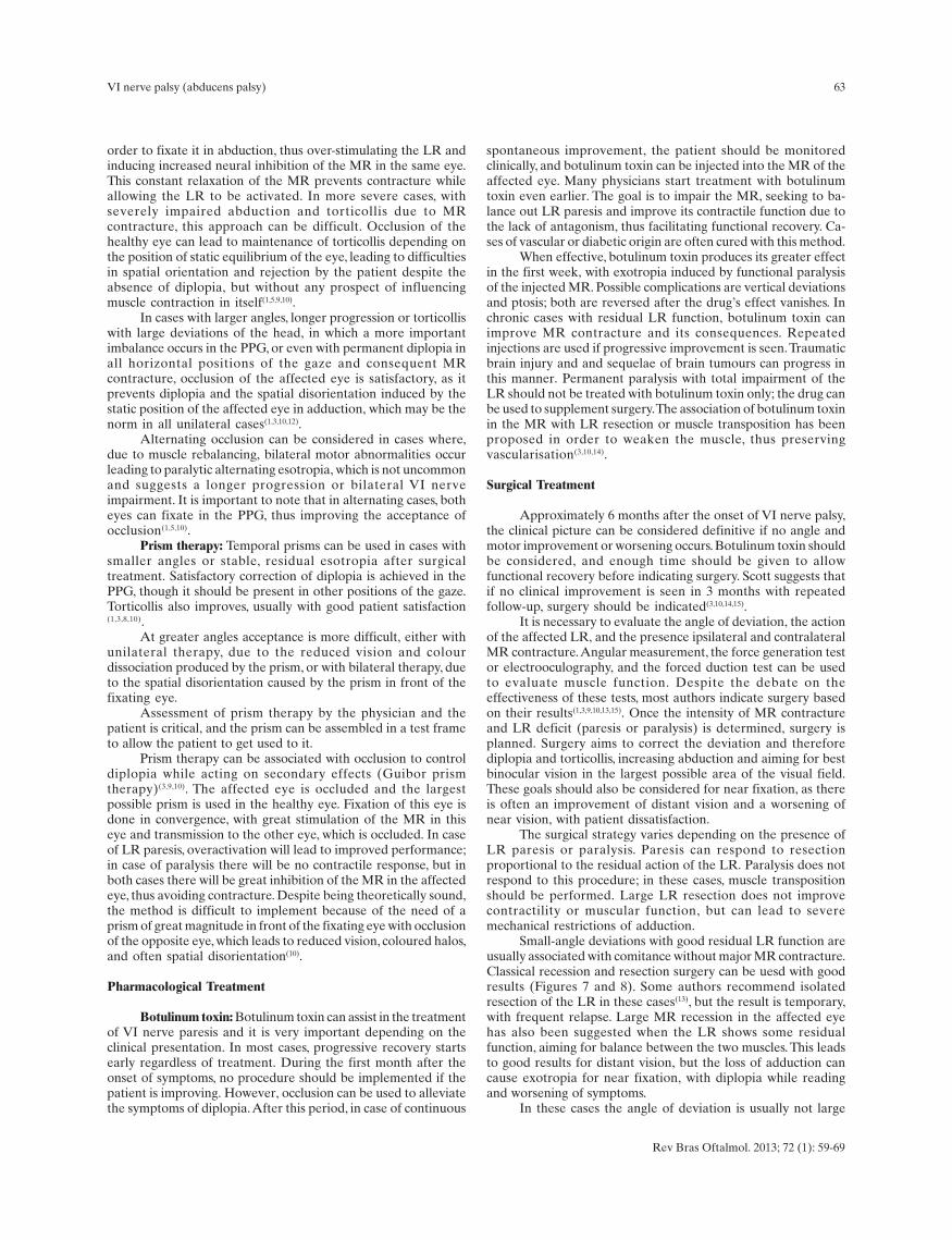
Small-angle deviations with good residual LR function areusually associated with comitance without major MR contracture.Classical recession and resection surgery can be uesd with goodresults (Figures 7 and 8). Some authors recommend isolatedresection of the LR in these cases(13), but the result is temporary,with frequent relapse. Large MR recession in the affected eyehas also been suggested when the LR shows some residualfunction, aiming for balance between the two muscles. This leadsto good results for distant vision, but the loss of adduction cancause exotropia for near fixation, with diplopia while readingand worsening of symptoms.
In these cases the angle of deviation is usually not large
64
Figure 7 – Right VI nerve paresis after peribulbar anaesthesia withbupivacaine for cataract surgery. Esotropia progressing to comitance(preoperative)
Figure 8 – The patient in Figure 6 after surgery, corrected with MRrecession and LR resection in the right eye
Rev Bras Oftalmol. 2013; 72 (1): 59-69
Curi RLN, Costa ICBO, Barroso TGM
and LR function is very good, with almost normal abductiondespite the deviation in the primary position. On the other hand,a decreasing angle can indicate spontaneous recovery; in thesecases, surgery should be postponed(1,5,10).
In similar cases with larger angles around 40-50 PD,recession/resection in the affected eye can be complemented withcontralateral MR recession, improving nerve induction of theparetic LR and improving its function as proposed by Horta-Barbosa(11,12). This author also recommends a large recession ofthe MR with the same goal (innervational surgery) (11).
In cases where LR function is absent or nearly absent,resection has no effect and muscle transposition is preferred. Still,Murray(16) and several other authors suggest a large (12 mm)recession of the MR associated with exaggerated resection ofthe LR (12 to 14 mm) in these cases. This approach leads to goodresults in the primary position but causes other complications.The first is relapse due to failure of LR elasticity, which initiallyacts as a mechanical restriction, holding the eye in the PPG, butsubsides with time. Also, while the mechanical action of theresected LR persists, there is great impairment of adduction sincethe MR was also largely recessed. This also impairs near fixationpermanently, because even without the restrictive action of theLR, the recessed MR loses its adduction function. Therefore littleeye movement remains, the binocular visual field is very smalland, as the other eye moves freely, diplopia occurs almostconstantly, with great variability and discomfort as it ishomonymous, with esotropia with the gaze toward the affectedLR and exotropia in the opposite direction.
Transposition of the vertical recti to the temporal side usingany of the known techniques is the ideal approach in these cases,as it produces elastic and innervational actions that antagonisethe action of the MR, avoiding deviation. Elastic factors due tovertical recti distension or transposition to the temporal sidesupport LR action and may be responsible for the slight abductionachieved, since attempted abduction causes relaxation of the MRand vertical recti (adductors), facilitating the performance of theseelastic components. Innervational factors produce a clinicalpicture similar to Duane Syndrome, i.e., during attemptedadduction stimulation of the MR is also transferred to the verti-cal recti (adductors) which antagonise the MR and, oncecontracted, prevent adduction of the eye. This co-contraction notonly stabilises eye movement but also acts in conjunction withelastic factors to prevent MR contracture.
RM recession associated with transposition of the verticalrecti has become a more frequent procedure, regardless of thetechnique used for transposition, showing good results regardingparallelism in the primary position and more stability, with fewerrelapses. Techniques of total transposition of the vertical rectitend to increase the risk of anterior segment ischemia.
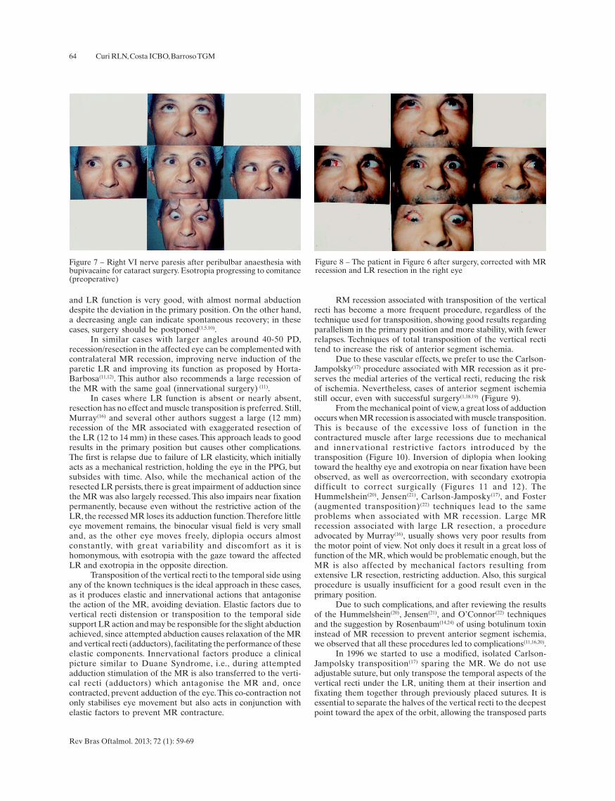
Due to these vascular effects, we prefer to use the Carlson-Jampolsky(17) procedure associated with MR recession as it pre-serves the medial arteries of the vertical recti, reducing the riskof ischemia. Nevertheless, cases of anterior segment ischemiastill occur, even with successful surgery(1,18,19) (Figure 9).
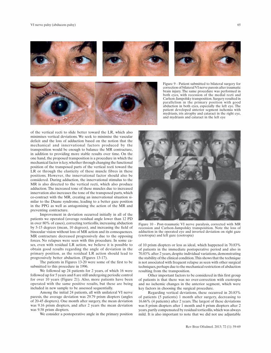
From the mechanical point of view, a great loss of adductionoccurs when MR recession is associated with muscle transposition.This is because of the excessive loss of function in thecontractured muscle after large recessions due to mechanicaland innervational restrictive factors introduced by thetransposition (Figure 10). Inversion of diplopia when lookingtoward the healthy eye and exotropia on near fixation have beenobserved, as well as overcorrection, with secondary exotropiadifficult to correct surgically (Figures 11 and 12). TheHummelshein(20), Jensen(21), Carlson-Jamposky(17), and Foster(augmented transposition)(22) techniques lead to the sameproblems when associated with MR recession. Large MRrecession associated with large LR resection, a procedureadvocated by Murray(16), usually shows very poor results fromthe motor point of view. Not only does it result in a great loss offunction of the MR, which would be problematic enough, but theMR is also affected by mechanical factors resulting fromextensive LR resection, restricting adduction. Also, this surgicalprocedure is usually insufficient for a good result even in theprimary position.
Due to such complications, and after reviewing the resultsof the Hummelshein(20), Jensen(21), and O’Connor(22) techniquesand the suggestion by Rosenbaum(14,24) of using botulinum toxininstead of MR recession to prevent anterior segment ischemia,we observed that all these procedures led to complications(11,16,20).
In 1996 we started to use a modified, isolated Carlson-Jampolsky transposition(17) sparing the MR. We do not useadjustable suture, but only transpose the temporal aspects of thevertical recti under the LR, uniting them at their insertion andfixating them together through previously placed sutures. It isessential to separate the halves of the vertical recti to the deepestpoint toward the apex of the orbit, allowing the transposed parts
65
Figure 9 - Patient submitted to bilateral surgery forcorrection of bilateral VI nerve paresis after traumaticbrain injury. The same procedure was performed inboth eyes, with recession of the medial recti andCarlson-Jampolsky transposition. Surgery resulted inparallelism in the primary position with goodabduction in both eyes, especially the left eye. Thepatient developed anterior segment ischemia withmydriasis, iris atrophy and cataract in the right eye,and mydriasis and cataract in the left eye
Figure 10 - Post-traumatic VI nerve paralysis, corrected with MRrecession and Carlson-Jampolsky transposition. Note the loss ofadduction in the operated eye and inverted deviation on right gaze(exotropia) and left gaze (esotropia)
Rev Bras Oftalmol. 2013; 72 (1): 59-69
VI nerve palsy (abducens palsy)
of the vertical recti to slide better toward the LR, which alsominimises vertical deviations. We seek to minimise the vasculardeficit and the loss of adduction based on the notion that themechanical and innervational factors produced by thetransposition would be enough to balance the MR contracture,in addition to providing more stable results over time. On theone hand, the proposed transposition is a procedure in which themechanical factor is key, whether through changing the functionalposition of the transposed parts of the vertical recti toward theLR or through the elasticity of these muscle fibres in thesepositions. However, the innervational factor should also beconsidered. During adduction, the innervational stimulus to theMR is also directed to the vertical recti, which also produceadduction. The increased tone of these muscles due to increasedinnervation also increases the tone of the transposed parts, whichco-contract with the MR, creating an innervational situation si-milar to the Duane syndrome, leading to a better gaze positionin the PPG as well as antagonising the action of the MR andpreventing contracture.
Improvement in deviation occurred initially in all of thepatients we operated (average residual angle lower than 12 PDin over 80% of cases), correcting torticollis, increasing abductionby 5-15 degrees (mean, 10 degrees), and increasing the field ofbinocular vision without loss of MR action and its consequences.MR contracture decreased progressively due to the opposingforces. No relapses were seen with this procedure. In some ca-ses, even with residual LR action, we believe it is possible toobtain good results regarding the angle of deviation in theprimary position, as the residual LR action should lead toprogressively better abduction. (Figures 13-17).
The patients in Figures 13-20 were some of the first to besubmitted to this procedure in 1996.
We followed up 24 patients for 2 years, of which 16 werefollowed up for 5 years and 8 are still undergoing periodic controlfor over 10 years (Figure 21). Also, more patients have beenoperated with the same positive results, but these are beingincluded in new sample to be assessed sequentially.
Among the initial 24 patients, all with unilateral VI nerveparesis, the average deviation was 29.79 prism diopters (anglesof 20-45 diopters). One month after surgery, the mean deviationwas 9.16 prism diopters, and after 2 years the mean deviationwas 9.58 prism diopters.
We consider a postoperative angle in the primary position
of 10 prism diopters or less as ideal, which happened in 70.83%of patients in the immediate postoperative period and also in70.83% after 2 years, despite individual variations, demonstratingthe stability of the clinical condition. This shows that the techniqueis not associated with frequent relapse as seen with other surgicaltechniques, perhaps due to the mechanical restriction of abductionresulting from the transposition.
Other important factors to be considered in this first groupof patients is that there was no over-correction with exotropiaand no ischemic changes in the anterior segment, which werekey factors in choosing the surgical procedure.
Regarding vertical deviations, these occurred in 20.83%of patients (5 patients) 1 month after surgery, decreasing to16.66% (4 patients) after 2 years. The largest of these deviationswas 8 prism diopters after 1 month and 6 prism diopters after 2years, partly compensated by residual torticollis, which was alwaysmild. It is also important to note that we did not use adjustable
66
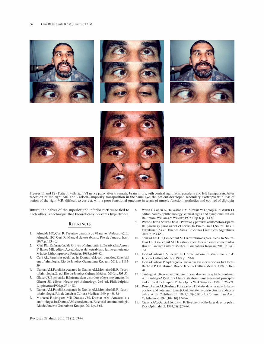
Figures 11 and 12 - Patient with right VI nerve palsy after traumatic brain injury, with central right facial paralysis and left hemiparesis. Afterrecession of the right MR and Carlson-Jampolsky transposition in the same eye, the patient developed secondary exotropia with loss ofaction of the right MR, difficult to correct, with a poor functional outcome in terms of muscle function, aesthetics and control of diplopia
Rev Bras Oftalmol. 2013; 72 (1): 59-69
Curi RLN, Costa ICBO, Barroso TGM
suture; the halves of the superior and inferior recti were tied toeach other, a technique that theoretically prevents hypertropia.
REFERENCES
1. Almeida HC, Curi R. Paresia e paralisia do VI nervo (abducente). In:Almeida HC, Curi R. Manual de estrabismo. Rio de Janeiro: [s.n.];1997. p. 133-40.
2. Curi RL. Enfermedad de Graves: oftalmopatia infiltrativa. In: ArroyoY, llanes ME, editor. Actualidades del estrabismo latino-americano.México: Lithoimpresora Portales; 1998. p.169-82.
3. Curi RL. Paralisias oculares. In: Dantas AM, coordenador. Essencialem oftalmologia. Rio de Janeiro: Guanabara Koogan; 2011. p. 1112-39.
4. Dantas AM. Paralisias oculares. In: Dantas AM, Monteiro MLR. Neuro-oftalmologia. 2a ed. Rio de Janeiro: Cultura Médica; 2010. p. 503-55.
5. Glaser JS, Bachynsky B. Infranuclear disorders of eye movements. In:Glaser JS, editor. Neuro-ophthalmology. 2nd ed. Philadelphia:Lippincott; c1990. p. 361-418.
6. Dantas AM. Paralisias oculares. In: Dantas AM, Monteiro MLR. Neuro-oftalmologia. Rio de Janeiro: Cultura Médica; 1999. p. 468-524.
7. Morterá-Rodrigues MP, Dantas JM, Dantas AM. Anatomia eembriologia. In: Dantas AM, coordenador. Essencial em oftalmologia.Rio de Janeiro: Guanabara Koogan; 2011. p. 3-61.
8. Walsh T, Cohen K, Helveston EM, Stewart W. Diplopia. In: Walsh TJ,editor. Neuro-ophthalmology: clinical signs and symptoms. 4th ed.Baltimore: Williams & Wilkins; 1997. Cap. 6. p. 114-80.
9. Prieto-Diaz J, Souza-Dias C. Paresias y parálisis oculomotoras: parteIII: paresias y parálisis del VI nervio. In: Prieto-Diaz J, Souza-Dias C.Estrabismo. 5a ed. Buenos Aires: Ediciones Cientificas Argentinas;2005. p. 354-65.
10. Souza-Dias CR, Goldchmit M. Os estrabismos paralíticos. In: Souza-Dias CR, Goldchmit M. Os estrabismos: teoria e casos comentados.Rio de Janeiro: Cultura Médica / Guanabara Koogan; 2011. p. 243-351.
11. Horta-Barbosa P. VI nervo. In: Horta-Barbosa P. Estrabismo. Rio deJaneiro: Cultura Médica; 1997. p. 163-8.
12. Horta-Barbosa P. Aplicações clínicas das leis inervacionais. In: Horta-Barbosa P. Estrabismo. Rio de Janeiro: Cultura Médica; 1997. p. 169-70.
13. Santiago AP, Rosenbaum AL. Sixth cranial nerve palsy. In: RosenbaumAL, Santiago AP, editors. Clinical strabismus management: principlesand surgical techniques. Philadelphia: W.B. Saunders; 1999. p. 259-71.
14. Rosembaum AL, Kushner BJ, Kirschen D. Vertical rectus muscle trans-position and botulinum toxin (Oculinum) to medial rectus for abducenspalsy. Arch Ophthalmol. 1989;107(6):820-3. Comment in ArchOphthalmol. 1991;109(10):1345-6.
15. Ciancia AO, Garcia HA, Lavin R. Treatment of the lateral rectus palsy.Doc Ophthalmol. 1984;58(1):57-64.
67
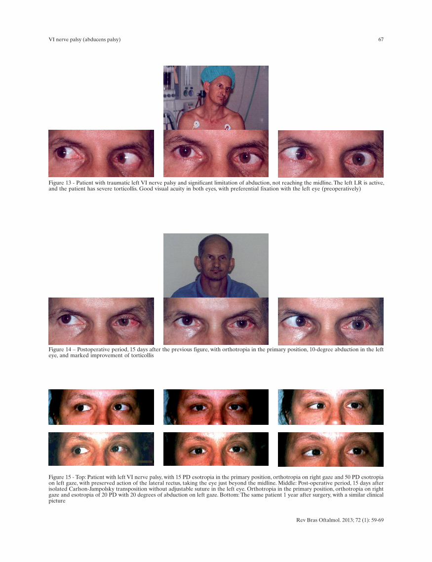
Figure 13 - Patient with traumatic left VI nerve palsy and significant limitation of abduction, not reaching the midline. The left LR is active,and the patient has severe torticollis. Good visual acuity in both eyes, with preferential fixation with the left eye (preoperatively)
Figure 14 – Postoperative period, 15 days after the previous figure, with orthotropia in the primary position, 10-degree abduction in the lefteye, and marked improvement of torticollis
Figure 15 - Top: Patient with left VI nerve palsy, with 15 PD esotropia in the primary position, orthotropia on right gaze and 50 PD esotropiaon left gaze, with preserved action of the lateral rectus, taking the eye just beyond the midline. Middle: Post-operative period, 15 days afterisolated Carlson-Jampolsky transposition without adjustable suture in the left eye. Orthotropia in the primary position, orthotropia on rightgaze and esotropia of 20 PD with 20 degrees of abduction on left gaze. Bottom: The same patient 1 year after surgery, with a similar clinicalpicture
Rev Bras Oftalmol. 2013; 72 (1): 59-69
VI nerve palsy (abducens palsy)
68
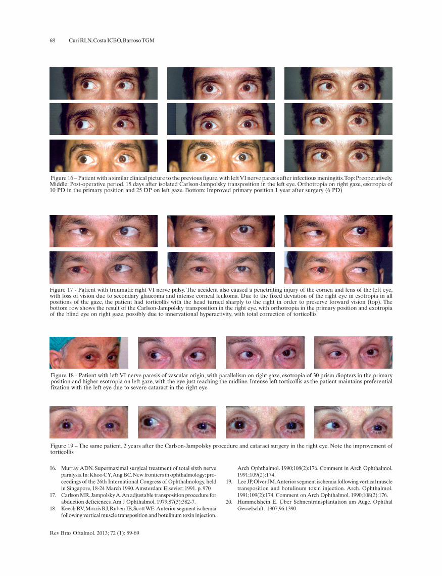
Figure 17 - Patient with traumatic right VI nerve palsy. The accident also caused a penetrating injury of the cornea and lens of the left eye,with loss of vision due to secondary glaucoma and intense corneal leukoma. Due to the fixed deviation of the right eye in esotropia in allpositions of the gaze, the patient had torticollis with the head turned sharply to the right in order to preserve forward vision (top). Thebottom row shows the result of the Carlson-Jampolsky transposition in the right eye, with orthotropia in the primary position and exotropiaof the blind eye on right gaze, possibly due to innervational hyperactivity, with total correction of torticollis
Figure 18 - Patient with left VI nerve paresis of vascular origin, with parallelism on right gaze, esotropia of 30 prism diopters in the primaryposition and higher esotropia on left gaze, with the eye just reaching the midline. Intense left torticollis as the patient maintains preferentialfixation with the left eye due to severe cataract in the right eye
Figure 19 – The same patient, 2 years after the Carlson-Jampolsky procedure and cataract surgery in the right eye. Note the improvement oftorticollis
Figure 16 – Patient with a similar clinical picture to the previous figure, with left VI nerve paresis after infectious meningitis. Top: Preoperatively.Middle: Post-operative period, 15 days after isolated Carlson-Jampolsky transposition in the left eye. Orthotropia on right gaze, esotropia of10 PD in the primary position and 25 DP on left gaze. Bottom: Improved primary position 1 year after surgery (6 PD)
Rev Bras Oftalmol. 2013; 72 (1): 59-69
Curi RLN, Costa ICBO, Barroso TGM
16. Murray ADN. Supermaximal surgical treatment of total sixth nerveparalysis. In: Khoo CY, Ang BC. New frontiers in ophthalmology: pro-ceedings of the 26th International Congress of Ophthalmology, heldin Singapore, 18-24 March 1990. Amsterdan: Elsevier; 1991. p. 970
17. Carlson MR, Jampolsky A. An adjustable transposition procedure forabduction deficiences. Am J Ophthalmol. 1979;87(3):382-7.
18. Keech RV, Morris RJ, Ruben JB, Scott WE. Anterior segment ischemiafollowing vertical muscle transposition and botulinum toxin injection.
Arch Ophthalmol. 1990;108(2):176. Comment in Arch Ophthalmol.1991;109(2):174.
19. Lee JP, Olver JM. Anterior segment ischemia following vertical muscletransposition and botulinum toxin injection. Arch. Ophthalmol.1991;109(2):174. Comment on Arch Ophthalmol. 1990;108(2):176.
20. Hummelshein E. Über Sehnentransplantation am Auge. OphthalGesselschft. 1907;96:1390.
69
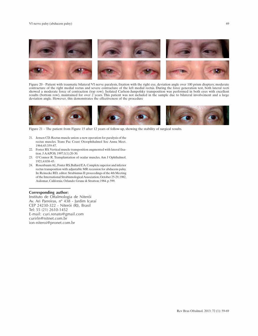
Figure 20 - Patient with traumatic bilateral VI nerve paralysis, fixation with the right eye, deviation angle over 100 prism diopters; moderatecontracture of the right medial rectus and severe contracture of the left medial rectus. During the force generation test, both lateral rectishowed a moderate force of contraction (top row). Isolated Carlson-Jampolsky transposition was performed in both eyes with excellentresults (bottom row), maintained for over 2 years. This patient was not included in the sample due to bilateral involvement and a largedeviation angle. However, this demonstrates the effectiveness of the procedure
Figure 21 – The patient from Figure 15 after 12 years of follow-up, showing the stability of surgical results.
Corresponding author:Instituto de Oftalmologia de NiteróiAv. Ari Parreiras, nº 438 - Jardim IcaraíCEP 24230-322 - Niterói (RJ), BrasilTel: 55 (21) 2610-1452E-mail: [email protected]@[email protected]
Rev Bras Oftalmol. 2013; 72 (1): 59-69
VI nerve palsy (abducens palsy)
21. Jensen CD. Rectus muscle union: a new operation for paralysis of therectus muscles. Trans Pac Coast Otoophthalmol Soc Annu Meet.1964;45:359-87.
22. Foster RS. Vertical muscle transposition augmented with lateral fixa-tion. J AAPOS. 1997;1(1):20-30.
23. O’Connor R. Transplantation of ocular muscles. Am J Ophthalmol.1921;4:838-45.
24. Rosenbaum AL, Foster RS, Ballard EA. Complete superior and inferiorrectus transposition with adjustable MR recession for abducens palsy.In: Reinecke RD, editor. Strabismus II: proceedings of the 4th Meetingof the International Strabismological Association, October 25-29, 1982,Asilomar, California. Orlando: Grune & Stratton; 1984. p. 599.