Embed Size (px)
Citation preview
ORIGINALRESEARCH
Velocity Measurements in the Middle CerebralArteries of Healthy Volunteers Using 3D RadialPhase-Contrast HYPRFlow: Comparison withTranscranial Doppler Sonography and2D Phase-Contrast MR Imaging
W. ChangB. Landgraf
K.M. JohnsonS. Kecskemeti
Y. WuJ. VelikinaH. RowleyO. Wieben
C. MistrettaP. Turski
BACKGROUND AND PURPOSE: We have developed PC HYPRFlow, a comprehensive MRA techniquethat includes a whole-brain CE dynamic series followed by PC velocity-encoding, yielding a time seriesof high-resolution morphologic angiograms with associated velocity information. In this study, wepresent velocity data acquired by using the PC component of PC HYPRFlow (PC-VIPR).
MATERIALS AND METHODS: Ten healthy volunteers (6 women, 4 men) were scanned by using PCHYPRFlow and 2D-PC imaging, immediately followed by velocity measurements by using TCD.Velocity measurements were made in the M1 segments of the MCAs from the PC-VIPR, 2D-PC, andTCD examinations.
RESULTS: PC-VIPR showed approximately 30% lower mean velocity compared with TCD, consistentwith other comparisons of TCD with PC-MRA. The correlation with TCD was r � 0.793, and thecorrelation of PC-VIPR with 2D-PC was r � 0.723.
CONCLUSIONS: PC-VIPR is a technique capable of acquiring high-resolution MRA of diagnostic qualitywith velocity data comparable with TCD and 2D-PC. The combination of velocity information and fasthigh-resolution whole-brain morphologic angiograms makes PC HYPRFlow an attractive alternative tocurrent MRA methods.
ABBREVIATIONS: CE � contrast-enhanced; CE-VIPR � contrast-enhanced vastly undersampledisotropic projection reconstruction; 2D-PC � 2D phase-contrast; HYPR-LR � highly constrainedlocal projection reconstruction; MCA � middle cerebral artery; MRA � MR angiography; PC �phase-contrast; PC HYPRFlow � time-resolved MRA using highly constrained projection recon-struction and PC-VIPR data for the reconstruction convolution; PC-MRA � phase-contrast MRangiography; PC-VIPR � phase-contrast vastly undersampled isotropic projection reconstruction;SNR � signal intensity–to-noise ratio; TCD � transcranial Doppler sonography; VENC � velocityencoding; WSS � wall shear stress
The acquisition of intracranial velocity measurements andvelocity derivatives (WSS) is clinically useful for the eval-
uation of neurovascular disorders such as vasospasm, steno-ses, and aneurysms, but measurement of intracranial velocityhas proved challenging. TCD has been used clinically for de-cades and allows the acquisition of velocity measurements inthe MCAs and several other vessels through the temporal win-dow. However, the sonic properties of the cranial vault pre-vent measurement of velocity in many intracranial arteries byTCD. 2D-PC MR imaging has been used to obtain hemody-namic data for the past 20 years but has limited coverage and
exhibits partial volume effects.1 3D and 4D methods havehigher SNR, fewer partial volume effects, and improved spatialresolution but have an increased scanning time.2 Previous im-plementation of whole-brain cardiac-gated PC Cartesian 4DMR imaging has resulted in scanning times too long to beclinically useful.3,4 Advances in PC Cartesian acquisition havereduced scanning times and increased resolution to some de-gree. Investigators have used view-sharing,5 spatial harmon-ics,6 and parallel imaging7 to reduce scanning time. With thesemethods, in-plane spatial resolutions on the order of 0.6 �1 mm with scanning times in the 8- to 12-minute range can beachieved.8
However, obtaining high-resolution whole-brain angio-grams with velocity information within clinically useful im-aging times has been challenging. Recently, we imple-mented radial imaging techniques that are particularlywell-suited for neurovascular MRA. Because MRA is sparse,consisting of few changing nonzero elements, acceleratedacquisitions can often be performed with tolerable artifacts.For example, azimuthally undersampled radial acquisitionscan be used to acquire images in much shorter scanningtimes than Cartesian acquisitions of a similar resolutionwith acceptable SNR and image quality,9 due to the rela-
Received March 15, 2010; accepted after revision June 1.
From the Departments of Medical Physics (K.M.J., S.K., Y.W., J.V., O.W., C.M.), Radiology(B.L., H.R., C.M., P.T.), and Biomedical Engineering (K.M.J., C.M.), University of Wisconsin,Madison, Wisconsin; and University of Wisconsin School of Medicine and Public Health(W.C.), Madison, Wisconsin.
This work was supported by National Institutes of Health grant R21 EB009441 to PatrickTurski.
Please address correspondence to Warren Chang, MD, University of Wisconsin School ofMedicine and Public Health, 750 Highland Ave, Madison, WI 53705; e-mail: [email protected]
Indicates open access to non-subscribers at www.ajnr.org
DOI 10.3174/ajnr.A2240
54 Chang � AJNR 32 � Jan 2011 � www.ajnr.org
tively benign nature of streaklike artifacts caused by theundersampling.10,11
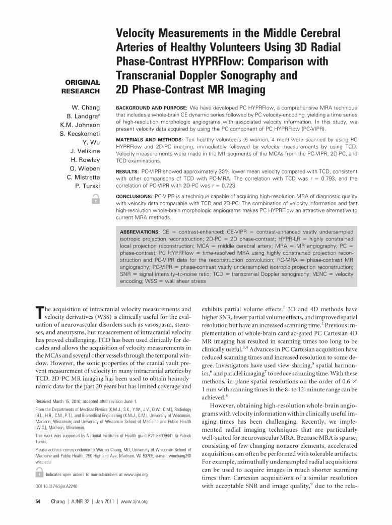
We have combined radial undersampling with a novelconstrained image reconstruction technique to create PCHYPRFlow,9,12 a comprehensive MRA technique. Figure 1shows a flow chart of how images are acquired and recon-structed by using PC HYPRFlow. A series of low-resolution3D-radial CE source images are acquired (CE-VIPR), followedby a high-resolution velocity-encoded PC acquisition (PC-VIPR13). The low-resolution source images are then con-volved with the high-resolution PC velocity acquisition by us-ing a technique called HYPR-LR14 to yield a time series ofhigh-resolution morphologic angiograms with associated ve-locity information. With current protocols, PC HYPRFlowprovides whole-brain angiograms with excellent spatial reso-lution (0.68 � 0.68 � 0.68 mm3) with scanning times of 5– 6minutes.9
Because PC HYPRFlow is a highly accelerated technique,it may be susceptible to errors. To ensure diagnostic confi-dence, we have conducted several studies to validate imagequality and velocity measurements from PC HYPRFlow.9,15-17
We previously reported that PC HYPRFlow provides imagesof diagnostic quality in medium- and large-sized intracranialvessels.9 In this study, we investigated velocity informationacquired by using the PC component of PC HYPRFlow. Wevalidated velocity measurements acquired by using PC-VIPRin the MCAs of healthy volunteers by comparing them withvelocity measurements from 2 reference standards, TCD andCartesian 2D-PC-MRA. The purpose of this comparison wasto define the relationship of PC HYPRFlow velocity measure-ments to other established modalities.
Materials and MethodsVolunteer studies were performed in compliance with Health Insur-
ance Portability and Accountability Act regulations and by using a
protocol approved by the local institutional review board. Ten healthy
volunteers ranging from 19 to 58 years of age were imaged (6 women,
4 men) with a clinical 3T MR imaging system (MR HD 750; GE
Healthcare, Milwaukee, Wisconsin) with a 8-channel head coil (Ex-
cite HD Brain Coil, GE Healthcare). Before contrast injection, a fast
2D-PC scan of each MCA was obtained to estimate the velocity to
determine optimum VENC to prevent aliasing, followed by a PC
HYPRFlow acquisition. Two fast low-resolution scans (2 � 2 � 2
mm3) were used in the time-resolved multiecho 3D radial acquisition
(CE-VIPR).18 Contrast was injected during the second scan. Subse-
quently, velocity encoding was performed by using a high-resolution
dual-echo 3D-radial PC acquisition (PC-VIPR). The PC-VIPR data
were used as a composite image (angiographic constraint) for HYPR-
LR reconstruction and for hemodynamic evaluation.14 Immediately
following the MR imaging examination, the volunteers underwent
TCD scanning.
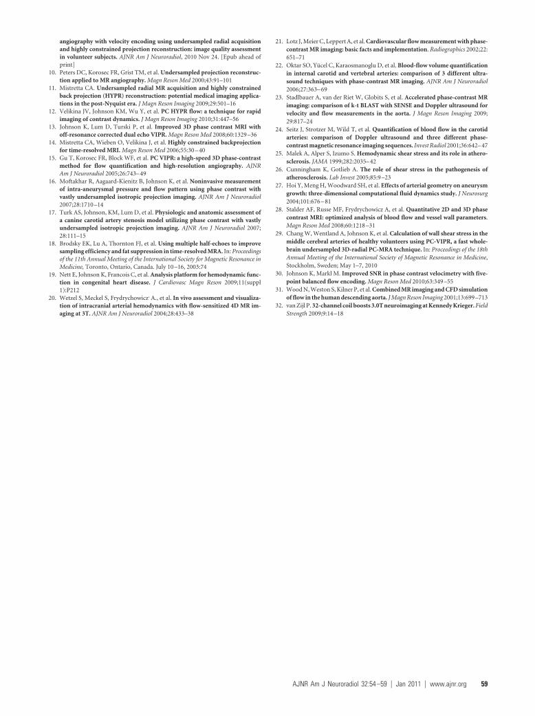
MR Imaging ProtocolImaging parameters for CE-VIPR were the following: FOV � 26 �
26 � 26 cm2, TR/TE � 3.0/0.4 ms, bandwidth � 125 kHz. For each
projection, there were 64 points from the center to the edge of the
k-space, with a frame update time of 0.75 seconds. Scanning param-
eters for postcontrast PC-VIPR were the following: FOV � 22 � 22 �
22 cm2, TR/TE � 12.5/4.8 ms, VENC � 80 –150 cm/s, bandwidth �
83.3 kHz, readout matrix � 320 points per projection, spatial resolu-
tion for the composite image � 0.68 � 0.68 � 0.68 mm3. Seven
thousand projections were acquired within 5 minutes. Gadobenate
dimeglumine (MultiHance; Bracco Diagnostics, Princeton, New Jer-
sey) was injected at 3 mL/s, and the contrast dose was 0.1 mm/kg
followed by a 20-mL saline flush. The MR imaging acquisitions were
cardiac gated by using chest leads (Table).
A series of 3D time-resolved velocity images were reconstructed
from the PC-VIPR data. A MatLab inhouse developed software filter
with 50 ms at a low spatial frequency and 130 ms at a high spatial
frequency was applied to improve the SNR. A vascular mask was
generated by applying a signal-intensity threshold on the complex
difference image. Subsequently, a region of interest, including ap-
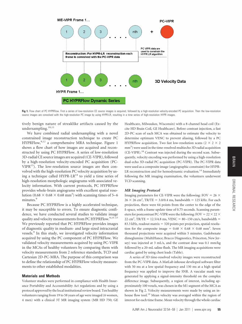
proximately 100 voxels, was chosen in the M1 segment of the MCA as
shown in Fig 2. Velocity measurements were made by using an in-
house flow tool.19 Mean velocity was averaged within the region of
interest for each time frame. Mean velocity through the whole cardiac
Fig 1. Flow chart of PC HYPRFlow. First a series of low-resolution CE source images is acquired, followed by a high-resolution velocity-encoded PC acquisition. Then the low-resolutionsource images are convolved with the high-resolution PC image by using HYPR-LR, resulting in a time series of high-resolution HYPR images.
BRA
INORIGIN
ALRESEARCH
AJNR Am J Neuroradiol 32:54 –59 � Jan 2011 � www.ajnr.org 55
cycle was calculated for each volunteer. The maximum velocity over
the cardiac cycle was designated as the peak systolic velocity, and the
minimum velocity was designated as the minimum diastolic velocity.
This process was repeated for data from the 2D-PC acquisition.
TCD ProtocolTCD imaging was performed in the MCAs bilaterally by using an
Acuson-Sequoia sonography scanner (Siemens, Malvern, Pennsylva-
nia) and a 4V1 MHz vector transducer. Depths for MCA interroga-
tion were retrieved from the subjects’ MRAs and given to the sonog-
rapher. The sonographer would attempt to replicate the depths when
obtaining spectral Doppler signals. Color and spectral Doppler im-
ages were obtained via the transtemporal window. Once spectral
Doppler images were obtained, mean, peak systolic, and end diastolic
velocities were recorded. Three velocity measurements were obtained
in each MCA.
Statistical MethodsCorrelations were acquired between PC-VIPR and TCD and PC-
VIPR and 2D-PC. Bland-Altman plots were generated comparing
PC-VIPR and TCD and PC-VIPR and 2D-PC, and the bias and limits
of agreement were calculated for each Bland-Altman plot. Statistics
were calculated by using Excel 2007 (Microsoft, Bothell, Washing-
ton). The Pearson r was used for correlation analysis, and a P value �
.05 for the correlation coefficient was considered statistically
significant.
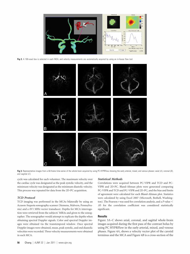
ResultsFigure 3A–C shows axial, coronal, and sagittal whole-brainimages acquired during the first pass of the contrast bolus byusing PC HYPRFlow in the early arterial, mixed, and venousphases. Figure 4A, shows a velocity vector plot of the carotidterminus and the MCA and Figure 4B is a cross-section of the
Fig 2. A 100-voxel box is selected in each MCA, and velocity measurements are automatically acquired by using an in-house flow tool.
Fig 3. Representative images from a 60-frame time-series of the whole brain acquired by using PC HYPRFlow showing the early arterial, mixed, and venous phases: axial (A ), coronal (B ),and sagittal (C ).
56 Chang � AJNR 32 � Jan 2011 � www.ajnr.org
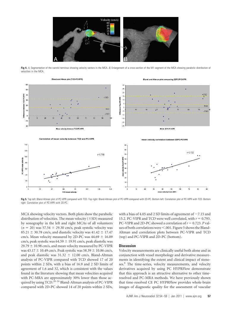
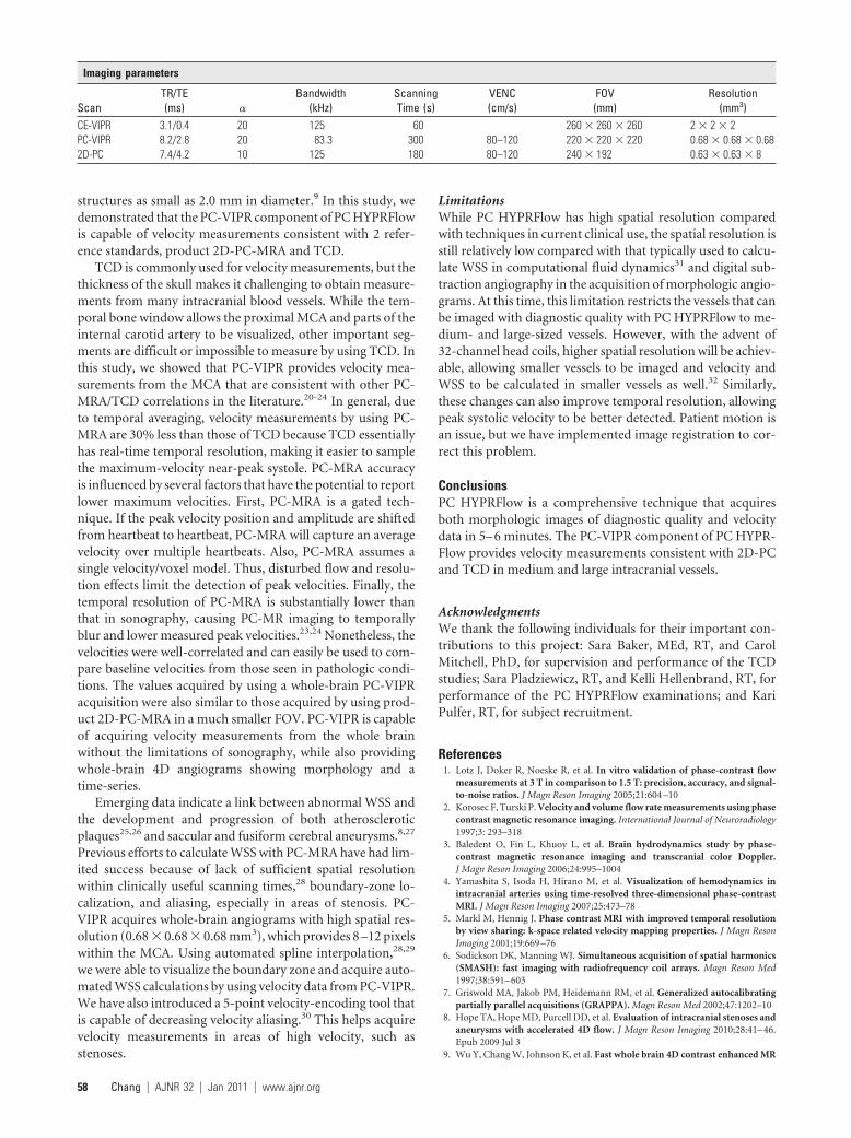
MCA showing velocity vectors. Both plots show the parabolicdistribution of velocities. The mean velocity (�SD) measuredby sonography in the left and right MCAs of all volunteers(n � 20) was 57.54 � 29.30 cm/s, peak systolic velocity was85.21 � 30.78 cm/s, and diastolic velocity was 41.42 � 17.47cm/s. Mean velocity measured by 2D-PC was 44.69 � 16.09cm/s, peak systolic was 64.59 � 19.91 cm/s, peak diastolic was29.79 � 10.98 cm/s, and mean velocity measured by PC-VIPRwas 43.17 � 10.49 cm/s. Peak systolic was 58.39 � 10.86 cm/s,and peak diastolic was 31.32 � 12.00 cm/s. Bland-Altmananalysis of PC-VIPR compared with TCD showed 17 of 20points within 2 SDs, with a bias of 16.9 and 2 SD limits ofagreement of 1.6 and 32, which is consistent with the valuesfound in the literature showing that mean velocities acquiredwith PC-MRA are approximately 30% lower than those ac-quired by using TCD.20-24 Bland-Altman analysis of PC-VIPRcompared with 2D-PC showed 14 of 20 points within 2 SDs,
with a bias of 4.05 and 2 SD limits of agreement of �7.15 and15.2. PC-VIPR and TCD were well correlated, with r � 0.793.PC-VIPR and 2D-PC showed a correlation of r � 0.723. P val-ues of both correlations were �.001. Figure 5 shows the Bland-Altman and correlation plots between PC-VIPR and TCD(top) and PC-VIPR and 2D-PC (bottom).
DiscussionVelocity measurements are clinically useful both alone and inconjunction with vessel morphology and derivative measure-ments in identifying the extent and clinical impact of steno-ses.8 The time-series, velocity measurements, and velocityderivatives acquired by using PC HYPRFlow demonstratethat this approach is an attractive alternative to other time-resolved and PC-MRA methods. We have previously shownthat time-resolved CE PC HYPRFlow provides whole-brainimages of diagnostic quality for the assessment of vascular
Fig 4. A, Segmentation of the carotid terminus showing velocity vectors in the MCA. B, Enlargement of a cross-section of the M1 segment of the MCA showing parabolic distribution ofvelocities in the MCA.
Fig 5. Top left: Bland-Altman plot of PC-VIPR compared with TCD. Top right: Bland-Altman plot of PC-VIPR compared with 2D-PC. Bottom left: Correlation plot of PC-VIPR with TCD. Bottomright: Correlation plot of PC-VIPR with 2D-PC.
AJNR Am J Neuroradiol 32:54 –59 � Jan 2011 � www.ajnr.org 57
structures as small as 2.0 mm in diameter.9 In this study, wedemonstrated that the PC-VIPR component of PC HYPRFlowis capable of velocity measurements consistent with 2 refer-ence standards, product 2D-PC-MRA and TCD.
TCD is commonly used for velocity measurements, but thethickness of the skull makes it challenging to obtain measure-ments from many intracranial blood vessels. While the tem-poral bone window allows the proximal MCA and parts of theinternal carotid artery to be visualized, other important seg-ments are difficult or impossible to measure by using TCD. Inthis study, we showed that PC-VIPR provides velocity mea-surements from the MCA that are consistent with other PC-MRA/TCD correlations in the literature.20-24 In general, dueto temporal averaging, velocity measurements by using PC-MRA are 30% less than those of TCD because TCD essentiallyhas real-time temporal resolution, making it easier to samplethe maximum-velocity near-peak systole. PC-MRA accuracyis influenced by several factors that have the potential to reportlower maximum velocities. First, PC-MRA is a gated tech-nique. If the peak velocity position and amplitude are shiftedfrom heartbeat to heartbeat, PC-MRA will capture an averagevelocity over multiple heartbeats. Also, PC-MRA assumes asingle velocity/voxel model. Thus, disturbed flow and resolu-tion effects limit the detection of peak velocities. Finally, thetemporal resolution of PC-MRA is substantially lower thanthat in sonography, causing PC-MR imaging to temporallyblur and lower measured peak velocities.23,24 Nonetheless, thevelocities were well-correlated and can easily be used to com-pare baseline velocities from those seen in pathologic condi-tions. The values acquired by using a whole-brain PC-VIPRacquisition were also similar to those acquired by using prod-uct 2D-PC-MRA in a much smaller FOV. PC-VIPR is capableof acquiring velocity measurements from the whole brainwithout the limitations of sonography, while also providingwhole-brain 4D angiograms showing morphology and atime-series.
Emerging data indicate a link between abnormal WSS andthe development and progression of both atheroscleroticplaques25,26 and saccular and fusiform cerebral aneurysms.8,27
Previous efforts to calculate WSS with PC-MRA have had lim-ited success because of lack of sufficient spatial resolutionwithin clinically useful scanning times,28 boundary-zone lo-calization, and aliasing, especially in areas of stenosis. PC-VIPR acquires whole-brain angiograms with high spatial res-olution (0.68 � 0.68 � 0.68 mm3), which provides 8 –12 pixelswithin the MCA. Using automated spline interpolation,28,29
we were able to visualize the boundary zone and acquire auto-mated WSS calculations by using velocity data from PC-VIPR.We have also introduced a 5-point velocity-encoding tool thatis capable of decreasing velocity aliasing.30 This helps acquirevelocity measurements in areas of high velocity, such asstenoses.
LimitationsWhile PC HYPRFlow has high spatial resolution comparedwith techniques in current clinical use, the spatial resolution isstill relatively low compared with that typically used to calcu-late WSS in computational fluid dynamics31 and digital sub-traction angiography in the acquisition of morphologic angio-grams. At this time, this limitation restricts the vessels that canbe imaged with diagnostic quality with PC HYPRFlow to me-dium- and large-sized vessels. However, with the advent of32-channel head coils, higher spatial resolution will be achiev-able, allowing smaller vessels to be imaged and velocity andWSS to be calculated in smaller vessels as well.32 Similarly,these changes can also improve temporal resolution, allowingpeak systolic velocity to be better detected. Patient motion isan issue, but we have implemented image registration to cor-rect this problem.
ConclusionsPC HYPRFlow is a comprehensive technique that acquiresboth morphologic images of diagnostic quality and velocitydata in 5– 6 minutes. The PC-VIPR component of PC HYPR-Flow provides velocity measurements consistent with 2D-PCand TCD in medium and large intracranial vessels.
AcknowledgmentsWe thank the following individuals for their important con-tributions to this project: Sara Baker, MEd, RT, and CarolMitchell, PhD, for supervision and performance of the TCDstudies; Sara Pladziewicz, RT, and Kelli Hellenbrand, RT, forperformance of the PC HYPRFlow examinations; and KariPulfer, RT, for subject recruitment.
References1. Lotz J, Doker R, Noeske R, et al. In vitro validation of phase-contrast flow
measurements at 3 T in comparison to 1.5 T: precision, accuracy, and signal-to-noise ratios. J Magn Reson Imaging 2005;21:604 –10
2. Korosec F, Turski P. Velocity and volume flow rate measurements using phasecontrast magnetic resonance imaging. International Journal of Neuroradiology1997;3: 293–318
3. Baledent O, Fin L, Khuoy L, et al. Brain hydrodynamics study by phase-contrast magnetic resonance imaging and transcranial color Doppler.J Magn Reson Imaging 2006;24:995–1004
4. Yamashita S, Isoda H, Hirano M, et al. Visualization of hemodynamics inintracranial arteries using time-resolved three-dimensional phase-contrastMRI. J Magn Reson Imaging 2007;25:473–78
5. Markl M, Hennig J. Phase contrast MRI with improved temporal resolutionby view sharing: k-space related velocity mapping properties. J Magn ResonImaging 2001;19:669 –76
6. Sodickson DK, Manning WJ. Simultaneous acquisition of spatial harmonics(SMASH): fast imaging with radiofrequency coil arrays. Magn Reson Med1997;38:591– 603
7. Griswold MA, Jakob PM, Heidemann RM, et al. Generalized autocalibratingpartially parallel acquisitions (GRAPPA). Magn Reson Med 2002;47:1202–10
8. Hope TA, Hope MD, Purcell DD, et al. Evaluation of intracranial stenoses andaneurysms with accelerated 4D flow. J Magn Reson Imaging 2010;28:41– 46.Epub 2009 Jul 3
9. Wu Y, Chang W, Johnson K, et al. Fast whole brain 4D contrast enhanced MR
Imaging parameters
ScanTR/TE(ms) �
Bandwidth(kHz)
ScanningTime (s)
VENC(cm/s)
FOV(mm)
Resolution(mm3)
CE-VIPR 3.1/0.4 20 125 60 260 � 260 � 260 2 � 2 � 2PC-VIPR 8.2/2.8 20 83.3 300 80–120 220 � 220 � 220 0.68 � 0.68 � 0.682D-PC 7.4/4.2 10 125 180 80–120 240 � 192 0.63 � 0.63 � 8
58 Chang � AJNR 32 � Jan 2011 � www.ajnr.org
angiography with velocity encoding using undersampled radial acquisitionand highly constrained projection reconstruction: image quality assessmentin volunteer subjects. AJNR Am J Neuroradiol, 2010 Nov 24. [Epub ahead ofprint]
10. Peters DC, Korosec FR, Grist TM, et al. Undersampled projection reconstruc-tion applied to MR angiography. Magn Reson Med 2000;43:91–101
11. Mistretta CA. Undersampled radial MR acquisition and highly constrainedback projection (HYPR) reconstruction: potential medical imaging applica-tions in the post-Nyquist era. J Magn Reson Imaging 2009;29:501–16
12. Velikina JV, Johnson KM, Wu Y, et al. PC HYPR flow: a technique for rapidimaging of contrast dynamics. J Magn Reson Imaging 2010;31:447–56
13. Johnson K, Lum D, Turski P, et al. Improved 3D phase contrast MRI withoff-resonance corrected dual echo VIPR. Magn Reson Med 2008;60:1329 –36
14. Mistretta CA, Wieben O, Velikina J, et al. Highly constrained backprojectionfor time-resolved MRI. Magn Reson Med 2006;55:30 – 40
15. Gu T, Korosec FR, Block WF, et al. PC VIPR: a high-speed 3D phase-contrastmethod for flow quantification and high-resolution angiography. AJNRAm J Neuroradiol 2005;26:743– 49
16. Moftakhar R, Aagaard-Kienitz B, Johnson K, et al. Noninvasive measurementof intra-aneurysmal pressure and flow pattern using phase contrast withvastly undersampled isotropic projection imaging. AJNR Am J Neuroradiol2007;28:1710 –14
17. Turk AS, Johnson, KM, Lum D, et al. Physiologic and anatomic assessment ofa canine carotid artery stenosis model utilizing phase contrast with vastlyundersampled isotropic projection imaging. AJNR Am J Neuroradiol 2007;28:111–15
18. Brodsky EK, Lu A, Thornton FJ, et al. Using multiple half-echoes to improvesampling efficiency and fat suppression in time-resolved MRA. In: Proceedingsof the 11th Annual Meeting of the International Society for Magnetic Resonance inMedicine, Toronto, Ontario, Canada. July 10 –16, 2003:74
19. Nett E, Johnson K, Francois C, et al. Analysis platform for hemodynamic func-tion in congenital heart disease. J Cardiovasc Magn Reson 2009;11(suppl1):P212
20. Wetzel S, Meckel S, Frydrychowicz, A., et al. In vivo assessment and visualiza-tion of intracranial arterial hemodynamics with flow-sensitized 4D MR im-aging at 3T. AJNR Am J Neuroradiol 2004;28:433–38
21. Lotz J, Meier C, Leppert A, et al. Cardiovascular flow measurement with phase-contrast MR imaging: basic facts and implementation. Radiographics 2002;22:651–71
22. Oktar SO, Yucel C, Karaosmanoglu D, et al. Blood-flow volume quantificationin internal carotid and vertebral arteries: comparison of 3 different ultra-sound techniques with phase-contrast MR imaging. AJNR Am J Neuroradiol2006;27:363– 69
23. Stadlbauer A, van der Riet W, Globits S, et al. Accelerated phase-contrast MRimaging: comparison of k-t BLAST with SENSE and Doppler ultrasound forvelocity and flow measurements in the aorta. J Magn Reson Imaging 2009;29:817–24
24. Seitz J, Strotzer M, Wild T, et al. Quantification of blood flow in the carotidarteries: comparison of Doppler ultrasound and three different phase-contrast magnetic resonance imaging sequences. Invest Radiol 2001;36:642– 47
25. Malek A, Alper S, Izumo S. Hemodynamic shear stress and its role in athero-sclerosis. JAMA 1999;282:2035– 42
26. Cunningham K, Gotlieb A. The role of shear stress in the pathogenesis ofatherosclerosis. Lab Invest 2005;85:9 –23
27. Hoi Y, Meng H, Woodward SH, et al. Effects of arterial geometry on aneurysmgrowth: three-dimensional computational fluid dynamics study. J Neurosurg2004;101:676 – 81
28. Stalder AF, Russe MF, Frydrychowicz A, et al. Quantitative 2D and 3D phasecontrast MRI: optimized analysis of blood flow and vessel wall parameters.Magn Reson Med 2008;60:1218 –31
29. Chang W, Wentland A, Johnson K, et al. Calculation of wall shear stress in themiddle cerebral arteries of healthy volunteers using PC-VIPR, a fast whole-brain undersampled 3D-radial PC-MRA technique. In: Proceedings of the 18thAnnual Meeting of the International Society of Magnetic Resonance in Medicine,Stockholm, Sweden; May 1–7, 2010
30. Johnson K, Markl M. Improved SNR in phase contrast velocimetry with five-point balanced flow encoding. Magn Reson Med 2010;63:349 –55
31. Wood N, Weston S, Kilner P, et al. Combined MR imaging and CFD simulationof flow in the human descending aorta. J Magn Reson Imaging 2001;13:699 –713
32. van Zijl P. 32-channel coil boosts 3.0T neuroimaging at Kennedy Krieger. FieldStrength 2009;9:14 –18
AJNR Am J Neuroradiol 32:54 –59 � Jan 2011 � www.ajnr.org 59


![Challenges and Future Prospects for Pulmonary Delivery … · Prospects for Pulmonary Delivery of Biologics ... Cerebral arteries / brain ... Interferon-omega Respimat® [54]](https://img.dokumen.tips/doc/110x75/5adaad287f8b9ae1768d7623/challenges-and-future-prospects-for-pulmonary-delivery-for-pulmonary-delivery.jpg)
![New INDEX [jpet.aspetjournals.org]jpet.aspetjournals.org/content/jpet/252/3/local/back... · 2006. 2. 15. · 1388 Index Vol.252 taglandins, cerebral and mesenteric arteries (dogs,](https://img.dokumen.tips/doc/110x75/606ca57a2c3b6b406f164e65/new-index-jpet-jpet-2006-2-15-1388-index-vol252-taglandins-cerebral-and.jpg)


![Review Article Genetics of Cerebral Vasospasmdownloads.hindawi.com/journals/nri/2013/291895.pdf · muscle contraction of cerebral arteries in mammals [ ]. A ... key signaling mediators](https://img.dokumen.tips/doc/110x75/5fb3faedae6f48547d41b67f/review-article-genetics-of-cerebral-muscle-contraction-of-cerebral-arteries-in-mammals.jpg)