Embed Size (px)
Citation preview
ANTIMICROBIAL AGENTS AND CHEMOTHERAPY, Apr. 2011, p. 1650–1660 Vol. 55, No. 40066-4804/11/$12.00 doi:10.1128/AAC.01368-10Copyright © 2011, American Society for Microbiology. All Rights Reserved.
Vaginal Microbicide Gel for Delivery of IQP-0528, a PyrimidinedioneAnalog with a Dual Mechanism of Action against HIV-1�†
Alamelu Mahalingam,1 Adam P. Simmons,2 Shweta R. Ugaonkar,2 Karen M. Watson,3Charlene S. Dezzutti,4 Lisa C. Rohan,4 Robert W. Buckheit, Jr.,3 and Patrick F. Kiser1,2*
Department of Pharmaceutics and Pharmaceutical Chemistry, University of Utah, 20 S 2030 E, Salt Lake City, Utah 841121;Department of Bioengineering, University of Utah, 20 S 2030 E Salt Lake City, Utah 841122; ImQuest BioSciences, Inc.,
7340 Executive Way, Suite R, Frederick, Maryland 217043; and Department of Obstetrics, Gynecology, andReproductive Sciences, University of Pittsburgh, Magee-Womens Research Institute, 204 Craft Ave.,
Room 542, Pittsburgh, Pennsylvania 152134
Received 6 October 2010/Returned for modification 17 November 2010/Accepted 12 January 2011
Pyrimidinediones, a novel class of compounds, have previously been shown to possess antiviral activity atnanomolar concentrations. One member of this class of compounds, IQP-0528, was selected as the lead moleculefor formulation development owing to its stability at physiologically relevant conditions, wide therapeutic window,and antiviral activity in the nanomolar range. Here, we report the development of two vaginal gels—3.0% hydroxy-ethyl cellulose (HEC) formulation and a 0.65% Carbopol formulation—for the sustained delivery of IQP-0528.Stability studies under accelerated conditions confirmed the chemical stability of IQP-0528 and mechanical stabilityof the gel formulation for 3 months. In vitro release studies revealed that diffusion-controlled release of IQP-0528occurred over 6 h, with an initial lag time of approximately 1 h. Based on the drug release profile, the 3.0% HECgel was selected as the lead formulation for safety and activity evaluations. The in vitro and ex vivo safety evaluationsshowed no significant loss in cell viability or significant inflammatory response after treatment with a 3.0% HEC gelcontaining 0.25% IQP-0528. In an in vitro HIV-1 entry inhibition assay, the lead formulation showed an 50% effectiveconcentration of 0.14 �g/ml for gel in culture media, which corresponds to �0.001 �M IQP-0528. The antiviralactivity was further confirmed by using polarized cervical explants, in which the formulation showed completeprotection against HIV infection. In summary, these results are encouraging and warrant further evaluation ofIQP-0528 gel formulations in in vivo models, as well as the development of alternative formulations for the deliveryof IQP-0528 as a microbicide.
The need for prophylactic strategies to prevent heterosexualtransmission of human immunodeficiency virus (HIV) was rec-ognized more than 20 years ago (30). Since then researchersfrom various fields have actively been developing technologiesto combat the rapid spread of HIV. These strategies includepre-exposure prophylactics (28, 41) and microbicides (14, 31)designed to topically deliver single or combination antiviralagents. Currently, microbicide delivery systems under develop-ment include gels, rings, films, and suppositories. Of thesesystems, vaginal gels remain the preferred choice for the firstline of microbicide product development. This can be attrib-uted to the ease of vaginal gel development, the extensive workreported in the literature using this dosage form, and theexistence of several vaginal semisolid products in the market.Microbicide administration via the vaginal route is advanta-geous since it allows local, noninvasive delivery of antiviralagents and enhanced biodistribution of drugs facilitated by therich underlying blood supply (37, 40). Furthermore, vaginalgels can be self-administered, which is critical in the develop-
ment of woman-controlled preventive strategies such as vagi-nal microbicides (37).
An effective prevention strategy will undoubtedly require amultitargeted approach that can be achieved either by com-bining multiple antiviral agents or by using drugs that possessmultiple mechanisms of inhibiting HIV transmission (20). Py-rimidinedione [PYD; 1-(3-cyclopenten-1-ylmethyl)-5-ethyl-6-(3,5-dimethylbenzoyl)-2,4(1H,3H)] derivatives constitute aunique class of non-nucleoside reverse transcriptase inhibitorsthat also inhibit HIV entry through an unknown mechanismthought to target a conformational epitope formed prior to thefusion of the viral envelope with the host cellular membrane(5). The dual mechanisms of action, high potency, minimaltoxicity to vaginal cells and natural flora and broad range ofactivity against wild-type and drug-resistant clinical virusesmake these molecules attractive candidates for microbicidedevelopment. Previous work by Buckheit et al. reported adetailed exploration of the structure-function relationship ofPYD analogs (3, 4). All of the PYD derivatives demonstratedinhibition of both reverse transcriptase and virus entry; how-ever, the effective concentration for the inhibition of cell entryand reverse transcriptase varied depending on the chemicalmodifications to 2,4(1H,3H)-pyrimidinedione (3, 4). In thepresent study, we report a preformulation screen of nine PYDswith reverse transcriptase inhibition activity in the nanomolarrange and therapeutic indices of �10,000 (see Table S2 in thesupplemental material) (4).
This work focuses on the development of an inexpensive
* Corresponding author. Mailing address: Department of Bioengi-neering, University of Utah, 20 S 2030 E Room 108, Salt Lake City, UT84112. Phone: (801) 505-6881. Fax: (801) 585-5151. E-mail: [email protected].
† Supplemental material for this article may be found at http://aac.asm.org/.
� Published ahead of print on 18 January 2011.
1650
vaginal gel formulation for the sustained delivery of PYD usingGRAS (generally regarded as safe) ingredients—polyacrylicacid (PAA) and a cellulose derivative (15, 27, 34, 38). In par-ticular, hydroxylethyl cellulose (HEC) has been used in severalvaginal gel formulations, including the universal placebo gel(32). Chemical stability of the active compound and mechan-ical stability of the gel formulation was evaluated under accel-erated conditions for 3 months. The release kinetics of theactive compound from the gel matrices were assessed using invitro and ex vivo drug release studies.
In addition to careful selection of the drug delivery vehicleand the active ingredient, a detailed exploration of the safetyand efficacy of the end formulation is critical to the develop-ment of a microbicide. Particularly after the clinical failures ofcellulose sulfate, N-9, Savvy, and several other microbicides, invitro safety and efficacy evaluations have become a major com-ponent of topical microbicide product development (6, 16, 24).These studies offer a cost-effective method of evaluating mi-crobicide formulations prior to more complex and expensive invivo studies (36). We evaluated the safety and efficacy of thePYD gel formulation using reconstructed VEC-100 tissue(MatTek Corp.) and human polarized ectocervical tissue. TheVEC-100 tissue recapitulates only the morphological charac-teristics of the stratified squamous epithelial layer, whereas thepolarized ectocervical explants contains both epithelium andrelevant immune cells (2, 7, 8, 26). In an in vitro HIV-1 entryinhibition assay the 3.0% HEC gel with 0.25% IQP-0528 dem-onstrated a 50% effective concentration (EC50) of 0.14 �g/mlof gel in culture media, corresponding to �0.001 �M IQP-0528, and showed complete protection of the human polarizedectocervical tissue against HIV infection.
MATERIALS AND METHODS
Materials. The PYD derivatives were provided by ImQuest BioSciences, Inc.(Frederick, MD). HEC 250 HX PHARM was purchased from Ashland, Inc.(Wilmington, DE). Carbopol 974P NF was purchased from Lubrizol (Wickliffe,OH). Glycerin was supplied by Mallinckrodt Baker, Inc. (Phillipsburg, NJ).Hydrogen peroxide (ACS grade), 19-norethindrone (purity � 98%), bovineserum albumin, methylparaben, and propylparaben were obtained from Sigma-Aldrich Corp. (St. Louis, MO). Acetonitrile and isopropyl alcohol (high-pressureliquid chromatography [HPLC] grade) were obtained from Fisher Scientific(Houston, TX). Lecithin and nonoxynol-9 (N-9) were purchased from SpectrumChemicals (Gardena, CA). Solutol HS-15 was purchased from BASF (FlorhamPark, NJ). The 3-(4,5-dimethylthiazol-2-yl)-5-(3-carboxymethoxyphenyl)-2-(4-sulfophenyl)-2H-tetrazolium (MTS) kit was obtained from Promega (Madison,WI), and all cell culture medium supplies were purchased from Invitrogen Corp.(Carlsbad, CA). VEC-100 tissues were purchased from MatTek Corp. (Ashland,MA).
Selection of lead PYD analog. We investigated the stability of nine PYDanalogs (see Table S2 in the supplemental material) to select a lead candidate forformulation development. The stability studies were conducted under variousphysiologically relevant conditions including pH 7.0 (neutral pH), pH 4.2 (vag-inal pH) (34), and pH 4.2 with a 0.1% hydrogen peroxide solution (simulatingvaginal environment). All of the samples were stored at 40°C and 75% relativehumidity to evaluate the stability of the PYD analogs under accelerated condi-tions. Stock solutions of PYD were prepared in methanol. Dilutions were madewith 100 mM phosphate buffer (pH 7.0) containing 2% Solutol HS-15 to obtaina final concentration of 30 �M. Similarly, solutions were made in 100 mM acetatebuffer (pH 4.2) containing 2% Solutol HS-15 for the pH 4.2 conditions. SolutolHS-15 was added to the buffers to keep the compounds solubilized after dilution.Samples were collected at 0, 1, 2, and 4 weeks and analyzed by using HPLC. Allsamples were run on a Zorbax ODS 5-�m, 4.6-by-150-mm C18 column at 37°Cwith a flow rate of 1.0 ml/min. A 12-min isocratic method was run at a 65:35water/acetonitrile ratio. PYD was detected at 267 nm. PYD quantification wasperformed on an Agilent 1200 Series HPLC equipped with ChemStation32
software. For each sample, the concentration of analyte was determined from astandard curve relating peak area to analyte concentration. Percentage recoveryof the PYD at each time point was calculated by normalizing the recovery to timezero. Samples were run in triplicate.
Preparation of the gel formulation. Appropriate amounts of the gelling agent(Carbopol, 0.65%; HEC, 3.0%) were added to 80% 18 M� distilled water andstirred for 45 min. To increase the buffering capacity of the gel, acetate buffer (25mM, pH 5.2) was used instead of distilled water in the preparation of the HECgel. A paste of the active compound (0.25%) in glycerin (3.5%) was added to thesolution containing the gelling agent. The preservatives methylparaben (0.15%)and propylparaben (0.05%) were weighed and mixed with preheated glycerin(1.5%) until all solids had dissolved. The paste obtained was added to thesolution containing the gelling agent, with constant stirring using an overheadstirrer equipped with a paddle-shaped propeller. Lastly, the pH of the gel wasadjusted to 5.2 � 0.2, and the total weight was adjusted to 100%. All gels wereallowed to equilibrate overnight and were retested for pH before use.
Chemical stability of the active compound in the gel formulation. Uponidentification of the lead PYD molecule, the drug (at a concentration of 0.25%)was incorporated into a gel containing 3.0% HEC gel or a gel containing 0.65%Carbopol 974P. Gel formulations were evaluated for stability under acceleratedconditions, which would be predictive of the long-term stability of the formula-tion at room temperature. Aliquots of the gel formulations were prepared inamber vials with rubber stoppers and aluminum crimps. The vials were stored at40°C and 75% relative humidity (RH; Caron, Marietta, OH) and at 50°C in a dryconvection oven (Thermo Haake, Waltham, MA). Samples were collected at 0,1, 2, 4, 8, and 12 weeks, and IQP-0528 was extracted for quantification by usingHPLC.
IQP-0528 extraction was performed in the presence of an internal standard(19-norethindrone). Required amounts (100 �l) of 19-norethindrone stock inmethanol were added to each vial containing the gel. The contents of the vialswere transferred into 10-ml volumetric flasks and sonicated for 30 min. Approx-imately 1 ml of the supernatant was filtered through a 0.2-�m-pore-size PTFEfilter and analyzed by HPLC. To determine the extraction efficiency of theabove-mentioned method, we created controls consisting of placebo gels spikedwith known amounts of IQP-0528. The extraction efficiency of the method wasdetermined using percent recovery of the internal standard and IQP-0528 fromthe controls. Samples and controls were run in triplicate.
Mechanical stability of the gel formulation. The viscosity of the gel formula-tions was measured by using a stress-controlled AR 550 rheometer equippedwith 20-mm 4° steel cone geometry. A sample of the gel (150 �l) was allowed toequilibrate for 2 min on a Peltier plate at 37°C, after which a steady-state shearwas applied to the gel to obtain viscosity profiles. Viscosity was measured intriplicate over the range of 1 to 100 s�1 to simulate physiological conditions (25).The mechanical stability of the gel formulations stored at 40°C in 75% RH andat 50°C in a dry atmosphere was evaluated at 0, 1, 2, 4, 8, and 12 weeks. Viscositymeasured at each time point was compared to viscosity at time zero.
In vitro release study. A continuous flow in-line Franz cell was used to performthe in vitro release study. The gel (200 �l) was placed on the donor compartmentby using a positive-displacement pipette. The donor and receptor compartmentswere separated by a 13-mm-pore-size nylon membrane (Millipore, Billerica,MA) prewetted by soaking in the receptor solution for 1 h. Three different sinkconditions were evaluated: 2% Solutol HS-15 in 100 mM acetate buffer (pH 4.2),liposomes (prepared from soy lecithin) (17) in acetate buffer (pH 4.2), and 50:50isopropyl alcohol-phosphate buffer (IPA-PBS; pH 7.0, 100 mM). The receptorsolution was circulated at a flow rate of 0.18 ml/min and collected at 2, 4, 6, 8, and24 h for IQP-0528 content analysis using HPLC (n � 5).
IQP-0528 uptake study. Porcine vaginal tracts were collected immediatelyafter sacrifice, placed in Kreb’s buffer, and transported to the laboratory atambient temperature within 1 h (21). Small sections of the tissue were dissectedby using surgical scissors, snap-frozen, and stored at �80°C until testing. Toconfirm the integrity of the porcine vaginal tissue after a single freeze-thaw cycle,permeability studies were performed with the model compound caffeine. Aspreviously reported, results from the permeability measurements indicated nodifference between fresh and frozen vaginal tissue (35). The frozen tissue wasthawed prior to use. To obtain consistent tissue thickness, the tissue sampleswere sliced by using a Thomas-Stadie-Riggs tissue slicer. The epithelium wasseparated by using blunt tweezers and gently pulled up to generate a final tissuethickness of approximately 500 �m (500 � 200 �m). A biopsy punch was used toobtain tissues with the desired cross-sectional diameter.
IQP-0528 uptake by the porcine vaginal tissue was determined by using acontinuous flow Franz cell. Gel (20 �l) was placed on the donor compartment byusing a positive-displacement pipette. Donor and receptor compartments wereseparated using a 6-mm biopsy specimen of porcine vaginal tissue. Phosphate-
VOL. 55, 2011 VAGINAL MICROBICIDE GEL FOR DELIVERY OF IQP-0528 1651
buffered saline (PBS) was circulated continuously through the receptor compart-ment to keep the tissue hydrated. Tissue samples were collected after 2, 4, and6 h and washed three times with PBS to remove any residual gel on the tissuebefore IQP-0528 content analysis.
Determination of IQP-0528 content in porcine vaginal tissue. Porcine vaginaltissue was placed in a 2-ml flat-bottom tube with a lock. To create a calibrationcurve, 1 to 300 �l of a 3 mM IQP-0528 stock in methanol was added to blanktissue to create eight spiked samples of known concentrations. To precipitateproteins, 800 �l of acetonitrile and 200 �l of a 5% trichloroacetic acid solutionwere added to each tube, followed by incubation for 5 min. Finally, the sampleswere homogenized by using a Qiagen TissueLyser for 5 min. Samples werecentrifuged at 14,000 � g for 10 min, and the supernatant was filtered andanalyzed for IQP-0528 content by using HPLC. The extraction efficiency forIQP-0528 from tissue samples was determined as 92% � 4%, with 19-norethin-drone as the internal standard according to the method described above forextraction of the drug from gels. A calibration curve was created using thetheoretical and experimental concentrations of IQP-0528 in the tissue sampleafter extraction. The actual concentration of IQP-0528 in the tissue was calcu-lated by using the calibration curve. All samples were run with n � 5.
IQP-0528 permeability studies using human ectocervical tissue. Freshly ex-cised human ectocervical tissue was obtained from the Tissue ProcurementFacility at the Magee Women’s Hospital, in accordance with institutional reviewboard (IRB) protocol number MWH-98-065. All tissue samples were from pre-menopausal women undergoing hysterectomy for benign conditions. The tissuewas immersed in Dulbecco modified Eagle medium (DMEM; Mediatech Cell-gro; Fisher Scientific, Pittsburgh, PA) and used within 30 min of retrieval. Excessstromal tissue from cervical tissue was removed by using a Thomas-Sadie slicer(Thomas Scientific, Swedesboro, NJ). The thickness of the tissue was determinedby placing the tissue between two premeasured histology slides, and it wasremeasured using calipers.
Permeability studies were conducted using Franz cells (PermeGear, Nazareth,PA). Cervical tissue was placed between the donor and receptor compartmentsof the apparatus. The test compound was introduced into the donor compart-ment of the experimental apparatus. DMEM without phenol red (HyClone) wasused as the receptor solution. Samples were obtained from the receptor com-partment at the predetermined time intervals of 0 min, 15 min, 30 min, and 1 to6 h. The drug permeability of the formulation across the tissue and the antiviralactivity of the receptor solution were examined for both the Carbopol and HECformulations. Receptor solutions were split; half of each solution was used forquantification of the drug by HPLC and the other half was used for bioactivitytesting.
Drug uptake and permeability studies on VEC-100 vaginal tissue. To deter-mine the ability of IQP-0528 to permeate and accumulate in human vaginaltissue, studies were performed on VEC-100 tissue, a stratified, well-studiedthree-dimensional model of human vaginal tissue. Based on the results from thein vitro release study and the permeability studies using human ectocervicaltissue, the 3.0% HEC gel with higher IQP-0528 release was selected for theVEC-100 permeability studies. The 3.0% HEC gel was applied to the tissuesamples in the inserts and cultured as recommended by the suppliers (2). Tissuesamples were washed three times with 100 �l of PBS, and trans-epithelial elec-trical resistance (TEER) measurements were recorded to ensure the integrity ofthe tissue during the study. After a washing step, fresh gel was applied to thetissue. This process was repeated for 3 days to study the effect of the gel on tissueafter three repeated exposures. Culture medium was collected every 24 h for 3days for IQP-0528 quantification (n � 6). Drug content in the tissue samplesafter 3 days was extracted and quantified using the same method as used with theporcine vaginal tissue.
In vitro safety and HIV-1 antiviral activity. A Vk2/E6E7 human vaginal cellline and the VEC-100 tissue model were used to evaluate the safety of theIQP-0528 gel formulation in vitro after a single exposure and three repeatedexposures. Diluted gel samples were added to the Vk2/E6E7 cells and incubatedfor 24 h. After exposure, cell viability was assessed by using an MTS assay. N-9and the universal placebo gel (at a 1:100 dilution) were used as toxic andnon-toxic controls, respectively.
To evaluate the safety of the formulation in the VEC-100 tissue, gel wasapplied to the tissue samples each day for 3 days. After three repeated exposures,tissue samples were analyzed for viability using 3-(4,5-dimethylthiazol-2-yl)-2,5-diphenyltetrazolium bromide (MTT). Tissues were also fixed in a 10% formalinsolution and processed to evaluate tissue morphology. After paraffin embedding,sections were cut and stained with hematoxylin and eosin. Toxicity was deter-mined based on observations of loss or damage to the epithelial layer. Culturemedium collected every 24 h for 3 days was evaluated for the induction ofinflammatory cytokines (interleukin-8 [IL-8], IL-1, IL-6, and tumor necrosis
factor alpha [TNF-]) using enzyme-linked immunosorbent assay (ELISA) kits(R&D Systems, Minneapolis, MN). These cytokines were chosen because theyhave been reported to be consistent indicators of cumulative mucosal toxicity andinflammatory responses (12). All samples were evaluated in triplicate except forthe samples undergoing cytokine analysis, which were evaluated with six repli-cates (n � 6). N-(2-Hydroxypropyl) methacrylamide polymer (pHPMA) (33) and1% Triton X-100 were used as the nontoxic and toxic controls, respectively.
We also evaluated the in vitro antiviral activity of the 3.0% HEC formulationusing a luminescence assay as described previously (6). The HEC placebo gel wasused as a negative control and Chicago sky blue was used as a positive control.Gels were serially diluted (625 to 200,000 times) to generate solutions that couldbe directly added to the cells. MAGI cells were seeded in a 96-well microtiterplate at a density of 10,000 cells/well for 24 h prior to the assay. After theovernight incubation at 37°C in 5% CO2, diluted test samples were added to thecells in triplicate. HIV-1IIIB was diluted in the assay medium to generate the de-sired virus titer and was then added to the cells. Cells were incubated with thetest sample for 2 h, after which the cell monolayers were washed three times andincubated for an additional 48 h. The inhibitory activity of the gels was evaluatedby chemiluminescence detection with the Gal-Screen system, and toxicity wasevaluated by using the tetrazolium dye 2,3-bis-(2-methoxy-4-nitro-5-sulfenyl)-(2H)-tetrazolium-5-carboxanilide (XTT). We also assayed the activity of the APIand the gel formulation in the presence of 25% seminal fluid.
In a separate assay, we preincubated the cells with the gel formulation for 1 h,after which the virus was added to the cells, followed by incubation for anadditional 2 h. In addition to evaluating the residual activity of the formulation,after preincubation of the formulation with the cells, we washed the cell mono-layer to remove the formulation. The virus was then added and incubated for 2 h.The antiviral activity was assayed after an additional 48 h of incubation bymeasuring the -galactosidase activity. Antiviral and toxicity results were re-ported as the EC50 and the 50% toxic concentration (TC50).
Safety and antiviral activity in polarized ectocervical explants. Evaluations ofthe safety and antiviral activity of the lead formulation, IQP-0528, were per-formed as previously described (7, 26). Briefly, the explant was placed in atranswell with the luminal side up. The edges around the explant were sealedwith Matrigel (BD Biosciences, San Jose, CA). The explants were maintainedwith the luminal surface at the air-liquid interface. The lamina propria wasimmersed in culture medium. Cultures were maintained at 37°C in a 5% CO2
atmosphere.For the safety evaluation, the explants were prepared in duplicate on the day
of surgery. To ensure an even spread of the gels and to allow them to be mixedwith HIV-1 for the efficacy evaluation (below), a 1:5 dilution of IQP-0528 orHEC placebo gels was applied to the apical side of the explants for 18 h. Controlexplants were untreated or treated with an apically applied 1:5 dilution of 2%N-9 gel (Gynol II; purchased over the counter). The following day, the explantswere washed and safety was evaluated by using an MTT [1-(4,5-dimethylthiazol-2-yl)-3,5-diphenylformazan] assay and histology methods (7, 26).
For the evaluation of antiviral activity, a 1:5 dilution of IQP-0528 or HECplacebo gel was mixed with HIV-1BaL and added to the apical side of theexplants. At 18 h after application, the explants were washed, and fresh culturemedium was added to the basolateral compartment. Every 3 to 4 days over a3-week period, the supernatant was collected and stored at �80°C for HIV-1 p24gag analysis, and fresh culture medium was replenished. Immunohistochemistry(IHC) was performed at the study endpoint for HIV-1-infected cells by stainingfor p24 gag.
Statistical analysis. Decreases in the drug content and viscosity of the gelstored under accelerated conditions were evaluated by comparison to levels attime zero by using a Student t test. Viability, TEER, and cytokine levels werecompared to the untreated (naive) controls by using a Student t test. Differencesin antiviral activity between the gel formulation and the control medium weredetermined by the Mann-Whitney U test. P values of �0.05 were consideredsignificant.
RESULTS
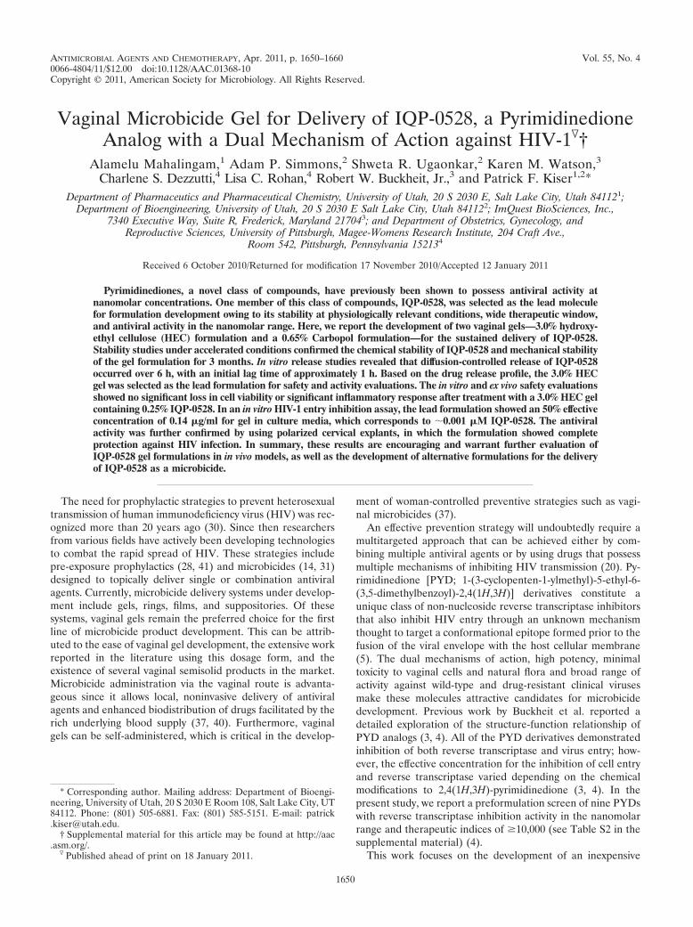
Selection of lead PYD analog. As shown in Fig. 1a and b,the majority of compounds stored at pH 4.2 and pH 7.0 at40°C and 75% RH in the dark showed no significant degra-dation after 4 weeks compared to their concentrations onday 0. However, the recovery of IQP-0558, -0410, and -1187was significantly reduced (Student t test, P � 0.05) (Fig. 1a)compared to day 0 concentrations. After 4 weeks at pH 4.2
1652 MAHALINGAM ET AL. ANTIMICROB. AGENTS CHEMOTHER.
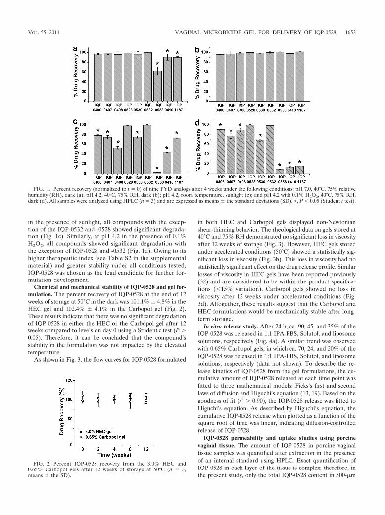
in the presence of sunlight, all compounds with the excep-tion of the IQP-0532 and -0528 showed significant degrada-tion (Fig. 1c). Similarly, at pH 4.2 in the presence of 0.1%H2O2, all compounds showed significant degradation withthe exception of IQP-0528 and -0532 (Fig. 1d). Owing to itshigher therapeutic index (see Table S2 in the supplementalmaterial) and greater stability under all conditions tested,IQP-0528 was chosen as the lead candidate for further for-mulation development.
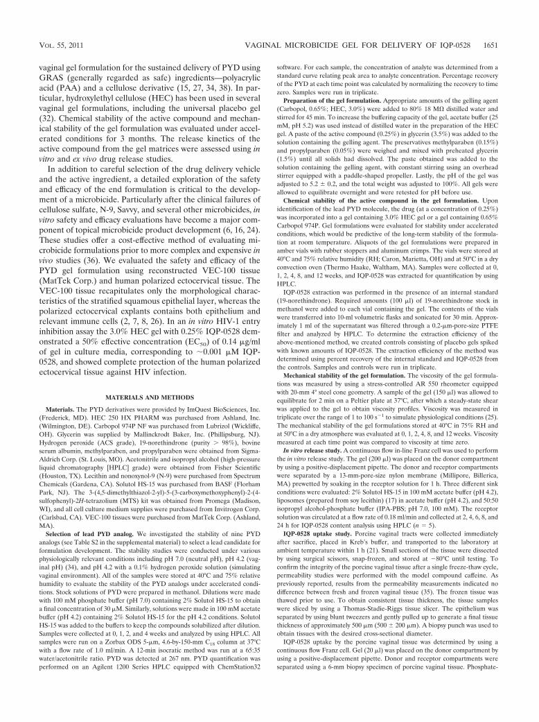
Chemical and mechanical stability of IQP-0528 and gel for-mulation. The percent recovery of IQP-0528 at the end of 12weeks of storage at 50°C in the dark was 101.1% � 4.8% in theHEC gel and 102.4% � 4.1% in the Carbopol gel (Fig. 2).These results indicate that there was no significant degradationof IQP-0528 in either the HEC or the Carbopol gel after 12weeks compared to levels on day 0 using a Student t test (P �0.05). Therefore, it can be concluded that the compound’sstability in the formulation was not impacted by the elevatedtemperature.
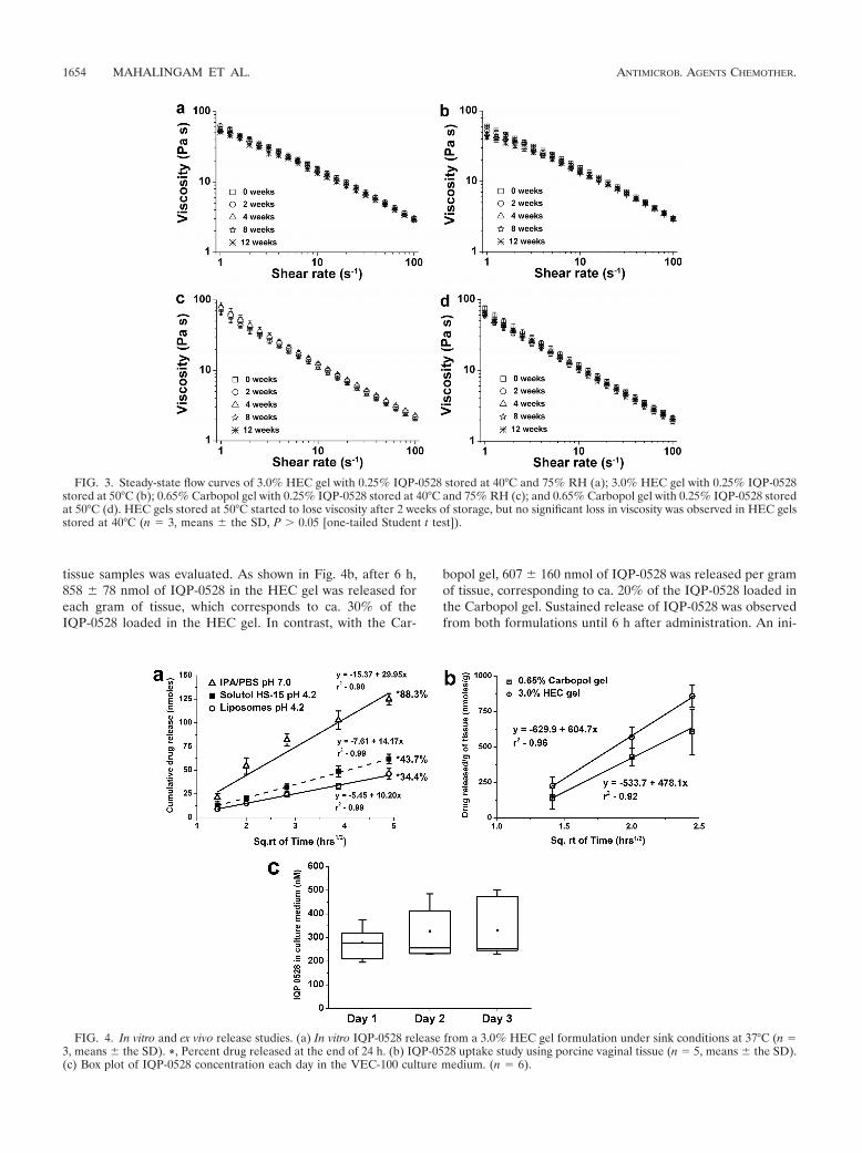
As shown in Fig. 3, the flow curves for IQP-0528 formulated
in both HEC and Carbopol gels displayed non-Newtonianshear-thinning behavior. The rheological data on gels stored at40°C and 75% RH demonstrated no significant loss in viscosityafter 12 weeks of storage (Fig. 3). However, HEC gels storedunder accelerated conditions (50°C) showed a statistically sig-nificant loss in viscosity (Fig. 3b). This loss in viscosity had nostatistically significant effect on the drug release profile. Similarlosses of viscosity in HEC gels have been reported previously(32) and are considered to be within the product specifica-tions (�15% variation). Carbopol gels showed no loss inviscosity after 12 weeks under accelerated conditions (Fig.3d). Altogether, these results suggest that the Carbopol andHEC formulations would be mechanically stable after long-term storage.
In vitro release study. After 24 h, ca. 90, 45, and 35% of theIQP-0528 was released in 1:1 IPA-PBS, Solutol, and liposomesolutions, respectively (Fig. 4a). A similar trend was observedwith 0.65% Carbopol gels, in which ca. 70, 24, and 20% of theIQP-0528 was released in 1:1 IPA-PBS, Solutol, and liposomesolutions, respectively (data not shown). To describe the re-lease kinetics of IQP-0528 from the gel formulations, the cu-mulative amount of IQP-0528 released at each time point wasfitted to three mathematical models: Ficks’s first and secondlaws of diffusion and Higuchi’s equation (13, 19). Based on thegoodness of fit (r2 � 0.90), the IQP-0528 release was fitted toHiguchi’s equation. As described by Higuchi’s equation, thecumulative IQP-0528 release when plotted as a function of thesquare root of time was linear, indicating diffusion-controlledrelease of IQP-0528.
IQP-0528 permeability and uptake studies using porcinevaginal tissue. The amount of IQP-0528 in porcine vaginaltissue samples was quantified after extraction in the presenceof an internal standard using HPLC. Exact quantification ofIQP-0528 in each layer of the tissue is complex; therefore, inthe present study, only the total IQP-0528 content in 500-�m
FIG. 1. Percent recovery (normalized to t � 0) of nine PYD analogs after 4 weeks under the following conditions: pH 7.0, 40°C, 75% relativehumidity (RH), dark (a); pH 4.2, 40°C, 75% RH, dark (b); pH 4.2, room temperature, sunlight (c); and pH 4.2 with 0.1% H2O2, 40°C, 75% RH,dark (d). All samples were analyzed using HPLC (n � 3) and are expressed as means � the standard deviations (SD). *, P � 0.05 (Student t test).
FIG. 2. Percent IQP-0528 recovery from the 3.0% HEC and0.65% Carbopol gels after 12 weeks of storage at 50°C (n � 3,means � the SD).
VOL. 55, 2011 VAGINAL MICROBICIDE GEL FOR DELIVERY OF IQP-0528 1653
tissue samples was evaluated. As shown in Fig. 4b, after 6 h,858 � 78 nmol of IQP-0528 in the HEC gel was released foreach gram of tissue, which corresponds to ca. 30% of theIQP-0528 loaded in the HEC gel. In contrast, with the Car-
bopol gel, 607 � 160 nmol of IQP-0528 was released per gramof tissue, corresponding to ca. 20% of the IQP-0528 loaded inthe Carbopol gel. Sustained release of IQP-0528 was observedfrom both formulations until 6 h after administration. An ini-
FIG. 3. Steady-state flow curves of 3.0% HEC gel with 0.25% IQP-0528 stored at 40°C and 75% RH (a); 3.0% HEC gel with 0.25% IQP-0528stored at 50°C (b); 0.65% Carbopol gel with 0.25% IQP-0528 stored at 40°C and 75% RH (c); and 0.65% Carbopol gel with 0.25% IQP-0528 storedat 50°C (d). HEC gels stored at 50°C started to lose viscosity after 2 weeks of storage, but no significant loss in viscosity was observed in HEC gelsstored at 40°C (n � 3, means � the SD, P � 0.05 [one-tailed Student t test]).
FIG. 4. In vitro and ex vivo release studies. (a) In vitro IQP-0528 release from a 3.0% HEC gel formulation under sink conditions at 37°C (n �3, means � the SD). *, Percent drug released at the end of 24 h. (b) IQP-0528 uptake study using porcine vaginal tissue (n � 5, means � the SD).(c) Box plot of IQP-0528 concentration each day in the VEC-100 culture medium. (n � 6).
1654 MAHALINGAM ET AL. ANTIMICROB. AGENTS CHEMOTHER.
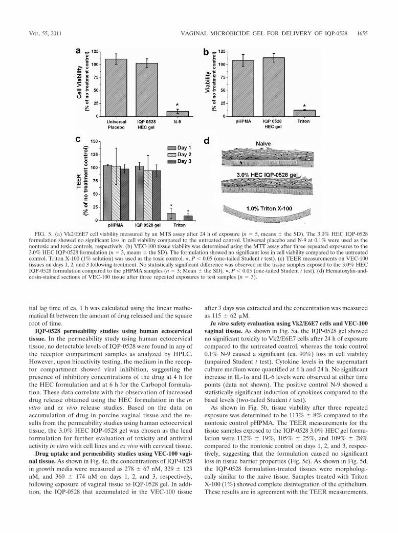
tial lag time of ca. 1 h was calculated using the linear mathe-matical fit between the amount of drug released and the squareroot of time.
IQP-0528 permeability studies using human ectocervicaltissue. In the permeability study using human ectocervicaltissue, no detectable levels of IQP-0528 were found in any ofthe receptor compartment samples as analyzed by HPLC.However, upon bioactivity testing, the medium in the recep-tor compartment showed viral inhibition, suggesting thepresence of inhibitory concentrations of the drug at 4 h forthe HEC formulation and at 6 h for the Carbopol formula-tion. These data correlate with the observation of increaseddrug release obtained using the HEC formulation in the invitro and ex vivo release studies. Based on the data onaccumulation of drug in porcine vaginal tissue and the re-sults from the permeability studies using human ectocervicaltissue, the 3.0% HEC IQP-0528 gel was chosen as the leadformulation for further evaluation of toxicity and antiviralactivity in vitro with cell lines and ex vivo with cervical tissue.
Drug uptake and permeability studies using VEC-100 vagi-nal tissue. As shown in Fig. 4c, the concentrations of IQP-0528in growth media were measured as 278 � 67 nM, 329 � 123nM, and 360 � 174 nM on days 1, 2, and 3, respectively,following exposure of vaginal tissue to IQP-0528 gel. In addi-tion, the IQP-0528 that accumulated in the VEC-100 tissue
after 3 days was extracted and the concentration was measuredas 115 � 62 �M.
In vitro safety evaluation using Vk2/E6E7 cells and VEC-100vaginal tissue. As shown in Fig. 5a, the IQP-0528 gel showedno significant toxicity to Vk2/E6E7 cells after 24 h of exposurecompared to the untreated control, whereas the toxic control0.1% N-9 caused a significant (ca. 90%) loss in cell viability(unpaired Student t test). Cytokine levels in the supernatantculture medium were quantified at 6 h and 24 h. No significantincrease in IL-1 and IL-6 levels were observed at either timepoints (data not shown). The positive control N-9 showed astatistically significant induction of cytokines compared to thebasal levels (two-tailed Student t test).
As shown in Fig. 5b, tissue viability after three repeatedexposure was determined to be 113% � 8% compared to thenontoxic control pHPMA. The TEER measurements for thetissue samples exposed to the IQP-0528 3.0% HEC gel formu-lation were 112% � 19%, 105% � 25%, and 109% � 28%compared to the nontoxic control on days 1, 2, and 3, respec-tively, suggesting that the formulation caused no significantloss in tissue barrier properties (Fig. 5c). As shown in Fig. 5d,the IQP-0528 formulation-treated tissues were morphologi-cally similar to the naive tissue. Samples treated with TritonX-100 (1%) showed complete disintegration of the epithelium.These results are in agreement with the TEER measurements,
FIG. 5. (a) Vk2/E6E7 cell viability measured by an MTS assay after 24 h of exposure (n � 5, means � the SD). The 3.0% HEC IQP-0528formulation showed no significant loss in cell viability compared to the untreated control. Universal placebo and N-9 at 0.1% were used as thenontoxic and toxic controls, respectively. (b) VEC-100 tissue viability was determined using the MTT assay after three repeated exposures to the3.0% HEC IQP-0528 formulation (n � 3, means � the SD). The formulation showed no significant loss in cell viability compared to the untreatedcontrol. Triton X-100 (1% solution) was used as the toxic control. *, P � 0.05 (one-tailed Student t test). (c) TEER measurements on VEC-100tissues on days 1, 2, and 3 following treatment. No statistically significant difference was observed in the tissue samples exposed to the 3.0% HECIQP-0528 formulation compared to the pHPMA samples (n � 3; Mean � the SD). *, P � 0.05 (one-tailed Student t test). (d) Hematoxylin-and-eosin-stained sections of VEC-100 tissue after three repeated exposures to test samples (n � 3).
VOL. 55, 2011 VAGINAL MICROBICIDE GEL FOR DELIVERY OF IQP-0528 1655
which showed no evidence of structural damage from exposureto the IQP-0528 gel.
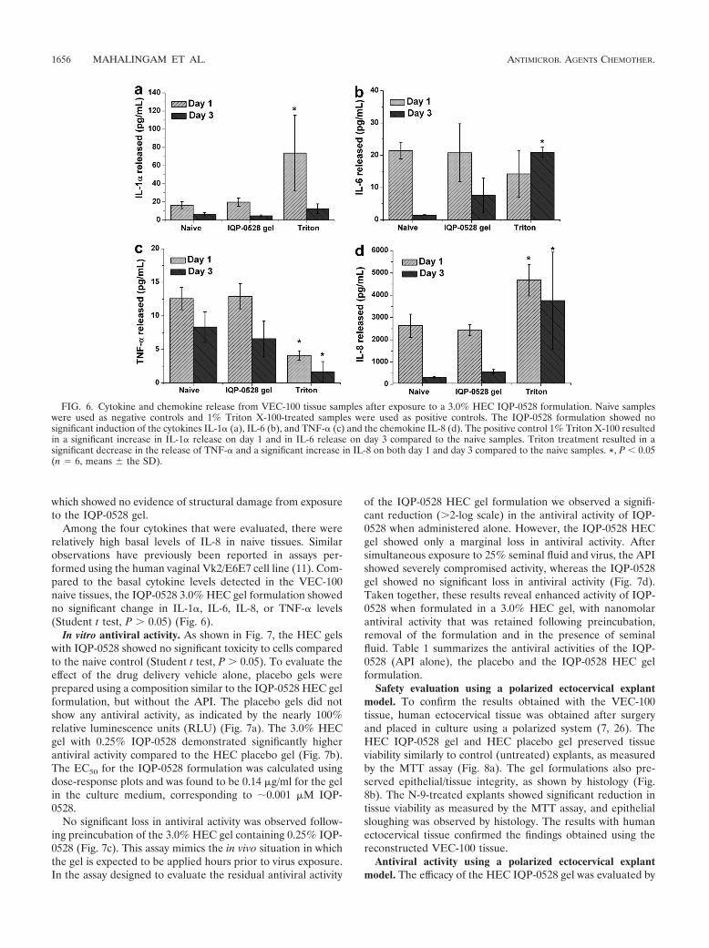
Among the four cytokines that were evaluated, there wererelatively high basal levels of IL-8 in naive tissues. Similarobservations have previously been reported in assays per-formed using the human vaginal Vk2/E6E7 cell line (11). Com-pared to the basal cytokine levels detected in the VEC-100naive tissues, the IQP-0528 3.0% HEC gel formulation showedno significant change in IL-1, IL-6, IL-8, or TNF- levels(Student t test, P � 0.05) (Fig. 6).
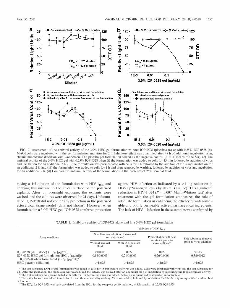
In vitro antiviral activity. As shown in Fig. 7, the HEC gelswith IQP-0528 showed no significant toxicity to cells comparedto the naive control (Student t test, P � 0.05). To evaluate theeffect of the drug delivery vehicle alone, placebo gels wereprepared using a composition similar to the IQP-0528 HEC gelformulation, but without the API. The placebo gels did notshow any antiviral activity, as indicated by the nearly 100%relative luminescence units (RLU) (Fig. 7a). The 3.0% HECgel with 0.25% IQP-0528 demonstrated significantly higherantiviral activity compared to the HEC placebo gel (Fig. 7b).The EC50 for the IQP-0528 formulation was calculated usingdose-response plots and was found to be 0.14 �g/ml for the gelin the culture medium, corresponding to �0.001 �M IQP-0528.
No significant loss in antiviral activity was observed follow-ing preincubation of the 3.0% HEC gel containing 0.25% IQP-0528 (Fig. 7c). This assay mimics the in vivo situation in whichthe gel is expected to be applied hours prior to virus exposure.In the assay designed to evaluate the residual antiviral activity
of the IQP-0528 HEC gel formulation we observed a signifi-cant reduction (�2-log scale) in the antiviral activity of IQP-0528 when administered alone. However, the IQP-0528 HECgel showed only a marginal loss in antiviral activity. Aftersimultaneous exposure to 25% seminal fluid and virus, the APIshowed severely compromised activity, whereas the IQP-0528gel showed no significant loss in antiviral activity (Fig. 7d).Taken together, these results reveal enhanced activity of IQP-0528 when formulated in a 3.0% HEC gel, with nanomolarantiviral activity that was retained following preincubation,removal of the formulation and in the presence of seminalfluid. Table 1 summarizes the antiviral activities of the IQP-0528 (API alone), the placebo and the IQP-0528 HEC gelformulation.
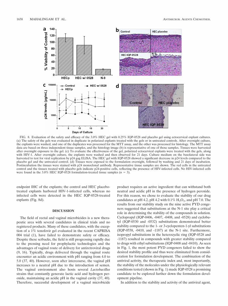
Safety evaluation using a polarized ectocervical explantmodel. To confirm the results obtained with the VEC-100tissue, human ectocervical tissue was obtained after surgeryand placed in culture using a polarized system (7, 26). TheHEC IQP-0528 gel and HEC placebo gel preserved tissueviability similarly to control (untreated) explants, as measuredby the MTT assay (Fig. 8a). The gel formulations also pre-served epithelial/tissue integrity, as shown by histology (Fig.8b). The N-9-treated explants showed significant reduction intissue viability as measured by the MTT assay, and epithelialsloughing was observed by histology. The results with humanectocervical tissue confirmed the findings obtained using thereconstructed VEC-100 tissue.
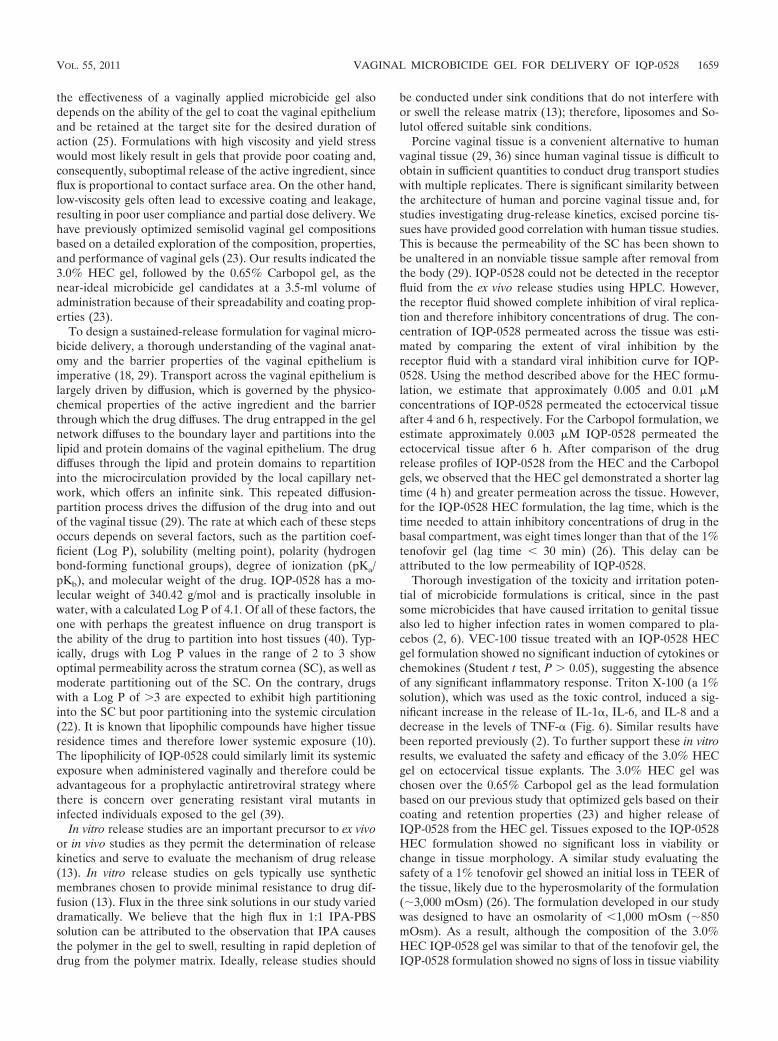
Antiviral activity using a polarized ectocervical explantmodel. The efficacy of the HEC IQP-0528 gel was evaluated by
FIG. 6. Cytokine and chemokine release from VEC-100 tissue samples after exposure to a 3.0% HEC IQP-0528 formulation. Naive sampleswere used as negative controls and 1% Triton X-100-treated samples were used as positive controls. The IQP-0528 formulation showed nosignificant induction of the cytokines IL-1 (a), IL-6 (b), and TNF- (c) and the chemokine IL-8 (d). The positive control 1% Triton X-100 resultedin a significant increase in IL-1 release on day 1 and in IL-6 release on day 3 compared to the naive samples. Triton treatment resulted in asignificant decrease in the release of TNF- and a significant increase in IL-8 on both day 1 and day 3 compared to the naive samples. *, P � 0.05(n � 6, means � the SD).
1656 MAHALINGAM ET AL. ANTIMICROB. AGENTS CHEMOTHER.
mixing a 1:5 dilution of the formulation with HIV-1BaL andapplying this mixture to the apical surface of the polarizedexplants. After an overnight exposure, the explants werewashed, and the cultures were observed for 21 days. Unformu-lated IQP-0528 did not confer any protection in the polarizedectocervical tissue model (data not shown). However, whenformulated in a 3.0% HEC gel, IQP-0528 conferred protection
against HIV infection as indicated by a �1 log reduction inHIV-1 p24 antigen levels by day 21 (Fig. 8c). This significantreduction in HIV-1 p24 (P � 0.007, Mann-Whitney test) aftertreatment with the gel formulation emphasizes the role ofadequate formulation in enhancing the efficacy of water-insol-uble and poorly permeable active pharmaceutical ingredients.The lack of HIV-1 infection in these samples was confirmed by
FIG. 7. Assessment of the antiviral activity of the 3.0% HEC gel formulation without IQP-0528 (placebo) (a) or with 0.25% IQP-0528 (b).MAGI cells were incubated with the gel formulation and virus for 2 h. Inhibitory effect was quantified after 48 h of additional incubation usingchemiluminescence detection with Gal-Screen. The placebo gel formulation served as the negative control (n � 3, means � the SD). (c) Theantiviral activity of the 3.0% HEC gel with 0.25% IQP-0528 when (i) the formulation was added to cells for 15 min followed by addition of virusand incubation for an additional 2 h, (ii) the formulation was preincubated with cells for 1 h followed by the addition of virus and incubation foran additional 2 h, and (iii) the formulation was added to cells for 1 h and then removed by washing, followed by addition of virus and incubationfor an additional 2 h. (d) Comparative antiviral activity of the formulations in the presence of 25% seminal fluid.
TABLE 1. Inhibitory activity of IQP-0528 alone and in a 3.0% HEC gel formulation
Assay conditions
Inhibition of HIV-1IIIB
Simultaneous addition of virus andtest substancea Preincubation with test
substance prior tovirus additionb
Test substance removedprior to virus additionc
Without seminalfluid
With 25% seminalfluid
IQP-0528 (API alone) (EC50 ��g/ml ) �0.0001 0.05 0.05 �0.17IQP-0528 HEC gel formulation (EC50 ��g/ml )/
IQP-0528 when formulated (EC50 ��g/ml )d0.14/0.0003 0.21/0.0005 0.26/0.0006 0.5/0.0012
HEC placebo (dilution) �1:625 �1:625 �1:625 �1:625
a The test substance (API or gel formulation) was added to cells for 15 min before the virus was added. Cells were incubated with virus and the test substance for2 h. After the incubation, the monolayer was washed, and the activity was assayed after an additional 48 h of incubation by measuring the -galactosidase activity.
b The test substance was preincubated with cells for 1 h before the virus was added. Activity was quantified as described in footnote a.c The test substance was added to cells for 1 h and then removed by washing. Virus was added, followed by incubation for 2 h. Activity was quantified as described
in footnote a.d The EC50 for IQP-0528 was back-calculated from the EC50 for the complete gel formulation, which consists of 0.25% IQP-0528.
VOL. 55, 2011 VAGINAL MICROBICIDE GEL FOR DELIVERY OF IQP-0528 1657
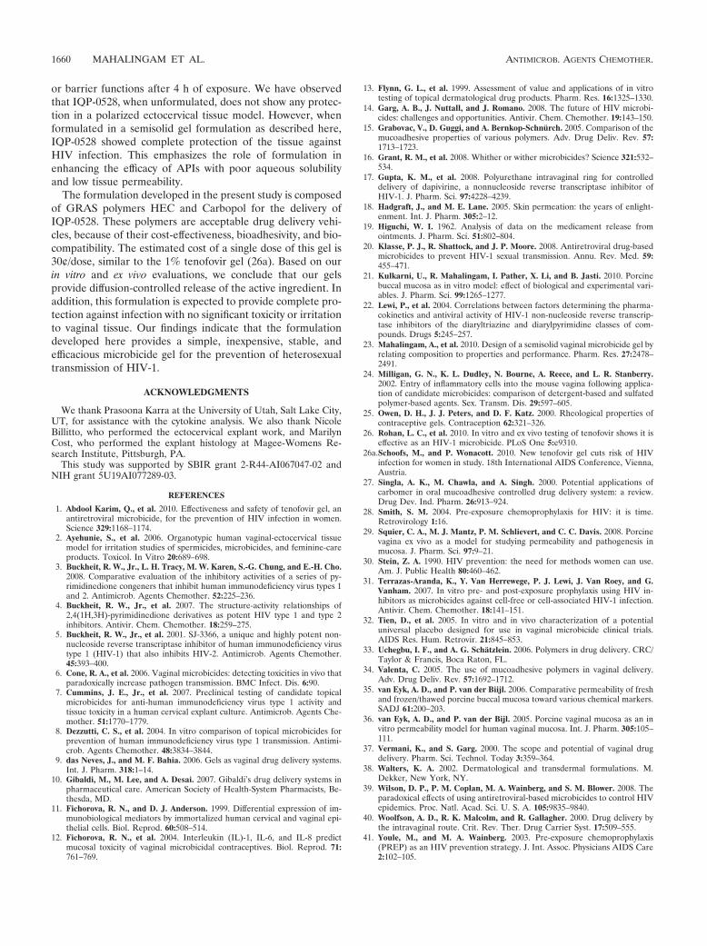
endpoint IHC of the explants; the control and HEC placebo-treated explants harbored HIV-1-infected cells, whereas noinfected cells were detected in the HEC IQP-0528-treatedexplants (Fig. 8d).
DISCUSSION
The field of rectal and vaginal microbicides is a new thera-peutic area with several candidates in clinical trials and noregistered products. Many of these candidates, with the excep-tion of a 1% tenofovir gel evaluated in the recent CAPRISA004 trial (1), have failed to demonstrate safety or efficacy.Despite these setbacks, the field is still progressing rapidly dueto the pressing need for prophylactic technologies and theadvantages of vaginal route of delivery for antiretroviral drugs(9, 16). Typically, drugs delivered through the vaginal routeencounter an acidic environment with pH ranging from 4.0 to5.0 (37, 40). However, soon after intercourse, the vaginal pHincreases to a neutral pH due to the introduction of semen.The vaginal environment also hosts several Lactobacillusstrains that constantly generate lactic acid and hydrogen per-oxide, maintaining an acidic pH in the vaginal cavity (37, 40).Therefore, successful development of a vaginal microbicide
product requires an active ingredient that can withstand bothneutral and acidic pH in the presence of hydrogen peroxide.For this reason, we chose to evaluate the stability of our drugcandidates at pH 4.2, pH 4.2 with 0.1% H2O2, and pH 7.0. Theresults from our stability study on the nine active PYD conge-ners suggested that substitutions at the N-1 site play a criticalrole in determining the stability of the compounds in solution.Cyclopropyl (IQP-0406, -0407, -0408, and -0528) and cyclobu-tyl (IQP-0530 and -0532) substitutions demonstrated betterstability compared to the 1- or 3-cyclopenten-1-yl substitutions(IQP-0558, -0410, and -1187) at the N-1 site. Furthermore,isopropyl substitutions in the heterocyclic ring (IQP-0528 and-1187) resulted in compounds with greater stability comparedto drugs with ethyl substitutions (IQP-0408 and -0410). As seenin Fig. 1, the most potent PYD congeners failed to show thedesired stability profile and thus were eliminated from consid-eration for formulation development. The combination of theantiviral activity, the therapeutic index and, most importantly,the stability of the molecules under the physiologically relevantconditions tested (shown in Fig. 1) made IQP-0528 a promisingcandidate to be explored further down the formulation devel-opment pipeline.
In addition to the stability and activity of the antiviral agent,
FIG. 8. Evaluation of the safety and efficacy of the 3.0% HEC gel with 0.25% IQP-0528 and placebo gel using ectocervical explant cultures.(a) The safety of the gels was evaluated in duplicate in polarized explants treated with the gels or in untreated controls. After overnight culture,the explants were washed, and one of the duplicates was processed for the MTT assay, and the other was processed for histology. The MTT assaydata are based on three independent tissue samples, and the histology image (b) is representative of one of those samples. Tissues were harvestedafter overnight exposure to the gel. (c) To evaluate the effectiveness of the gel, polarized ectocervical explants were treated with the gels, alongwith HIV-1. After overnight culture, the explants were washed and then observed for 21 days. Culture medium on the basolateral side washarvested to test for viral replication by p24 gag ELISA. The HEC gel with IQP-0528 showed a significant decrease in p24 levels compared to theplacebo gel and the untreated control. (d) Tissues were exposed to the formulation overnight, followed by washing and 21 days of incubation.Postincubation the tissues were stained with p24 monoclonal antibody. Representative tissue samples are shown. The red cells in the untreatedcontrol and the tissues treated with placebo gels indicate p24-positive cells, reflecting the presence of HIV-infected cells. No HIV-infected cellswere found in the 3.0% HEC IQP-0528 formulation-treated tissue samples (n � 3).
1658 MAHALINGAM ET AL. ANTIMICROB. AGENTS CHEMOTHER.
the effectiveness of a vaginally applied microbicide gel alsodepends on the ability of the gel to coat the vaginal epitheliumand be retained at the target site for the desired duration ofaction (25). Formulations with high viscosity and yield stresswould most likely result in gels that provide poor coating and,consequently, suboptimal release of the active ingredient, sinceflux is proportional to contact surface area. On the other hand,low-viscosity gels often lead to excessive coating and leakage,resulting in poor user compliance and partial dose delivery. Wehave previously optimized semisolid vaginal gel compositionsbased on a detailed exploration of the composition, properties,and performance of vaginal gels (23). Our results indicated the3.0% HEC gel, followed by the 0.65% Carbopol gel, as thenear-ideal microbicide gel candidates at a 3.5-ml volume ofadministration because of their spreadability and coating prop-erties (23).
To design a sustained-release formulation for vaginal micro-bicide delivery, a thorough understanding of the vaginal anat-omy and the barrier properties of the vaginal epithelium isimperative (18, 29). Transport across the vaginal epithelium islargely driven by diffusion, which is governed by the physico-chemical properties of the active ingredient and the barrierthrough which the drug diffuses. The drug entrapped in the gelnetwork diffuses to the boundary layer and partitions into thelipid and protein domains of the vaginal epithelium. The drugdiffuses through the lipid and protein domains to repartitioninto the microcirculation provided by the local capillary net-work, which offers an infinite sink. This repeated diffusion-partition process drives the diffusion of the drug into and outof the vaginal tissue (29). The rate at which each of these stepsoccurs depends on several factors, such as the partition coef-ficient (Log P), solubility (melting point), polarity (hydrogenbond-forming functional groups), degree of ionization (pKa/pKb), and molecular weight of the drug. IQP-0528 has a mo-lecular weight of 340.42 g/mol and is practically insoluble inwater, with a calculated Log P of 4.1. Of all of these factors, theone with perhaps the greatest influence on drug transport isthe ability of the drug to partition into host tissues (40). Typ-ically, drugs with Log P values in the range of 2 to 3 showoptimal permeability across the stratum cornea (SC), as well asmoderate partitioning out of the SC. On the contrary, drugswith a Log P of �3 are expected to exhibit high partitioninginto the SC but poor partitioning into the systemic circulation(22). It is known that lipophilic compounds have higher tissueresidence times and therefore lower systemic exposure (10).The lipophilicity of IQP-0528 could similarly limit its systemicexposure when administered vaginally and therefore could beadvantageous for a prophylactic antiretroviral strategy wherethere is concern over generating resistant viral mutants ininfected individuals exposed to the gel (39).
In vitro release studies are an important precursor to ex vivoor in vivo studies as they permit the determination of releasekinetics and serve to evaluate the mechanism of drug release(13). In vitro release studies on gels typically use syntheticmembranes chosen to provide minimal resistance to drug dif-fusion (13). Flux in the three sink solutions in our study varieddramatically. We believe that the high flux in 1:1 IPA-PBSsolution can be attributed to the observation that IPA causesthe polymer in the gel to swell, resulting in rapid depletion ofdrug from the polymer matrix. Ideally, release studies should
be conducted under sink conditions that do not interfere withor swell the release matrix (13); therefore, liposomes and So-lutol offered suitable sink conditions.
Porcine vaginal tissue is a convenient alternative to humanvaginal tissue (29, 36) since human vaginal tissue is difficult toobtain in sufficient quantities to conduct drug transport studieswith multiple replicates. There is significant similarity betweenthe architecture of human and porcine vaginal tissue and, forstudies investigating drug-release kinetics, excised porcine tis-sues have provided good correlation with human tissue studies.This is because the permeability of the SC has been shown tobe unaltered in an nonviable tissue sample after removal fromthe body (29). IQP-0528 could not be detected in the receptorfluid from the ex vivo release studies using HPLC. However,the receptor fluid showed complete inhibition of viral replica-tion and therefore inhibitory concentrations of drug. The con-centration of IQP-0528 permeated across the tissue was esti-mated by comparing the extent of viral inhibition by thereceptor fluid with a standard viral inhibition curve for IQP-0528. Using the method described above for the HEC formu-lation, we estimate that approximately 0.005 and 0.01 �Mconcentrations of IQP-0528 permeated the ectocervical tissueafter 4 and 6 h, respectively. For the Carbopol formulation, weestimate approximately 0.003 �M IQP-0528 permeated theectocervical tissue after 6 h. After comparison of the drugrelease profiles of IQP-0528 from the HEC and the Carbopolgels, we observed that the HEC gel demonstrated a shorter lagtime (4 h) and greater permeation across the tissue. However,for the IQP-0528 HEC formulation, the lag time, which is thetime needed to attain inhibitory concentrations of drug in thebasal compartment, was eight times longer than that of the 1%tenofovir gel (lag time � 30 min) (26). This delay can beattributed to the low permeability of IQP-0528.
Thorough investigation of the toxicity and irritation poten-tial of microbicide formulations is critical, since in the pastsome microbicides that have caused irritation to genital tissuealso led to higher infection rates in women compared to pla-cebos (2, 6). VEC-100 tissue treated with an IQP-0528 HECgel formulation showed no significant induction of cytokines orchemokines (Student t test, P � 0.05), suggesting the absenceof any significant inflammatory response. Triton X-100 (a 1%solution), which was used as the toxic control, induced a sig-nificant increase in the release of IL-1, IL-6, and IL-8 and adecrease in the levels of TNF- (Fig. 6). Similar results havebeen reported previously (2). To further support these in vitroresults, we evaluated the safety and efficacy of the 3.0% HECgel on ectocervical tissue explants. The 3.0% HEC gel waschosen over the 0.65% Carbopol gel as the lead formulationbased on our previous study that optimized gels based on theircoating and retention properties (23) and higher release ofIQP-0528 from the HEC gel. Tissues exposed to the IQP-0528HEC formulation showed no significant loss in viability orchange in tissue morphology. A similar study evaluating thesafety of a 1% tenofovir gel showed an initial loss in TEER ofthe tissue, likely due to the hyperosmolarity of the formulation(�3,000 mOsm) (26). The formulation developed in our studywas designed to have an osmolarity of �1,000 mOsm (�850mOsm). As a result, although the composition of the 3.0%HEC IQP-0528 gel was similar to that of the tenofovir gel, theIQP-0528 formulation showed no signs of loss in tissue viability
VOL. 55, 2011 VAGINAL MICROBICIDE GEL FOR DELIVERY OF IQP-0528 1659
or barrier functions after 4 h of exposure. We have observedthat IQP-0528, when unformulated, does not show any protec-tion in a polarized ectocervical tissue model. However, whenformulated in a semisolid gel formulation as described here,IQP-0528 showed complete protection of the tissue againstHIV infection. This emphasizes the role of formulation inenhancing the efficacy of APIs with poor aqueous solubilityand low tissue permeability.
The formulation developed in the present study is composedof GRAS polymers HEC and Carbopol for the delivery ofIQP-0528. These polymers are acceptable drug delivery vehi-cles, because of their cost-effectiveness, bioadhesivity, and bio-compatibility. The estimated cost of a single dose of this gel is30¢/dose, similar to the 1% tenofovir gel (26a). Based on ourin vitro and ex vivo evaluations, we conclude that our gelsprovide diffusion-controlled release of the active ingredient. Inaddition, this formulation is expected to provide complete pro-tection against infection with no significant toxicity or irritationto vaginal tissue. Our findings indicate that the formulationdeveloped here provides a simple, inexpensive, stable, andefficacious microbicide gel for the prevention of heterosexualtransmission of HIV-1.
ACKNOWLEDGMENTS
We thank Prasoona Karra at the University of Utah, Salt Lake City,UT, for assistance with the cytokine analysis. We also thank NicoleBillitto, who performed the ectocervical explant work, and MarilynCost, who performed the explant histology at Magee-Womens Re-search Institute, Pittsburgh, PA.
This study was supported by SBIR grant 2-R44-AI067047-02 andNIH grant 5U19AI077289-03.
REFERENCES
1. Abdool Karim, Q., et al. 2010. Effectiveness and safety of tenofovir gel, anantiretroviral microbicide, for the prevention of HIV infection in women.Science 329:1168–1174.
2. Ayehunie, S., et al. 2006. Organotypic human vaginal-ectocervical tissuemodel for irritation studies of spermicides, microbicides, and feminine-careproducts. Toxicol. In Vitro 20:689–698.
3. Buckheit, R. W., Jr., L. H. Tracy, M. W. Karen, S.-G. Chung, and E.-H. Cho.2008. Comparative evaluation of the inhibitory activities of a series of py-rimidinedione congeners that inhibit human immunodeficiency virus types 1and 2. Antimicrob. Agents Chemother. 52:225–236.
4. Buckheit, R. W., Jr., et al. 2007. The structure-activity relationships of2,4(1H,3H)-pyrimidinedione derivatives as potent HIV type 1 and type 2inhibitors. Antivir. Chem. Chemother. 18:259–275.
5. Buckheit, R. W., Jr., et al. 2001. SJ-3366, a unique and highly potent non-nucleoside reverse transcriptase inhibitor of human immunodeficiency virustype 1 (HIV-1) that also inhibits HIV-2. Antimicrob. Agents Chemother.45:393–400.
6. Cone, R. A., et al. 2006. Vaginal microbicides: detecting toxicities in vivo thatparadoxically increase pathogen transmission. BMC Infect. Dis. 6:90.
7. Cummins, J. E., Jr., et al. 2007. Preclinical testing of candidate topicalmicrobicides for anti-human immunodeficiency virus type 1 activity andtissue toxicity in a human cervical explant culture. Antimicrob. Agents Che-mother. 51:1770–1779.
8. Dezzutti, C. S., et al. 2004. In vitro comparison of topical microbicides forprevention of human immunodeficiency virus type 1 transmission. Antimi-crob. Agents Chemother. 48:3834–3844.
9. das Neves, J., and M. F. Bahia. 2006. Gels as vaginal drug delivery systems.Int. J. Pharm. 318:1–14.
10. Gibaldi, M., M. Lee, and A. Desai. 2007. Gibaldi’s drug delivery systems inpharmaceutical care. American Society of Health-System Pharmacists, Be-thesda, MD.
11. Fichorova, R. N., and D. J. Anderson. 1999. Differential expression of im-munobiological mediators by immortalized human cervical and vaginal epi-thelial cells. Biol. Reprod. 60:508–514.
12. Fichorova, R. N., et al. 2004. Interleukin (IL)-1, IL-6, and IL-8 predictmucosal toxicity of vaginal microbicidal contraceptives. Biol. Reprod. 71:761–769.
13. Flynn, G. L., et al. 1999. Assessment of value and applications of in vitrotesting of topical dermatological drug products. Pharm. Res. 16:1325–1330.
14. Garg, A. B., J. Nuttall, and J. Romano. 2008. The future of HIV microbi-cides: challenges and opportunities. Antivir. Chem. Chemother. 19:143–150.
15. Grabovac, V., D. Guggi, and A. Bernkop-Schnurch. 2005. Comparison of themucoadhesive properties of various polymers. Adv. Drug Deliv. Rev. 57:1713–1723.
16. Grant, R. M., et al. 2008. Whither or wither microbicides? Science 321:532–534.
17. Gupta, K. M., et al. 2008. Polyurethane intravaginal ring for controlleddelivery of dapivirine, a nonnucleoside reverse transcriptase inhibitor ofHIV-1. J. Pharm. Sci. 97:4228–4239.
18. Hadgraft, J., and M. E. Lane. 2005. Skin permeation: the years of enlight-enment. Int. J. Pharm. 305:2–12.
19. Higuchi, W. I. 1962. Analysis of data on the medicament release fromointments. J. Pharm. Sci. 51:802–804.
20. Klasse, P. J., R. Shattock, and J. P. Moore. 2008. Antiretroviral drug-basedmicrobicides to prevent HIV-1 sexual transmission. Annu. Rev. Med. 59:455–471.
21. Kulkarni, U., R. Mahalingam, I. Pather, X. Li, and B. Jasti. 2010. Porcinebuccal mucosa as in vitro model: effect of biological and experimental vari-ables. J. Pharm. Sci. 99:1265–1277.
22. Lewi, P., et al. 2004. Correlations between factors determining the pharma-cokinetics and antiviral activity of HIV-1 non-nucleoside reverse transcrip-tase inhibitors of the diaryltriazine and diarylpyrimidine classes of com-pounds. Drugs 5:245–257.
23. Mahalingam, A., et al. 2010. Design of a semisolid vaginal microbicide gel byrelating composition to properties and performance. Pharm. Res. 27:2478–2491.
24. Milligan, G. N., K. L. Dudley, N. Bourne, A. Reece, and L. R. Stanberry.2002. Entry of inflammatory cells into the mouse vagina following applica-tion of candidate microbicides: comparison of detergent-based and sulfatedpolymer-based agents. Sex. Transm. Dis. 29:597–605.
25. Owen, D. H., J. J. Peters, and D. F. Katz. 2000. Rheological properties ofcontraceptive gels. Contraception 62:321–326.
26. Rohan, L. C., et al. 2010. In vitro and ex vivo testing of tenofovir shows it iseffective as an HIV-1 microbicide. PLoS One 5:e9310.
26a.Schoofs, M., and P. Wonacott. 2010. New tenofovir gel cuts risk of HIVinfection for women in study. 18th International AIDS Conference, Vienna,Austria.
27. Singla, A. K., M. Chawla, and A. Singh. 2000. Potential applications ofcarbomer in oral mucoadhesive controlled drug delivery system: a review.Drug Dev. Ind. Pharm. 26:913–924.
28. Smith, S. M. 2004. Pre-exposure chemoprophylaxis for HIV: it is time.Retrovirology 1:16.
29. Squier, C. A., M. J. Mantz, P. M. Schlievert, and C. C. Davis. 2008. Porcinevagina ex vivo as a model for studying permeability and pathogenesis inmucosa. J. Pharm. Sci. 97:9–21.
30. Stein, Z. A. 1990. HIV prevention: the need for methods women can use.Am. J. Public Health 80:460–462.
31. Terrazas-Aranda, K., Y. Van Herrewege, P. J. Lewi, J. Van Roey, and G.Vanham. 2007. In vitro pre- and post-exposure prophylaxis using HIV in-hibitors as microbicides against cell-free or cell-associated HIV-1 infection.Antivir. Chem. Chemother. 18:141–151.
32. Tien, D., et al. 2005. In vitro and in vivo characterization of a potentialuniversal placebo designed for use in vaginal microbicide clinical trials.AIDS Res. Hum. Retrovir. 21:845–853.
33. Uchegbu, I. F., and A. G. Schatzlein. 2006. Polymers in drug delivery. CRC/Taylor & Francis, Boca Raton, FL.
34. Valenta, C. 2005. The use of mucoadhesive polymers in vaginal delivery.Adv. Drug Deliv. Rev. 57:1692–1712.
35. van Eyk, A. D., and P. van der Biijl. 2006. Comparative permeability of freshand frozen/thawed porcine buccal mucosa toward various chemical markers.SADJ 61:200–203.
36. van Eyk, A. D., and P. van der Bijl. 2005. Porcine vaginal mucosa as an invitro permeability model for human vaginal mucosa. Int. J. Pharm. 305:105–111.
37. Vermani, K., and S. Garg. 2000. The scope and potential of vaginal drugdelivery. Pharm. Sci. Technol. Today 3:359–364.
38. Walters, K. A. 2002. Dermatological and transdermal formulations. M.Dekker, New York, NY.
39. Wilson, D. P., P. M. Coplan, M. A. Wainberg, and S. M. Blower. 2008. Theparadoxical effects of using antiretroviral-based microbicides to control HIVepidemics. Proc. Natl. Acad. Sci. U. S. A. 105:9835–9840.
40. Woolfson, A. D., R. K. Malcolm, and R. Gallagher. 2000. Drug delivery bythe intravaginal route. Crit. Rev. Ther. Drug Carrier Syst. 17:509–555.
41. Youle, M., and M. A. Wainberg. 2003. Pre-exposure chemoprophylaxis(PREP) as an HIV prevention strategy. J. Int. Assoc. Physicians AIDS Care2:102–105.
1660 MAHALINGAM ET AL. ANTIMICROB. AGENTS CHEMOTHER.