Embed Size (px)
Citation preview

Vaccination with a Porcine Reproductive and Respiratory Syndrome(PRRS) Modified Live Virus Vaccine Followed by Challenge withPRRS Virus and Porcine Circovirus Type 2 (PCV2) Protects againstPRRS but Enhances PCV2 Replication and Pathogenesis Compared toResults for Nonvaccinated Cochallenged Controls
Megan C. Niederwerder,a Bhupinder Bawa,a Nick V. L. Serão,b Benjamin R. Trible,a Maureen A. Kerrigan,a Joan K. Lunney,c
Jack C. M. Dekkers,b Raymond R. R. Rowlanda
Department of Diagnostic Medicine/Pathobiology, College of Veterinary Medicine, Kansas State University, Manhattan, Kansas, USAa; Department of Animal Science, IowaState University, Ames, Iowa, USAb; U.S. Department of Agriculture, Agricultural Research Service, Beltsville Agricultural Research Center, Beltsville, Maryland, USAc
Coinfections involving porcine reproductive and respiratory syndrome virus (PRRSV) and porcine circovirus type 2 (PCV2)contribute to a group of disease syndromes known as porcine circovirus-associated disease (PCVAD). Presumably, PRRSV infec-tion enhances PCV2 replication as a result of modulation of host immunity. The purpose of this study was to evaluate PCV2 rep-lication and pathogenesis in pigs vaccinated with a PRRS modified live virus (MLV) vaccine and subsequently challenged with acombination of PRRSV and PCV2. During the early postchallenge period, the number of pigs with PRRSV-associated clinicalsigns was decreased, and average daily gain (ADG) was increased, in the vaccinated group, demonstrating the protective effect ofPRRS vaccination. However, during the later postchallenge period, more pigs in the vaccinated group showed increased PCV2viremia, decreased ADG, increased PCVAD clinical signs, and increased mortality. In this disease model, the early benefits ofPRRSV vaccination were outweighed by the later amplification of PCVAD.
Porcine circovirus type 2 (PCV2), a single-stranded DNA virusin the family Circoviridae, contributes to a group of syn-
dromes collectively termed porcine circovirus-associated disease(PCVAD) (1). Two important clinical syndromes associated withPCVAD are PCV2-associated pneumonia and postweaning mul-tisystemic wasting syndrome (PMWS) (1, 2). Management ofPCV2 through the use of inactivated and subunit vaccines has ledto the effective control of PCVAD in North America and Europe.However, the emergence of new PCV2 strains and the lack ofPCV2 vaccination programs in other countries create an uncer-tain future for continued disease control.
Porcine reproductive and respiratory syndrome virus (PRRSV) isa single-stranded RNA virus in the family Arteriviridae (3, 4). Forthe past 20 years, PRRSV has remained the most costly diseaseaffecting swine production worldwide (5). PRRSV infection con-tributes to a number of immunological outcomes that increase thesusceptibility of the host to secondary infections by primary andsecondary pathogens (6–8). PRRSV is frequently isolated alongwith PCV2 (9) and is one of the major cofactors linked with in-creasing PCV2 replication and pathogenesis (10–12). Previouswork by us and others has shown that a principal contribution ofPRRSV is to increase PCV2 viremia (13). Increased PCV2 replica-tion is likely the result of immune stimulation that results in morePCV2-permissive cells combined with PRRSV-induced immuno-modulation. The complex etiology of PCVAD, including the roleof PRRSV infection, has yet to be fully understood. In an extensivebody of work, we identified the aberrant recognition of a nonneu-tralizing decoy epitope on the PCV2 capsid protein (CP) as a con-tributing factor in PCVAD immunopathogenesis. Natural PCV2infection of a population produces a mixture of pigs that recog-nize the decoy and neutralizing epitopes, which may explain why
only a subpopulation of infected pigs goes on to develop PCVAD(13–15).
In this study, we took advantage of a host genetics study toevaluate clinical and virological outcomes after experimental chal-lenge with PCV2 and PRRSV in pigs with or without prior vacci-nation with a commercial PRRS modified live virus (MLV). Theresults demonstrate the protective properties of vaccination; how-ever, the short-term benefit is outweighed by the longer-term im-pact of MLV on PCVAD.
MATERIALS AND METHODSAnimals and housing. Experiments involving animals and viruses wereperformed in accordance with the Guide for the Care and Use of Agricul-tural Animals in Research and Teaching by the Federation of Animal Sci-ence Societies (FASS) (16) and with the USDA Animal Welfare Act andAnimal Welfare Regulations and were approved by the Kansas State Uni-versity Institutional Animal Care and Use Committees and Institutional
Received 30 July 2015 Returned for modification 24 August 2015Accepted 1 October 2015
Accepted manuscript posted online 7 October 2015
Citation Niederwerder MC, Bawa B, Serão NVL, Trible BR, Kerrigan MA, Lunney JK,Dekkers JCM, Rowland RRR. 2015. Vaccination with a porcine reproductive andrespiratory syndrome (PRRS) modified live virus vaccine followed by challengewith PRRS virus and porcine circovirus type 2 (PCV2) protects against PRRS butenhances PCV2 replication and pathogenesis compared to results fornonvaccinated cochallenged controls. Clin Vaccine Immunol 22:1244 –1254.doi:10.1128/CVI.00434-15.
Editor: D. W. Pascual
Address correspondence to Megan C. Niederwerder,[email protected].
Copyright © 2015, American Society for Microbiology. All Rights Reserved.
1244 cvi.asm.org December 2015 Volume 22 Number 12Clinical and Vaccine Immunology
on March 17, 2016 by D
igiTop -U
SD
A's D
igital Desktop Library
http://cvi.asm.org/
Dow
nloaded from
Biosafety Committees. The study was conducted as part of the evaluationof a previously described genomic marker, WUR (17). The populationused in this study was composed of pigs with two genotypes: 50% hadWUR genotype AA, and 50% had WUR genotype AB or BB. The AB andBB genotypes were predicted to have beneficial effects on the response toPRRSV infection. Both the vaccine group and the nonvaccine group werebalanced according to WUR genotype; therefore, WUR was not a factor inthe comparison of the outcomes of the vaccine and nonvaccine groups.Three-week-old barrows (n � 226; average age, 19.4 � 1.8 days) wereobtained from a high-health commercial source negative for PRRSV.While the pigs were derived from a sow herd previously vaccinated with aPCV2 capsid subunit vaccine, the piglets were not vaccinated for PCV2and were obtained after weaning without regard to maternal antibodylevels. All pigs were housed in two environmentally controlled rooms atthe Kansas State University Large Animal Research Center and weremaintained under biosafety level 2 (BSL-2) conditions. Rooms werechemically disinfected, cleaned with a high-heat pressure washer, and gasdecontaminated with vaporized hydrogen peroxide prior to use. Bothrooms were empty for at least 19 days prior to the start of the study. Pigswere housed in 20 pens, each with an area of 144 ft2, with 11 to 12 pigs perpen. Pigs were given access to food and water ad libitum.
Experimental design. A total of 226 pigs were randomly allocated totwo identical rooms by use of a random number assignment protocol andwere housed in groups of 11 to 12 pigs per pen. After acclimation for 4days, 115 pigs in one room were vaccinated with a 2-ml dose of a com-mercial PRRS MLV vaccine (Ingelvac PRRS MLV; Boehringer IngelheimAnimal Health; GenBank accession no. AF159149) administered intra-muscularly according to the vaccine label instructions. At 28 days post-vaccination (dpv), all pigs in both rooms were challenged with a combi-nation of PRRSV and PCV2b. Individual body weights were determinedon days �3, 0, 7, 14, 21, 28, 35, 42, 49, 56, 63, and 70 postvaccination.Blood samples were collected from all pigs at 0, 4, 7, 11, 14, 21, 28, 35, and42 days postinfection (dpi). Blood was also collected from the vaccinatedgroup at 0, 4, 7, 11, 14, and 21 dpv. At 11 dpi, 10 vaccinated and 10nonvaccinated pigs were randomly selected for humane euthanasia, andcomplete necropsies were performed. Between days 32 and 42 postinfec-tion, 11 pigs showing clinical signs of PCVAD and 7 pigs not showing suchsigns were humanely euthanized, and complete necropsies were per-formed. These pigs were selected on the basis of clinical disease withoutregard to vaccine status.
Challenge inoculum. The PRRSV and PCV2b isolates used to preparethe inoculum were originally derived from the lymph node of a pig withsevere PMWS, as described previously (13, 14). PRRSV (isolate KS62;GenBank accession no. KM035803) was isolated by propagation onMARC-145 cells. The PRRSV component of the challenge inoculum,KS62, shared 88.06% identity with the MLV (GenBank accession no.AF159149) at the peptide sequence level of GP5. Since wild-type PCV2b(GenBank accession no. JQ692110) does not propagate to high levels incell culture, we took advantage of the heat stability of PCV2 to make avirus preparation from a lymph node suspension enriched for PCV2. Thesuspension was heat treated at 55°C for 30 min to remove PRRSV, bacte-ria, and other heat-labile agents. The treated homogenate was recombinedwith the isolated PRRSV in order to infect cesarean-derived, colostrum-deprived (CD/CD) pigs. A combination lung/lymph node homogenatewas prepared from the CD/CD pigs, and PRRSV and PCV2 were isolatedfrom the homogenate by the methods described above. Analysis of theheat-treated preparation for common agents showed that the preparationwas negative for most heat-stable agents, such as parvovirus, but still pos-itive for torque teno sus virus (TTSuV) and porcine oncovirus (PCOV),which are ubiquitous.
The titers of PRRSV were determined on MARC-145 cells. Briefly, thevirus was serially diluted 1:10 in minimal essential medium (MEM; Corn-ing) supplemented with 7% fetal bovine serum (FBS; Sigma-Aldrich),penicillin-streptomycin (Pen Strep; 80 U/ml and 80 �g/ml, respectively;Gibco), 3 �g/ml amphotericin B (Fungizone) (Gibco), and 25 mM
HEPES (Life Technologies). The dilutions were then added in quadrupli-cate to confluent MARC-145 cells in a 96-well tissue culture plate (BDFalcon). Following a 4-day incubation at 37°C under 5% CO2, wells wereexamined for PRRSV-induced cytopathic effects, and the 50% tissue cul-ture infectious dose (TCID50) per milliliter was calculated using themethod of Reed and Muench (18).
The quantity of PCV2 was determined by titration on swine testicle(ST) cells. Briefly, serial 10-fold dilutions of the PCV2 challenge stockwere plated in quadruplicate into rapidly dividing ST cells in a 96-welltissue culture plate (BD Falcon). Dilutions were prepared in Eagle’s min-imal essential medium (EMEM; Sigma-Aldrich) supplemented with 7%FBS (Sigma-Aldrich) and 50 �g/ml of gentamicin (Lonza). Following a3-day incubation at 37°C under 5% CO2, the cells were fixed, permeabil-ized with 80% acetone, and then stained with fluorescein isothiocyanate(FITC)-labeled porcine anti-PCV (Veterinary Medical Research and De-velopment, Inc.). Infected cells were visualized using an inverted fluores-cence microscope, and the TCID50 per milliliter was calculated using themethod of Reed and Muench (18).
The challenge viruses were recombined to yield a 2-ml dose consistingof 103.6 TCID50 PCV2 and 105 TCID50 PRRSV in MEM. The 2-ml dosewas split, with 1 ml administered intranasally and the remaining 1 mladministered intramuscularly.
Clinical evaluation. Pigs were evaluated daily for the presence of clin-ical signs associated with PCVAD, including dyspnea, aural cyanosis,coughing, nasal discharge, open-mouth breathing, poor body condition,muscle wasting, pallor or jaundice, lameness, joint effusion, depression,and lethargy. Each pig was visually examined by a veterinarian or veteri-nary assistant each day during the study period. Appropriate treatmentswere initiated for pigs that presented with moderate to severe clinicaldisease. Examples of clinical presentations where treatment was adminis-tered included (i) difficult respiration, (ii) mucoid nasal discharge, (iii)lameness with associated joint effusion, (iv) pallor or jaundice associatedwith muscle wasting, and (v) lethargy or depression, with a rectal temper-ature of �104°F. For clinically affected pigs, antibiotic therapy was ad-ministered, including ceftiofur hydrochloride for respiratory or systemicdisease, oxytetracycline for infectious arthritis, and enrofloxacin for casesunresponsive to the previous two antibiotics. All pigs with overt clinicaldisease and rectal temperatures of �104°F were administered flunixinmeglumine, a nonsteroidal anti-inflammatory drug (NSAID). Pigs withintractable fevers of �4 days’ duration were given a 2-day washout periodand were then administered oral meloxicam. All treatments were admin-istered as directed by a veterinarian. Clinical signs and systemic treat-ments unrelated to PRRSV or PCVAD (e.g., lacerations, dermatitis, hoofwounds, congenital hernias) were documented but were not included inthe data analysis related to clinical outcomes. Animals were humanelyeuthanized with pentobarbital sodium. Pigs that died or were humanelyeuthanized due to circumstances unrelated to the effects of coinfectionwere excluded from the mortality analysis. Average daily gain (ADG) wascalculated as the change in weight divided by the number of days and wasreported in kilograms per day.
Gross pathology and histopathology. Lungs were removed in totoimmediately after euthanasia. Gross lung lesions were scored using twotechniques. First, the percentage of the lung affected by pneumonia wasestimated for both the dorsal and ventral aspects of each lung lobe duringgross necropsy. The results were reported as the percentage of the wholelung affected by pneumonia (ranging from 0 to 100%) (19). Second, thedorsal and ventral aspects of the whole lung were photographed (with anOlympus Stylus 7010 camera), and digital images were evaluated aftergross necropsy using a photo scoring system. Gross anatomical photoscores were determined on a scale of 0 to 4, as follows: 0, no macroscopiclesions; 1, pneumonia affecting �25% of gross lung; 2, pneumonia affect-ing 25 to 50% of gross lung; 3, pneumonia affecting 50 to 75% of grosslung; 4, pneumonia affecting �75% of gross lung. The evaluator wasblinded to the sources of the lung pictures.
For histopathology, tissues collected from the lung, tracheobronchial
PRRS Vaccination Enhances PCVAD
December 2015 Volume 22 Number 12 cvi.asm.org 1245Clinical and Vaccine Immunology
on March 17, 2016 by D
igiTop -U
SD
A's D
igital Desktop Library
http://cvi.asm.org/
Dow
nloaded from

lymph node, and inguinal lymph node were immediately placed in 10%neutral buffered formalin and were allowed to fix for at least 7 days. Fixedtissues were processed in an automated tissue processor and were embed-ded in paraffin. Slide-mounted tissue sections were stained with hematox-ylin and eosin (H&E) and were evaluated by a blinded board-certifiedpathologist. Microscopic lung lesions were estimated based on the follow-ing scoring system: 0, no significant microscopic lesions; 1, mild intersti-tial pneumonia with �50% lung lobe involvement; 2, mild to moderatemultifocal interstitial pneumonia with 50 to 75% lung lobe involvement;3, moderate to severe multifocal interstitial pneumonia with 50 to 75%lung lobe involvement; 4, severe diffuse interstitial pneumonia with�75% lung lobe involvement. The final score assigned to each pig was anaverage from two separate evaluations by the same pathologist, who re-mained blinded to the source of the lung tissue.
PCV2 immunohistochemical (IHC) staining. PCV2 antigen stainingin paraffin-embedded thin sections of tissue was performed by personnelin the Kansas State Veterinary Diagnostic Laboratory. Briefly, deparaf-finized slide-mounted thin sections were first treated with proteinase K(1.2 mg/ml diluted in Bond enzyme diluent with 0.35% ProClin 950) for10 min at room temperature (Bond enzyme pretreatment kit; Leica Bio-systems). A rabbit anti-PCV2 antibody (Iowa State University) was di-luted 1:500 in Bond primary antibody diluent (Leica Biosystems) and wasapplied to the tissue section for 15 min at room temperature. Boundantibody was detected by incubation with 25 �g/ml Poly-AP anti-rabbitIgG (Leica Biosystems) in antibody diluent for 25 min at room tempera-ture. The complex was visualized using Fast Red chromogen (Bond Poly-mer Refine Red Detection kit; Leica Biosystems) and was counterstainedwith hematoxylin.
Measurement of PRRSV and PCV2 viremia. Viral DNA and RNAwere extracted simultaneously from 50 �l of serum by using Ambion’sMagMAX-96 viral isolation kit (Applied Biosystems) in accordance withthe manufacturer’s instructions. PRRS viral RNA was quantified by usingEZ-PRRSV MPX 4.0 real-time reverse transcription-PCR (RT-PCR) tar-get-specific reagents (Tetracore) according to the manufacturer’s instruc-tions. For consistency, each plate contained Tetracore quantification stan-dards and control sets for use with EZ-PRRSV MPX 4.0 RT-PCR reagents.All PCRs were carried out on a CFX96 Touch real-time PCR detectionsystem (Bio-Rad) in a 96-well format using the recommended cyclingparameters. PCV2 DNA was quantified using SsoAdvanced UniversalSYBR green supermix (Bio-Rad). The forward and reverse PCR primerswere 5=-AATGCAGAGGCGTGATTGGA-3= and 5=-CCAGTATGTGGTTTCCGGGT-3=, respectively. The primers were used at a final concentra-tion of 300 �M. Nuclease-free water was used to bring the master mixvolume to 18 �l per reaction. The addition of 2 �l of template nucleic acidbrought the final reaction volume for each sample to 20 �l. Standardcurves and positive and negative controls were included on each plate.Plasmid DNA was used for the PCV2 standard curve and positive-controltemplate. DNA inserted into the plasmid was obtained from a field strainof PCV2 (PCV2b 321/393). Plasmid DNA was isolated by using thePureYield Plasmid Miniprep system (Promega) according to the manu-facturer’s instructions. The DNA for the standard curve was quantifiedusing a NanoDrop 8000 spectrophotometer. The standard curve was pro-duced by diluting the purified plasmid DNA 1:1,000 in nuclease-free wa-ter, followed by five serial 1:10 dilutions in nuclease-free water. The finalstandard curve contained 6 points ranging from 107 to 102 copies of tem-plate DNA, which produced threshold crossing values between 15 and 33cycles. Standard curves were run in duplicate with nuclease-free water as anegative control. The PCV2 PCR was carried out on a CFX96 Touchreal-time PCR detection system using the following settings: activation at98°C for 2 min, followed by 40 cycles of denaturation at 98°C for 5 s andannealing/extension at 60°C for 10 s. The melting curve was performedbetween 65 and 95°C using 0.5°C increments. The PCR assay results werereported as log10 PRRSV RNA starting quantity (copy number) per 50-�lreaction volume or log10 PCV2 DNA starting quantity per 20-�l reactionvolume.
Microsphere immunoassay for detection of anti-PCV2 antibodies.PCV2b capsid protein (CP) polypeptide fragments CP(43–233) andCP(160 –233) were cloned and expressed in the Escherichia coli vectorpHUE as described previously (14). For protein expression, bacteria weregrown in Luria-Bertani (LB) broth plus ampicillin (0.01 mg/ml) and wereincubated at 37°C with shaking. When the optical density at 600 nm(OD600) reached 0.4 to 0.6, protein expression was induced with isopropyl�-D-1-thiogalactopyranoside (IPTG; final concentration, 1 mM), andbacteria were harvested 4 h later. Protein was purified by using the USBPrepEase histidine-tagged protein purification kit (Affymetrix) undernondenaturing conditions, according to the manufacturer’s directions.Purity was assessed by SDS-PAGE, and total protein was measured usingthe Bio-Rad protein assay.
Proteins were coupled to carboxylated Luminex MagPlex polystyrenemicrospheres according to the manufacturer’s directions. For the assay,approximately 2,500 antigen-coated beads, suspended in 50 �l phos-phate-buffered saline with 10% goat serum (PBS-GS), were placed in eachwell of a 96-well round-bottom polystyrene plate (Costar). Sera were di-luted 1:400 in PBS-GS, and 50 �l was added to each well. The plate waswrapped in foil and was incubated for 30 min at room temperature withgentle shaking. The plate was placed on a magnet, and beads were washedthree times with 190 �l of PBS-GS. For the detection of IgG, 50 �l of aBiotin-SP-conjugated, affinity-purified goat anti-swine secondary anti-body (IgG; Jackson ImmunoResearch) was diluted to 2 �g/ml in PBS-GS,and 100 �l was added to each well. The plate was incubated at roomtemperature for 30 min and was washed three times, followed by theaddition of 50 �l of streptavidin-conjugated phycoerythrin (SAPE) (2�g/ml in PBS-GS). After 30 min, the plate was washed, and the micro-spheres were resuspended in 100 �l of PBS-GS. The microspheres wereanalyzed using a Magpix instrument (Luminex) and Luminex xPONENTsoftware, version 4.2. A minimum of 100 microspheres were used for thecalculation of mean fluorescence intensity (MFI). The sample-to-positive(S/P) ratio was calculated as (MFI of sample � MFI of negative control)/(MFI of standard positive control � MFI of negative control).
Statistical analyses. A logistic mixed model was used to evaluate theeffect of vaccination on the binary traits of blue ear, veterinary treatmentfor PCVAD, and mortality. The model included the design effects of vac-cination, WUR, and the interaction between vaccination and WUR, withinitial body weight as a covariate. Random effects included sire, dam, andpen. Odds ratios for the effect of vaccination were estimated as the vacci-nated group over the nonvaccinated group (treatment and mortality) andthe nonvaccinated group over the vaccinated group (blue ear). Odds ra-tios included Wald’s confidence intervals (CI).
Data on the percentage of lung lobe involvement were analyzed as anormal quantitative response variable, whereas gross anatomical photoscores and microscopic lung lesion scores were analyzed as ordinal cate-gorical variables in a mixed multinomial regression. The model includedthe fixed effects of PRRS vaccination, WUR, their interaction, and clinicalsigns (presence/absence). Sire and dam were included as random effects.All analyses were performed using the GLIMMIX procedure of SAS, ver-sion 9.3 (Statistical Analysis System Institute, Inc.). Comparisons of aver-age daily gain, viremia, and antibody response between groups were per-formed using GraphPad Prism software, version 5.00, using the unpairedt test.
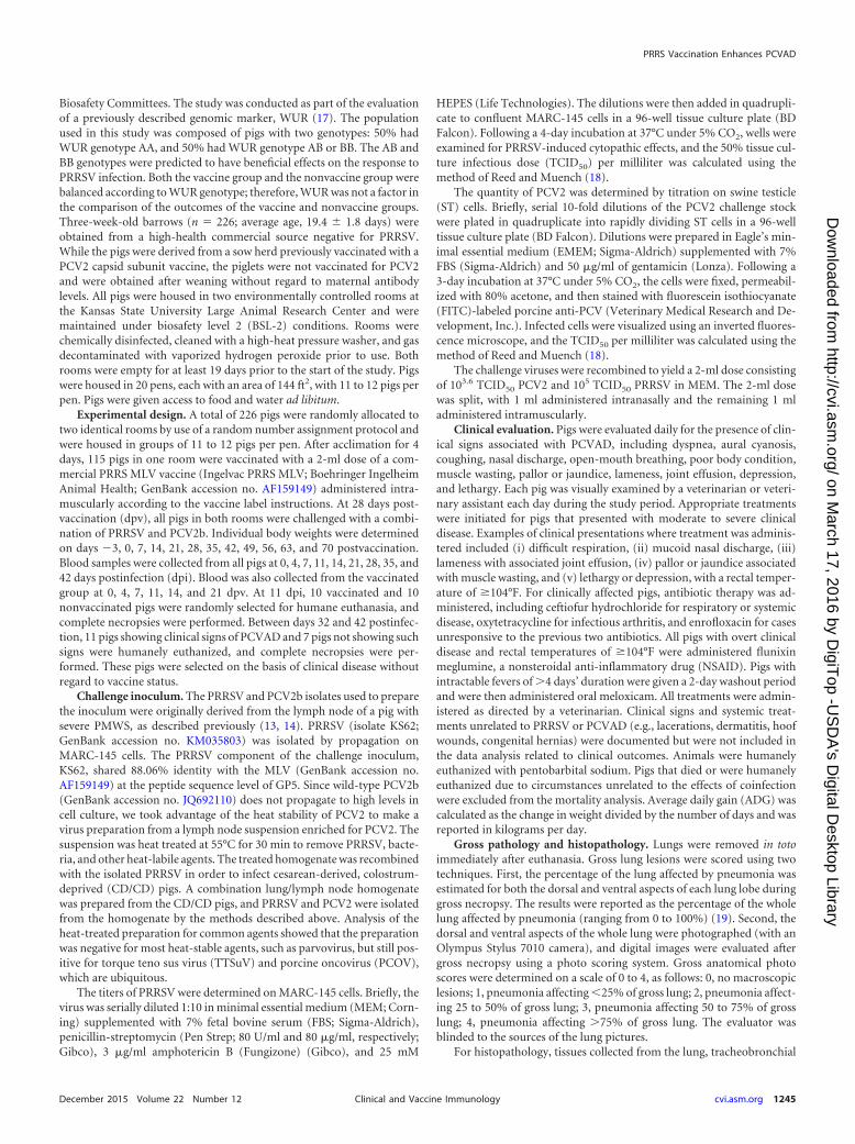
RESULTSPRRSV viremia is decreased after PRRS vaccination, but PCV2viremia is increased. PRRS MLV replication in the vaccine groupprior to challenge was assessed by RT-PCR on serum samplescollected at 11 dpv. Of the 84 pig sera tested, 78 (93%) had detect-able levels of the vaccine virus, confirming that the pigs supportedactive MLV replication (Fig. 1). The mean level of viremia at 11dpv was 2.7 � 1.7 log10 templates per PCR. Prior to challenge, thenonvaccine group was negative for PRRSV nucleic acid in serum(data not shown).
Niederwerder et al.
1246 cvi.asm.org December 2015 Volume 22 Number 12Clinical and Vaccine Immunology
on March 17, 2016 by D
igiTop -U
SD
A's D
igital Desktop Library
http://cvi.asm.org/
Dow
nloaded from
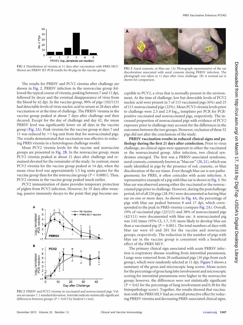
The results for PRRSV and PCV2 viremia after challenge areshown in Fig. 2. PRRSV infection in the nonvaccine group fol-lowed the typical course of viremia, peaking between 7 and 11 dpi,followed by decay and the eventual disappearance of virus fromthe blood by 42 dpi. In the vaccine group, 90% of pigs (102/113)had detectable levels of virus nucleic acid in serum at 28 days aftervaccination or at the time of challenge. The PRRSV viremia in thevaccine group peaked at about 7 days after challenge and thendecayed. Except for the day of challenge and day 42, the meanPRRSV level was significantly lower on all days in the vaccinegroup (Fig. 2A). Peak viremia for the vaccine group at days 7 and11 was reduced by �1 log unit from that for nonvaccinated pigs.The results demonstrated that vaccination was effective in reduc-ing PRRS viremia in a heterologous challenge model.
Mean PCV2 viremia levels for the vaccine and nonvaccinegroups are presented in Fig. 2B. In the nonvaccine group, meanPCV2 viremia peaked at about 21 days after challenge and re-mained elevated for the remainder of the study. In contrast, meanPCV2 viremia for the vaccine group peaked at 14 dpi, when themean virus level was approximately 1.5 log units greater for thevaccine group than for the nonvaccine group (P � 0.0001). Thus,PCV2 viremia in the vaccine group peaked much earlier.
PCV2 immunization of dams provides temporary protectionof piglets from PCV2 infection. However, by 35 days after wean-ing, passive immunity decays to the point that pigs become sus-
ceptible to PCV2, a virus that is normally present in the environ-ment. At the time of challenge, low but detectable levels of PCV2nucleic acid were present in 7 of 115 vaccinated pigs (6%) and 25of 111 nonvaccinated pigs (22%). Mean PCV2 viremia levels priorto challenge were 2.3 and 2.9 log10 templates per PCR for PCR-positive vaccinated and nonvaccinated pigs, respectively. The in-creased proportion of nonvaccinated pigs with evidence of PCV2exposure prior to challenge may account for the differences in theoutcomes between the two groups. However, exclusion of these 32pigs did not alter the conclusions of the study.
PRRSV vaccination results in reduced clinical signs and pa-thology during the first 21 days after coinfection. Prior to viruschallenge, no clinical signs were apparent in either the vaccinatedor the nonvaccinated group. After infection, two clinical syn-dromes emerged. The first was a PRRSV-associated syndrome,aural cyanosis, commonly known as “blue ear” (20, 21), which waseasily identified in pigs by the presence of red, cyanotic, or bluediscoloration of the ear tissue. Even though blue ear is not patho-gnomonic for PRRS, it often coincides with acute infection. Arepresentative example of a pig with blue ear is shown in Fig. 3. Noblue ear was observed among either the vaccinated or the nonvac-cinated pigs prior to challenge. However, during the postchallengeperiod, 64 of all 226 pigs (28.3%) were documented as having blueear on one or more days. As shown in Fig. 4A, the percentage ofpigs with blue ear peaked between 8 and 17 dpi, which corre-sponded to the peak in PRRS viremia (compare Fig. 2A). Overall,19% of vaccinated pigs (22/115) and 38% of nonvaccinated pigs(42/111) were documented with blue ear. A nonvaccinated pigwas 3.02 times (95% CI, 1.7, 5.9) more likely to develop blue earthan a vaccinated pig (P � 0.001). The total numbers of days withblue ear were 65 and 201 for the vaccine and nonvaccinegroups, respectively. The reduction in the number of pigs withblue ear in the vaccine group is consistent with a beneficialeffect of the PRRS MLV.
The primary clinical sign associated with acute PRRSV infec-tion is respiratory disease resulting from interstitial pneumonia.Lungs were removed from 20 euthanized pigs (10 pigs from eachgroup), which were randomly selected at 11 dpi. Figure 5 shows asummary of the gross and microscopic lung scores. Mean scoresfor the percentage of gross lung lobe involvement and microscopicscoring for interstitial pneumonia were higher in the nonvaccinegroup; however, the differences were not statistically significant(P � 0.62 for the percentage of lung involvement and 0.30 for thehistopathology score). Together, the results showed that vaccina-tion with the PRRS MLV had an overall protective effect by reduc-ing PRRSV viremia and decreasing PRRS-associated clinical signs.
FIG 1 Distribution of viremia at 11 days after vaccination with PRRS MLV.Shown are PRRSV RT-PCR results for 84 pigs in the vaccine group.
FIG 2 PRRSV and PCV2 viremia in vaccinated and nonvaccinated pigs. Val-ues are means � 1 standard deviation. Asterisks indicate statistically significantdifferences between groups (P � 0.015 by Student’s t test).
FIG 3 Aural cyanosis, or blue ear. (A) Photograph representative of the eardiscoloration associated with aural cyanosis during PRRSV infection. Thephotograph was taken at 11 days after virus challenge. (B) A normal ear isshown for comparison.
PRRS Vaccination Enhances PCVAD
December 2015 Volume 22 Number 12 cvi.asm.org 1247Clinical and Vaccine Immunology
on March 17, 2016 by D
igiTop -U
SD
A's D
igital Desktop Library
http://cvi.asm.org/
Dow
nloaded from

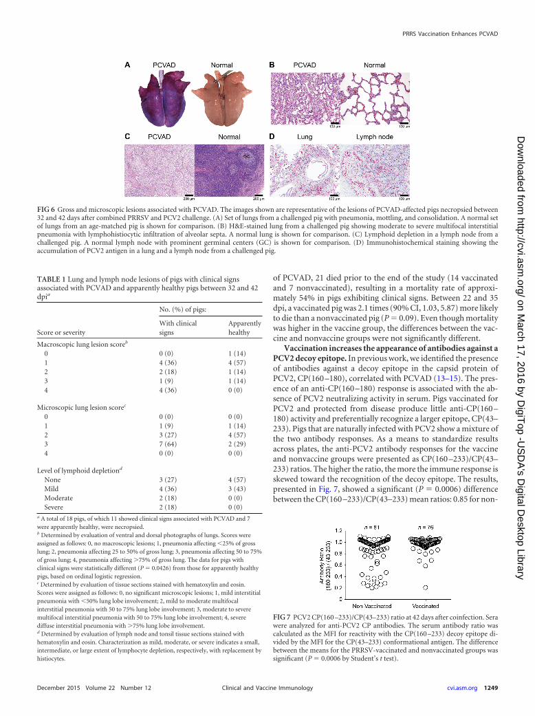
PRRS vaccination results in increased clinical signs and pa-thology at 22 to 42 days after coinfection. Beyond the acute pe-riod of infection, a second clinical syndrome appeared, which firstbecame apparent by an increase in the number of pigs receivingsystemic veterinary treatment due to clinical signs associated withPCVAD, such as tachypnea, dyspnea, pyrexia, loss of condition,muscle wasting, mucoid nasal discharge, lethargy, and pallor orjaundice (Fig. 4B). Lesions typical of PCVAD were found by grossanatomical and microscopic examinations of lungs and lymphnodes from pigs that died or were euthanized. Representative pic-tures and photomicrographs showing the lesions associated withclinically affected pigs are presented in Fig. 6. Lungs showed mul-tifocal to diffuse interstitial pneumonia with mottling of lung tis-sue, hemorrhage, and consolidation (Fig. 6A). At the microscopiclevel, multifocal to diffuse interstitial pneumonia with lymphohis-tiocytic infiltration into the alveolar septa and peribronchiolarareas was easily visible (Fig. 6B). The lymph nodes of affected pigsshowed depletion of lymphocytes (Fig. 6C). Positive staining forPCV2 antigen was observed in the lymph nodes and lungs of af-fected pigs (Fig. 6D). Analysis of gross and microscopic lesionscombined with the accumulation of PCV2 antigen in target organsconfirmed the presence of PCVAD.
The number of pigs undergoing treatment as a result ofPCVAD-associated clinical signs peaked between 22 and 35 dpi(Fig. 4B). During this time, 39 pigs received at least 1 day of vet-erinary treatment, including 12 nonvaccinated pigs (12/101[12%]) and 27 pigs in the vaccine group (27/105 [26%]). A vacci-nated pig was 2.67 times (95% CI, 1.23, 5.80) more likely to receiveveterinary treatment during peak PCVAD than a nonvaccinatedpig (P � 0.01). Large amounts of PCV2 in serum were associatedwith the 39 pigs that went on to develop PCVAD. At 14 dpi, sig-nificantly higher levels of circulating PCV2 were present in the 39PCVAD-affected pigs (mean, 5.8 log10 templates/PCR) than in the
163 pigs without clinical signs (mean, 4.8 log10 templates/PCR)(P � 0.004). The different treatments administered to the 39 pigswith clinical signs included a single antibiotic and an NSAID(16/39 [41%]), multiple antibiotics and an NSAID (7/39 [18%]), asingle antibiotic (6/39 [15%]), multiple antibiotics (3/39 [8%]),and an NSAID alone (3/39 [8%]). Four of the 39 pigs (10%) werehumanely euthanized after the initial treatment due to the severityof the clinical presentations. The decline in the percentage of pigswith PCVAD clinical signs was largely the result of increased mor-tality or the euthanization of pigs that were moribund or nonre-sponsive to treatment (compare Fig. 4B and C). Over the entirestudy period, 49 pigs received at least 1 day of systemic veterinarytreatment: 16% in the nonvaccine group (18/111) and 27% in thevaccine group (31/115). A vaccinated pig was 1.79 times (90% CI,0.99, 3.25) more likely to receive veterinary treatment than a non-vaccinated pig (P � 0.11) over the entire study period.
Macroscopic and microscopic changes in organs and tissueswere evaluated between 32 and 42 dpi for 11 clinically affectedpigs, which were humanely euthanized as a result of failure torespond to treatment. For comparison, 7 pigs without clinicalsigns were also necropsied. As summarized in Table 1, all 11 pigswith clinical signs showed some form of macroscopic lung in-volvement, as determined by the photographic score. The macro-scopic scores for the group without clinical signs were significantlylower (P � 0.04). A similar trend for clinically affected versusnonaffected pigs appeared at the microscopic level; however, thedifference was not statistically significant (P � 0.16). Mild to se-vere lymphoid depletion was observed in 8 of the 11 pigs withclinical signs (73%), compared to only 3 of the 7 (43%) pigs with-out clinical signs. Even though the pigs without clinical signs ap-peared normal, almost all showed some form of pathology relatedto PCVAD, such as mild to moderate pneumonia and/or mildlymphoid depletion.
Effect of PRRS MLV vaccination on mortality. As shown inFig. 4C, of the 101 nonvaccinated pigs, 9 died, resulting in anoverall survival rate of 91.1%. Of the 105 vaccinated pigs, 14 died,for an overall survival rate of 86.7% (Fig. 4C). A vaccinated pig was1.7 times (95% CI, 0.89, 3.72) more likely to die during the overallstudy period than a nonvaccinated pig (P � 0.35). Increased mor-tality became apparent after 20 days and was associated with theappearance of PCVAD. Of the 39 pigs that developed clinical signs
FIG 4 Clinical outcomes for the vaccinated and nonvaccinated groups after adual challenge with PRRSV and PCV2. Panel A shows the percentage of pigswith aural cyanosis, a PRRSV-associated syndrome. Clinical signs were as-sessed as described in Materials and Methods.
FIG 5 Assessment of lung pathology at 11 days after PRRSV/PCV2 challenge.Ten vaccinated pigs (filled bars) and 10 nonvaccinated pigs (shaded bars) wererandomly removed from the study at 11 days and were assessed for pneumo-nia. Results (means � standard deviations) are presented as the percentage ofgross lung involvement and the histopathology score. The differences betweenthe vaccine and nonvaccine groups were not significant (P � 0.05).
Niederwerder et al.
1248 cvi.asm.org December 2015 Volume 22 Number 12Clinical and Vaccine Immunology
on March 17, 2016 by D
igiTop -U
SD
A's D
igital Desktop Library
http://cvi.asm.org/
Dow
nloaded from
of PCVAD, 21 died prior to the end of the study (14 vaccinatedand 7 nonvaccinated), resulting in a mortality rate of approxi-mately 54% in pigs exhibiting clinical signs. Between 22 and 35dpi, a vaccinated pig was 2.1 times (90% CI, 1.03, 5.87) more likelyto die than a nonvaccinated pig (P � 0.09). Even though mortalitywas higher in the vaccine group, the differences between the vac-cine and nonvaccine groups were not significantly different.
Vaccination increases the appearance of antibodies against aPCV2 decoy epitope. In previous work, we identified the presenceof antibodies against a decoy epitope in the capsid protein ofPCV2, CP(160 –180), correlated with PCVAD (13–15). The pres-ence of an anti-CP(160 –180) response is associated with the ab-sence of PCV2 neutralizing activity in serum. Pigs vaccinated forPCV2 and protected from disease produce little anti-CP(160 –180) activity and preferentially recognize a larger epitope, CP(43–233). Pigs that are naturally infected with PCV2 show a mixture ofthe two antibody responses. As a means to standardize resultsacross plates, the anti-PCV2 antibody responses for the vaccineand nonvaccine groups were presented as CP(160 –233)/CP(43–233) ratios. The higher the ratio, the more the immune response isskewed toward the recognition of the decoy epitope. The results,presented in Fig. 7, showed a significant (P � 0.0006) differencebetween the CP(160 –233)/CP(43–233) mean ratios: 0.85 for non-
FIG 6 Gross and microscopic lesions associated with PCVAD. The images shown are representative of the lesions of PCVAD-affected pigs necropsied between32 and 42 days after combined PRRSV and PCV2 challenge. (A) Set of lungs from a challenged pig with pneumonia, mottling, and consolidation. A normal setof lungs from an age-matched pig is shown for comparison. (B) H&E-stained lung from a challenged pig showing moderate to severe multifocal interstitialpneumonia with lymphohistiocytic infiltration of alveolar septa. A normal lung is shown for comparison. (C) Lymphoid depletion in a lymph node from achallenged pig. A normal lymph node with prominent germinal centers (GC) is shown for comparison. (D) Immunohistochemical staining showing theaccumulation of PCV2 antigen in a lung and a lymph node from a challenged pig.
TABLE 1 Lung and lymph node lesions of pigs with clinical signsassociated with PCVAD and apparently healthy pigs between 32 and 42dpia
Score or severity
No. (%) of pigs:
With clinicalsigns
Apparentlyhealthy
Macroscopic lung lesion scoreb
0 0 (0) 1 (14)1 4 (36) 4 (57)2 2 (18) 1 (14)3 1 (9) 1 (14)4 4 (36) 0 (0)
Microscopic lung lesion scorec
0 0 (0) 0 (0)1 1 (9) 1 (14)2 3 (27) 4 (57)3 7 (64) 2 (29)4 0 (0) 0 (0)
Level of lymphoid depletiond
None 3 (27) 4 (57)Mild 4 (36) 3 (43)Moderate 2 (18) 0 (0)Severe 2 (18) 0 (0)
a A total of 18 pigs, of which 11 showed clinical signs associated with PCVAD and 7were apparently healthy, were necropsied.b Determined by evaluation of ventral and dorsal photographs of lungs. Scores wereassigned as follows: 0, no macroscopic lesions; 1, pneumonia affecting �25% of grosslung; 2, pneumonia affecting 25 to 50% of gross lung; 3, pneumonia affecting 50 to 75%of gross lung; 4, pneumonia affecting �75% of gross lung. The data for pigs withclinical signs were statistically different (P � 0.0426) from those for apparently healthypigs, based on ordinal logistic regression.c Determined by evaluation of tissue sections stained with hematoxylin and eosin.Scores were assigned as follows: 0, no significant microscopic lesions; 1, mild interstitialpneumonia with �50% lung lobe involvement; 2, mild to moderate multifocalinterstitial pneumonia with 50 to 75% lung lobe involvement; 3, moderate to severemultifocal interstitial pneumonia with 50 to 75% lung lobe involvement; 4, severediffuse interstitial pneumonia with �75% lung lobe involvement.d Determined by evaluation of lymph node and tonsil tissue sections stained withhematoxylin and eosin. Characterization as mild, moderate, or severe indicates a small,intermediate, or large extent of lymphocyte depletion, respectively, with replacement byhistiocytes.
FIG 7 PCV2 CP(160 –233)/CP(43–233) ratio at 42 days after coinfection. Serawere analyzed for anti-PCV2 CP antibodies. The serum antibody ratio wascalculated as the MFI for reactivity with the CP(160 –233) decoy epitope di-vided by the MFI for the CP(43–233) conformational antigen. The differencebetween the means for the PRRSV-vaccinated and nonvaccinated groups wassignificant (P � 0.0006 by Student’s t test).
PRRS Vaccination Enhances PCVAD
December 2015 Volume 22 Number 12 cvi.asm.org 1249Clinical and Vaccine Immunology
on March 17, 2016 by D
igiTop -U
SD
A's D
igital Desktop Library
http://cvi.asm.org/
Dow
nloaded from

vaccinated pigs versus 0.97 for vaccinated pigs. Exclusion of the 32pigs that showed the presence of PCV2 nucleic acid at the time ofchallenge changed the ratios but did not affect the conclusion;CP(160 –233)/CP(43–233) mean ratios were 0.92 for nonvacci-nated pigs (n � 61) and 0.98 for vaccinated pigs (n � 72) (P �0.03).
As discussed above, previous work showed that pigs with lowCP(160 –233)/CP(43–233) ratios are protected from disease.Therefore, nonvaccinated pigs with ratios of �0.5 (n � 13) werecompared to nonvaccinated pigs with PCV2 antibody ratios of�0.5 (n � 68). Pigs with antibody ratios of �0.5 had significantlylower levels of circulating PCV2 in the serum at 21 (P � 0.0001),28 (P � 0.0008), 35 (P � 0.007), and 42 (P � 0.03) dpi than pigswith higher ratios. The results confirm earlier findings describingthe nonprotective effect of anti-CP(160 –233) antibodies (13).
Effect of PRRS MLV vaccination on ADG. Over the entire70-day study period, the mean average daily gain (ADG) for thevaccine group (n � 91) was 0.65 � 0.11 kg, compared to 0.68 �0.10 kg for the nonvaccine group (n � 92). The means differedsignificantly between the two groups (P � 0.029). Decreased meanADG was also observed in the vaccine group during the 42-daypost–virus challenge period. However, the difference between thevaccine group (ADG, 0.82 � 0.14 kg) (n � 91) and the nonvaccinegroup (ADG, 0.86 � 0.14 kg) (n � 92) was not statistically signif-icant (P � 0.061). In addition, ADG differences between the twogroups during the 70-day study period were no longer significantafter the exclusion of the 32 pigs with PCV2 detected prior tochallenge.
Therefore, a more-detailed analysis was conducted by calculat-ing ADG on a weekly basis (Fig. 8). The results showed that ADGdifferences between the nonvaccine and vaccine groups could bedivided into three distinct phases. In the first phase, covering theprechallenge period, mean ADG after vaccination was reduced,with a significant difference between the vaccinated and nonvac-cinated groups appearing at 3 weeks after vaccination. The secondphase covered the period of acute PRRSV infection. At 1 weekafter virus challenge, mean ADG was significantly increased forthe vaccine group. ADG remained higher for the vaccine group inthe second week postchallenge, but the difference was not signif-
icant. The improved ADG for the vaccinated pigs likely resultedfrom the positive effect of vaccination in reducing PRRS-associ-ated clinical signs and virus load (Fig. 2A and 4A). The third phasecovered the period from the onset of PCVAD, beginning at about3 weeks after virus challenge, to the end of the study. During thisphase, ADG was lower in the vaccinated group at every time point,with significantly lower mean ADG values on week 6 after viruschallenge. A significant decrease in ADG was also initially detectedfor vaccinated pigs on week 3 postchallenge; however, this differ-ence was no longer significant after the 32 PCV2-positive pigs hadbeen excluded (P � 0.08). The lower mean ADG values are con-sistent with the effects of PCVAD, which include poor growthperformance and muscle wasting.
The negative effect of vaccination on ADG could have been theresult of increased numbers of pigs with clinically apparentPCVAD. Therefore, a separate analysis was performed after the 26pigs with clinical signs that survived the length of the study hadbeen excluded from the vaccine and nonvaccine groups. The re-moval of pigs with clinical signs increased the ADGs of the twogroups to 0.69 � 0.09 kg for nonvaccinated pigs without clinicalsigns (n � 83) and 0.66 � 0.10 kg for vaccinated pigs withoutclinical signs (n � 74) (P � 0.047). However, even in the absenceof overt clinical signs, PRRSV vaccination had a negative effect onweight gain.
DISCUSSION
Enhanced PCV2 infection leading to PCVAD is typically associ-ated with immune stimulation (22, 23). PCV2 replication is lo-cated in the nuclei of permissive cells and is dependent on cellularenzymes expressed during the S phase of the cell cycle (24). It ispresumed that actively dividing lymphocytes, in response to animmune stimulus, provide a cellular environment ideal for sup-porting PCV2 replication. PRRSV, porcine parvovirus, and My-coplasma hyopneumoniae are common copathogens linked withlymphoproliferation and increased PCV2 pathogenesis (10–13,25–31). Examples of noninfectious immunostimulators includeimmunization with keyhole limpet hemocyanin in incompleteFreund’s adjuvant (22, 32) and inactivated vaccines, such as My-coplasma hyopneumoniae (32–34) and Actinobacillus pleuropneu-
FIG 8 Weekly average daily gain (ADG) before and after virus challenge. Mean ADG values for the nonvaccinated (shaded bars) and vaccinated (filled bars)groups were calculated on a weekly basis. The data show means and standard deviations. The numbers of pigs used in the analysis are given below the bar graph.Asterisks indicate statistically significant differences in ADG between groups (P � 0.03 by Student’s t test).
Niederwerder et al.
1250 cvi.asm.org December 2015 Volume 22 Number 12Clinical and Vaccine Immunology
on March 17, 2016 by D
igiTop -U
SD
A's D
igital Desktop Library
http://cvi.asm.org/
Dow
nloaded from

moniae (33, 34). The results of this study showed that the PRRSMLV initially had a beneficial effect in reducing PRRS-associatedclinical signs and PRRS viremia; however, PRRSV-vaccinated pigsshowed increased PCV2 replication, reduced average daily gain,and increased clinical signs associated with PCVAD.
Previous experimental studies documenting interactions be-tween the PRRS MLV and PCV2 infection have yielded conflictingresults. Allan et al. (35) found that colostrum-deprived, specific-pathogen-free (SPF) pigs infected with PCV2 at the age of 5 weeksand administered the PRRS MLV 1 week later had greateramounts of PCV2 antigen in tissues and more-severe histologiclesions, characteristic of PMWS, than pigs infected with PCV2alone. However, the pigs failed to exhibit clinical signs or grosslesions typical of PCVAD (35). In contrast, Opriessnig et al. (36)evaluated the effects of PCV2 infection on the efficacy of the PRRSMLV in groups of 10 early-weaned SPF pigs. Pigs were inoculatedwith PCV2 at the age of 6 weeks, vaccinated with the PRRS MLV 2weeks later, and then challenged with PRRSV at the age of 12weeks. The group with PCV2 and the MLV exhibited lower ADGand more-severe lung lesions after PRRSV challenge than thegroup that was vaccinated and received a PRRSV challenge with-out PCV2. Because PCV2 was not detected in affected lungs byIHC, the authors attributed the lesions to PRRSV infection andfurther concluded that the effect of PCV2 was to reduce the effi-cacy of the PRRS MLV (36). Park et al. (37) investigated the po-tential for the PRRS MLV to reduce PRRSV-associated amplifica-tion of PCV2 pathogenesis after coinfection with PRRSV andPCV2. Groups of 8 conventional pigs were subjected to a variety oftreatments involving different combinations of the PRRS MLV,wild-type PRRSV, a PCV2 vaccine, and PCV2. Pigs were vacci-nated with the PRRS MLV, a PCV2 subunit vaccine, or both andwere then challenged, 4 weeks later, with PRRSV, PCV2, or both.The group that received the PRRS MLV followed by coinfectionshowed no differences in PCV2 viremia, PCV2-associated pathol-ogy, or the number of PCV2-positive cells in lymph nodes andlungs from the coinfected group that was not vaccinated with thePRRS MLV (37). In contrast to our study, in which vaccinationenhanced PCVAD, these authors found that coinfected pigs, withor without previous PRRS MLV vaccination, had similar PCV2replication and pathogenesis.
Field studies have also yielded conflicting results. A survey con-ducted on 70 pig farms in the Netherlands included questionsregarding PRRS MLV use on farms with and without PMWS orPCVAD. The results showed that the PRRS MLV is a significantrisk factor for PMWS outbreaks (38). In contrast, an analysis ofthe effect of the PRRS MLV on farms affected by PCVAD in theUnited States found that farms incorporating the PRRS MLV havesignificantly lower levels of PCV2 viremia than nonvaccinatingfarms in the age group in which peak wasting disease occurred(39). In that study, quantitative PCR (qPCR) was used to measurePCV2 in serum samples collected at different time points from 6herds vaccinated with the PRRS MLV and 12 nonvaccinatedherds. These results suggest that the PRRS MLV can reduce PCV2viremia (39).
The failure of experimental studies to find a consistent linkbetween the PRRS MLV and PCVAD was likely due in part to thelength of the observation period. For example, the studies de-scribed above were terminated on day 25 after infection withPCV2 (35) and 21 days after challenge with PRRSV and PCV2(37). In the current study, peak PCVAD occurred between 22 and
35 days after challenge with PRRSV and PCV2. Another impor-tant difference is the sizes of the experimental groups. Typically,the prevalence of clinically ill pigs on farms affected by PCVAD isonly 2 to 25% (40–42). In small groups of pigs, animals with clin-ical disease may not be apparent or present. The current studyutilized more than 200 pigs, mimicking the environment found inthe field and providing the depth of data required for the obser-vation and quantification of low-percentage outcomes. PCVADmorbidity rates were 11.9% and 25.7% in nonvaccinated and vac-cinated pigs, respectively. Therefore, PCVAD should be assessedby comparing mortality rates, clinical disease presentation,viremia, and weight gain in relatively large groups evaluated forseveral weeks after infection.
Because large groups of pigs were required to ensure clinicaldisease expression, some control groups, including pigs chal-lenged with PCV2 or PRRSV alone, were not incorporated intothe study design. Therefore, conclusions are based on the findingsfor vaccinated and nonvaccinated cochallenged pigs. Cautionshould be exercised in generalizing results to single infections (i.e.,the effect of the PRRS MLV on PCV2-challenged pigs).
PCV2 is ubiquitous in swine populations, and elimination ofthe virus from the environment is extremely difficult. As demon-strated in this study, 32 of 226 pigs had detectable PCV2 in serumat the time of challenge. Although these pigs were in the minority(14%), and group titers were relatively low (2.3 and 2.9 log10 tem-plates per PCR for vaccinated and nonvaccinated pigs, respec-tively), the presence of PCV2 prior to challenge had to be consid-ered a possible factor in postchallenge response. This wasespecially true due to the difference between the proportions ofPCV2-positive pigs in the two groups, despite randomized alloca-tion of pigs and balanced genotypes. This difference was likely dueto the failure of randomization to distribute PCV2-positive pigsequally across the two groups. Therefore, analyses were also com-pleted after the 32 initially PCV2 positive pigs were excluded, andall conclusions of the study were confirmed. Regardless, this high-lights the difficulty of eliminating PCV2 from the environmentand also suggests that pigs should be balanced according to PCV2status prior to challenge.
Significantly increased levels of PCV2 viremia in PRRS MLV-treated pigs were observed at 11 and 14 days after challenge, butnot at later time points (Fig. 2B). This effect of the PRRS MLV onPCV2 infection is similar to that seen in a previous study by us,which showed a significant increase in the level of PCV2 viremia at23 days after coinfection with PRRSV and PCV2 (13). In the cur-rent study, it is noteworthy that PCV2 viremia levels did not differsignificantly between the vaccine and nonvaccine groups duringpeak PCVAD. However, the 39 clinically ill pigs did maintain sig-nificantly higher levels (P � 0.02) of circulating PCV2 duringthese later time points (21 and 35 dpi) (data not shown). Theincreased incidence of PCVAD in vaccinated pigs between 22 and35 dpi may also be the result of greater levels of localized PCV2 intissues. Further studies are needed to assess differences in thequantity and tissue distribution of PCV2 between vaccinated andnonvaccinated pigs.
Average daily gain (ADG) is used in swine production as anobjective measure of overall health and performance. The nega-tive effect of the PRRS MLV on growth performance is well doc-umented. For example, Opriessnig et al. (43) reported that pigsvaccinated with the PRRS MLV at the age of 2 weeks exhibitedsignificantly lower ADG than nonvaccinated pigs. Pretzer et al.
PRRS Vaccination Enhances PCVAD
December 2015 Volume 22 Number 12 cvi.asm.org 1251Clinical and Vaccine Immunology
on March 17, 2016 by D
igiTop -U
SD
A's D
igital Desktop Library
http://cvi.asm.org/
Dow
nloaded from
(44) found that PRRS MLV-vaccinated weaned pigs had lowerADG between 0 and 14 dpv than nonvaccinated pigs. While thiseffect on ADG was no longer apparent between 21 and 42 dpv, thevaccinated pigs maintained lighter weights overall (44). We con-firmed the negative effect of the PRRS MLV on ADG during the28-day period prior to coinfection; significantly reduced ADG wasobserved during the third week after vaccination (Fig. 8). Thebenefit of PRRSV vaccination was documented during the first 2weeks after PCV2 and PRRSV challenge, when the MLV had apositive effect on ADG. Although vaccinated pigs in this study hadincreased ADG in the first 2 weeks postchallenge, this effect wasquickly outweighed when ADG was decreased in the presence ofPCVAD. Vaccination decreased ADG in both clinically affectedpigs and pigs without clinical signs during the study period, dem-onstrating that poor growth performance may be a subclinicalmanifestation of PCVAD in apparently healthy pigs.
At least three mechanisms may be involved in the enhance-ment of PCVAD following PRRSV vaccination. First, the PRRSMLV may function to stimulate the immune system and increasethe number of PCV2-permissive cells. As with wild-type viruses,lymphocytes undergo mitosis in response to vaccination with thePRRS MLV, thereby increasing the population of cells with theability to support PCV2 replication. In addition, the vaccine likelystimulates PRRSV-specific lymphocyte populations that are re-stimulated after challenge with a wild-type PRRSV. Second, likewild-type PRRS viruses, the PRRS MLV may suppress innate im-munity, thereby blocking anti-PCV2 responses. For example,PRRSV nonstructural proteins, such as nsp1 and nsp2, block theinduction of interferon and response of cells to interferon (5).Viral proteins such as nsp1 and nsp1� antagonize the type Iinterferon response by degrading key components needed for in-terferon gene expression and inhibiting interferon signaling path-ways (45, 46). Finally, the third mechanism is based on the possi-bility that the PRRS MLV may skew the immune response towardthe production of nonneutralizing PCV2-specific antibodies.
PCV2 has circulated in the swine population for at least 25years. In 2005, the emergence of PCV2b in North America wasattributed to outbreaks of PCVAD (47–49). Since then, the diseasehas been effectively managed through the use of PCV2 vaccines(50–52). Therefore, the negative effect of the MLV on PCV2 in-fection may not be relevant. However, there remain several coun-tries in which PRRS MLV vaccination is in wide use, but in theabsence of a comprehensive PCVAD vaccination program. Fur-ther, there is the potential for new and emerging PCV2 strains toescape current vaccine protection. Emerging PCV2 mutant strainshave been documented in China (53) and more recently have beenassociated with PCVAD outbreaks in vaccinated herds in theUnited States and Korea (54, 55). Overall, this study supports thenotion that maintaining a successful PCV2 control program andassessing the risk of virulent PRRSV exposure is critical to weigh-ing the benefits of the PRRS MLV.
ACKNOWLEDGMENT
This work was supported by USDA NIFA award 2013-68004-20362.
REFERENCES1. Baekbo P, Kristensen CS, Larsen LE. 2012. Porcine circovirus diseases: a
review of PMWS. Transbound Emerg Dis 59:60 – 67. http://dx.doi.org/10.1111/j.1865-1682.2011.01288.x.
2. Ramamoorthy S, Meng XJ. 2009. Porcine circoviruses: a minuscule yet
mammoth paradox. Anim Health Res Rev 10:1–20. http://dx.doi.org/10.1017/S1466252308001461.
3. Benfield DA, Nelson E, Collins JE, Harris L, Goyal SM, Robison D,Christianson WT, Morrison RB, Gorcyca D, Chladek D. 1992. Charac-terization of swine infertility and respiratory syndrome (SIRS) virus (iso-late ATCC VR-2332). J Vet Diagn Invest 4:127–133. http://dx.doi.org/10.1177/104063879200400202.
4. Conzelmann KK, Visser N, Van Woensel P, Thiel HJ. 1993. Molecularcharacterization of porcine reproductive and respiratory syndrome virus,a member of the arterivirus group. Virology 193:329 –339. http://dx.doi.org/10.1006/viro.1993.1129.
5. Chand RJ, Trible BR, Rowland RR. 2012. Pathogenesis of porcine re-productive and respiratory syndrome virus. Curr Opin Virol 2:256 –263.http://dx.doi.org/10.1016/j.coviro.2012.02.002.
6. Gómez-Laguna J, Salguero FJ, Pallares FJ, Carrasco L. 2013. Immuno-pathogenesis of porcine reproductive and respiratory syndrome in therespiratory tract of pigs. Vet J 195:148 –155. http://dx.doi.org/10.1016/j.tvjl.2012.11.012.
7. Opriessnig T, Gimenez-Lirola LG, Halbur PG. 2011. Polymicrobialrespiratory disease in pigs. Anim Health Res Rev 12:133–148. http://dx.doi.org/10.1017/S1466252311000120.
8. Renukaradhya GJ, Alekseev K, Jung K, Fang Y, Saif LJ. 2010. Porcinereproductive and respiratory syndrome virus-induced immunosuppres-sion exacerbates the inflammatory response to porcine respiratory coro-navirus in pigs. Viral Immunol 23:457– 466. http://dx.doi.org/10.1089/vim.2010.0051.
9. Pallarés FJ, Halbur PG, Opriessnig T, Sorden SD, Villar D, Janke BH,Yaeger MJ, Larson DJ, Schwartz KJ, Yoon KJ, Hoffman LJ. 2002.Porcine circovirus type 2 (PCV-2) coinfections in US field cases ofpostweaning multisystemic wasting syndrome (PMWS). J Vet Diagn In-vest 14:515–519. http://dx.doi.org/10.1177/104063870201400614.
10. Allan GM, McNeilly F, Ellis J, Krakowka S, Meehan B, McNair I,Walker I, Kennedy S. 2000. Experimental infection of colostrum de-prived piglets with porcine circovirus 2 (PCV2) and porcine reproductiveand respiratory syndrome virus (PRRSV) potentiates PCV2 replication.Arch Virol 145:2421–2429. http://dx.doi.org/10.1007/s007050070031.
11. Harms PA, Sorden SD, Halbur PG, Bolin SR, Lager KM, Morozov I,Paul PS. 2001. Experimental reproduction of severe disease in CD/CDpigs concurrently infected with type 2 porcine circovirus and porcine re-productive and respiratory syndrome virus. Vet Pathol 38:528 –539. http://dx.doi.org/10.1354/vp.38-5-528.
12. Rovira A, Balasch M, Segales J, Garcia L, Plana-Duran J, Rosell C,Ellerbrok H, Mankertz A, Domingo M. 2002. Experimental inoculationof conventional pigs with porcine reproductive and respiratory syndromevirus and porcine circovirus 2. J Virol 76:3232–3239. http://dx.doi.org/10.1128/JVI.76.7.3232-3239.2002.
13. Trible BR, Ramirez A, Suddith A, Fuller A, Kerrigan M, Hesse R,Nietfeld J, Guo B, Thacker E, Rowland RR. 2012. Antibody responsesfollowing vaccination versus infection in a porcine circovirus-type 2(PCV2) disease model show distinct differences in virus neutralizationand epitope recognition. Vaccine 30:4079 – 4085. http://dx.doi.org/10.1016/j.vaccine.2012.04.022.
14. Trible BR, Kerrigan M, Crossland N, Potter M, Faaberg K, Hesse R,Rowland RR. 2011. Antibody recognition of porcine circovirus type 2capsid protein epitopes after vaccination, infection, and disease. Clin Vac-cine Immunol 18:749 –757. http://dx.doi.org/10.1128/CVI.00418-10.
15. Trible BR, Suddith AW, Kerrigan MA, Cino-Ozuna AG, Hesse RA,Rowland RR. 2012. Recognition of the different structural forms of thecapsid protein determines the outcome following infection with porcinecircovirus type 2. J Virol 86:13508 –13514. http://dx.doi.org/10.1128/JVI.01763-12.
16. Federation of Animal Science Societies. 2010. Guide for the care and useof agricultural animals in research and teaching. Federation of AnimalScience Societies, Champaign, IL.
17. Boddicker N, Waide EH, Rowland RR, Lunney JK, Garrick DJ, ReecyJM, Dekkers JC. 2012. Evidence for a major QTL associated with hostresponse to porcine reproductive and respiratory syndrome virus chal-lenge. J Anim Sci 90:1733–1746. http://dx.doi.org/10.2527/jas.2011-4464.
18. Reed LJ, Muench H. 1938. A simple method of estimating fifty per centendpoints. Am J Hyg 27:493– 497.
19. Halbur PG, Paul PS, Frey ML, Landgraf J, Eernisse K, Meng XJ, LumMA, Andrews JJ, Rathje JA. 1995. Comparison of the pathogenicity oftwo US porcine reproductive and respiratory syndrome virus isolates with
Niederwerder et al.
1252 cvi.asm.org December 2015 Volume 22 Number 12Clinical and Vaccine Immunology
on March 17, 2016 by D
igiTop -U
SD
A's D
igital Desktop Library
http://cvi.asm.org/
Dow
nloaded from
that of the Lelystad virus. Vet Pathol 32:648 – 660. http://dx.doi.org/10.1177/030098589503200606.
20. Done SH, Paton DJ. 1995. Porcine reproductive and respiratory syn-drome: clinical disease, pathology and immunosuppression. Vet Rec 136:32–35. http://dx.doi.org/10.1136/vr.136.2.32.
21. Paton DJ, Brown IH, Edwards S, Wensvoort G. 1991. ‘Blue ear’ diseaseof pigs. Vet Rec 128:617. http://dx.doi.org/10.1136/vr.128.26.617.
22. Krakowka S, Ellis JA, McNeilly F, Ringler S, Rings DM, Allan G. 2001.Activation of the immune system is the pivotal event in the production ofwasting disease in pigs infected with porcine circovirus-2 (PCV-2). VetPathol 38:31– 42. http://dx.doi.org/10.1354/vp.38-1-31.
23. Opriessnig T, Halbur PG. 2012. Concurrent infections are important forexpression of porcine circovirus associated disease. Virus Res 164:20 –32.http://dx.doi.org/10.1016/j.virusres.2011.09.014.
24. Tischer I, Peters D, Rasch R, Pociuli S. 1987. Replication of porcinecircovirus: induction by glucosamine and cell cycle dependence. Arch Vi-rol 96:39 –57. http://dx.doi.org/10.1007/BF01310989.
25. Opriessnig T, Thacker EL, Yu S, Fenaux M, Meng XJ, Halbur PG. 2004.Experimental reproduction of postweaning multisystemic wasting syn-drome in pigs by dual infection with Mycoplasma hyopneumoniae andporcine circovirus type 2. Vet Pathol 41:624 – 640. http://dx.doi.org/10.1354/vp.41-6-624.
26. Allan GM, Kennedy S, McNeilly F, Foster JC, Ellis JA, Krakowka SJ,Meehan BM, Adair BM. 1999. Experimental reproduction of severe wast-ing disease by co-infection of pigs with porcine circovirus and porcineparvovirus. J Comp Pathol 121:1–11. http://dx.doi.org/10.1053/jcpa.1998.0295.
27. Krakowka S, Ellis JA, Meehan B, Kennedy S, McNeilly F, Allan G. 2000.Viral wasting syndrome of swine: experimental reproduction ofpostweaning multisystemic wasting syndrome in gnotobiotic swine bycoinfection with porcine circovirus 2 and porcine parvovirus. Vet Pathol37:254 –263. http://dx.doi.org/10.1354/vp.37-3-254.
28. Ellis J, Krakowka S, Lairmore M, Haines D, Bratanich A, Clark E, AllanG, Konoby C, Hassard L, Meehan B, Martin K, Harding J, Kennedy S,McNeilly F. 1999. Reproduction of lesions of postweaning multisystemicwasting syndrome in gnotobiotic piglets. J Vet Diagn Invest 11:3–14. http://dx.doi.org/10.1177/104063879901100101.
29. Kennedy S, Moffett D, McNeilly F, Meehan B, Ellis J, Krakowka S, AllanGM. 2000. Reproduction of lesions of postweaning multisystemic wastingsyndrome by infection of conventional pigs with porcine circovirus type 2alone or in combination with porcine parvovirus. J Comp Pathol 122:9 –24. http://dx.doi.org/10.1053/jcpa.1999.0337.
30. Fan P, Wei Y, Guo L, Wu H, Huang L, Liu J, Liu C. 2013. Synergisticeffects of sequential infection with highly pathogenic porcine reproduc-tive and respiratory syndrome virus and porcine circovirus type 2. Virol J10:265. http://dx.doi.org/10.1186/1743-422X-10-265.
31. Sinha A, Shen HG, Schalk S, Beach NM, Huang YW, Meng XJ, HalburPG, Opriessnig T. 2011. Porcine reproductive and respiratory syndromevirus (PRRSV) influences infection dynamics of porcine circovirus type 2(PCV2) subtypes PCV2a and PCV2b by prolonging PCV2 viremia andshedding. Vet Microbiol 152:235–246. http://dx.doi.org/10.1016/j.vetmic.2011.05.005.
32. Krakowka S, Ellis J, McNeilly F, Waldner C, Rings DM, Allan G. 2007.Mycoplasma hyopneumoniae bacterins and porcine circovirus type 2(PCV2) infection: induction of postweaning multisystemic wasting syn-drome (PMWS) in the gnotobiotic swine model of PCV2-associated dis-ease. Can Vet J 48:716 –724.
33. Allan GM, McNeilly F, Kennedy S, Meehan B, Ellis J, Krakowka S. 2000.Immunostimulation, PCV-2 and PMWS. Vet Rec 147:170 –171.
34. Opriessnig T, Yu S, Gallup JM, Evans RB, Fenaux M, Pallares F,Thacker EL, Brockus CW, Ackermann MR, Thomas P, Meng XJ,Halbur PG. 2003. Effect of vaccination with selective bacterins on con-ventional pigs infected with type 2 porcine circovirus. Vet Pathol 40:521–529. http://dx.doi.org/10.1354/vp.40-5-521.
35. Allan GM, Caprioli A, McNair I, Lagan-Tregaskis P, Ellis J, KrakowkaS, McKillen J, Ostanello F, McNeilly F. 2007. Porcine circovirus 2replication in colostrum-deprived piglets following experimental infec-tion and immune stimulation using a modified live vaccine against por-cine respiratory and reproductive syndrome virus. Zoonoses PublicHealth 54:214 –222. http://dx.doi.org/10.1111/j.1863-2378.2007.01041.x.
36. Opriessnig T, McKeown NE, Harmon KL, Meng XJ, Halbur PG. 2006.Porcine circovirus type 2 infection decreases the efficacy of a modified live
porcine reproductive and respiratory syndrome virus vaccine. Clin Vac-cine Immunol 13:923–929. http://dx.doi.org/10.1128/CVI.00074-06.
37. Park C, Oh Y, Seo HW, Han K, Chae C. 2013. Comparative effects ofvaccination against porcine circovirus type 2 (PCV2) and porcine repro-ductive and respiratory syndrome virus (PRRSV) in a PCV2-PRRSV chal-lenge model. Clin Vaccine Immunol 20:369 –376. http://dx.doi.org/10.1128/CVI.00497-12.
38. De Jong MF, Elbers A, Wellenberg GJ. 2003. Factors associated withPMWS and PDNS: a case-control study, p 215. In Proceedings of the 4thInternational Symposium on Emerging and Re-emerging Pig Diseases—Rome, 29 June to 2 July 2003.
39. Genzow M, Schwartz K, Gonzalez G, Anderson G, Chittick W. 2009.The effect of vaccination against porcine reproductive and respiratorysyndrome virus (PRRSV) on the porcine circovirus-2 (PCV-2) load inporcine circovirus associated disease (PCVAD) affected pigs. Can J VetRes 73:87–90.
40. Sorden SD, Harms PA, Sirinarumitr T, Morozov I, Halbur PG, YoonK-J, Paul PS. 1998. Porcine circovirus and PRRS virus co-infection in pigswith chronic bronchointerstitial pneumonia and lymphoid depletion: anemerging syndrome in Midwestern swine, p 75. In Proceedings of the 41stAnnual Meeting of the American Association of Veterinary LaboratoryDiagnosticians. American Association of Veterinary Laboratory Diagnos-ticians, Visalia, CA.
41. Quintana J, Segales J, Rosell C, Calsamiglia M, Rodriguez-Arrioja GM,Chianini F, Folch JM, Maldonado J, Canal M, Plana-Duran J, DomingoM. 2001. Clinical and pathological observations on pigs with postweaningmultisystemic wasting syndrome. Vet Rec 149:357–361. http://dx.doi.org/10.1136/vr.149.12.357.
42. Kyriakis SC, Saoulidis K, Lekkas S, Miliotis CC, Papoutsis PA, KennedyS. 2002. The effects of immuno-modulation on the clinical and patholog-ical expression of postweaning multisystemic wasting syndrome. J CompPathol 126:38 – 46. http://dx.doi.org/10.1053/jcpa.2001.0520.
43. Opriessnig T, Pallares FJ, Nilubol D, Vincent AL, Thacker EL, VaughnEM, Roof M, Halbur PG. 2005. Genomic homology of ORF 5 genesequence between modified live vaccine virus and porcine reproductiveand respiratory syndrome virus challenge isolates is not predictive of vac-cine efficacy. J Swine Health Production 13:246 –253.
44. Pretzer SD, Claussen KM, Bergstrom JR, Henry SC, Phillips R, TokachMD, Goodband RD, Nelssen JL, Dritz SS. 1996. The effects of porcinereproductive and respiratory syndrome (PRRS) vaccination on postwean-ing growth performance, p 83– 86. In Swine Day, 1996. Kansas State Uni-versity, Agricultural Experiment Station and Cooperative Extension Ser-vice, Manhattan, KS.
45. Chen Z, Lawson S, Sun Z, Zhou X, Guan X, Christopher-Hennings J,Nelson EA, Fang Y. 2010. Identification of two auto-cleavage products ofnonstructural protein 1 (nsp1) in porcine reproductive and respiratorysyndrome virus infected cells: nsp1 function as interferon antagonist. Vi-rology 398:87–97. http://dx.doi.org/10.1016/j.virol.2009.11.033.
46. Han M, Yoo D. 2014. Modulation of innate immune signaling by non-structural protein 1 (nsp1) in the family Arteriviridae. Virus Res 194:100 –109. http://dx.doi.org/10.1016/j.virusres.2014.09.007.
47. Carman S, Cai HY, DeLay J, Youssef SA, McEwen BJ, Gagnon CA,Tremblay D, Hazlett M, Lusis P, Fairles J, Alexander HS, van DreumelT. 2008. The emergence of a new strain of porcine circovirus-2 in Ontarioand Quebec swine and its association with severe porcine circovirus asso-ciated disease—2004 –2006. Can J Vet Res 72:259 –268.
48. Horlen KP, Schneider P, Anderson J, Nietfeld JC, Henry SC, TokachLM, Rowland RRR. 2007. A cluster of farms experiencing severe porcinecircovirus associated disease: clinical features and association with thePCV2b genotype. J Swine Health Production 15:270 –278.
49. Cheung AK, Lager KM, Kohutyuk OI, Vincent AL, Henry SC, BakerRB, Rowland RR, Dunham AG. 2007. Detection of two porcine circovi-rus type 2 genotypic groups in United States swine herds. Arch Virol152:1035–1044. http://dx.doi.org/10.1007/s00705-006-0909-6.
50. Horlen KP, Dritz SS, Nietfeld JC, Henry SC, Hesse RA, Oberst R, HaysM, Anderson J, Rowland RR. 2008. A field evaluation of mortality rateand growth performance in pigs vaccinated against porcine circovirustype 2. J Am Vet Med Assoc 232:906 –912. http://dx.doi.org/10.2460/javma.232.6.906.
51. Velasova M, Alarcon P, Werling D, Nevel A, Wieland B. 2013. Effec-tiveness of porcine circovirus type 2 vaccination in reducing the severity ofpost-weaning multisystemic wasting syndrome in pigs. Vet J 197:842–847. http://dx.doi.org/10.1016/j.tvjl.2013.05.043.
PRRS Vaccination Enhances PCVAD
December 2015 Volume 22 Number 12 cvi.asm.org 1253Clinical and Vaccine Immunology
on March 17, 2016 by D
igiTop -U
SD
A's D
igital Desktop Library
http://cvi.asm.org/
Dow
nloaded from
52. Kixmöller M, Ritzmann M, Eddicks M, Saalmuller A, Elbers K, Fach-inger V. 2008. Reduction of PMWS-associated clinical signs and co-infections by vaccination against PCV2. Vaccine 26:3443–3451. http://dx.doi.org/10.1016/j.vaccine.2008.04.032.
53. Guo LJ, Lu YH, Wei YW, Huang LP, Liu CM. 2010. Porcine circovirustype 2 (PCV2): genetic variation and newly emerging genotypes in China.Virol J 7:273. http://dx.doi.org/10.1186/1743-422X-7-273.
54. Seo HW, Park C, Kang I, Choi K, Jeong J, Park SJ, Chae C. 2014.
Genetic and antigenic characterization of a newly emerging porcinecircovirus type 2b mutant first isolated in cases of vaccine failure inKorea. Arch Virol 159:3107–3111. http://dx.doi.org/10.1007/s00705-014-2164-6.
55. Opriessnig T, Xiao CT, Gerber PF, Halbur PG. 2013. Emergence of anovel mutant PCV2b variant associated with clinical PCVAD in two vac-cinated pig farms in the U.S. concurrently infected with PPV2. Vet Micro-biol 163:177–183. http://dx.doi.org/10.1016/j.vetmic.2012.12.019.
Niederwerder et al.
1254 cvi.asm.org December 2015 Volume 22 Number 12Clinical and Vaccine Immunology
on March 17, 2016 by D
igiTop -U
SD
A's D
igital Desktop Library
http://cvi.asm.org/
Dow
nloaded from