Embed Size (px)
Citation preview
UvA-DARE is a service provided by the library of the University of Amsterdam (http://dare.uva.nl)
UvA-DARE (Digital Academic Repository)
Gastrointestinal polyposis syndromes
Keller, J.J.
Link to publication
Citation for published version (APA):Keller, J. J. (2003). Gastrointestinal polyposis syndromes.
General rightsIt is not permitted to download or to forward/distribute the text or part of it without the consent of the author(s) and/or copyright holder(s),other than for strictly personal, individual use, unless the work is under an open content license (like Creative Commons).
Disclaimer/Complaints regulationsIf you believe that digital publication of certain material infringes any of your rights or (privacy) interests, please let the Library know, statingyour reasons. In case of a legitimate complaint, the Library will make the material inaccessible and/or remove it from the website. Please Askthe Library: https://uba.uva.nl/en/contact, or a letter to: Library of the University of Amsterdam, Secretariat, Singel 425, 1012 WP Amsterdam,The Netherlands. You will be contacted as soon as possible.
Download date: 18 May 2020

PartPart ï Chemoprevention of the Adenoma-Carcinoma Sequence
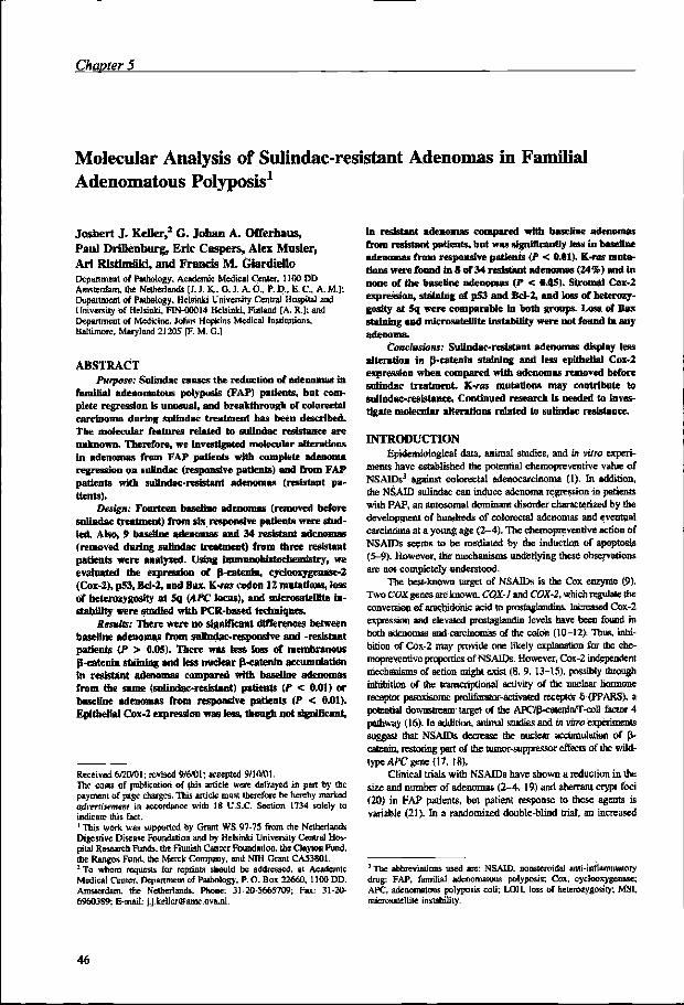
Chapterr 5
Molecularr Analysis of Sulindac-Resistant Adenomas in Familial Adenomatous Polyposis
Josbertt J. Keller, G. Jofaan A. Offerhaus, Paul Drillenburg, Eric Caspérs, Alex Muster, Arii Ristimaki, Francis M. Giardiello
DepartmentDepartment of Pathology, Academic Medical Center, Amsterdam, The Netherlands DepartmentDepartment of Medicine, Johns Hopkins University Hospital, Baltimore, MD, USA DepartmentDepartment of Pathology, Helsinki University Central Hospital, Helsinki, Finland
ClinicalClinical Cancer Research 2001; 7:4000-7
ChapterChapter 5
Molecularr Analysis of Sulindac-resistant Adenomas in Familial Adenomatouss Polyposis1
Josbertt J. Keller,2 G. Johan A. Orferhaus, Paull Drillenburg , Eric Caspers, Alex Musler, Ar ii Ristimaki, and Francis M. Giardiello Departmentt of Pathology, Academic Medical Center, 1100 DD Amsterdam,, the Netherlands [J. J. K., G. J. A- 0„ P. D„ E. C„ A. M-); Departmentt of Pathology. Helsinki University Central Hospital and universityy of Helsinki. FIN-00014 Helsinki, Finland [A. R.]; and Departmentt of Medicine. Johns Hopkin; Medical Institutions, Baltimore,, Maryland 21205 IF, M. G.]
ABSTRAC T T Purpose:Purpose: Sulindac causes the reduction of adenomas in
familia ll adenomatous polyposis (FAP) patients, bot com-pletee regression is unusual, and breakthrough of colorectal carcinomaa durin g sulindac treatment has been described. Hi ee molecular features related to sullndac resistance are unknown.. Therefore, we investigated molecular alterations inn adenomas from FAP patients with complete adenoma regressionn on stiHndac (responsive patients) and from FAP patientss with suSndac-resistant adenomas (resistant pa-tients). .
Design;Design; Fourteen baseline adenomas (removed before Bulindacc treatment) from six responsive patients were stud-ied.. Also, 9 baseHne adenomas and 34 resistant adenomas (removedd durin g sufindac treatment) from three resistant patientss were analyzed. Using mimunwlri^tocbeinistry , we evaluatedd me expression of B-catenin, cydooxygenase-2 (Cox-2),, p53, Bd-2, and Bax. K-ras cedon 12 mutations, loss off heterozygosity at 5q (APC loens), and mkrosateUlte in-stabilit yy were studied with PCR-based techniques.
ResuUs:ResuUs: There were no significant differences between baselinee adenomas from sidrndac-responstve and -resistant patientss (P > 0.05). There was (ess loss of membranous B-cateninn staining and less nuclear B-catenin accumulation inn resistant adenomas compared with baseline adenomas fromm the same (sulindac-resistant) patients (P < 0.01) or baselinee adenomas from responsive patients (P < 0.01). Epitheliall Cox-2 expression was less, though not significant,
Receivedd 6/20/01; revised 9/6/01; accepted 9/10701. Thee costs of publication of this article were defrayed in part by the paymentt of page chaises, This article most therefore be hereby marked advertisementadvertisement in accordance with 18 U.S.C. Section 1734 solely to indicatee this fact. 11 This work was supported by Grant WS 97-75 from the Netherlands Digestivee Disease Foundation and by Helsinki University Centra) Hos-pitall Research Funds, the Finnish Cancer Foundation, the Clayton Fund, thee Ranges Fond, the Merck Company, and NIH Grant CA53801. 22 To Whom requests for reprints should be addressed, at Academic Medicall Center. Department of Pathology. P. O. BOK 22660. 1100 DD, Amsterdam,, the Netherlands. Phone: 31-20-5665709; Fax: 31-20-6960389;; E-mail: j.j,keller<gamc.uvajil.
inn resistant adenomas compared with baseline adenomas fromm resistant patients, but was significantly less in basenue adenomass from responsive patients (P < 0.01). K-ras muta-tionss were found in 8 of 34 resistant adenomas (24%) and In nonee of the baseline adenomas (P < 0.05). Stromal Cox-2 expression,, staining of p53 and Bd-2, and loss of heterozy-gosityy at 5q were comparable in both groups. Loss of Bax stainingg and microsatellite instabilit y were not found in any adenoma. .
Conclusions:Conclusions: Sulindac-resistant adenomas display less alterationn in B-catenin staining and less epithelial Cox-2 expressionn when compared with adenomas removed before suüodacc treatment. K-ras mutations may contribut e to suhndac-resistance.. Continued research is needed to inves-tigatee molecular alterations related to sulindac resistance.
INTRODUCTIO N N Epidemiologicall data, animal studies, and in vitro experi-
mentss have established trie potential chemopreventive value of NSAIDs** against colorectal adenocarcinoma (1). In addition, thee NSAID sulindac can induce adenoma regression in patients withh FAP, as autosomal dominant disorder characterized by the developmentt of hundreds of colorectal adenomas and eventual carcinomaa at a young age (2-4). The chemopreventive action of NSAIDss seems to be mediated by the induction of apoptosis (5-9).. However, the mechanisms underlying these observations aree not completely understood.
Thee best-known target of NSAIDs is the Cox enzyme (9). Twoo COX genes are known, COX-J and COX-2, which regulate the conversionn of arachidonic acid to prostaglandins. Increased Cox-2 expressionn and elevated prostaglandin levels have been found is bod]] adenomas and carcinomas of the colon (10-12), Thus, inhi-bitionn of Cox-2 may provide one likely explanation for the che-mopreventivee properdes of NSAIDs, However, Cox-2 independent mechanismss of action might exist (8, 9, 13-15), possibly through inhibitionn of the iranscripöonal activity of the nuclear hormone receptorr peroxisome proliferator-activated receptor 6 (PPARS), a potentiall downstream target of the APO^caienin/T-cell factor 4 pathwayy (16). m addition, animal studies and in vitro experiments suggestt that NSAIDs decrease die nuclear accumulation of B-calenjn,, restoring part of the tumor-suppressor effects of the wild-typee APC gene (17, 18).
Clinicall trials with NSAIDs have shown a reduction in the sizee and number of adenomas (2-4, 19) and aberrant crypt foci (20)) in PAP patients, but patient response to these agents is variablee (21). In a randomized double-blind trial, an increased
311 The abbreviations used arc: NSAID. nonsteroidal anb-inflamrnalory drug;; FAP, familial adenomatous polyposis; Cox, cyckexygenase; AFC,, adenomatous polyposis coli; LOH. loss of heterozygosity; MSI, mkrosatellitee instability.
46 6

Sulindac-resistantSulindac-resistant adenomas
TableTable 1 Characteristics of patients with adenomas responsive and resistant to sulindac treatment
Responsivee patients 1 1 n n m m IV V V V VI I
Resistantt patients
vn n vm m Df f
Durationn of treatment t
122 mo 366 mo 544 mo 455 mo 388 mo 99 mo
488 mo 300 mo 99 mo
No,, of polyps studied
Pretreauneni i
2 2 2 2 4 4 1 1 2 2 3 3
i i I I 7 7
Fosttreatment t
31 1 1 1 2 2
Age e
32 2 42 2 52 2 24 4 35 5 23 3
36 6 25 5 23 3
Sex x
F F F F F F F F F F F F
M M F F M M
APCAPC mutation
Nott tested Code** 423 Nott tested Segment* * Nott tested Codon54I I
Codoo827 7 Codonn 1061 Codon625 5
Qeorcctall anastomosis/ intactt colon
IRA" " IRA A IRA A IRA A IRA A IRA A
IRA A IRA A Intactt colon
Baselinee polyp count/size e
(mm) )
1972.2 2 47/2J J 10/2.2 2 29/3.5 5 16/2.1 1
una una 80/3,8 8 7/3.1 1
34/4 4
"" IRA, tleoreetal anastomosis. ** Segment 1. codons 1-804 of the APC gene.
numberr of adenomas was noted between 6 and 9 months of sulindacc therapy (2), suggesting the development or selection of resistantt adenomas during long-term sulindac treatment. More-over,, breakthrough cancers in the rectal stump during sulindac cternopreventivee therapy have been reported (22r-24). This lit -eraturee highlights potential limitations of NSAJJBis as chemopre-ventivee agente, and the need for hiomarfcers. which predict resistancee to chemopreventive treatment Recent in vitro data showw that the loss of functional Bax results in resistance for NSAlD-indueedd apoptosis (25). In addition, K-im-trahsforroed cellss are relatively refractory to stuindac-toduced apoptosis (26). Thesee studies suggest that the lack of adenoma regression on sulindacc may be related to SAX or K-ras mutations.
Thiss study describes the iimnunobistochemical and molec-ularr features of adenomas removed at baseline (before treatment withh sulindac) from patients with complete polyp regression on sulindacc (responsive patients) and adenomas removed at base-linee and during sulindac treatment from individuals with sulin-dac-resistantt adenomas (resistant patients). We investigated al-terationss possibly involved in the mechanisms underlying NSAJD-inducedd chemoprevention of colorectal carcinoma, in-cludingg B-catenin expression, LOH at the APC locus 5q, and Cox-22 expression, or potentially related to resistance, such as K-rasK-ras mutations and the loss of Bax expression. In addition, apoptosis-relatedd protein expression was assessed (p53 and Bcl-2).. MSI was studied, because a relationship seems to exist betweenn MSI and low Cox-2 expression (27).
MATERIAL SS AND METHOD S Studyy Population. The study population consisted of
ninee Caucasian FAP patients, of whom eight had undergone a colectomyy and ileorectal anastomosis. Patient characteristics are shownn in Table 1. Al l patients had adenomas in the rectum and weree treated with sulindac ISO mg p.o. twice a day for at least 99 months. Informed consent was obtained in accordance with approvall by the Johns Hopkins University Joint Committee on Clinicall Investigation (Institutional Review Board). Medication compliancee was assessed by pill count, and all patients took >80%% of the scheduled doses. Patients underwent flexible sig-moidoscopyy at baseline and every 3 months, using an Olympus flexiblee sigmoidoscope after preparation with a clear liquid diet
andd oral cathartic solution. Enemas that could influence mucosal biochemistryy were not given. Biopsies taken from adenomas at baselinee (before sulindac treatment) and during sulindac treat-mentt were formalin fixed, paraffin embedded, and H&E stained forr histological examination. Baseline adenomas from six PAP patientss with complete polyp regression during treatment with sulindacc (responsive patients) were studied (patients I-VI , Table 1).. Three of the nine patients (patients VII-IX , Table 1) showed incompletee or no response to treatment with suHndac (resistant patiënte).. Adenomas from those individuals, removed before treatmentt with sulindac (baseline) and daring treatment with sulindacc (resistant adenomas) were included. Adenomas from differentt groups were matched for size, architecture, and degree off dysplasia.
Responsivee Patients. The control group consisted of 14 adenomass from the rectal stump of six patients (patients 1-VT) withh complete regression of adenomas within 6 months of treatmentt with sulindac. Adenomas removed before treatment withh sulindac (baseline) were studied.
Resistantt Patients. Patient VTJ had a baseline sigmoid-oscopyy (before drug treatment) revealing 80 adenomas with an averagee size of 3.8 mm in the rectal stump. Initially, sulindac administrationn induced a regression in both number and size of adenomas.. However, after 2 years on drug treatment, the num-berr of adenomas began to increase. After 4 years of sulindac therapy,, this patient was withdrawn from the trial because of accelerationn of adenoma development and referred for surgical removall of the rectal stump to prevent rectal cancer. From patientt VH, one adenoma removed before treatment with sulin-dacc was studied. In addition, 31 adenomas from seven randomly chosenn time points between 5 months and 4 years of treatment withh sulindac were analyzed (resistant adenomas).
Patientt VHI initially had seven adenomas with an average sizee of 3.1 mm in the rectal stump. After 3 months of sulindac treatment,, there was complete adenoma regression. At 2 years of sulindacc treatment, an ulcerative lesion was seen in the rectal stumpp at sigmoidoscopy. Histological examination at the ulcer marginn revealed a tubular adenoma, which was analyzed in the presentt study (resistant adenoma). One adenoma removed be-foree treatment with sulindac was studied.
Patientt IX had an intact colon and was treated with sulin-
47 7

ChapterChapter 5
dacc for 9 months. At baseline, 34 adenomas with an average size off 3 mm were noted. Sulindac did not affect number and size of thee adenomas. Seven rectal adenomas removed before treatment withh sulindac and two adenomas removed after 3 and 6 months off treatment with sulindac (resistant adenomas) were analyzed.
Tissuee Preparation. Formalin-fixed, paraffin-embedded sampless were cut into 5-ujn sections and mounted on glass slides.. For DNA isolation, slides were stained with hematoxylin, andd dysplastic and normal epithelium were microdissected. A standardd proteinase-K digestion was used lo isolate DNA.
Immunohistocfaemistryy for pVCatenin; Cox-2; p53; Bcl-2;; and Bax. Immunohistochemistiy was performed on unstainedd 5-p.m sections as described previously (28). For an-tigenn enhancement, the slides were submerged in citrate buffer andd heated for 10 min at 100"C, Immunostaining was done usingg primary monoclonal antibodies against B-catenin, clone 144 (Transduction Laboratories, Lexington. KY), at a dilution of 1:1000;; Cox-2, no. 160112 (Cayman Chemical. Ann Arbor, MI) ,, at a dilution of 1:100: p53, D07 (Dako. Glostrup, Den-mark),, at a dilution of 1:200; Bcl-2, clone 124 (Dako), at a dilutionn of 1:12.5; and against Bax, die polyclonal rabbit IgG p-199 (Santa Cruz Biotechnology. Santa Cruz, CA), at a dilution off 1:100. Primary antibodies against B-catenin and Cox-2 were incubatedd overnight at 4°C; incubation with antibodies against p53,, Bcl-2, and Bax was done for 1 h at room temperature. Primaryy antibodies were replaced by PBS (137 mM NaCl, 2.7 DIMM KC1, 10 mM Na2HP04. and 1.8 mM KHaPOj in negative controll slides. A known p53-positive colorectal carcinoma was usedd as a positive control for pS3 staining. The staining pattern inn the adjacent normal mucosa was used as a marker for the specificc staining of fl-catenin, Cox-2, Bcl-2, and Bax. To assess thee specificity of the Cox-2 antibody, a subset of adenomas was stainedd with the primary antibody after preadsorption of a hu-mann Cox-2 control peptide (Cayman Chemical) for 1 h at room temperature.. This resulted in me blocking of the Cox-2 staining patternn described below.
Immunostainedd slides were scored by two observers (G.. J. A. O. and J. J. K.) in a coded fashion. For fi-caienih, membranous,, cytoplasmic, and nuclear staining were assessed separately.. Membranous and cytoplasmic staining were com-paredd with the normal mucosa and assessed as decreased ex-pressionn (loss) or overexpiession, respectively; nuclear staining (absentt in normal tissue) was scored seraiquantiiatively using a scalee from 1 to 4 (1, no expression; 2, <5% positive nuclei; 3, <25%% positive nuclei; and 4, >25% positive nuclei). Cox-2 stainingg 'm the stroma underlying the epithelium and in the epitheliumm was assessed separately in a semiquantitative manner onn a scale from 1 to 4 (1. no expression; 2, weak staining; 3, moderatee staining; and 4. intense staining). p53 immunostaining wass considered positive when >10% of nuclei stained positive (29).. Bel-2 staining was compared with the normal mucosa for assessmentt of overexpression, and Bax-stained adenomas were evaluatedd for loss of expression, lmmunohistochemical staining wass completely absent in one responsive adenoma, possibly becausee of a fixation error. Also, there was limited tissue avail-ablee from another responsive adenoma, precluding assessment off Bax and Cox-2 staining.
K-Rass Codon 12 Analysis. K-ras codon 12 analysis was performedd as described previously (30). DNA samples were
usedd for amplification of K-ras codon 12-specific sequences by PCR.. PCR products were then digested with Mval. which only recognizess wild-type K-ras codon 12. Subsequently, a second-roundd PCR was performed on both the digested and undigested first-roundd PCR products. After denaturation, the undigested andd digested (mutant-enriched) PCR products were spotted onto aa nylon membrane and hybridized to each of the K-ras codon 12 mutation-specificc oligodeoxynuCleotides. Final stringency washess were carried out at 63°C before autoradiography. K-ras codonn 12 mutational analysis was performed twice, in indepen-dentt experiments.
LO HH and MSI Analysis. Analysis of LOH and screen-ingg for MSI was done as described previously (3D, comparing microdissectedd tumor tissue with normal tissue from the same patient.. LOH analysis of the APC locus at chromosome 5q was performedd with the markers D5S82. D5S107. and D5S346. MSI wass assessed with the B AT-26 marker.4 Cycling was performed inn a PTC 100 cycler (MJ Research, Inc.. Waltham, MA), and die PCRR products were analyzed using an automated ABB77 se-quencerr and the Genescan 2.1 software (PE Biosystems, Foster City.. CA).
Statistics.. Nonparametric tests were used. Comparisons betweenn groups were made by Mann*Whitney test for nuclear B-cateninn staining and epithelial and stroma) Cox-2 expression. TheThe nonparametric Fisher's exact test was used for analysis of differencess in B-catenin (toss of membranous staining and over-expressionn of cytoplasmic staining), Cox-2 (moderate/strong comparedd with weak/absent staining), p53, Bcl-2, K-ras muta-tions,, and 5q LOH. The relationship between membranous and nuclearr B-catenin staining was analyzed by Mann-Whitney test. Thee relationship between stromal and epithelial Cox-2 staining wass evaluated by Spearman's rank correlation test A P <0.05 wass considered statistically significant. Al l Ps were two-sided.
RESULTS S Al ll adenomas were tubular or tubulovillous lesions, <1
cm,, with mild to moderate dysplasia. No morphological differ-encess were noted between adenomas from responsive patients (nn = 14) and adenomas from resistant patients removed before (nn = 9) or during (w = 34) treatment with sulindac. The results off inmiunohistocheraistry (Fig. 1) for B-catenin, Cox-2, p53, Bcl-2,, and Bax, K-ras codon 12 mutational analysis (Fig. 2) and 5qq LOH and MSI are listed in Table 2.
Baselinee Adenomas from Sulindac-responsjve and Sulindac-resistantt Patients. There were no significant differ-encess in any molecular parameter analyzed in baseline adeno-mass (removed before treatment wim sulindac) from sulindac-responsivee patients compared with sulindac-resistant patients (P(P > 0.05; Table 2). In general, baseline adenomas displayed reducedd membranous S-catenin staining compared with the nor-mall mucosa and an accumulation of cytoplasmic and nuclear B-cateninn (Fig. 1,^-B) in accordance with literature reports (32, 33).. Very littl e Cox-2 staining was found in the normal mucosa. Thee Cox-2 staining pattern in adenomas (Fig. 1, £*-£) was
44 For primer sequence see the Genome Database: Internet address: htip://wwww .gdb.org-
48 8

Sulindac-resistantSulindac-resistant adenomas
Fig.Fig. I Immunohistochemical scainingg of P-catenin. Cox-2, p53.. bcl-2, and bax. d, dysplaslic epithelium:: n. nonnal rectal epi-thelium.. A-C. p-catenin staining, withh nuclear accumulation of P-cateninn and loss of membra-nouss staining in dysplaslic epi-theliumm Id) compared with adjacentt normal epithelium (n) inn baseline adenomas from sulindac-rcsponsivee (A) and -resistantt (B) patients. C. exam-plee of .sulindac-resistant ade-noma,, with strong membranous stainingg and very few nuclear P-cateninn in dysplastic crypts (d).(d). D-F, Cox-2 staining, with epitheliall and stromal (arrow) expressionn in baseline adenomas fromm sulindac-responsive (D) andd -resistant (£) patients. F, ex-amplee of a sulindac-resistant ad-enoma,, with stromal Cox-2 ex-pression.. G-l, Bax expression inn baseline adenomas from sulindac-responsivee (G) and -resistantt <lh patients, and in aa sulindac-resistant adenoma (/).. /. nuclear overexpression off p53 in dysplastic epithe-lium.. K, overexpression of bcl-22 in dysplaslic epithelium (d)(d) compared with adjacent normall epithelium (n).
similarr to that described by Chappie el al. (12), i.e., increased epitheliall staining and stromal staining beneath dysplastic sur-facee epithelium. In baseline adenomas, there was a positive correlationn between stromal and epithelial staining (P = 0.003). Althoughh epithelial Cox-2 overexpression seemed more pro-nouncedd in baseline adenomas from sulindac-responsive pa-tientss compared with baseline adenomas from sulindac-resistant patientss (P = 0.06). the frequency distribution of Cox-2 as-sessedd as moderate or strong (S3) compared with negative or weakk was not significantly different in both groups (P — 0.12). Stromall Cox-2 expression, positive staining for p53 (Fig. 1/), Bcl-22 overexpression (Fig. IK), and LOH at the APC-locus 5q weree comparable in both groups. Loss of Bax expression (Fig. 1, G-H),G-H), K-ras codon 12 mutations, and MSI were not found in anyy baseline adenoma.
Sulindac-resistantt Adenomas. Loss of membranous S-cateninn expression was found in 13 of 34 (38%) sulindac-resistantt adenomas, which was significantly less than in baseline adenomass from the same patients (88%: P = 0.001) and base-linee adenomas from responsive patients (85%; P = 0.008; Fig. 1,, A-C). In addition, there was significantly less nuclear accu-mulationn of B-catenin in resistant adenomas compared with baselinee adenomas from the same patients (P < 0.001) and baselinee adenomas from responsive patients (P = 0.002; Fig. 1, A-C).A-C). Cytoplasmic B-catenin staining was present in 33 of 34 (97%)) resistant adenomas and did not differ from baseline adenomas. .
Theree was less epithelial Cox-2 expression (Fig. 1, D-F) in resistantt adenomas compared with baseline adenomas from the samee patients, however, this difference was not significant (P = 0.32).. There was significantly less epithelial Cox-2 expression inn sulindac-resistant adenomas than in baseline adenomas from responsivee patients (P = 0.002). Moderate or strong epithelial Cox-22 staining (assessed as s3) was found in 13 of 34 (38%) resistantt adenomas compared with 5 of 9 (55%) baseline ade-nomass from resistant patients (P = 0.46) and 11 of 12 (92%) baselinee adenomas from responsive patients (P = 0.002). There wass also less stromal Cox-2 staining (Fig. 1, D-F) in resistant adenomass compared with baseline adenomas, however, the dif-ferencee was not significant (P = 0.14 when compared with baselinee adenomas from the same patients: and P = 0.098 when comparedd with baseline adenomas from responsive patients). Theree was a positive relationship between epithelial and stromal Cox-22 expression in resistant adenomas (P = 0.045). as was alsoo found in the baseline adenomas (see above).
Resistantt adenomas showed positive staining for p53 (Fig. 1/)) and overexpression of Bcl-2 (Fig. \K). comparable with baselinee adenomas from resistant and responsive patients. Loss off Bax expression (Fig. 1, G-l) was not found in resistant adenomas.. K-ras codon 12 mutations were found in 8 of 34 (24%)) resistant adenomas (Fig. 2). whereas no mutations were foundd in the nine baseline adenomas from the same patients (ƒ»» = 0.17) nor in the 14 baseline adenomas from responsive patientss (P = 0.09). When baseline adenomas from resistant and

ChapterChapter 5
WTT D 1
Fig.Fig. 2 Autoradiogram of K-ras codon 12 mutation analysis. Displayed aree the wild-type oligonucleotides (WT). the D mutation-specific oligo-nucleotidess (valine mutation: D). and the E mutation-specific oligonu-cleotidess (aspartic acid mutation: £). From each sample, nonenriched DNAA (left) and mutant-enriched DNA (right) are hybridized next to eachh other in a paired way. Each row contains three pairs of spots/ columnn with DNA from three different samples. Row I, hybridization controls,, DNA complementary to the labeled oligonucleotide. The eight detectedd mutations (6 D mutations and 2 E mutations) and a positive controll (marked with /) are shown as paired spots. DNA was replaced byy HaO as a negative control (blank spot in WT column marked with 2).
responsivee patients were grouped (n = 23). there was a statis-ticallyy significant difference in the K-ras codon 12 mutation rate betweenn resistant adenomas (8 of 34) and baseline adenomas (0 off 23; P = 0.02). LOH at the APC locus 5q was found in 7 of 322 (22%) informative resistant adenomas. LOH was found in resistantt adenomas with and without nuclear accumulation of P-catenin.. MSI was not found in resistant adenomas.
DISCUSSION N Althoughh NSAJDs have chemopreventive activity against
colorectall cancer, literature reports reveal that these agents do nott completely prevent the occurrence of colorectal carcinoma. Thiss emphasizes the importance of understanding the mecha-nismss of action of these drugs and identifying biomarkers, whichh predict resistance to chemopreventive treatment.
Thee present study found less loss of membranous p-cate-nin.. less nuclear accumulation of P-catenin, and less epithelial Cox-22 in sulindac-resistant adenomas compared with adenomas fromm the same patients, removed before treatment with sulindac (baseline),, and baseline adenomas from patients who showed a completee response after sulindac administration. In addition, K-rasK-ras codon 12 mutations were only noted in sulindac resistant adenomas.. These findings may indicate a role for these factors inn the mechanism underlying NSAID-induced chemopreven-tion.. Alternatively, our results might reflect a distinct molecular make-upp of sulindac-resistant tumors. The timing of the occur-rencerence of sulindac-resistant adenomas seems unpredictable, inas-muchh as two of three sulindac-resistant patients were initially in
completee remission during the first years of treatment. The resultss need to be interpreted with caution because of the small numberss of resistant patients from whom adenomas were avail-ablee for study. The vast majority of resistant adenomas are from onee patient, and potential confounding factors related to this patientt cannot be excluded. Also, our study design precludes definitivee conclusions between the possibilities that the ob-servedd differences are caused by treatment with sulindac or are relatedrelated to sulindac resistance. Ideally, individual polyps could bee marked and biopsied before and during sulindac therapy, comparingg molecular alterations in polyps that ultimately re-gresss to those that remain. Barriers to this methodology in FAP includee the small size of adenomas and the difficulty in obtain-ingg patient consent for such a protocol.
Noo significant differences were found between baseline adenomass from resistant patients and those with a complete responsee on sulindac. Thus, the studied markers could not predictt which patients would completely respond to treatment withh sulindac. The molecular alterations of baseline adenomas fromm sulindac-responsive and -resistant FAP patients were con-sistentt with previously reported alterations in FAP-related ade-nomas.. Loss of membranous P-catenin and translocation of P-cateninn to the nucleus has been described in colorectal ade-nomass and carcinomas (32-34). Indeed, most baseline adeno-mass showed reduced membranous B-catenin staining and nu-clearr accumulation of B-catenin. Interestingly, there was less losss of membranous B-catenin and less nuclear B-catenin in sulindac-resistantt adenomas.
FAPP patients have an inherited mutation of the APC gene. Normally,, wild-type APC regulates the degradation and nuclear exportt of B-catenin (35, 36), which is mainly localized at the celll membrane. Inacrivarion of APC results in increased cyto-plasmicc levels, loss of membranous expression, and nuclear accumulationn of B-catenin. Nuclear B-catenin binds to T-cell factorr 4 and activates the transcription of different oncogenic factorss (35). Animal studies and in vitro experiments have addressedd overexpression and nuclear translocation of B-catenin ass potential targets for the anticarcinogenic action of NSAIDs. Mahmoudd el al. (7) showed decreased expression of p-catenin afterr treatment with sulindac in the normal intestinal mucosa of ApcApcMm/+Mm/+ mice, a murine model of FAP. and Oshima et al. (37) showedd an increased membrane-bound fraction of p-catenin in adenomass of Ape*116 mice after 8 weeks of treatment with sulindac,, whereas no change in nuclear accumulation was noted. Inn addition. McEntee et al. (38) reported decreased nuclear and cytoplasmicc P-catenin expression in the small intestinal adeno-mass of ApcM,D/* mice after treatment with sulindac for 2-4 days,, but not after 20 days treatment. Adenomas which persist afterr 20 days treatment with sulindac could be considered re-sistant.. We found less nuclear p-catenin in sulindac-resistant adenomas,, whereas nuclear accumulation of p-catenin was un-alteredd in sulindac-resistant adenomas of Apc!Mn/+ mice and Apc*Apc*7i67i6 mice (37, 38). This discordance might reflect differ-encess between rectal adenomas in human FAP and small intes-tinall adenomas in these mice models. Recently. Brown et al. (17)) reported decreased nuclear accumulation of P-catenin in carcinogen-inducedd tumors in rats after treatment with sulindac, supportingg our present findings. Also, in vitro experiments have shownn that the NSAID indomethaein decreases nuclear P-cate-

Sulindac-resistantSulindac-resistant adenomas
TabUTabU 2 Molecular alterations in baseline and sulindac-resistanl adenomas Molecularr alterations in adenomas removed before freatment with sulindac (baseline) from patients with a complete response on sulindac (A)
andd sulindac-resistant patients (B) and in sulindac-resistant adenomas (C). Nuclear p-cateaia, and epithelial and stromal Cox-2 expression were assessedd in a semiquantitative maimer, and the median and range (lowest and highest scores) are presented.
Nuclearr fJ-catcain Epitheliall Cox-2 Stromall Cox-2
Membranouss p-catenin decreased Cytoplasmicc -catenin overexpression p533 overexpression Bel-22 overexpression Baxx loss of expression K-rasK-ras codon 12 mutation LOWW at 5q (APC locus) MSII (Bat-26)
A:: (n = 14)" Baselinee adenomas
Responsivee patients
Mediann (range)
3(2-*) ) 4(1-4) ) 3(1-*) )
n/nn/n tested (%)
11/133 (85%) 12/133 (92%) 5/133 (38%)
11/13(85%) ) 0/12 2 0/14(0%) ) 2/14(14%) ) 0/14 4
B:: (» = 9f Baselinee adenomas
Resistantt patients
Mediann (range) 3(3-4) ) 33 0-4) 3 (M ) )
n/nn tested (%)
9/9(100%) ) 7/99 (77%) 2/9(22%) ) 679(66%) ) G/9 9 0/9 9 1/2(50%) ) 0/9 9
C:: 0t = 34) Resistantt adenomas
Resistantt patients
Mediann (range)
22 0 ^ ) 2 ( M ) ) 3(l^t ) )
nfnnfn tested (%)
13/34(38%) ) 33/344 (97%) 6/34(18%) )
25/344 (74%) 0/34 4 8/34(24%) ) 7/322 (22%) 0/34 4
AA compared withC C
P P
PP = 0.002 p=aoo2 2 PP = 0.098
P P
PP = 0.008 PP = 0.48 PP = 0.25 PP = 0.70
PP = 0.09* PP = 0.70
BB compared withC C
P P PP < 0.001 PP = 0.32 PP = 0.14
P P
PP = 0.001 PP = 0.1I P== 1.00 PP = 0.69
PP = 0.17* PP = 0.42
"" There were no statistically significant differences in any molecular parameter between A and B (P > 0.05). ** Overall, the difference m K-rt o mutation r&te between bas e
andd resistant adenomas was statistically significant P = 0.02.
no)) expression in colorectal cancer ceil lines (18). Taken to-gether,, the translocation ofp-cateron. caused by the toss of APC tumor-suppressorr activity, might be a target of NSAIDs. possi-blyy by enhancing the nuclear export óf p-calenin through mech-anismss that interact with tt e nuclear export receptor CRMI (36).
Lesss epithelial Cox-2 was found in sulindac-resistant ade-nomass compared with baseline adenomas. Cox-2 expression can bee reduced by NSAIDs in vitro (39), and sulindac decreases the expressionn of Cox-2 in the normal mucosa of ApcMio/ + mice (40).. Our findings suggest that this effect also occurs in human adenomass of FAP patients treated with sulindac. Although di-minished,, nuclear [J-catenin and Cox-2 expression were still presentt in sulindac-resistant adenomas. Resistance to sulindac mightt be caused by failur e of this NSAID to completely abolish thesee oncogenic stimuli. Alternatively , sulindac therapy may be unablee to counteract the effect of an alternative (sulindac-resist-ant)) tumor progression pathway not dependent on Cox-2 and/or nuclearr pVcatenin signaling.
Severall molecular alterations potentially involved in sulin-dacc resistance were investigated1. Mutant p53 was found in both groups,, supporting in vitro findings that p53 is not involved in NSAJD-inducedd chemopreveniioa (8). Recently, BAX mutations havee been associated with sulindac resistance in vifr o (25). We foundd Bax expression in all (resistant) adenomas studied. How-ever,, irnmunohlstocbemistry might fail to detect loss of func-tionaltional Bax protein. Arber et al (26) showed that K-ras-trans-formedd cells were relatively resistant to sulmdac-induced apoptosis.. In this study, 8 K-ras codon 12 mutations were found inn 34 sulindac-resistant adenomas (24%), whereas those muta-tionss were not found in baseline adenomas from either sulindac-resistantt or -responsive patients. K-ras codon 12 mutations accountt for the majorit y of ras mutations in human colorectal carcinogenesiss and occur in <10% of small colorectal adeno-mass (<1 cm; Ref. 41). Thus, die frequency of K-ras codon 12
mutationss in sulindac-resistant adenomas was higher than ex-pected.. In addition, when baseline adenomas from sulindac-resistantt and -responsive patients were grouped, a significant differencee tn the K-ra* mutation rate between baseline and resistantt adenomas was noted, suggesting a role for K-ras mu-tationss in sulindac resistance. Our study does not determine whetherr the observed differences are caused by sulindac or are ann underlying cause of solmdac resistance. However, it is un-likelyy that sulindac treatment would induce ras mutations. More probably,, K-ras mutations constitute a point of no return beyond whichh chemopreventive treatment with NSAIDs is ineffective. Off interest wil l be die effect of combinatorial chemopreventive therapy;; in this regard, the work of Torrance ex al (42), describ-ingg almost complete adenoma regression in ApcMiB/~'' mice after treatmentt with a combination of sulindac and a specific epider-mall growth factor receptor kinase inhibitor seems promising.
I ss summary, our results show less oncogenic activation of p-cateninn and less Cox-2 in sulindac-resistant adenomas. One explanationn for these findings is that nuclear p-catenin accumu-lationn and Cox-2 expression are decreased with sulindac treat-ment.. However, resistant adenomas may occur from limited suppressionn of these factors caused by drug delivery and/or involvementt of an alternative (sulindac-resistant) tumor pro-gressionn pathway. K-ras mutations might be involved in sulin-dacc resistance. Additional research is needed to elucidate such ann alternative tumor progression pathway and to investigate whetherr nuclear p-catenin staining, epithelial Cox-2 overexpres-sion,, and K-ras codon 12 mutations can be useful biomarkers.
ACKNOWLEDGMENT S S Wee thank Linda Bylind for her help collecting tissue specimens.
WUfriedd Meun for his help preparing the figures, Prof. Willem Lam-menss van Bueren far statistical advice, and Marian Weterman for criticall reading of the manuscript
51 1
ChapterChapter 5
REFERENCES S 1.. Giardiello. F. M.. Offerhaus, G. I . and DuBois, R. N. The role of nonsteroidall anti-inflammatory drugs in colorectal cancer prevention. Eur.. J, Cancer, 31A: 1071-1076, 1995. 2.. Giardiello, F. M., Hamilton, S. R.. Krusn, A, J.. Piantadosi, S„ Hylind,, L. M.. Cclano. P., Booker. S. V.. Robinson. C. R„ and Offer* haus.. G. J. Treatment of colonic aw) rectal adenomas with sulindac In familiall adenomatous polyposis. N. Engl. J. Med., 328: 1313-1316. 1993. . 3.. Nugent, K. P., Farmer. K. C, Spigelman, A. D-, Williams. C. B-, and Phillips,, R, K. Randomized controlled trial of the effect of sulindac on duodenall and rectal polyposis and cell proliferation in patients with familiall adenomatous polyposis. Br. J. Surg.. 80: 1618-1619. 1993. 4.. Winde, G.. Schmid. K. W.. Brandt. B., Muller. O., and Osswald, H. Clinicall and genomic influence of sulindac on rectal mucosa m familial adenomatouss polyposis. Dis. Colon (tectum, 40: 1156-1169, 1997. 5.. Pasricha, P. J.. Bedi, A.. O'Connor. JC, RashkL A., Akhtar. A. J.. Zahurak,, M. L.. Piantadosi, S., Hamilton, S. R.. and Giardiello, F. M. Thee effects of sulindac on colorectal proliferation and apoptosis in familiall adenomatous polyposis. Gastroenterology, 109: 994 998. 1995. . 6.. Keller. J. J,. Offerhaus, G. J., Polak, M-, Goodman, S. N„ Zahurak. M.. L„ Hylind, L. M.. Hamilton. S. R., and Giardiello, F. M. Rectal epitheliall apoptosis in familial adenomatous polyposis patients treated withh sulindac. GuL 45: 822-888. 1999. 7.. Mahmoud. N. N„ Boolbot, S. K„ Bilinski. R. T„ Martucci. C, Chadbum,, A., and Bcrtagnolli. M. M. Ape gene mutation is associated withh a dominant-negative effect upon intestinal cell migration. Cancer Res... 57: 5045-5050, 1997. 8.. Piazza, G. A„ Rahm. A. K, Finn, T. S„ Fryer. B. H.. Li, H., Stoumen.. A. L., Pamukcu, R.. and Ahnen, D. J, Apoptosis primarily accountss for toe growth-inhibitory properties of sulindac metabolites andd involves a mechanism that is independent of cyclooxygenase inhi-bition,, cell cycle arrest, and p53 induction. Cancer Res., 57:2452-2459, 1997. . 9.. Stuff, S. J., and Rigas, B The role of cyclooxygenase inhibition in thee antineoplastic effects of nonsteroidal antiinflammatory drugs WSAID».. J. Exp. Med.. 190: 445-450. 1999. 10.. Eberhart, C. E., Coffey, R. J:, Radhika, A„ Giardiello, F. M., Ferrenbach.. S., and DuBois. R. N. Up-regulation of cyclooxygenase 2 genee expression in tinman colorectal adenomas and adenocarcinomas. Gastroenterology.. 107: 1183-1188, 1994. ]J.. Yang, V. W„ Shields, J. M.. Hamilton, S. R.. Spaunhake. E. W.. Hubbard.. W. C. Hylind. L. M.. Robinson. C. R., and Giardiello. F. M. Size-dependentt increase in prostanoid levels in adenomas of patients withh familial adenomatous polyposis. Cancer Res.. 58: 1750-1753, 1998. . 12.. Chappie, K. S„ Cartwright. E. J., Hawcroft. G.. Tisbury. A.. Boni-fer,, C, Scott, N., Windsor. A. C. Guiltou, P. J.. Markham. A. F.. Coletta,, P. L.. and Hull. M. A. Localization of cyclOoXygenase-2 in humann sporadic colorectal adenomas. Am. J. Pathol., 156: 545-553. 2000. . 13.. Thompson, H- J., Jiang, C, Lu, J., Mehta, R. G.. Piazza, G. A.. Paranka.. N. S., Pamukcu, R„ and Ahnen. D. J. Sulfonc metabolite of sulindacc inhibits mammary carcinogenesis. Cancer Res.. 57: 267-271. 1957. . 14.. Chiu. C. H.. McEntee. M. F.. and Wheian, J. Sulindac causes rapid regressionregression of preexisting tumors in Min/+ mice independent of pros-taglandinn biosynthesis. Cancer Res.. 57: 4267-4273. 1997. 15.. Hanif, R., Pittas, A.. Feng, Y.t Koutsos. M. I.. Qiao, L.. Staiano-Coico,, L.. Stuff, S. L and Rigas, B. Effects of nonsteroidal anti-inflammatoryy drugs on proliferation and on induction of apoptosis in colonn cancer cells by a prostaglandin-independcnt pathway. Biochem. Pharmacol.. 52: 237-245. 1996. 16.. He, T. C, Chan. T. A., Vogelstem, B., and Kinzlcr, K. W. PPAR& iss an APC-regutated urge! of nonsteroidal anti-inflammatory drugs. Cell,, 99: 335-345, 1999.
17.. Brown. W. A.. Skinner. S. A., Vogtagis, D„ and O'Brien, P. E. Inhibitionn of B-catenin translocation—a novel explanation for the pro-tectivee effect of NSAlDs to colorectal cancer. Gastroenterology, 118: A4867.. 2000. IS.. Smith, M. L-, Hawcroft G.. and Hull M. A. The effect of non-steroidall anti-inflammatory drugs on human colorectal cancer cells: evidencee of different mechanisms of action. Eur. J. Cancer, 36: 664-674,, 2000. 19.. Giardiello. F. M.. Spannhake, E. W., DuBois. R. N.. Hylind. L. M.. Robinson,, C. R., Hubbard, W. C, Hamilton. S. R., and Yang, V. W. Prostaglandinn levels in human colorectal mucosa: effects of sulindac in patientss with familial adenomatous polyposis. Dig. Dis. Sci„ 43: 311-316,, 1998. 20.. Sterabach, G„ Lynch. P. M. Phillips, R, K., Wallace, M. H„ Hawk, E.,, Gordon, G. B„ Wakabayashi, N.,Saunders. B.. Shea, Y„ Fujimura. T.TT Su. L. K-, and Levin, B. The effect of celecoxib. a cyclooxygenase-2 inhibitor,, in familial adenomatous polyposis: N. Engl. J. Med. 342: 1946-1952.. 2000. 21.. Takayama, T.. Katsuki, S., Takahashi, Y„ Ohi, M„ Nojiri, S-, Sakamaki,, S., Kato, J.. Kogawa. K„ Miyake, H„ and Niitsu. Y. Aberrant cryptt foci of the colon as precursors óf adenoma and cancer. N. Engl. J.. Med.. 339: 1277-1284. 1998. 22.. Giardiello, F. M., Offerhaus. J. A., Tersmette. A. C, Hylind. L. M.. Krush,, A. J., Brcnsingcr. J. D., Booker, S. V., and Hamilton. S. R, Sulindacc induced regression of colorectal adenomas in familial adenom-atouss polyposis: evaluation of predictive factors. Gut, 38: 578-581. 1996. . 23.. Thorson, A. G„ Lynch, H. T,. and Srayrk, T. C. Rectal cancer in FAPP patient after sulindac. Lancet, 343: 180, 1994. 24.. Niv, Y.. and Fraser. G. M. Adenocarcinoma in the rectal segment in familiall polyposis coli is not prevented by sulindac therapy. Gastroen-terology,, 107: 854-857. 1994. 25.. Zhang, L., Yu, J„ Park, B. H.. Kinder. K. W„ and Vogelstem, B. Rotee of BAX in the apoptotlc response to anticancer agents. Science (Wash.. DC), 290: 989-992. 2000. 26.. Arber, N., Han, E. K., Sgambalo. A.. Piazza. G. A., Delohery. T.. M., Begeraann. M,. WeghoxsL C. M„ Kim. N. H., Pamukcu, R.. Ahnen,, D. J., Reed, J, C. Weinstein, L B., and Holt, P. R. A K-rax oncogenee increases resistance to sulindac-induced apoptosis in rat en-tcrocytes.. Gastroenterology, 113: 1892-1900, 1997. 27.. Karnes, W. E.. Jr., Shattuck-Brandt, R,. Burgart. L. J.. DuBois. R.. N., Tester. D. J„ Cunningham. J. M., Kim. C. Y.. McDonnell, S. K.. Schaid,, D. J., and Thibodeau. S. N. Reduced COX-2 protein in colo-rectall cancer with defective mismatch repair. Cancer Res.. 55: 5473-5477.. 1998. 28.. Entius, M. M., Keller, J. J., DriUenburg. p„ Kaypers, K. C-. Gi-ardiello,, F. M-, and Offerhaus, G. J. Microsatellite instability and expressionn of hMLH-1 and hMSH-2 in sebaceous gland carcinomas as markerss for Muir-Torre syndrome. Clin. Cancer Res.. 6: 1784-1789. 2000. . 29.. Baas. 1. O.. Mulder, J. W.. Offerhaus. G. J., Vogelstem, B.. and Hamilton.. S. R. An evaluation of six antibodies for imniunohistochem-istryy of mutant p53 gene product in archival colorectal neoplasms. J.. Pathol.. 172: 5-12, 1994. 30.. Entius, M. M.. Wesierman. A. M., Giardiello, F. M.. van Velthuysen,, M. L„ Polak, M. M., Slebos, R. I , Wilson. 1. H.. Hamilton, S.. R-, and Offerhaus, G. J. Peutc-Jeghers polyps, dysplasia, and K-ras codonn 12 mutations. Gut 41: 320-322. 1997. 31.. van Rees, B. P.. Clcfon-Janscn, A. M„ Cense, H. A., Polak. M. M.. Clement.. M. I.. DriUenburg, P.. van Lanschot. J. J„ and Offerhaus. G. J. Molecularr evidence of Meld cancerization in a patient with 7 tumors of thee aerodigestive tract Hum. Pathol.. 31: 269-271. 2000. 32.. Inomata, M., Ochiai. A, Aki moto, 5., Kilano, S.. and Hirohashi, S. Alterationn of f -eatenin expression in colonic epithelial cells óf familial adenomatouss polyposis patients. Cancer Res.. 56: 2213-2217. 1996. 33.. Hao. X., Tomlinson, I., llyas, M., Palazzo, J. P., and Talbot, I. C. Reciprocityy between membranous and nuclear expression of B-catenin inn colorectal tumours. Virchows Arch., 431: 431: 167-172, 1997.
52 2

Sulindac-resistantSulindac-resistant adenomas
34.. Hugh, T. J.. Dillon. S. A., O'Dowd, G„ Getty, B-, Kgnatelli , M.. Poston,, G. J., and Kinselta, A. R. fi-Catenin expression in primar y and metastaticc colorectal carcinoma. Int . J. Cancer, 82: 504-511, 1999.
35.. Morin . P. J„ Sparks. A. B„ Korinek . V„ Barker, N„ Clevers, H., Vogclstein,, B., and Kinder . K. W. Activation of p-caimrih-Tcf signaling inn colon cancer by mutation» in $-catenin or APC. Science (Wash. DC), 275:275: 1787-1790. 1997,
36.. Henderson, B. R. Nuclear-cytoptasmic shuttling of APC regulates p-cateninn subcellular localization and turnover. Nat. Cell Biol., 2: 653-660.. 2000. 37.. Oshima, M., Murai , N„ Kargman, S.. Argneilo, M., Luk , P., Kwong,, E.. Taketo, M. M., and Evans. J. F. Cheraoprevention of intestinall polyposis in the Apc8716 mouse by rofecoxib, a specific cyclooxygenase-22 inhibitor . Cancer Res., 61: 1733-1740. 2001. 38.. MeEntce, M. F.. Chiu, C. H„ and Wnetan, J. Relationship of fK-aleninn and Bcl-2 expression to solindac-induced regression of intes-tinall tumors m Min mice. Carcinogenesis (Loud.), 20: 635-640,1999.
39.. Xu, X. M.. San&ores-G&rcia, L.. Chen, X. M-, Matijevic-Alekstc, N... Du, M., and Wu, K. K. Suppression of inducible cycloaxygenase 2 genee transcription by aspirin and sodium salicylate. Proc. Nail. Acad. Sci.. USA, 96: 5292-5297. 1999. 40.. Boofbol, S. K., Danneaberg, A. J., Chadbum, A , Martucci , C , Guo. X .. J., Ramonetti, J. T., Abreu-Goris, M.T Newmark, H. L,, Lipkin , M. L.. DeCosse.. J. S„ and Bertagnolti, M. M. Cycloaxygeaase-2 overexpres-itonn and tumor formation are blocked by suUndac in a murine model of familia ll adenomatous polyposis. Cancer Res.. 56: 2556-2560. 1996. 41.. Vogetstein, B., Fearon. E. R.. Hamilton, S. R., Kem. S. E.. Preis-inger,, A. C„ Leppen, M., Nakamura. Y., While, R-, Smits. A. M., and Bos,, J. L . Genetic alterations during colorectal-nunor development. N.. Engl, J. Med., 3)9: 525-532. 1988. 42.. Torrance, C. J., Jackson. P. E.. Montgomery, E,. Klnzler . K. W„ Vogelstem,, B„ Wissner, A.. Nunes, M., Frost, P., and Discafani, C. M. Combinatoriall cbemoprevention of intestinal neoplasia. Nat. Med-, 6: 1024-1028,, 2000.
53 3