Embed Size (px)
Citation preview
Using the EHR for the identification of patients at high risk for hereditary
breast and ovarian cancer.
Brian DrohanUniversity of Massachusetts
5/30/08
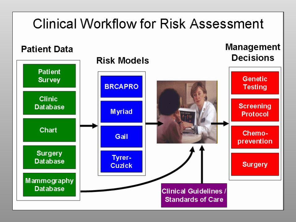
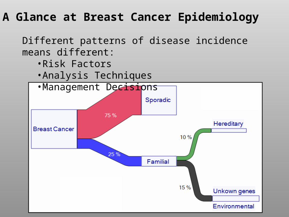
Different patterns of disease incidence means different:•Risk Factors•Analysis Techniques•Management Decisions
A Glance at Breast Cancer Epidemiology
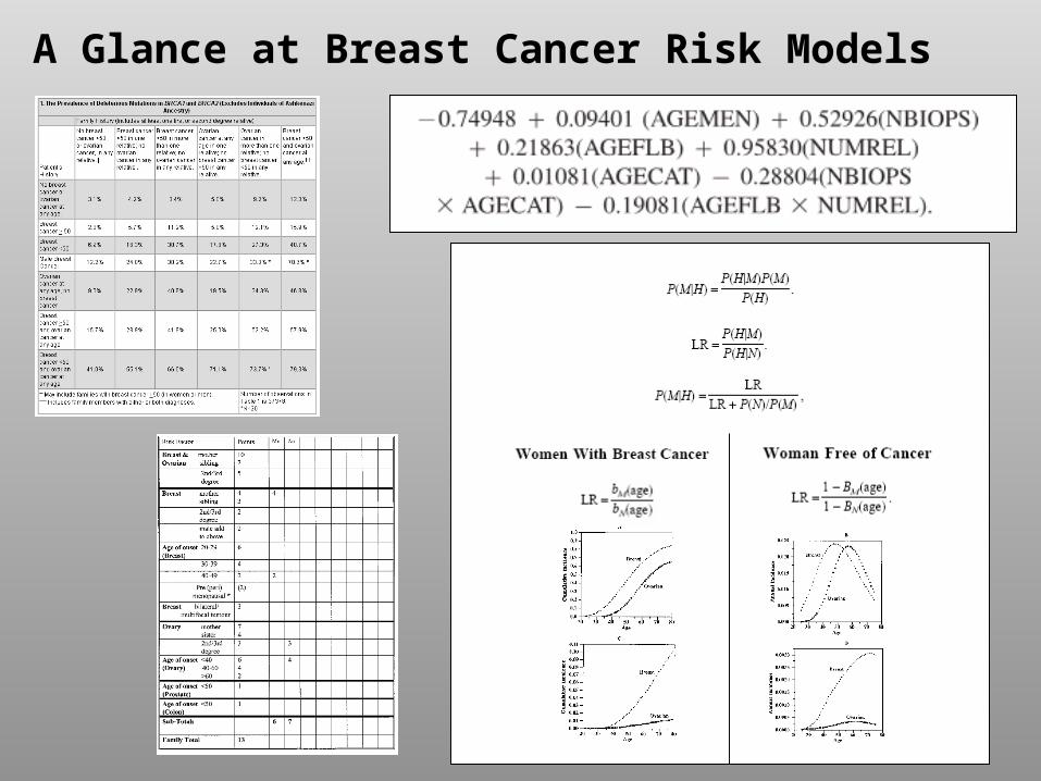
A Glance at Breast Cancer Risk Models
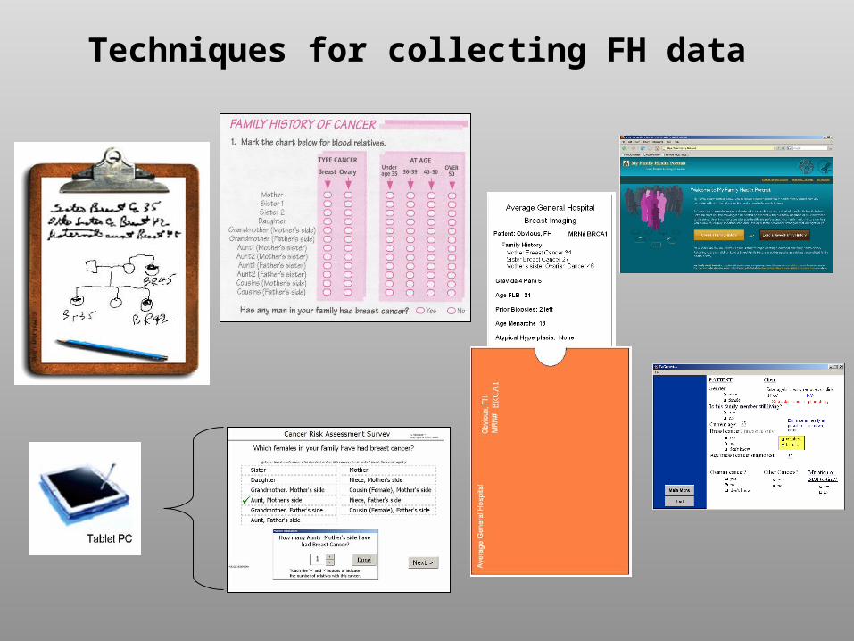
Techniques for collecting FH data
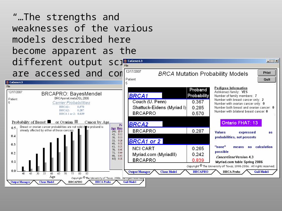
“…The strengths and weaknesses of the various models described here become apparent as the different output screens are accessed and compared…”
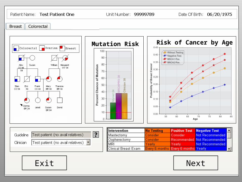
Breast Ovarian Colorectal
NextExit
Mutation Risk
?
Risk of Cancer by Age
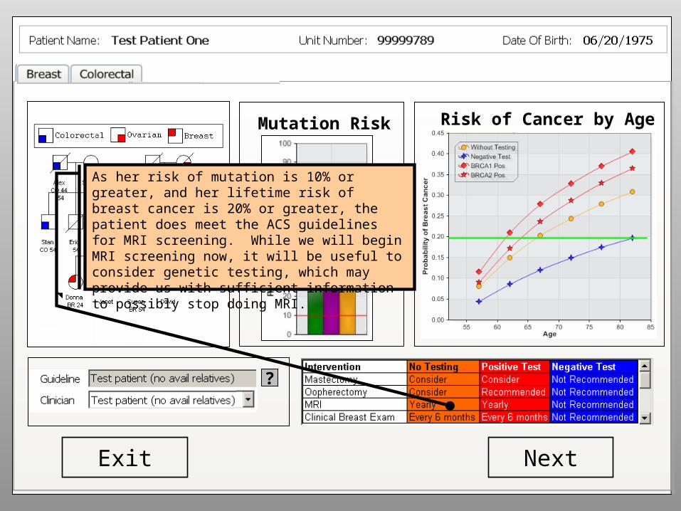
Breast Ovarian Colorectal
NextExit
Mutation Risk
?
Risk of Cancer by Age
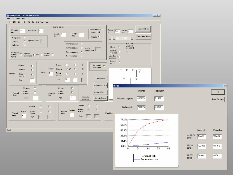
As her risk of mutation is 10% or greater, and her lifetime risk of breast cancer is 20% or greater, the patient does meet the ACS guidelines for MRI screening. While we will begin MRI screening now, it will be useful to consider genetic testing, which may provide us with sufficient information to possibly stop doing MRI.
Breast Ovarian Colorectal
NextExit
Mutation Risk
?
Risk of Cancer by Age
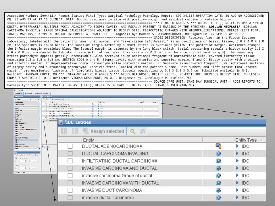
Accession Number: S99X41314 Report Status: Final Type: Surgical Pathology Pathology Report: S99-X41314 OPERATION DATE: 30 AUG 99 ACCESSIONED ON: 30 AUG 99 at 13:15 CLINICAL DATA: Ductal carcinoma in situ with positive margin and residual calcium on outside biopsy. =============================================================================== *** FINAL DIAGNOSIS *** BREAST (LEFT), RE-EXCISION: ATYPICAL DUCTAL HYPERPLASIA, MULTIPLE FOCI, ADJACENT TO BIOPSY CAVITY, AND FOCALLY PRESENT AT MARGINS OF THE SPECIMEN. LOBULAR NEOPLASIA (LOBULAR CARCINOMA IN-SITU). LARGE STROMAL CALCIFICATIONS ADJACENT TO BIOPSY SITE. FIBROCYSTIC CHANGES WITH MICROCALCIFICATIONS. BREAST (LEFT FINAL SHAVED MARGINS): ATYPICAL DUCTAL HYPERPLASIA, SMALL FOCI. Diagnosis by: MARYAM S. MOHAMMADKHANI, MD Signed On: 07 SEP 99 at 09:17 =============================================================================== GROSS DESCRIPTION: Received fresh in the Frozen Section Laboratory, labeled with the patient's name, unit number, and "re-excision left breast," is an ovoid piece of breast tissue, 5.0 X 4.0 X 2.0 cm, the specimen is inked black, the superior margin marked by a short stitch is overinked yellow; the posterior margin, overinked orange; the inferior margin overinked blue. The lateral margin is oriented by the long black stitch. Serial sectioning reveals a biopsy cavity 1.5 X 1.0 X 0.8 cm, surrounded by fibrotic tissue with fat necrosis. This cavity is 0.3 cm from the anterior (closest margin). The remaining breast parenchyma appears grossly unremarkable. Also received is an additional fragment of unremarkable skin- covered fibrofatty tissue measuring 2.2 x 1.5 x 0.8 cm. SECTION CODE A and D: Biopsy cavity with anterior and superior margin. B and C: Biopsy cavity with anterior and inferior margin. E: Representative normal parenchyma (also posterior margin). F: Separate skin-covered fragment. J-M: Additonal sections of biopsy cavity and surrounding parenchyma. Received fresh, labeled with the patient's name, unit number, and "left breast final shaved margin," are unoriented fragments of fibrofatty breast tissue, loosely aggregating to 3.5 X 3.0 X 0.7 cm. Submitted entirely in G-I. Resident: ANUPAMA GUPTA, MD *** INTRA-OPERATIVE DIAGNOSIS *** GROSS DIAGNOSIS, BREAST (LEFT), RE-EXCISION: PREVIOUS BIOPSY SITE: NO LESION GROSSLY SUSPICIOUS. O.R. Resident: VIKRAM DESHPANDE, MD O.R. Diagnosis by: Gunnlaugur P. Nielsen, MD =============================================================================== SOURCE CARE UNIT: SAME DAY SURGICAL UNIT - ACC3 REPORTS TO: Barbara Lynn Smith, M.D. PART A: BREAST (LEFT), RE-EXCISION PART B: BREAST (LEFT FINAL SHAVED MARGINS)

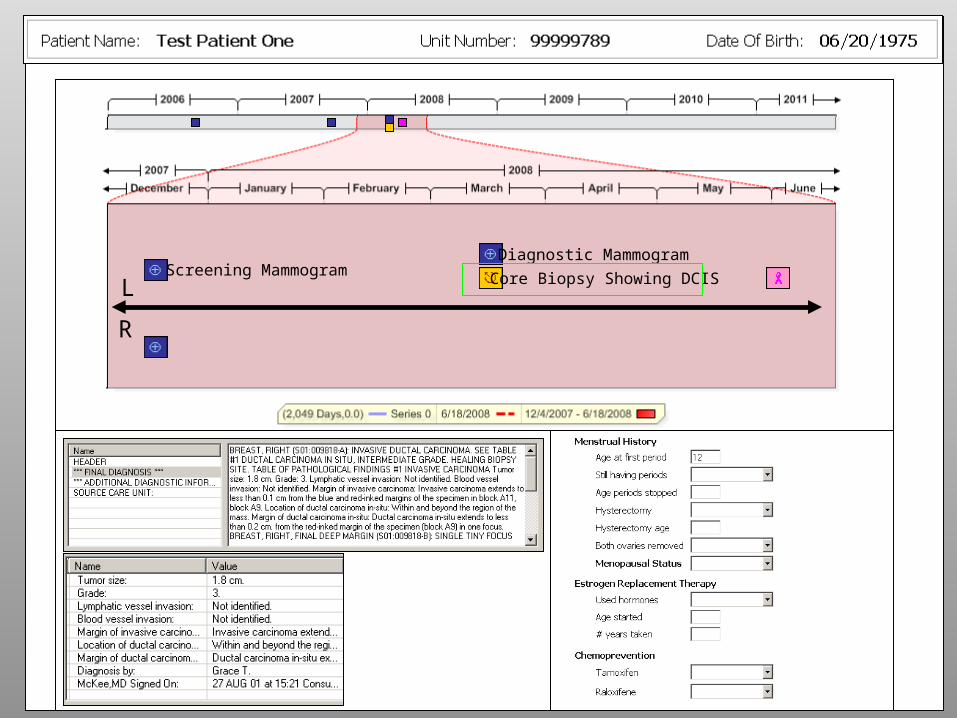
L
R
Screening MammogramDiagnostic Mammogram
Core Biopsy Showing DCIS
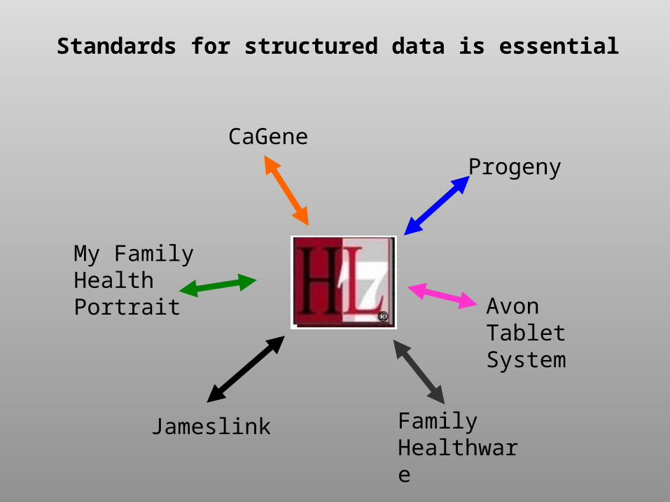
CaGene
My Family HealthPortrait
Jameslink Family Healthware
Avon Tablet System
Progeny
Standards for structured data is essential
Special Thanks
•Prof. Georges Grinstein•John Sharko•Christine Lawrence•Loura Costello•Mary Beth Smirtic•Others at IVPR
•Dr. Kevin Hughes•Connie Roche•Elissa Ozzane•Sherwood Hughes•Many more…
University of Massachusetts Massachusetts General Hospital