Embed Size (px)
Citation preview
Affymetrix® Cytogenetics Copy Number Assay User Guide
P/N 702607 Rev. 2
For research use only. Not for use in diagnostic procedures.
Trademarks
Affymetrix®, , GeneChip®, HuSNP®, GenFlex®, Flying Objective™, CustomExpress®, CustomSeq®, NetAffx™, Tools to Take You As Far As Your Vision®, The Way Ahead™, Powered by Affymetrix™, GeneChip-compatible™, and Command Console™ are trademarks of Affymetrix, Inc.
All other trademarks are the property of their respective owners.
Limited License
Subject to the Affymetrix terms and conditions that govern your use of Affymetrix products, Affymetrix grants you a non-exclusive, non-transferable, non-sublicensable license to use this Affymetrix product only in accordance with the manual and written instructions provided by Affymetrix. You understand and agree that except as expressly set forth in the Affymetrix terms and conditions, that no right or license to any patent or other intellectual property owned or licensable by Affymetrix is conveyed or implied by this Affymetrix product. In particular, no right or license is conveyed or implied to use this Affymetrix product in combination with a product not provided, licensed or specifically recommended by Affymetrix for such use.
Patents
Arrays: Products may be covered by one or more of the following patents: U.S. Patent Nos. 5,445,934; 5,744,305; 5,945,334; 6,140,044; 6,261,776; 6,291,183; 6,346,413; 6,399,365; 6,420,169; 6,551,817; 6,610,482; 6,733,977; 6,955,915 and D430,024 and other U.S. or foreign patents. Products are manufactured and sold under license from OGT under 5,700,637 and 6,054,270
Reagents: Products may be covered by one or more of the following patents: U.S. Patent Nos. 6,965,020; 6,864,059.
Copyright
© 2008 Affymetrix, Inc. All rights reserved.
CONTENTS
Chapter 1 Before You Start . . . . . . . . . . . . . . . . . . . . . . . . . . . . . . . . . . . . . . . . . . . . . . . 1
About the Affymetrix Cytogenetics Solution . . . . . . . . . . . . . . . . . . . . . . . . . . . . . . . . . . . .2Tips for Ensuring Successful Performance of the Protocol . . . . . . . . . . . . . . . . . . . . . . . . .2
Equipment and Calibration . . . . . . . . . . . . . . . . . . . . . . . . . . . . . . . . . . . . . . . . . . . . . . . .2Pipetting . . . . . . . . . . . . . . . . . . . . . . . . . . . . . . . . . . . . . . . . . . . . . . . . . . . . . . . . . . . . . .3Reagent Handling and Storage . . . . . . . . . . . . . . . . . . . . . . . . . . . . . . . . . . . . . . . . . . . .3Master Mix Preparation . . . . . . . . . . . . . . . . . . . . . . . . . . . . . . . . . . . . . . . . . . . . . . . . . .3Laboratory Workflow . . . . . . . . . . . . . . . . . . . . . . . . . . . . . . . . . . . . . . . . . . . . . . . . . . . .4Preparing the Work Area for Each Stage . . . . . . . . . . . . . . . . . . . . . . . . . . . . . . . . . . . . .4Thermal Cyclers, 96-well Plate, and Adhesive Seals . . . . . . . . . . . . . . . . . . . . . . . . . . . .4Program Your Thermal Cyclers . . . . . . . . . . . . . . . . . . . . . . . . . . . . . . . . . . . . . . . . . . . .5
Chapter 2 Laboratory Setup and Recommendations . . . . . . . . . . . . . . . . . . . . . . . . . . 7
Configuration 1 — Two Separate Rooms . . . . . . . . . . . . . . . . . . . . . . . . . . . . . . . . . . . . . .7Pre-PCR Clean Room . . . . . . . . . . . . . . . . . . . . . . . . . . . . . . . . . . . . . . . . . . . . . . . . . . . .8Post-PCR Room . . . . . . . . . . . . . . . . . . . . . . . . . . . . . . . . . . . . . . . . . . . . . . . . . . . . . . . .9
Configuration 2 — One Room . . . . . . . . . . . . . . . . . . . . . . . . . . . . . . . . . . . . . . . . . . . . . .10Pre-PCR Clean Area . . . . . . . . . . . . . . . . . . . . . . . . . . . . . . . . . . . . . . . . . . . . . . . . . . . .11Post-PCR Area . . . . . . . . . . . . . . . . . . . . . . . . . . . . . . . . . . . . . . . . . . . . . . . . . . . . . . . .11Single Direction Workflow . . . . . . . . . . . . . . . . . . . . . . . . . . . . . . . . . . . . . . . . . . . . . . .13
Contamination Prevention . . . . . . . . . . . . . . . . . . . . . . . . . . . . . . . . . . . . . . . . . . . . . . . . .14Safety Precautions . . . . . . . . . . . . . . . . . . . . . . . . . . . . . . . . . . . . . . . . . . . . . . . . . . . . . .14
Chapter 3 Genomic DNA General Requirements. . . . . . . . . . . . . . . . . . . . . . . . . . . . . 15
General Requirements . . . . . . . . . . . . . . . . . . . . . . . . . . . . . . . . . . . . . . . . . . . . . . . . . . .15Sources of Human Genomic DNA . . . . . . . . . . . . . . . . . . . . . . . . . . . . . . . . . . . . . . . . . . .16Genomic DNA Extraction/Purification Methods . . . . . . . . . . . . . . . . . . . . . . . . . . . . . . . .16DNA Cleanup . . . . . . . . . . . . . . . . . . . . . . . . . . . . . . . . . . . . . . . . . . . . . . . . . . . . . . . . . . .16References . . . . . . . . . . . . . . . . . . . . . . . . . . . . . . . . . . . . . . . . . . . . . . . . . . . . . . . . . . . .16
Chapter 4 Affymetrix® Cytogenetics Copy Number Assay . . . . . . . . . . . . . . . . . . . . . 19
About the Protocol . . . . . . . . . . . . . . . . . . . . . . . . . . . . . . . . . . . . . . . . . . . . . . . . . . . . . .19About the Illustrations in this Chapter . . . . . . . . . . . . . . . . . . . . . . . . . . . . . . . . . . . . . .20About the Reagents, Equipment and Consumables Specified in this Chapter . . . . . . .20
Workflows . . . . . . . . . . . . . . . . . . . . . . . . . . . . . . . . . . . . . . . . . . . . . . . . . . . . . . . . . . . . .21

ii Affymetrix® Cytogenetics Copy Number Assay User Guide
Genomic DNA Preparation . . . . . . . . . . . . . . . . . . . . . . . . . . . . . . . . . . . . . . . . . . . . . . . .23About this Stage . . . . . . . . . . . . . . . . . . . . . . . . . . . . . . . . . . . . . . . . . . . . . . . . . . . . . .23Location and Duration . . . . . . . . . . . . . . . . . . . . . . . . . . . . . . . . . . . . . . . . . . . . . . . . . .23Input Required . . . . . . . . . . . . . . . . . . . . . . . . . . . . . . . . . . . . . . . . . . . . . . . . . . . . . . . .23Equipment and Consumables Required . . . . . . . . . . . . . . . . . . . . . . . . . . . . . . . . . . . .24Reagents Required . . . . . . . . . . . . . . . . . . . . . . . . . . . . . . . . . . . . . . . . . . . . . . . . . . . .24Preparing the Genomic DNA . . . . . . . . . . . . . . . . . . . . . . . . . . . . . . . . . . . . . . . . . . . . .25Aliquoting the Prepared Genomic DNA and Controls . . . . . . . . . . . . . . . . . . . . . . . . . .26What To Do Next . . . . . . . . . . . . . . . . . . . . . . . . . . . . . . . . . . . . . . . . . . . . . . . . . . . . . .27
Stage 1: Nsp and Sty Restriction Enzyme Digest . . . . . . . . . . . . . . . . . . . . . . . . . . . . . . .28About this Stage . . . . . . . . . . . . . . . . . . . . . . . . . . . . . . . . . . . . . . . . . . . . . . . . . . . . . .28Location and Duration . . . . . . . . . . . . . . . . . . . . . . . . . . . . . . . . . . . . . . . . . . . . . . . . . .28Input Required From Previous Stage . . . . . . . . . . . . . . . . . . . . . . . . . . . . . . . . . . . . . . .28Equipment and Consumables Required . . . . . . . . . . . . . . . . . . . . . . . . . . . . . . . . . . . .29Reagents Required . . . . . . . . . . . . . . . . . . . . . . . . . . . . . . . . . . . . . . . . . . . . . . . . . . . .29Prepare the Reagents, Equipment and Consumables . . . . . . . . . . . . . . . . . . . . . . . . . .30Prepare the Nsp Digest Master Mix . . . . . . . . . . . . . . . . . . . . . . . . . . . . . . . . . . . . . . .31Add Nsp Digest Master Mix to Samples . . . . . . . . . . . . . . . . . . . . . . . . . . . . . . . . . . . .31Prepare the Sty Digest Master Mix . . . . . . . . . . . . . . . . . . . . . . . . . . . . . . . . . . . . . . . .32Add Sty Digest Master Mix to Samples . . . . . . . . . . . . . . . . . . . . . . . . . . . . . . . . . . . .33Load Nsp and Sty Samples onto the Thermal Cycler . . . . . . . . . . . . . . . . . . . . . . . . . .33What To Do Next . . . . . . . . . . . . . . . . . . . . . . . . . . . . . . . . . . . . . . . . . . . . . . . . . . . . . .33
Stage 2: Nsp and Sty Ligation . . . . . . . . . . . . . . . . . . . . . . . . . . . . . . . . . . . . . . . . . . . . . .34About this Stage . . . . . . . . . . . . . . . . . . . . . . . . . . . . . . . . . . . . . . . . . . . . . . . . . . . . . .34Location and Duration . . . . . . . . . . . . . . . . . . . . . . . . . . . . . . . . . . . . . . . . . . . . . . . . . .34Input Required From Previous Stage . . . . . . . . . . . . . . . . . . . . . . . . . . . . . . . . . . . . . . .34Equipment and Consumables Required . . . . . . . . . . . . . . . . . . . . . . . . . . . . . . . . . . . .35Reagents Required . . . . . . . . . . . . . . . . . . . . . . . . . . . . . . . . . . . . . . . . . . . . . . . . . . . .35Prepare the Reagents, Consumables and Other Components . . . . . . . . . . . . . . . . . . .36Prepare the Nsp Ligation Master Mix . . . . . . . . . . . . . . . . . . . . . . . . . . . . . . . . . . . . . .37Add Nsp Ligation Master Mix to Reactions . . . . . . . . . . . . . . . . . . . . . . . . . . . . . . . . . .38Prepare the Sty Ligation Master Mix . . . . . . . . . . . . . . . . . . . . . . . . . . . . . . . . . . . . . . .38Add Sty Ligation Master Mix to Reactions . . . . . . . . . . . . . . . . . . . . . . . . . . . . . . . . . .39Load the Nsp and Sty Samples Onto the Thermal Cycler . . . . . . . . . . . . . . . . . . . . . . .40Dilute the Ligated Samples . . . . . . . . . . . . . . . . . . . . . . . . . . . . . . . . . . . . . . . . . . . . . .40What To Do Next . . . . . . . . . . . . . . . . . . . . . . . . . . . . . . . . . . . . . . . . . . . . . . . . . . . . . .40
Stage 3: Nsp and Sty PCR . . . . . . . . . . . . . . . . . . . . . . . . . . . . . . . . . . . . . . . . . . . . . . . . .41About this Stage . . . . . . . . . . . . . . . . . . . . . . . . . . . . . . . . . . . . . . . . . . . . . . . . . . . . . .41Location and Duration . . . . . . . . . . . . . . . . . . . . . . . . . . . . . . . . . . . . . . . . . . . . . . . . . .41Input Required from Previous Stage . . . . . . . . . . . . . . . . . . . . . . . . . . . . . . . . . . . . . . .41Equipment and Materials Required . . . . . . . . . . . . . . . . . . . . . . . . . . . . . . . . . . . . . . . .42Reagents Required . . . . . . . . . . . . . . . . . . . . . . . . . . . . . . . . . . . . . . . . . . . . . . . . . . . .43Gels and Related Materials Required . . . . . . . . . . . . . . . . . . . . . . . . . . . . . . . . . . . . . .43Important Information About This Stage . . . . . . . . . . . . . . . . . . . . . . . . . . . . . . . . . . . .43Prepare the Reagents, Consumables and Other Components . . . . . . . . . . . . . . . . . . .44Transfer Diluted Ligated Samples to the PCR Plate . . . . . . . . . . . . . . . . . . . . . . . . . . .45Prepare the PCR Master Mix . . . . . . . . . . . . . . . . . . . . . . . . . . . . . . . . . . . . . . . . . . . . .46

contents iii
Add PCR Master Mix to Each Sample . . . . . . . . . . . . . . . . . . . . . . . . . . . . . . . . . . . . . .46Load PCR Plate onto a Thermal Cycler . . . . . . . . . . . . . . . . . . . . . . . . . . . . . . . . . . . . .47Check the PCR Reaction by Running a Gel . . . . . . . . . . . . . . . . . . . . . . . . . . . . . . . . . .48What To Do Next . . . . . . . . . . . . . . . . . . . . . . . . . . . . . . . . . . . . . . . . . . . . . . . . . . . . . .50
Stage 4: PCR Product Purification . . . . . . . . . . . . . . . . . . . . . . . . . . . . . . . . . . . . . . . . . . .51About this Stage . . . . . . . . . . . . . . . . . . . . . . . . . . . . . . . . . . . . . . . . . . . . . . . . . . . . . .51Location and Duration . . . . . . . . . . . . . . . . . . . . . . . . . . . . . . . . . . . . . . . . . . . . . . . . . .51Input Required from Previous Stage . . . . . . . . . . . . . . . . . . . . . . . . . . . . . . . . . . . . . . .51Equipment and Consumables Required . . . . . . . . . . . . . . . . . . . . . . . . . . . . . . . . . . . .52Reagents Required . . . . . . . . . . . . . . . . . . . . . . . . . . . . . . . . . . . . . . . . . . . . . . . . . . . .52Important Information About This Stage . . . . . . . . . . . . . . . . . . . . . . . . . . . . . . . . . . . .53Prepare the 75% EtOH . . . . . . . . . . . . . . . . . . . . . . . . . . . . . . . . . . . . . . . . . . . . . . . . .53Pool the PCR Products . . . . . . . . . . . . . . . . . . . . . . . . . . . . . . . . . . . . . . . . . . . . . . . . .53Purify the Pooled PCR Products . . . . . . . . . . . . . . . . . . . . . . . . . . . . . . . . . . . . . . . . . .54What To Do Next . . . . . . . . . . . . . . . . . . . . . . . . . . . . . . . . . . . . . . . . . . . . . . . . . . . . . .57
Stage 5: Quantitation . . . . . . . . . . . . . . . . . . . . . . . . . . . . . . . . . . . . . . . . . . . . . . . . . . . . .58About this Stage . . . . . . . . . . . . . . . . . . . . . . . . . . . . . . . . . . . . . . . . . . . . . . . . . . . . . .58Location and Duration . . . . . . . . . . . . . . . . . . . . . . . . . . . . . . . . . . . . . . . . . . . . . . . . . .58Input Required from Previous Stage . . . . . . . . . . . . . . . . . . . . . . . . . . . . . . . . . . . . . . .58Equipment and Consumables Required . . . . . . . . . . . . . . . . . . . . . . . . . . . . . . . . . . . .58Reagents Required . . . . . . . . . . . . . . . . . . . . . . . . . . . . . . . . . . . . . . . . . . . . . . . . . . . .58Important Information About This Stage . . . . . . . . . . . . . . . . . . . . . . . . . . . . . . . . . . . .59Prepare the Reagents, Equipment and Consumables . . . . . . . . . . . . . . . . . . . . . . . . . .59Procedure if Using a Microplate Spectrophotometer . . . . . . . . . . . . . . . . . . . . . . . . . .59Procedure if Using a NanoDrop . . . . . . . . . . . . . . . . . . . . . . . . . . . . . . . . . . . . . . . . . . .61Assess the OD Readings . . . . . . . . . . . . . . . . . . . . . . . . . . . . . . . . . . . . . . . . . . . . . . . .62What To Do Next . . . . . . . . . . . . . . . . . . . . . . . . . . . . . . . . . . . . . . . . . . . . . . . . . . . . . .62
Stage 6: Fragmentation . . . . . . . . . . . . . . . . . . . . . . . . . . . . . . . . . . . . . . . . . . . . . . . . . . .63About this Stage . . . . . . . . . . . . . . . . . . . . . . . . . . . . . . . . . . . . . . . . . . . . . . . . . . . . . .63Location and Duration . . . . . . . . . . . . . . . . . . . . . . . . . . . . . . . . . . . . . . . . . . . . . . . . . .63Input Required from Previous Stage . . . . . . . . . . . . . . . . . . . . . . . . . . . . . . . . . . . . . . .63Equipment and Consumables Required . . . . . . . . . . . . . . . . . . . . . . . . . . . . . . . . . . . .64Reagents Required . . . . . . . . . . . . . . . . . . . . . . . . . . . . . . . . . . . . . . . . . . . . . . . . . . . .64Gels and Related Materials Required . . . . . . . . . . . . . . . . . . . . . . . . . . . . . . . . . . . . . .65Important Information About This Stage . . . . . . . . . . . . . . . . . . . . . . . . . . . . . . . . . . . .65Prepare the Reagents, Consumables and Other Components . . . . . . . . . . . . . . . . . . .66Prepare the Samples for Fragmentation . . . . . . . . . . . . . . . . . . . . . . . . . . . . . . . . . . . .67What To Do Next . . . . . . . . . . . . . . . . . . . . . . . . . . . . . . . . . . . . . . . . . . . . . . . . . . . . . .69Check the Fragmentation Reaction by Running a Gel . . . . . . . . . . . . . . . . . . . . . . . . . .70
Stage 7: Labeling . . . . . . . . . . . . . . . . . . . . . . . . . . . . . . . . . . . . . . . . . . . . . . . . . . . . . . . .71About this Stage . . . . . . . . . . . . . . . . . . . . . . . . . . . . . . . . . . . . . . . . . . . . . . . . . . . . . .71Location and Duration . . . . . . . . . . . . . . . . . . . . . . . . . . . . . . . . . . . . . . . . . . . . . . . . . .71Input Required from Previous Stage . . . . . . . . . . . . . . . . . . . . . . . . . . . . . . . . . . . . . . .71Equipment and Consumables Required . . . . . . . . . . . . . . . . . . . . . . . . . . . . . . . . . . . .71Reagents Required . . . . . . . . . . . . . . . . . . . . . . . . . . . . . . . . . . . . . . . . . . . . . . . . . . . .72Prepare the Reagents, Consumables and Other Components . . . . . . . . . . . . . . . . . . .72Prepare the Labeling Master Mix . . . . . . . . . . . . . . . . . . . . . . . . . . . . . . . . . . . . . . . . .73
iv Affymetrix® Cytogenetics Copy Number Assay User Guide
Add the Labeling Master Mix to the Samples . . . . . . . . . . . . . . . . . . . . . . . . . . . . . . . .73What To Do Next . . . . . . . . . . . . . . . . . . . . . . . . . . . . . . . . . . . . . . . . . . . . . . . . . . . . . .74
Stage 8: Target Hybridization . . . . . . . . . . . . . . . . . . . . . . . . . . . . . . . . . . . . . . . . . . . . . .75About this Stage . . . . . . . . . . . . . . . . . . . . . . . . . . . . . . . . . . . . . . . . . . . . . . . . . . . . . .75Location and Duration . . . . . . . . . . . . . . . . . . . . . . . . . . . . . . . . . . . . . . . . . . . . . . . . . .75Input Required from Previous Stage . . . . . . . . . . . . . . . . . . . . . . . . . . . . . . . . . . . . . . .75Equipment and Consumables Required . . . . . . . . . . . . . . . . . . . . . . . . . . . . . . . . . . . .75Reagents Required . . . . . . . . . . . . . . . . . . . . . . . . . . . . . . . . . . . . . . . . . . . . . . . . . . . .76Important Information About This Stage . . . . . . . . . . . . . . . . . . . . . . . . . . . . . . . . . . . .77Prepare the Reagents, Consumables and Other Components . . . . . . . . . . . . . . . . . . .77Prepare the Arrays . . . . . . . . . . . . . . . . . . . . . . . . . . . . . . . . . . . . . . . . . . . . . . . . . . . . .78Prepare the Hybridization Master Mix . . . . . . . . . . . . . . . . . . . . . . . . . . . . . . . . . . . . . .79
Chapter 5 Washing, Staining and Scanning Arrays . . . . . . . . . . . . . . . . . . . . . . . . . . 83
Equipment and Consumables Required . . . . . . . . . . . . . . . . . . . . . . . . . . . . . . . . . . . . . .83Reagents Required . . . . . . . . . . . . . . . . . . . . . . . . . . . . . . . . . . . . . . . . . . . . . . . . . . . . . .84Reagent Preparation . . . . . . . . . . . . . . . . . . . . . . . . . . . . . . . . . . . . . . . . . . . . . . . . . . . . .84
Wash A: Non-Stringent Wash Buffer . . . . . . . . . . . . . . . . . . . . . . . . . . . . . . . . . . . . . . .84Wash B: Stringent Wash Buffer . . . . . . . . . . . . . . . . . . . . . . . . . . . . . . . . . . . . . . . . . .850.5 mg/mL Anti-Streptavidin Antibody . . . . . . . . . . . . . . . . . . . . . . . . . . . . . . . . . . . . . .8512X MES Stock Buffer . . . . . . . . . . . . . . . . . . . . . . . . . . . . . . . . . . . . . . . . . . . . . . . . . .851X Array Holding Buffer . . . . . . . . . . . . . . . . . . . . . . . . . . . . . . . . . . . . . . . . . . . . . . . . .85
Fluidics Station and Scanner Control Software . . . . . . . . . . . . . . . . . . . . . . . . . . . . . . . . .86Register a New Experiment or Sample . . . . . . . . . . . . . . . . . . . . . . . . . . . . . . . . . . . . . . .86Prime the Fluidics Station . . . . . . . . . . . . . . . . . . . . . . . . . . . . . . . . . . . . . . . . . . . . . . . . .86Wash and Stain Arrays . . . . . . . . . . . . . . . . . . . . . . . . . . . . . . . . . . . . . . . . . . . . . . . . . . .87
Prepare Arrays for Washing and Staining . . . . . . . . . . . . . . . . . . . . . . . . . . . . . . . . . . .87Prepare Buffers and Solutions . . . . . . . . . . . . . . . . . . . . . . . . . . . . . . . . . . . . . . . . . . . .87Washing and Staining Arrays . . . . . . . . . . . . . . . . . . . . . . . . . . . . . . . . . . . . . . . . . . . . .89
Scanning Arrays . . . . . . . . . . . . . . . . . . . . . . . . . . . . . . . . . . . . . . . . . . . . . . . . . . . . . . . . .91Prepare the Scanner . . . . . . . . . . . . . . . . . . . . . . . . . . . . . . . . . . . . . . . . . . . . . . . . . . .91Prepare Arrays for Scanning . . . . . . . . . . . . . . . . . . . . . . . . . . . . . . . . . . . . . . . . . . . . .91Scanning the Array . . . . . . . . . . . . . . . . . . . . . . . . . . . . . . . . . . . . . . . . . . . . . . . . . . . . .92
Shutting Down the Fluidics Station . . . . . . . . . . . . . . . . . . . . . . . . . . . . . . . . . . . . . . . . . .92Data Analysis . . . . . . . . . . . . . . . . . . . . . . . . . . . . . . . . . . . . . . . . . . . . . . . . . . . . . . . . . . .93
Chapter 6 Troubleshooting . . . . . . . . . . . . . . . . . . . . . . . . . . . . . . . . . . . . . . . . . . . . . . 95
General Assay Performance Recommendations . . . . . . . . . . . . . . . . . . . . . . . . . . . . . . . .95Troubleshooting the Cytogenetics Copy Number Assay . . . . . . . . . . . . . . . . . . . . . . . . . .97OD Troubleshooting Guidelines . . . . . . . . . . . . . . . . . . . . . . . . . . . . . . . . . . . . . . . . . . . .99Affymetrix Instruments . . . . . . . . . . . . . . . . . . . . . . . . . . . . . . . . . . . . . . . . . . . . . . . . . .100
contents v
Chapter 7 Fluidics Station Care and Maintenance. . . . . . . . . . . . . . . . . . . . . . . . . . . 101
General Fluidics Station Care . . . . . . . . . . . . . . . . . . . . . . . . . . . . . . . . . . . . . . . . . . . . .101Fluidics Station Bleach Protocol . . . . . . . . . . . . . . . . . . . . . . . . . . . . . . . . . . . . . . . . . . .101
The Bleach Cycle . . . . . . . . . . . . . . . . . . . . . . . . . . . . . . . . . . . . . . . . . . . . . . . . . . . . .102The Rinse Cycle . . . . . . . . . . . . . . . . . . . . . . . . . . . . . . . . . . . . . . . . . . . . . . . . . . . . . .105
Appendix A Guidelines for Processing 16 Samples . . . . . . . . . . . . . . . . . . . . . . . . . . . 107
Digestion, Ligation and PCR . . . . . . . . . . . . . . . . . . . . . . . . . . . . . . . . . . . . . . . . . . . . . .108
Appendix B Guidelines for Processing 24 Samples . . . . . . . . . . . . . . . . . . . . . . . . . . . 111
Digestion, Ligation and PCR . . . . . . . . . . . . . . . . . . . . . . . . . . . . . . . . . . . . . . . . . . . . . .112
Appendix C Reagents, Equipment, and Consumables . . . . . . . . . . . . . . . . . . . . . . . . . 115
About this Appendix . . . . . . . . . . . . . . . . . . . . . . . . . . . . . . . . . . . . . . . . . . . . . . . . . . . .115From Affymetrix . . . . . . . . . . . . . . . . . . . . . . . . . . . . . . . . . . . . . . . . . . . . . . . . . . . . . . .115Equipment Required from Other Suppliers . . . . . . . . . . . . . . . . . . . . . . . . . . . . . . . . . . .117
Pre-PCR Clean Area Equipment Required . . . . . . . . . . . . . . . . . . . . . . . . . . . . . . . . . .117Post-PCR Area Equipment Required . . . . . . . . . . . . . . . . . . . . . . . . . . . . . . . . . . . . . .118
Reagents Required from Other Suppliers . . . . . . . . . . . . . . . . . . . . . . . . . . . . . . . . . . . .119Consumables Required from Other Suppliers . . . . . . . . . . . . . . . . . . . . . . . . . . . . . . . . .120Supplier Contact List . . . . . . . . . . . . . . . . . . . . . . . . . . . . . . . . . . . . . . . . . . . . . . . . . . . .121
Appendix D Thermal Cycler Programs. . . . . . . . . . . . . . . . . . . . . . . . . . . . . . . . . . . . . . 123
Cyto Digest . . . . . . . . . . . . . . . . . . . . . . . . . . . . . . . . . . . . . . . . . . . . . . . . . . . . . . . . .123Cyto Ligate . . . . . . . . . . . . . . . . . . . . . . . . . . . . . . . . . . . . . . . . . . . . . . . . . . . . . . . . . .123Cyto PCR . . . . . . . . . . . . . . . . . . . . . . . . . . . . . . . . . . . . . . . . . . . . . . . . . . . . . . . . . . .124Cyto Fragment . . . . . . . . . . . . . . . . . . . . . . . . . . . . . . . . . . . . . . . . . . . . . . . . . . . . . . .125Cyto Label . . . . . . . . . . . . . . . . . . . . . . . . . . . . . . . . . . . . . . . . . . . . . . . . . . . . . . . . . .125Cyto Hyb . . . . . . . . . . . . . . . . . . . . . . . . . . . . . . . . . . . . . . . . . . . . . . . . . . . . . . . . . . .125

vi Affymetrix® Cytogenetics Copy Number Assay User Guide
Chapte
r1 BEFORE YOU STARTTopics in this chapter include:
• About the Affymetrix Cytogenetics Solution on page 2
• Tips for Ensuring Successful Performance of the Protocol on page 2
IMPORTANT: The Cytogenetics Copy Number assay protocol is optimized for processingfrom 4 to 24 samples at a time to obtain copy number results. This protocol is not intendedfor genome-wide association studies.
An assay protocol for processing 48 samples is described in the Affymetrix® Genome-WideHuman SNP Nsp/Sty 6.0 User Guide, P/N 702504.
2 Affymetrix® Cytogenetics Copy Number Assay User Guide
About the Affymetrix Cytogenetics SolutionCytogenetics studies are performed to identify structural changes in DNA, such as copy number changes. Individuals typically have two copies of the genome in each of their cells: one inherited from the mother, and one inherited from the father. Chromosomal abnormalities are common in several disease states such as:
• Deletions
When one or both copies of a particular chromosome region are lost.
• Gains
When a chromosome or chromosomal region is duplicated or multiplied.
• Uniparental Disomies (UPDs)
When two copies of a chromosome or chromosomal region are present, but both have been inherited from a single parent.
Traditional cytogenetics techniques, such as karyotyping and fluorescent in situ hybridization (FISH) have been used to study chromosomal abnormalities for decades. However, karyotyping only detects abnormalities at low resolutions (larger than ~5 Mb), and FISH is a more focused and targeted approach without the benefit of genome-wide analysis. Further, these techniques are limited to only providing copy number information so that UPDs cannot be identified.
The combination of Affymetrix SNP 6.0 arrays, the Cytogenetics Copy Number Assay, and Genotyping Console 2.1 software allows you to perform high-resolution genome-wide DNA copy number analysis. The Affymetrix solution for cytogenetics also provides genotyping information, enabling detection of loss of heterozygosity (LOH), which can be used to detect UPDs. The combined high resolution DNA copy number data and the ability to detect gains, losses, and UPDs on a single array makes the Affymetrix Cytogenetics Solution a great tool for next generation cytogenetics studies.
Tips for Ensuring Successful Performance of the ProtocolSuccessful performance of the Cytogenetics Copy Number Assay requires accuracy and attention to detail. Many stages involve specific yet distinct enzymatic reactions. For example, in stage 1, genomic DNA is digested with the restriction enzymes NspI and StyI. In stage 2, it is ligated to a common adaptor with T4 DNA ligase. Following ligation, the template undergoes PCR using TITANIUM™ Taq DNA polymerase. Once the product has been purified, it is then fragmented and end-labeled using terminal deoxynucleotidyl transferase.
The stages involving enzymatic reactions are the most critical of the assay. Thus, it is important to carefully monitor and control any variables such as pH, salt concentrations, time, and temperature, all of which can adversely modulate enzyme activity.
Equipment and CalibrationKeep dedicated equipment in each of the areas used for this protocol including pipettors, ice buckets, coolers, etc. To avoid contamination, do not move equipment from one area to another.
Along with the enzymatic stages, lab instrumentation plays an important role in the successful completion of this assay. To aid in maintaining consistency across samples and operators, all equipment should be well maintained and calibrated, including:
• All thermal cyclers
• GeneChip® Hybridization Oven
• GeneChip® Fluidics Station
• GeneChip® Scanner 3000 7G
• Plate spectrophotometer or NanoDrop
• All multi-channel pipets
chapter 1 | Before You Start 3
PipettingSince the Cytogenetics Copy Number Assay involves a series of ordered stages, the output of one stage directly impacts the performance of the subsequent stage. For example, the quantity and purity of the DNA after purification can affect the kinetics of the Fragmentation Reagent (enzyme) during the subsequent fragmentation stage.
To efficiently process samples:
• Always use pipets that have been calibrated to ± 5%.
• It is essential that you be proficient with the use of single- and multi-channel pipets.
To familiarize yourself with the use of multi-channel pipets, we strongly recommend practicing several times before processing actual samples. You can use water to get a feel for aspirating and dispensing solutions to multiple wells simultaneously.
Reagent Handling and Storage
Successful sample processing can be achieved by incorporating the following principles:
• Use only fresh reagents from the recommended vendors to help eliminate changes in pH or the salt concentration of buffers.
• Properly store all enzyme reagents. Storage methods can profoundly impact activity.
• Store the reagents used for the digestion, ligation and PCR in the Pre-PCR Clean Area.
• Consult the appropriate MSDS for reagent storage and handling requirements.
When Using Reagents at the Lab Bench• Properly chill essential equipment such as cooling chambers and reagent coolers before use.
• Unless otherwise indicated, keep all reagents (except enzymes) on ice, or in a cooling chamber/block that has been chilled to 4 °C on ice or in a refrigerator.
• Ensure that enzymes are kept at –20 °C until needed. When removed from the freezer, immediately place in a cooler that has been chilled to –20 °C.
• Keep all tubes, master mixes and working solutions in chilled cooling chambers on ice.
• Since enzyme activity is a function of temperature, ensure that all temperature transitions are rapid and/or well-controlled to help maintain consistency across samples.
Master Mix PreparationCarefully follow each master mix recipe. Use pipets that have been calibrated to ± 5%. When molecular biology-grade water is specified, be sure to use the AccuGENE® water listed in Appendix C. Using in-house ddH2O or other water can negatively affect your results. The enzymatic reaction in Stage 6: Fragmentation is particularly sensitive to pH and metal ion contamination.
If you run out of master mix during any of these procedures, a volume error has been made or the pipets are not accurate. We recommend that you stop and repeat the experiment.
IMPORTANT: Always use the 30 reaction Genome-Wide Human SNP Nsp/Sty Assay Kit5.0/6.0 (P/N 901013) for this protocol. This kit has been tested for multiple freeze/thawcycles.
You can freeze/thaw the reagents in the 30 reaction kit ≤ 8 times.
4 Affymetrix® Cytogenetics Copy Number Assay User Guide
Laboratory Workflow• Maintain a single direction workflow. Do not re-enter the Pre-PCR Clean Area after entering the Post-
PCR Area until you have showered and changed into freshly laundered clothing.
• Never bring amplified products into the Pre-PCR Clean Area.
• Keep dedicated equipment in each room or area used for this protocol. To avoid contamination, do not move equipment between the Pre-PCR Clean Area and the Post-PCR Area.
Preparing the Work Area for Each StageMany of the stages in the Cytogenetics Copy Number Assay must be performed rapidly and on ice to carefully control enzyme activity and temperature transitions. Therefore, we recommend that you set up all of the equipment, consumables and reagents (except for the enzymes) prior to beginning each stage.
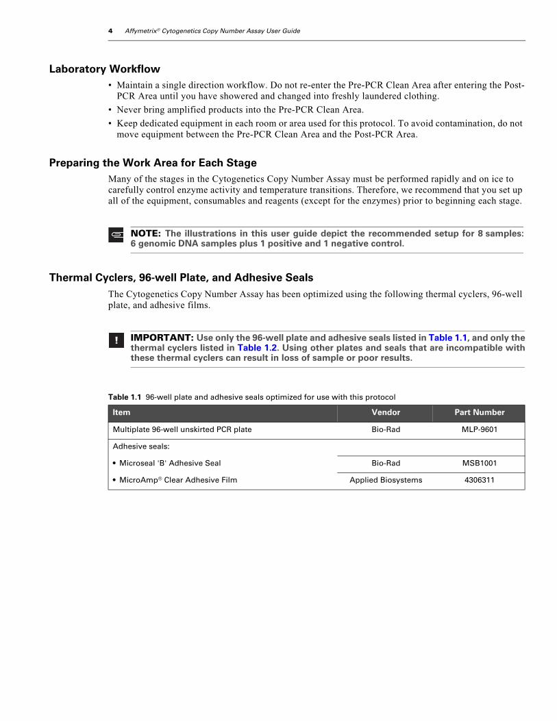
Thermal Cyclers, 96-well Plate, and Adhesive SealsThe Cytogenetics Copy Number Assay has been optimized using the following thermal cyclers, 96-well plate, and adhesive films.
NOTE: The illustrations in this user guide depict the recommended setup for 8 samples:6 genomic DNA samples plus 1 positive and 1 negative control.
IMPORTANT: Use only the 96-well plate and adhesive seals listed in Table 1.1, and only thethermal cyclers listed in Table 1.2. Using other plates and seals that are incompatible withthese thermal cyclers can result in loss of sample or poor results.
Table 1.1 96-well plate and adhesive seals optimized for use with this protocol
Item Vendor Part Number
Multiplate 96-well unskirted PCR plate Bio-Rad MLP-9601
Adhesive seals:
• Microseal 'B' Adhesive Seal Bio-Rad MSB1001
• MicroAmp® Clear Adhesive Film Applied Biosystems 4306311
chapter 1 | Before You Start 5
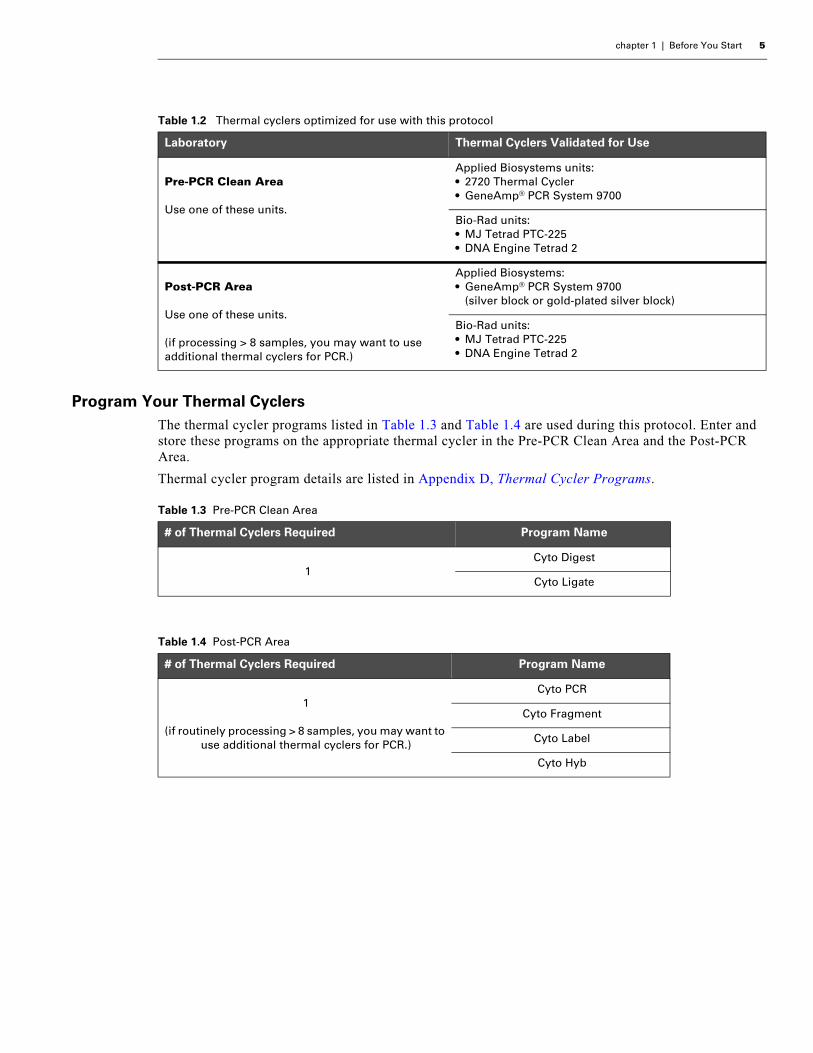
Program Your Thermal CyclersThe thermal cycler programs listed in Table 1.3 and Table 1.4 are used during this protocol. Enter and store these programs on the appropriate thermal cycler in the Pre-PCR Clean Area and the Post-PCR Area.
Thermal cycler program details are listed in Appendix D, Thermal Cycler Programs.
Table 1.2 Thermal cyclers optimized for use with this protocol
Laboratory Thermal Cyclers Validated for Use
Pre-PCR Clean Area
Use one of these units.
Applied Biosystems units: • 2720 Thermal Cycler • GeneAmp® PCR System 9700
Bio-Rad units:• MJ Tetrad PTC-225• DNA Engine Tetrad 2
Post-PCR Area
Use one of these units.
(if processing > 8 samples, you may want to use additional thermal cyclers for PCR.)
Applied Biosystems: • GeneAmp® PCR System 9700
(silver block or gold-plated silver block)
Bio-Rad units:• MJ Tetrad PTC-225• DNA Engine Tetrad 2
Table 1.3 Pre-PCR Clean Area
# of Thermal Cyclers Required Program Name
1Cyto Digest
Cyto Ligate
Table 1.4 Post-PCR Area
# of Thermal Cyclers Required Program Name
1
(if routinely processing > 8 samples, you may want to use additional thermal cyclers for PCR.)
Cyto PCR
Cyto Fragment
Cyto Label
Cyto Hyb

6 Affymetrix® Cytogenetics Copy Number Assay User Guide

Chapte
r2 LABORATORY SETUP AND RECOMMENDATIONSThis chapter provides an overview of two laboratory setups that can used when performing the Affymetrix® Cytogenetics Copy Number Assay.
Configuration 1 — Two Separate RoomsThe use of two separate rooms greatly reduces the risk of sample contamination due to previously-amplified PCR products. These rooms are referred to as the:
• Pre-PCR Clean Room
• Post-PCR Room
The high-level steps performed in each room is presented in Table 2.1.
IMPORTANT: If possible, we strongly recommend using two separate rooms whenperforming this protocol.
Table 2.1 Assay workflow when two separate rooms are used
Room Template(Genomic DNA)
PCR Product
Pre-PCR Clean RoomAssay steps:
• Genomic DNA preparation• Digestion• Ligation• PCR setup only
Post-PCR Room Assay steps:
• PCR thermal cycling• Fragmentation• Labeling• Hybridization• Washing and staining• Scanning
8 Affymetrix® Cytogenetics Copy Number Assay User Guide
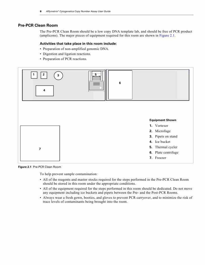
Pre-PCR Clean RoomThe Pre-PCR Clean Room should be a low copy DNA template lab, and should be free of PCR product (amplicons). The major pieces of equipment required for this room are shown in Figure 2.1.
Activities that take place in this room include:• Preparation of non-amplified genomic DNA.
• Digestion and ligation reactions.
• Preparation of PCR reactions.
To help prevent sample contamination:
• All of the reagents and master stocks required for the steps performed in the Pre-PCR Clean Room should be stored in this room under the appropriate conditions.
• All of the equipment required for the steps performed in this room should be dedicated. Do not move any equipment including ice buckets and pipets between the Pre- and the Post-PCR Rooms.
• Always wear a fresh gown, booties, and gloves to prevent PCR carryover, and to minimize the risk of trace levels of contaminants being brought into the room.
Figure 2.1 Pre-PCR Clean Room
Equipment Shown
1. Vortexer
2. Microfuge
3. Pipets on stand
4. Ice bucket
5. Thermal cycler
6. Plate centrifuge
7. Freezer
1 2 3
4
5
6
7
6
chapter 2 | Laboratory Setup and Recommendations 9
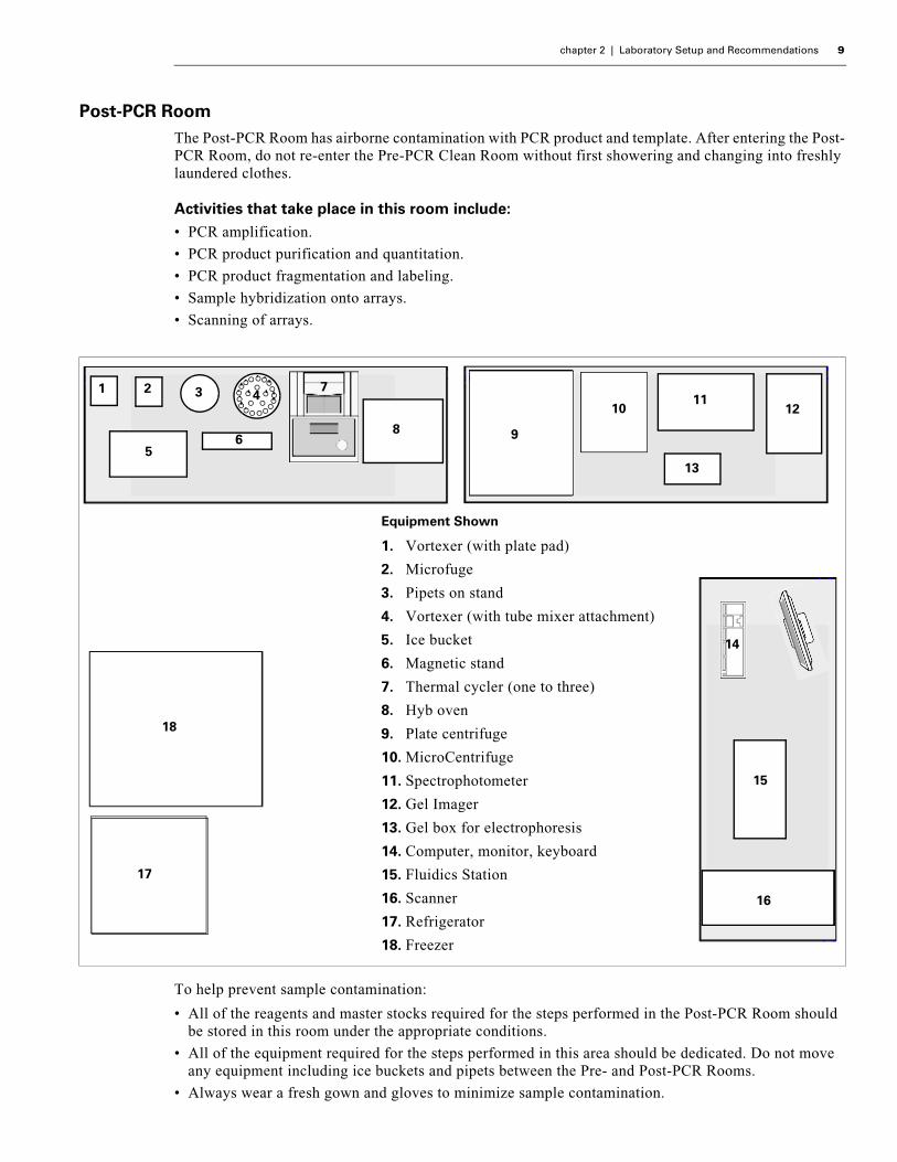
Post-PCR RoomThe Post-PCR Room has airborne contamination with PCR product and template. After entering the Post-PCR Room, do not re-enter the Pre-PCR Clean Room without first showering and changing into freshly laundered clothes.
Activities that take place in this room include:• PCR amplification.
• PCR product purification and quantitation.
• PCR product fragmentation and labeling.
• Sample hybridization onto arrays.
• Scanning of arrays.
To help prevent sample contamination:
• All of the reagents and master stocks required for the steps performed in the Post-PCR Room should be stored in this room under the appropriate conditions.
• All of the equipment required for the steps performed in this area should be dedicated. Do not move any equipment including ice buckets and pipets between the Pre- and Post-PCR Rooms.
• Always wear a fresh gown and gloves to minimize sample contamination.
Equipment Shown
1. Vortexer (with plate pad)
2. Microfuge
3. Pipets on stand
4. Vortexer (with tube mixer attachment)
5. Ice bucket
6. Magnetic stand
7. Thermal cycler (one to three)
8. Hyb oven
9. Plate centrifuge
10. MicroCentrifuge
11. Spectrophotometer
12. Gel Imager
13. Gel box for electrophoresis
14. Computer, monitor, keyboard
15. Fluidics Station
16. Scanner
17. Refrigerator
18. Freezer
1 2 3 7
5
4
6 8
911
13
14
8
16
17
18
12
9
10
15

10 Affymetrix® Cytogenetics Copy Number Assay User Guide
Configuration 2 — One RoomOne room with two distinctly separated areas: Pre-PCR Clean Area and Post-PCR Area.
Figure 2.2 One room configuration
2 3 4
6
5
7
Laminar Flow or PCR Cabinet
MARKING ON FLOOR TO DILINEATE PRE-PCR CLEAN AREA FROM POST-PCR AREA
1
Storage Area
8
1
11
2 3
45
678910
12
131415
16
17
18
Pre-PCR Clean Area
Post-PCR Area
7
1213
We strongly recommend the use of a laminar flow cabinet or a PCR cabinet when the entire assay is to be performed in one room.

chapter 2 | Laboratory Setup and Recommendations 11
Pre-PCR Clean AreaFor the best results, adhere to the following guidelines.
• Keep the Pre-PCR Clean Area
- Free of DNA template and PCR amplicons.
- Protected from contaminants by performing all steps inside a laminar flow cabinet or a PCR cabinet.
• If using a laminar flow cabinet, keep it turned on at all times.
• Keep the UV light in the laminar flow or PCR cabinet turned on when not in use.
• Always wear a gown, booties, and gloves to prevent PCR carryover, and to minimize the risk of trace levels of contaminants being brought into this area.
Equipment in Pre-PCR Clean Area
The equipment shown for the Pre-PCR Clean Area in Figure 2.2 on page 10 is listed below.
1. Laminar flow cabinet or PCR cabinet
2. Vortexer
3. Microfuge
4. Pipets on stand
5. Ice bucket
6. Thermal cycler
7. Plate centrifuge
8. Freezer
About Laminar Flow Cabinets
The air curtain from the laminar flow cabinet prevents the introduction of contaminants from the surrounding air into work area, particularly PCR products from the Post-PCR Area. Store master stocks of PCR primer and adaptor in the laminar flow cabinet.
Post-PCR AreaThe Post-PCR Area has airborne contamination with PCR product and template. After entering the Post-PCR Area it is inadvisable to re-enter the Pre-PCR Clean Area without first showering and changing into freshly laundered clothes.
The equipment shown for the Post-PCR Area in Figure 2.2 on page 10 consists of:
1. Computer, monitor and keyboard
2. Fluidics station
3. Scanner
4. Ice bucket
5. Magnetic stand
6. Vortexer with plate stand
IMPORTANT: We strongly recommend that each pre-PCR step be performed in a laminarflow or PCR cabinet, including reagent and master mix preparation. The use of this cabinetis essential for preventing sample contamination due to the introduction of PCR productsfrom the Post-PCR Area and DNA template.
All of the equipment required for the pre-PCR steps should be dedicated for pre-PCR andkept in the laminar flow or PCR cabinet. This equipment includes pipets and tips, the thermalcycler, and vortexer.

12 Affymetrix® Cytogenetics Copy Number Assay User Guide
7. Microfuge
8. Pipets on stand
9. Vortexer with tube mixer attachment
10. Thermal cycler (one to three)
11. Hybridization oven
12. Plate centrifuge
13. MicroCentrifuge
14. Spectrophotometer
15. Gel imager
16. Gel box for electrophoresis
17. Refrigerator
18. Freezer
chapter 2 | Laboratory Setup and Recommendations 13
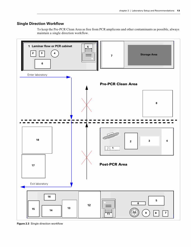
Single Direction WorkflowTo keep the Pre-PCR Clean Area as free from PCR amplicons and other contaminants as possible, always maintain a single direction workflow.
Figure 2.3 Single direction workflow
2 3 4
6
5
7
Laminar flow or PCR cabinet1
Storage Area
8
1
2 3 4
56
7891011
12
131415
16
17
18
Pre-PCR Clean Area
Post-PCR Area
Enter laboratory
Exit laboratory
7
1213

14 Affymetrix® Cytogenetics Copy Number Assay User Guide
Contamination PreventionCare should be taken to minimize possible sources of contamination that would reduce genotyping accuracy, call rate, and consequently, genetic power. To reduce the possibility of cross-contamination, Affymetrix strongly recommends that you maintain a single direction workflow: from the Pre-PCR Clean Area to the Post-PCR Area. Do not re-enter the Pre-PCR Clean Area from the Post-PCR Area.
Precautions that you can take to minimize contaminating pre-PCR steps with amplified PCR product include the following:
• Store reagents in the proper area according to the box label and reagent kit insert.
• Use proper gowning procedures.
• Print separate copies of the protocol for each room.
Safety PrecautionsThe Affymetrix® Genome-Wide Human SNP Nsp/Sty Assay Kit 5.0/6.0 as well as the Affymetrix® Genome-Wide Human SNP Array 6.0 are for research use only.
All blood and other potentially infectious materials should be handled as if capable of transmitting infection and disposed of with proper precautions in accordance with federal, state, and local regulations.
Wear appropriate personal protective equipment when performing this assay. At a minimum, safety glasses and chemical resistant gloves should be worn.
IMPORTANT:• The most likely potential source of contamination for the Cytogenetics Copy Number
Assay is previously amplified PCR product.
• Each area should contain dedicated equipment such as thermal cyclers, microfuges, pipetsand tips, ice buckets, etc.
• Once you enter the Post-PCR Area, do not return to the Pre-PCR Clean Area until you haveshowered and changed into freshly laundered clothing.
• Maintain an ambient laboratory environment throughout the procedure.
NOTE: Some components required for this assay may pose significant health risks. Followprudent laboratory practices when handling and disposing of carcinogens and toxins. Referto the manufacturer’s Material Safety Data Sheet for additional information.

Chapte
r3 GENOMIC DNA GENERAL REQUIREMENTSThe general requirements for genomic DNA sources and extraction methods are described in this chapter. The success of this assay requires the amplification of PCR fragments between 200 and 1100 bp in size throughout the genome. To achieve this, the genomic DNA must be of high quality, and must be free of contaminants that would affect the enzymatic reactions carried out.
For this protocol, you will use the Affymetrix® Genome-Wide Human SNP Nsp/Sty Assay Kit 5.0/6.0 (30 reaction; P/N 901013). This kit contains the genomic DNA control Reference Genomic DNA 103 (Ref 103). This control meets the requirements outlined below. The size of the starting genomic DNA can be compared with Ref103 DNA to assess the quality. The control DNA should also be used as a routine experimental positive control and for troubleshooting.
Assay performance may vary for genomic DNA samples that do not meet the general requirements described below. However, the reliability of any given result should be assessed in the context of overall experimental design and goals.
General Requirements• DNA must be double-stranded (not single-stranded).
This requirement relates to the restriction enzyme digestion step in the protocol.
• DNA must be free of PCR inhibitors.
Examples of inhibitors include high concentrations of heme (from blood) and high concentrations of chelating agents (i.e., EDTA). The genomic DNA extraction/purification method should render DNA that is generally salt-free because high concentrations of certain salts can also inhibit PCR and other enzyme reactions. DNA should be prepared as described in Chapter 4, Affymetrix® Cytogenetics Copy Number Assay.
• DNA must not be contaminated with other human genomic DNA sources, or with genomic DNA from other organisms.
PCR amplification of the ligated genomic DNA is not human specific, so sufficient quantities of non-human DNA may also be amplified and could potentially result in compromised genotype calls. Contaminated or mixed DNA may manifest as high detection rates and low call rates.
• DNA must not be highly degraded.
For any particular SNP, the genomic DNA fragment containing the SNP must have Nsp I (or Sty I) restriction sites intact so that ligation can occur on both ends of the fragment and PCR can be successful. The approximate average size of genomic DNA may be assessed on a 1% or 2% agarose gel using an appropriate size standard control. Ref 103 can be run on the same gel for side-by-side comparison. High quality genomic DNA will run as a major band at approximately 10-20 kb on the gel.
Pre-amplification methods or pre-digestion with restriction enzymes other than Nsp I or Sty I have not been tested by Affymetrix. If other methods are desired, we recommend conducting experiments to evaluate their performance with this assay.
16 Affymetrix® Cytogenetics Copy Number Assay User Guide
Sources of Human Genomic DNAThe following sources of human genomic DNA have been successfully tested in the laboratories at Affymetrix for DNA that meets the requirements described in the section General Requirements on page 15.
• blood
• cell lineSuccess with other types of samples such as saliva will depend on quality (degree of degradation, degree of inhibitors present, etc.), quantity of genomic DNA extracted, and purity of these types of samples, as described under General Requirements on page 15.
Genomic DNA Extraction/Purification MethodsGenomic DNA extraction and purification methods that meet the general requirements outlined above should yield successful results. Methods that include boiling or strong denaturants are not acceptable, because the DNA would be rendered single-stranded. Genomic DNA extracted using the following methods have been tested at Affymetrix:
1. SDS/ProK digestion, phenol-chloroform extraction, Microcon® or Centricon® (Millipore) ultrapurification and concentration.
2. QIAGEN; QIAamp® DNA Blood Maxi Kit.
DNA CleanupIf a genomic DNA preparation is suspected to contain inhibitors, the following cleanup procedure can be used:
1. Add 0.5 volumes of 7.5 M NH4OAc, 2.5 volumes of absolute ethanol (stored at –20°C), and 0.5 µL of glycogen (5 mg/mL) to 250 ng genomic DNA.
2. Vortex and incubate at –20°C for 1 hr.
3. Centrifuge at 12,000 x g in a microcentrifuge at room temperature for 20 min.
4. Remove supernatant and wash pellet with 0.5 mL of 80% ethanol.
5. Centrifuge at 12,000 g at room temperature for 5 min.
6. Remove the 80% ethanol and repeat the 80% ethanol wash one more time.
7. Resuspend the pellet in reduced EDTA TE buffer (10 mM Tris, pH 8.0, 0.1 mM EDTA, pH 8.0).
ReferencesFeigelson, H.S., Rodriguez, C., Robertson, A.S., Jacobs, E.J., Calle, E.E., Reid, Y.A., Thun, M.J. Determinants of DNA yield and quality from buccal cell samples collected with mouthwash. Cancer Epidemiol Biomarkers Prev. 10(9), 1005-8 (2001).
Heath, Ellen M., Morken, Nathaniel W., Campbell, Kristen A., Tkach, Dennis, Boyd, Erin A., Strom, Daniel A. Use of Buccal Cells Collected in Mouthwash as a Source of DNA for Clinical Testing. Arch Pathol Lab Med 125, 127-133 (2001).
King, I.B., Satia-Abouta, J., Thornquist, M.D., Bigler, J., Patterson, R.E., Kristal, A.R., Shattuck, A. L., Potter, J.D., White, E., Abouta, J.S. Buccal cell DNA yield, quality, and collection costs: comparison of methods for large-scale studies. Cancer Epidemiol Biomarkers Prev. 11(10 Pt 1), 1130-3 (2002).
Lench, N., Stanier, P., Williamson, R. Simple non-invasive method to obtain DNA for gene analysis. Lancet Jun 18;1(8599), 1356–1358 (1988).
Paez, J.G., Lin, M., Beroukhim, R., Lee, J.C., Zhao, X., Richter, D.J., Gabriel, S., Herman, P., Sasaki, H., Altshuler, D., Li, C., Meyerson, M., Sellers, W.R. Genome coverage and sequence fidelity of phi29 polymerase-based multiple strand displacement whole genome amplification. Nucleic Acids Research 32(9), (2004).

chapter 3 | Genomic DNA General Requirements 17
Tzvetkov, M.V., Becker, C., Kulle, B., Nurnberg, P., Brockmoller, J., Wojnowski, L. Genome-wide single-nucleotide polymorphism arrays demonstrate high fidelity of multiple displacement-based whole-genome amplification. Electrophoresis Feb;26(3):710-5 (2005).
Wong, K.K., Tsang, Y.T.M., Shen, J., Cheng, R.S., Chang, Y., Man, T., Lau, C.C. Allelic imbalance analysis by high-density single-nucleotide polymorphic allele (SNP) array with whole genome amplified DNA. Nucleic Acids Res. May 17;32(9):e69 (2004).

18 Affymetrix® Cytogenetics Copy Number Assay User Guide
Chapte
r4 AFFYMETRIX® CYTOGENETICS COPY NUMBER ASSAYAbout the ProtocolThe Affymetrix® Cytogenetics Copy Number Assay is designed for processing as few as four samples (including controls). The protocol is presented in the following stages:
• Genomic DNA Preparation on page 23
• Stage 1: Nsp and Sty Restriction Enzyme Digest on page 28
• Stage 2: Nsp and Sty Ligation on page 34
• Stage 3: Nsp and Sty PCR on page 41
• Stage 4: PCR Product Purification on page 51
• Stage 5: Quantitation on page 58
• Stage 6: Fragmentation on page 63
• Stage 7: Labeling on page 71
• Stage 8: Target Hybridization on page 75
IMPORTANT: The Cytogenetics Copy Number assay protocol is optimized for processingfrom 4 to 24 samples at a time to obtain copy number results. This protocol is not intendedfor genome-wide association studies.
An assay protocol for processing 48 samples is described in the Affymetrix® Genome-WideHuman SNP Nsp/Sty 6.0 User Guide, P/N 702504.
20 Affymetrix® Cytogenetics Copy Number Assay User Guide
About the Illustrations in this ChapterThis protocol has been optimized for processing 4 to 24 samples. The illustrations in this chapter are based on running 8 samples: 6 genomic DNA samples, plus 1 positive and 1 negative control. Use these illustrations as guidelines when processing 8 or fewer samples.
If processing 9 to 24 samples, refer to Appendix A, Guidelines for Processing 16 Samples or Appendix B, Guidelines for Processing 24 Samples. Important guidelines for plate layouts are included in these appendices.
About the Reagents, Equipment and Consumables Specified in this Chapter
Genome-Wide Human SNP Nsp/Sty Assay Kit 5.0/6.0 — 30 Reactions
Equipment, Consumables, and Other Reagents
IMPORTANT: Always use the 30 reaction Genome-Wide Human SNP Nsp/Sty Assay Kit5.0/6.0 for this protocol. This kit has been tested for multiple freeze/thaw cycles. You canfreeze/thaw the reagents in the 30 reaction kit ≤ 8 times.
IMPORTANT: This protocol has been optimized using the equipment, consumables andreagents listed herein. For the best results, we strongly recommend that you adhere to theprotocol as described (no deviations); do not substitute reagents.

chapter 4 | Affymetrix® Cytogenetics Copy Number Assay 21
Workflows
Recommended 4-Day WorkflowFigure 4.1 shows the recommended 4-day workflow for one operator processing four to 24 samples including controls.
Figure 4.1 Workflow recommended for processing one to 24 samples
22 Affymetrix® Cytogenetics Copy Number Assay User Guide
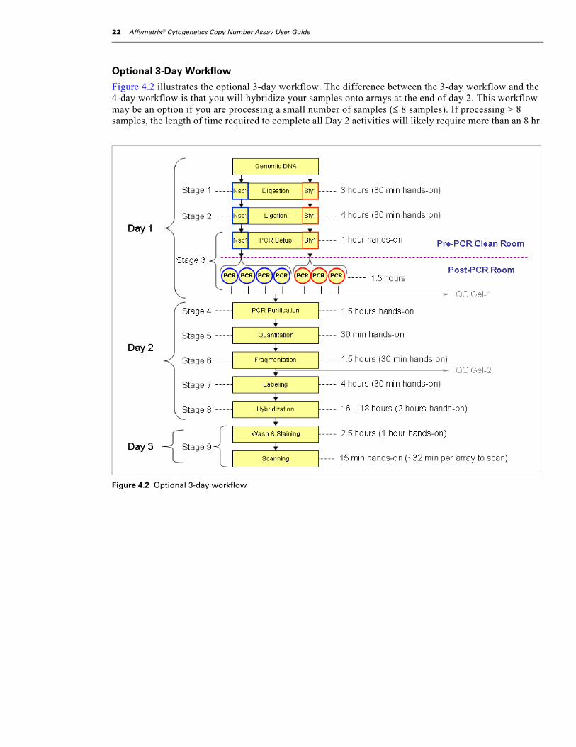
Optional 3-Day Workflow
Figure 4.2 illustrates the optional 3-day workflow. The difference between the 3-day workflow and the 4-day workflow is that you will hybridize your samples onto arrays at the end of day 2. This workflow may be an option if you are processing a small number of samples (≤ 8 samples). If processing > 8 samples, the length of time required to complete all Day 2 activities will likely require more than an 8 hr.
Figure 4.2 Optional 3-day workflow

chapter 4 | Affymetrix® Cytogenetics Copy Number Assay 23
Genomic DNA Preparation
About this StageThe human genomic DNA you will process using the Cytogenetics Copy Number Assay should meet the general requirements listed in Chapter 3, Genomic DNA General Requirements. During this stage, you will prepare the genomic DNA by:
1. Determining the concentration of each sample (if required).
2. Diluting each sample to 50 ng/µL using reduced EDTA TE buffer.
Location and Duration• Pre-PCR Clean Area
• Hands-on time: dependent upon number of samples to be processed
Input RequiredThe illustrations in this user guide depict the processing of eight samples: six genomic DNA samples, plus one positive and one negative control.
About Using ControlsWe recommend including one positive and one negative control with every set of samples processed. For the positive control, use the Ref 103 included in the Genome-Wide Human SNP Nsp/Sty Assay Kit 5.0/6.0. For the negative control, use water (AccuGENE).
Table 4.1 Input Required for Genomic DNA Preparation
Quantity Item
4 to 24 Genomic DNA samples that meet the requirements listed in Chapter 3, Genomic DNA General Requirements.

24 Affymetrix® Cytogenetics Copy Number Assay User Guide
Equipment and Consumables RequiredThe equipment and consumables listed in Table 4.2 are required for this stage.
Reagents RequiredThe following reagents are required for this stage.
Table 4.2 Equipment and Consumables Required for Genomic DNA Preparation
Quantity Item
As required Adhesive seals for 96-well plates
1 Cooling chamber, double, chilled to 4 °C on ice (do not freeze)
1 Ice bucket, filled with ice
2 Markers, red and blue, fine point, permanent
1 Mini microcentrifuge (microfuge)
1 Pipet, single channel P20
1 Pipet, single channel P100 or P200
As needed Pipet tips
2 Plate, Bio-Rad 96-well unskirted
1 Plate centrifuge
1 Plate spectrophotometer or NanoDrop(required only if no OD measurements available for samples)
1 Vortexer
** IMPORTANT Use only the thermal cyclers, 96-well plate, and adhesive films and listed under Thermal Cyclers, 96-well Plate, and Adhesive Seals on page 4.
Table 4.3 Reagents Required for Genomic DNA Preparation
Reagent
Reduced EDTA TE Buffer (0.1 mM EDTA, 10 mM Tris HCL, pH 8.0)
Reference Genomic DNA 103 (positive control)
AccuGENE water (negative control)

chapter 4 | Affymetrix® Cytogenetics Copy Number Assay 25
Preparing the Genomic DNAThis protocol has been optimized using UV absorbance to determine genomic DNA concentrations. Other quantitation methods such as PicoGreen may give different readings. Therefore, you should correlate readings from other methods to the equivalent UV absorbance reading.
Setup the Work Area
To setup the work area:
1. Place a double cooling chamber on ice (Figure 4.3).
2. Place a 96-well plate in the top half of the cooling chamber.
.
Dilute the Genomic DNA
To dilute the genomic DNA:
1. Thaw the genomic DNA (gDNA) and Ref 103 as follows:
A. Place on the bench top at room temperature until thawed.
B. Once thawed, place in the cooling chamber on ice.
2. Vortex the gDNA samples at high speed for 3 sec.
3. Spin down for 30 sec; then place back in the cooling chamber.
4. If sample concentration is unknown, take an OD measurement of each sample now.
Figure 4.3 Diluting genomic DNA samples to 50 ng/µL
NOTE: The illustrations in this user guide depict the setup recommended for eight samples:six genomic DNA samples plus one positive control and one negative control.
If running less than eight samples, follow the same plate layout.
If running more than eight samples, refer to Appendix A, Guidelines for Processing 16Samples or Appendix B, Guidelines for Processing 24 Samples for more information.
Reference Genomic DNA 103 — do NOT dilute
1
2
3
4
5
6
+
Genomic DNA samples
5
2
3
4
6
1
Reduced EDTA TE Buffer
26 Affymetrix® Cytogenetics Copy Number Assay User Guide
Apply the convention that 1 absorbance unit at 260 nm equals 50 µg/mL for double-stranded DNA. This convention assumes a path length of 1 cm. Consult your spectrophotometer handbook for more information. If using a method other than UV absorbance, correlate the reading to the equivalent UV absorbance reading.
5. Based on OD measurements, dilute each sample in a separate well of the 96-well plate to 50 ng/µL using reduced EDTA TE buffer.
6. Seal the plate, vortex at high speed for 3 sec; then spin down for 30 sec.
7. Place back on the cooling chamber.
Aliquoting the Prepared Genomic DNA and Controls
Setup the Work Area
To setup the work area:
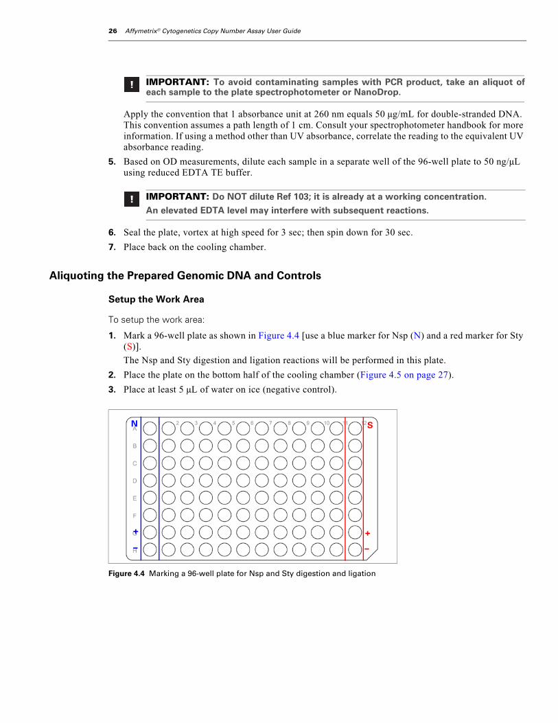
1. Mark a 96-well plate as shown in Figure 4.4 [use a blue marker for Nsp (N) and a red marker for Sty (S)].
The Nsp and Sty digestion and ligation reactions will be performed in this plate.
2. Place the plate on the bottom half of the cooling chamber (Figure 4.5 on page 27).
3. Place at least 5 µL of water on ice (negative control).
IMPORTANT: To avoid contaminating samples with PCR product, take an aliquot ofeach sample to the plate spectrophotometer or NanoDrop.
IMPORTANT: Do NOT dilute Ref 103; it is already at a working concentration.
An elevated EDTA level may interfere with subsequent reactions.
Figure 4.4 Marking a 96-well plate for Nsp and Sty digestion and ligation
N S
+
–
+
–

chapter 4 | Affymetrix® Cytogenetics Copy Number Assay 27
Aliquot the gDNA and Controls
To aliquot the prepared genomic DNA and controls:
1. Vortex the Ref 103 for 3 sec; then spin down for 30 sec.
2. Transfer two 5 µL aliquots of the first sample to wells A1 and A12 of the digest/ligate plate (Figure 4.5 on page 27).
3. Transfer two 5 µL aliquots of each remaining gDNA sample in the same manner.
4. For the controls, aliquot 5 µL of:
A. Ref 103 (+) to wells G1 and G12.
B. Water (–) to wells H1 and H12.
5. Tightly seal the digest/ligate plate.
What To Do NextDo one of the following:
• Proceed to Stage 1: Nsp and Sty Restriction Enzyme Digest on page 28.
• Store the prepared digest/ligate plate at –20 °C.
NOTE: 5 μL of the 50 ng/μL working stock is equivalent to 250 ng genomic DNA per well.
Figure 4.5 Setup for aliquoting diluted gDNA and controls to a 96-well plate labeled for Nsp and Sty digest/ligation
Diluted gDNAsamples
5
2
3
4
6
SN
1
+
–
+
–
96-well platelabeled for Nsp and
Sty digestion andligation
Water, AccuGENE
Ref 103 positive control
Transfer two 5 μL aliquots of each diluted gDNA to the digest/ligate plate — one for Nsp reactions; one for Sty reactions. + = positive control (5 μL Ref 103) – = negative control (5 μL water)

28 Affymetrix® Cytogenetics Copy Number Assay User Guide
Stage 1: Nsp and Sty Restriction Enzyme Digest
About this StageDuring this stage, one aliquot of each sample is digested by the NspI restriction enzyme; the other aliquot by the StyI restriction enzyme. You will:
1. Prepare a Nsp Digest Master Mix and add it to the samples in column 1.
2. Prepare a Sty Digest Master Mix and add it to the samples in column 12.
3. Place the samples onto a thermal cycler and run the Cyto Digest program.
Location and Duration• Pre-PCR Clean Area
• Hands-on time: 30 min
• Cyto Digest thermal cycler program time: 2.5 hr
Input Required From Previous StageThe input required is shown below.
Item
Plate containing two equal aliquots of each genomic DNA and each control prepared as instructed under Genomic DNA Preparation on page 23 (5 µL at 50 ng/µL in each well).

chapter 4 | Affymetrix® Cytogenetics Copy Number Assay 29
Equipment and Consumables RequiredThe following equipment and consumables are required for this stage.
Reagents RequiredThe following reagents are required for this stage.
Table 4.4 Equipment and Consumables Required for Stage 1: Nsp and Sty Restriction Enzyme Digest
Quantity Item
As required Adhesive seals for 96-well plates
1 Centrifuge, plate
1 Cooler, chilled to –20 °C
1 Cooling chamber, double, chilled to 4 °C on ice (do not freeze)
1 Ice bucket, filled with ice
1 Markers, blue and red, fine point, permanent
1 Mini centrifuge (microfuge)
1 Pipet, single channel P10
1 Pipet, single channel P100 or P200
As required Pipet tips for pipets listed above
1 Thermal cycler
2 Tubes, Eppendorf 1.5 mL
1 Vortexer
** IMPORTANT Use only the thermal cyclers, 96-well plate, and adhesive films and listed under Thermal Cyclers, 96-well Plate, and Adhesive Seals on page 4.
Table 4.5 Reagents Required for Stage 1: Nsp and Sty Restriction Enzyme Digest
Reagent
BSA (100X; 10 mg/mL)
NE Buffer 2 (10X)
NE Buffer 3 (10X)
NspI (10 U/µL; NEB)
StyI (10 U/µL; NEB)
AccuGENE® Water, molecular biology-grade
30 Affymetrix® Cytogenetics Copy Number Assay User Guide
Prepare the Reagents, Equipment and Consumables
Thaw Reagents and Genomic DNA
1. Allow the following reagents to thaw on ice:
• NE Buffer 2
• NE Buffer 3
• BSA
2. If the plate of genomic DNA and controls is frozen, allow it to thaw in a cooling chamber on ice.
Setup the Work Area
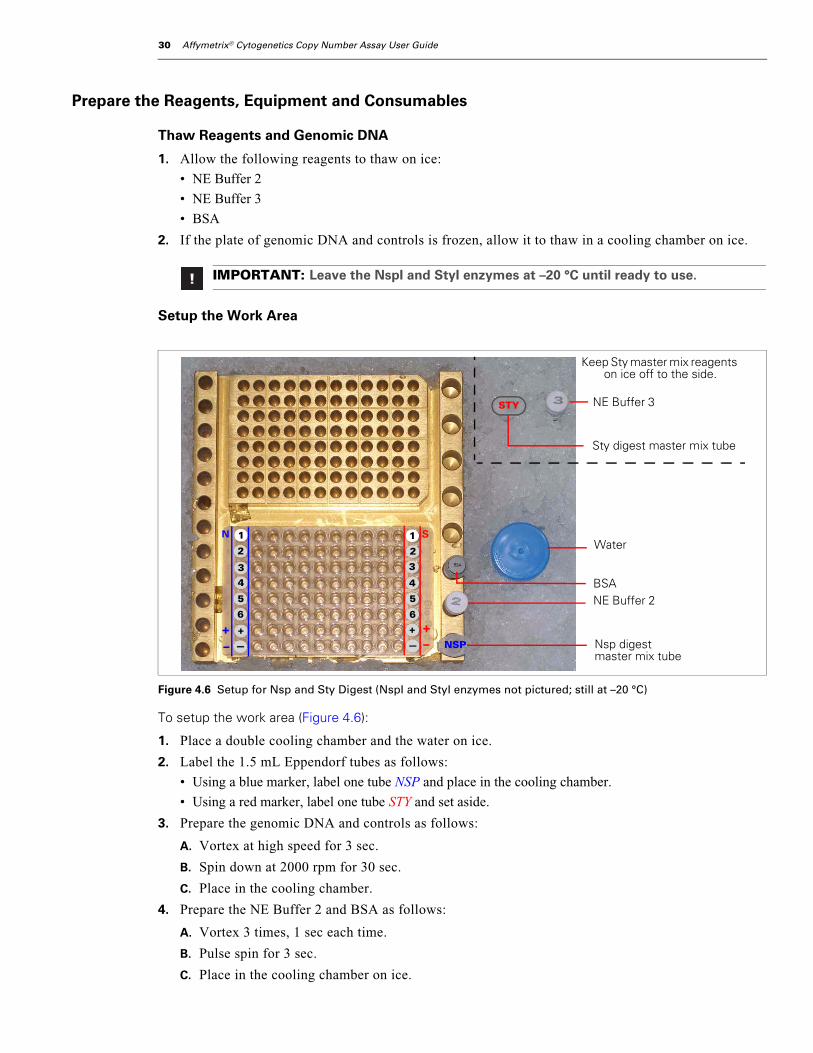
To setup the work area (Figure 4.6):
1. Place a double cooling chamber and the water on ice.
2. Label the 1.5 mL Eppendorf tubes as follows:
• Using a blue marker, label one tube NSP and place in the cooling chamber.
• Using a red marker, label one tube STY and set aside.
3. Prepare the genomic DNA and controls as follows:
A. Vortex at high speed for 3 sec.
B. Spin down at 2000 rpm for 30 sec.
C. Place in the cooling chamber.
4. Prepare the NE Buffer 2 and BSA as follows:
A. Vortex 3 times, 1 sec each time.
B. Pulse spin for 3 sec.
C. Place in the cooling chamber on ice.
IMPORTANT: Leave the NspI and StyI enzymes at –20 °C until ready to use.
Figure 4.6 Setup for Nsp and Sty Digest (NspI and StyI enzymes not pictured; still at –20 °C)
5
+
2
3
4
6
1
—
5
+
2
3
4
6
1
—
SN
+ +–– NSP
STY
Water
BSANE Buffer 2
Nsp digest master mix tube
NE Buffer 3
Sty digest master mix tube
Keep Sty master mix reagents on ice off to the side.

chapter 4 | Affymetrix® Cytogenetics Copy Number Assay 31
Preheat the Thermal Cycler Lid
Power on the thermal cycler to preheat the lid. Leave the block at room temperature.
Prepare the Nsp Digest Master Mix
Keeping all reagents and tubes on ice, prepare the Nsp Digest Master Mix as follows:
1. To the 1.5 mL Eppendorf tube labeled NSP, add the appropriate volumes of the following reagents (see Table 4.6):
• Water (AccuGENE)
• NE Buffer 2
• BSA
2. Place the master mix in the cooling chamber.
3. Remove the NspI enzyme from the freezer and immediately place in a cooler.
4. Pulse spin the enzyme for 3 sec.
5. Immediately add the enzyme to the master mix.
6. Return the enzyme to the cooler.
7. Vortex the master mix at high speed 3 times, 1 sec each time.
8. Pulse spin for 3 sec.
9. Place in the cooling chamber.
10. Proceed immediately to Add Nsp Digest Master Mix to Samples on page 31.
Add Nsp Digest Master Mix to Samples
To add Nsp Digest Master Mix to samples:
1. Aliquot 14.75 µL of Nsp Digest Master Mix to each sample and controls in column 1.
2. Return remaining NE Buffer 2 and NspI enzyme to the freezer.
3. Discard remaining Nsp Digest Master Mix.
Table 4.6 NspI Digest Master Mix
Reagent 1 Sample 4 Samples(25% extra**)
8 Samples(15% extra)
12 Samples(15% extra)
24 Samples(15% extra)
AccuGENE® Water 11.55 µL 57.8 µL 106.3 µL 159.4 µL 318.8 µL
NE Buffer 2 (10X) 2 µL 10 µL 18.4 µL 27.6 µL 55.2 µL
BSA (100X; 10 mg/mL) 0.2 µL 1 µL 1.8 µL 2.8 µL 5.5 µL
NspI (10 U/µL) 1 µL 5 µL 9.2 µL 13.8 µL 27.6 µL
Total 14.75 µL 73.8 µL 135.7 µL 203.6 µL 407.1 µL
** To avoid pipetting < 1 μL of BSA, prepare 25% extra when processing ≤ 4 samples.
Genomic DNA (50 ng/µL) 5.00 µL
Nsp Digest Master Mix 14.75 µL
Total Volume 19.75 µL

32 Affymetrix® Cytogenetics Copy Number Assay User Guide
Prepare the Sty Digest Master Mix
Keeping all reagents and tubes on ice, prepare the Sty Digest Master Mix as follows:
1. To the 1.5 mL Eppendorf tube labeled STY, add the appropriate volumes of the following reagents as shown in Table 4.7:
• Water (AccuGENE)
• NE Buffer 3
• BSA
2. Place the master mix in the cooling chamber.
3. Remove the StyI enzyme from the freezer and immediately place in a cooler.
4. Pulse spin the enzyme for 3 sec.
5. Immediately add the enzyme to the master mix.
6. Return remaining enzyme to the cooler.
7. Vortex the master mix at high speed 3 times, 1 sec each time.
8. Pulse spin for 3 sec.
9. Place in the cooling chamber.
10. Proceed immediately to Add Sty Digest Master Mix to Samples on page 33.
Figure 4.7 Adding Nsp Digest Master Mix to gDNA samples and controls
Table 4.7 StyI Digest Master Mix
Reagent 1 Sample 4 Samples(25% extra**)
8 Samples(15% extra)
12 Samples(15% extra)
24 Samples(15% extra)
AccuGENE® Water 11.55 µL 57.8 µL 106.3 µL 159.4 µL 318.8 µL
NE Buffer 3 (10X) 2 µL 10 µL 18.4 µL 27.6 µL 55.2 µL
BSA (100X; 10 mg/mL) 0.2 µL 1 µL 1.8 µL 2.8 µL 5.5 µL
StyI (10 U/µL) 1 µL 5 µL 9.2 µL 13.8 µL 27.6 µL
Total 14.75 µL 73.8 µL 135.7 µL 203.6 µL 407.1 µL
** 25% extra is required for 4 samples only. If processing 8 samples, 15% extra is sufficient.
N S
+
–
+
–
Add 14.75 μL Nsp DigestMaster Mix to each sample
and controls in column 1.

chapter 4 | Affymetrix® Cytogenetics Copy Number Assay 33
Add Sty Digest Master Mix to Samples
To add the Sty Digest Master Mix to samples:
1. Aliquot 14.75 µL of Sty Digest Master Mix to each sample and control in column 12.
The total volume in each well is now 19.75 µL.
2. Tightly seal the plate.
Load Nsp and Sty Samples onto the Thermal Cycler
1. Vortex the plate at high speed for 3 sec; then spin down at 2000 rpm for 30 sec.
2. Ensure that the lid of thermal cycler is preheated.
3. Load the plate onto the thermal cycler and run the Cyto Digest program (Table 4.8).
4. Return any remaining reagents to the freezer.
5. When the program is finished, remove the plate and spin down at 2000 rpm for 30 sec.
What To Do NextDo one of the following:
• If following the recommended workflow (Figure 4.1 on page 21), place the plate in a cooling chamber on ice and proceed immediately to Stage 2: Nsp and Sty Ligation on page 34.
• If not proceeding directly to the next step, store the plate at –20 °C.
Figure 4.8 Adding Sty Digest Master Mix
IMPORTANT: Ensure that the seal is not pulled off the wells when you close thethermal cycler lid.
Table 4.8 Cyto Digest Program
Cyto Digest Program
Temperature Time
37 °C 120 min
65 °C 20 min
4 °C Hold
N S
+
–
+
–
Add 14.75 μL Sty Digest Master Mix to each sample and control in column 12.
34 Affymetrix® Cytogenetics Copy Number Assay User Guide
Stage 2: Nsp and Sty Ligation
About this StageDuring this stage, the Nsp digested samples are ligated using the Nsp Adaptor; the Sty digested samples are ligated using the Sty Adaptor. You will:
1. Prepare a Nsp Ligation Master Mix and add it to the Nsp digested samples.
2. Prepare a Sty Ligation Master Mix and add it to the Sty digested samples.
3. Place samples onto a thermal cycler and run the Cyto Ligate program.
4. Dilute the ligated samples with water.
Location and Duration• Pre-PCR Clean Area
• Hands-on time: 30 min
• Cyto Ligate thermal cycler program time: 3.3 hr
Input Required From Previous StageThe input required from Stage 1: Nsp and Sty Restriction Enzyme Digest is:
Item
Plate of Nsp and Sty digested samples
chapter 4 | Affymetrix® Cytogenetics Copy Number Assay 35
Equipment and Consumables RequiredThe following equipment and consumables are required for this stage.
Reagents RequiredThe following reagents are required for this stage.
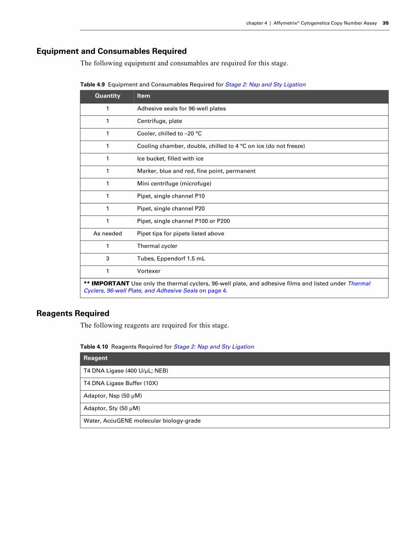
Table 4.9 Equipment and Consumables Required for Stage 2: Nsp and Sty Ligation
Quantity Item
1 Adhesive seals for 96-well plates
1 Centrifuge, plate
1 Cooler, chilled to –20 °C
1 Cooling chamber, double, chilled to 4 °C on ice (do not freeze)
1 Ice bucket, filled with ice
1 Marker, blue and red, fine point, permanent
1 Mini centrifuge (microfuge)
1 Pipet, single channel P10
1 Pipet, single channel P20
1 Pipet, single channel P100 or P200
As needed Pipet tips for pipets listed above
1 Thermal cycler
3 Tubes, Eppendorf 1.5 mL
1 Vortexer
** IMPORTANT Use only the thermal cyclers, 96-well plate, and adhesive films and listed under Thermal Cyclers, 96-well Plate, and Adhesive Seals on page 4.
Table 4.10 Reagents Required for Stage 2: Nsp and Sty Ligation
Reagent
T4 DNA Ligase (400 U/µL; NEB)
T4 DNA Ligase Buffer (10X)
Adaptor, Nsp (50 µM)
Adaptor, Sty (50 µM)
Water, AccuGENE molecular biology-grade
36 Affymetrix® Cytogenetics Copy Number Assay User Guide
Prepare the Reagents, Consumables and Other Components
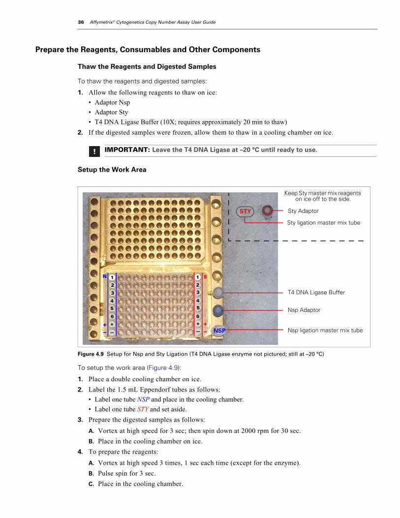
Thaw the Reagents and Digested Samples
To thaw the reagents and digested samples:
1. Allow the following reagents to thaw on ice:
• Adaptor Nsp
• Adaptor Sty
• T4 DNA Ligase Buffer (10X; requires approximately 20 min to thaw)
2. If the digested samples were frozen, allow them to thaw in a cooling chamber on ice.
Setup the Work Area
To setup the work area (Figure 4.9):
1. Place a double cooling chamber on ice.
2. Label the 1.5 mL Eppendorf tubes as follows:
• Label one tube NSP and place in the cooling chamber.
• Label one tube STY and set aside.
3. Prepare the digested samples as follows:
A. Vortex at high speed for 3 sec; then spin down at 2000 rpm for 30 sec.
B. Place in the cooling chamber on ice.
4. To prepare the reagents:
A. Vortex at high speed 3 times, 1 sec each time (except for the enzyme).
B. Pulse spin for 3 sec.
C. Place in the cooling chamber.
IMPORTANT: Leave the T4 DNA Ligase at –20 °C until ready to use.
Figure 4.9 Setup for Nsp and Sty Ligation (T4 DNA Ligase enzyme not pictured; still at –20 °C)
Nsp ligation master mix tube
Sty Adaptor
5
+
2
3
4
6
1
—
5
+
23
4
6
1
—
SN
NSP
T4 DNA Ligase Buffer
Nsp Adaptor
+–
+–
Sty ligation master mix tube
STY
Keep Sty master mix reagents on ice off to the side.

chapter 4 | Affymetrix® Cytogenetics Copy Number Assay 37
Preheat the Thermal Cycler LidPower on the thermal cycler to preheat the lid. Leave the block at room temperature.
The lid must be preheated before samples are loaded.
Prepare the Nsp Ligation Master Mix
Keeping all reagents and tubes on ice, prepare the Nsp Ligation Master Mix as follows:
1. To the 1.5 mL Eppendorf tube labeled NSP, add the following reagents based on the volumes shown in Table 4.11:
• T4 DNA Ligase Buffer (10X)
• Adaptor Nsp
2. Remove the T4 DNA Ligase from the freezer and immediately place in the cooler.
3. Pulse spin the T4 DNA Ligase for 3 sec.
4. Immediately add the T4 DNA Ligase to the master mix; then place back in the cooler.
5. Vortex the master mix at high speed 3 times, 1 sec each time.
6. Pulse spin for 3 sec.
7. Place the master mix on ice.
8. Proceed immediately to Add Nsp Ligation Master Mix to Reactions.
IMPORTANT: T4 DNA Ligase Buffer (10X) contains ATP and should be thawed on ice.Vortex the buffer as long as necessary before use to ensure precipitate is re-suspended and that the buffer is clear.
Table 4.11 NspI Ligation Master Mix
Reagent 1 Sample 4 Samples(15% extra)
8 Samples(15% extra)
12 Samples (15% extra)
24 Samples (15% extra)
T4 DNA Ligase Buffer (10X)
2.5 µL 11.5 µL 23.0 µL 34.5 µL 69 µL
Adaptor, Nsp(50 µM)
0.75 µL 3.45 µL 6.90 µL 10.35 µL 20.7 µL
T4 DNA Ligase (400 U/µL)
2 µL 9.2 µL 18.4 µL 27.6 µL 55.2 µL
Total 5.25 µL 24.15 µL 48.30 µL 72.45 µL 144.90 µL

38 Affymetrix® Cytogenetics Copy Number Assay User Guide
Add Nsp Ligation Master Mix to Reactions
To add Nsp Ligation Master Mix to samples:
1. Using a P20 pipet, aliquot 5.25 µL of Nsp Ligation Master Mix to each Nsp digested sample and control (Figure 4.10).
2. Discard any remaining Nsp Ligation Master Mix.
Prepare the Sty Ligation Master Mix
Keeping all reagents and tubes on ice, prepare the Sty Ligation Master Mix as follows:
1. To the 1.5 mL Eppendorf tube labeled STY, add the following reagents based on the volumes shown in Table 4.12 on page 39:
• T4 DNA Ligase Buffer (10X)
• Adaptor, Sty
2. Immediately add the T4 DNA Ligase to the master mix; then place back in the cooler.
3. Vortex the master mix at high speed 3 times, 1 sec each time.
4. Pulse spin for 3 sec.
5. Place the master mix on ice.
6. Proceed immediately to Add Sty Ligation Master Mix to Reactions.
Nsp Digested DNA 19.75 µL
Nsp Ligation Master Mix* 5.25 µL
Total 25.00 µL
* Contains ATP and DTT. Keep on ice.
Figure 4.10 Adding Nsp ligate master mix to Nsp digested samples and controls
N S
+
–
+
–
Add 5.25 μL Nsp LigationMaster Mix to each sample
and control in column 1.
chapter 4 | Affymetrix® Cytogenetics Copy Number Assay 39
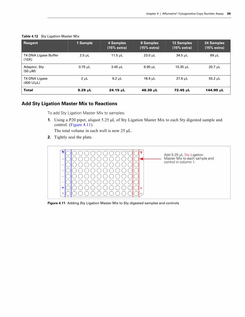
Add Sty Ligation Master Mix to Reactions
To add Sty Ligation Master Mix to samples:
1. Using a P20 pipet, aliquot 5.25 µL of Sty Ligation Master Mix to each Sty digested sample and control. (Figure 4.11).
The total volume in each well is now 25 µL.
2. Tightly seal the plate.
Table 4.12 Sty Ligation Master Mix
Reagent 1 Sample 4 Samples(15% extra)
8 Samples(15% extra)
12 Samples (15% extra)
24 Samples (15% extra)
T4 DNA Ligase Buffer (10X)
2.5 µL 11.5 µL 23.0 µL 34.5 µL 69 µL
Adaptor, Sty (50 µM)
0.75 µL 3.45 µL 6.90 µL 10.35 µL 20.7 µL
T4 DNA Ligase (400 U/µL)
2 µL 9.2 µL 18.4 µL 27.6 µL 55.2 µL
Total 5.25 µL 24.15 µL 48.30 µL 72.45 µL 144.90 µL
Figure 4.11 Adding Sty Ligation Master Mix to Sty digested samples and controls
N S
+
–
+
–
Add 5.25 μL Sty Ligation Master Mix to each sample and control in column 1.
40 Affymetrix® Cytogenetics Copy Number Assay User Guide
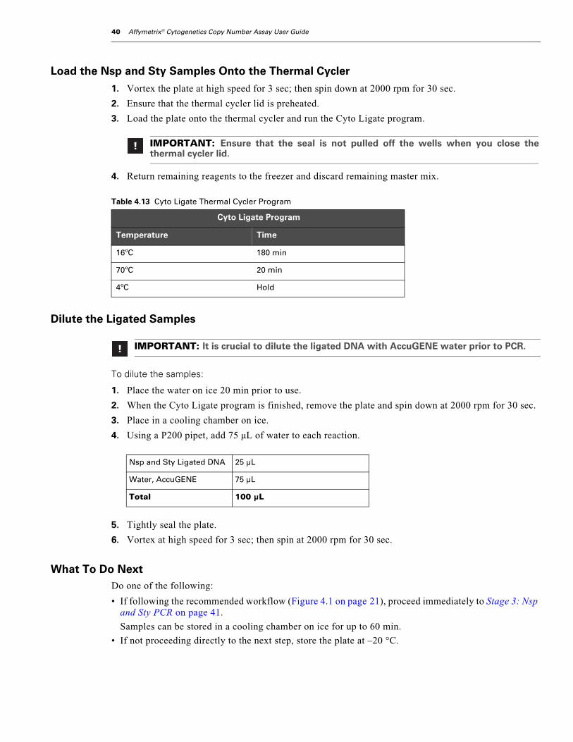
Load the Nsp and Sty Samples Onto the Thermal Cycler
1. Vortex the plate at high speed for 3 sec; then spin down at 2000 rpm for 30 sec.
2. Ensure that the thermal cycler lid is preheated.
3. Load the plate onto the thermal cycler and run the Cyto Ligate program.
4. Return remaining reagents to the freezer and discard remaining master mix.
Dilute the Ligated Samples
To dilute the samples:
1. Place the water on ice 20 min prior to use.
2. When the Cyto Ligate program is finished, remove the plate and spin down at 2000 rpm for 30 sec.
3. Place in a cooling chamber on ice.
4. Using a P200 pipet, add 75 µL of water to each reaction.
5. Tightly seal the plate.
6. Vortex at high speed for 3 sec; then spin at 2000 rpm for 30 sec.
What To Do NextDo one of the following:
• If following the recommended workflow (Figure 4.1 on page 21), proceed immediately to Stage 3: Nsp and Sty PCR on page 41.
Samples can be stored in a cooling chamber on ice for up to 60 min.
• If not proceeding directly to the next step, store the plate at –20 °C.
IMPORTANT: Ensure that the seal is not pulled off the wells when you close thethermal cycler lid.
Table 4.13 Cyto Ligate Thermal Cycler Program
Cyto Ligate Program
Temperature Time
16ºC 180 min
70ºC 20 min
4ºC Hold
IMPORTANT: It is crucial to dilute the ligated DNA with AccuGENE water prior to PCR.
Nsp and Sty Ligated DNA 25 µL
Water, AccuGENE 75 µL
Total 100 µL

chapter 4 | Affymetrix® Cytogenetics Copy Number Assay 41
Stage 3: Nsp and Sty PCR
About this StageDuring this stage, you will:
1. Transfer equal aliquots of each:
• Nsp diluted ligated sample into four wells of a 96-well plate.
• Sty diluted ligated sample into three wells of the same 96-well plate.
2. Prepare a PCR Master Mix and add it to each ligated sample.
3. Place the samples onto a thermal cycler and run the Cyto PCR program.
4. Confirm each PCR reaction by running 3 µL of each PCR product on a gel.
Location and Duration• Pre-PCR Clean Area
- PCR Master Mix preparation
- PCR set up
• Post-PCR Area: samples placed on thermal cycler
• Hands-on time: 1 hr
• Cyto PCR thermal cycler program time: 1.5 hr
Samples can be held overnight at 4 °C.
Input Required from Previous StageThe input required from Stage 2: Nsp and Sty Ligation is:
Item
Diluted ligated Nsp and Sty samples
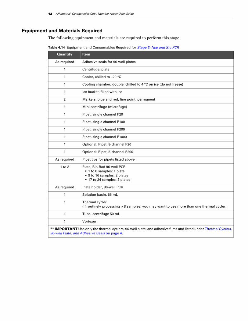
42 Affymetrix® Cytogenetics Copy Number Assay User Guide
Equipment and Materials RequiredThe following equipment and materials are required to perform this stage.
Table 4.14 Equipment and Consumables Required for Stage 3: Nsp and Sty PCR
Quantity Item
As required Adhesive seals for 96-well plates
1 Centrifuge, plate
1 Cooler, chilled to –20 °C
1 Cooling chamber, double, chilled to 4 °C on ice (do not freeze)
1 Ice bucket, filled with ice
2 Markers, blue and red, fine point, permanent
1 Mini centrifuge (microfuge)
1 Pipet, single channel P20
1 Pipet, single channel P100
1 Pipet, single channel P200
1 Pipet, single channel P1000
1 Optional: Pipet, 8-channel P20
1 Optional: Pipet, 8-channel P200
As required Pipet tips for pipets listed above
1 to 3 Plate, Bio-Rad 96-well PCR• 1 to 8 samples: 1 plate • 9 to 16 samples: 2 plates• 17 to 24 samples: 3 plates
As required Plate holder, 96-well PCR
1 Solution basin, 55 mL
1 Thermal cycler(If routinely processing > 8 samples, you may want to use more than one thermal cycler.)
1 Tube, centrifuge 50 mL
1 Vortexer
** IMPORTANT Use only the thermal cyclers, 96-well plate, and adhesive films and listed under Thermal Cyclers, 96-well Plate, and Adhesive Seals on page 4.
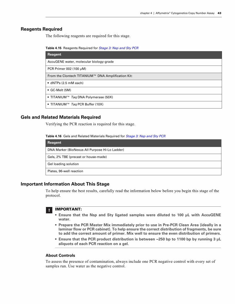
chapter 4 | Affymetrix® Cytogenetics Copy Number Assay 43
Reagents RequiredThe following reagents are required for this stage.
Gels and Related Materials RequiredVerifying the PCR reaction is required for this stage.
Important Information About This StageTo help ensure the best results, carefully read the information below before you begin this stage of the protocol.
About ControlsTo assess the presence of contamination, always include one PCR negative control with every set of samples run. Use water as the negative control.
Table 4.15 Reagents Required for Stage 3: Nsp and Sty PCR
Reagent
AccuGENE water, molecular biology-grade
PCR Primer 002 (100 µM)
From the Clontech TITANIUM™ DNA Amplification Kit:
• dNTPs (2.5 mM each)
• GC-Melt (5M)
• TITANIUM™ Taq DNA Polymerase (50X)
• TITANIUM™ Taq PCR Buffer (10X)
Table 4.16 Gels and Related Materials Required for Stage 3: Nsp and Sty PCR
Reagent
DNA Marker (BioNexus All Purpose Hi-Lo Ladder)
Gels, 2% TBE (precast or house-made)
Gel loading solution
Plates, 96-well reaction
IMPORTANT:• Ensure that the Nsp and Sty ligated samples were diluted to 100 μL with AccuGENE
water.
• Prepare the PCR Master Mix immediately prior to use in Pre-PCR Clean Area (ideally in alaminar flow or PCR cabinet). To help ensure the correct distribution of fragments, be sureto add the correct amount of primer. Mix well to ensure the even distribution of primers.
• Ensure that the PCR product distribution is between ~250 bp to 1100 bp by running 3 μLaliquots of each PCR reaction on a gel.
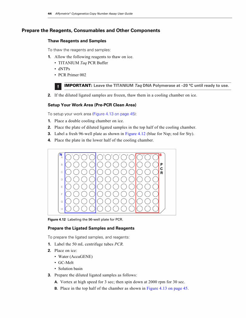
44 Affymetrix® Cytogenetics Copy Number Assay User Guide
Prepare the Reagents, Consumables and Other Components
Thaw Reagents and Samples
To thaw the reagents and samples:
1. Allow the following reagents to thaw on ice.
• TITANIUM Taq PCR Buffer
• dNTPs
• PCR Primer 002
2. If the diluted ligated samples are frozen, thaw them in a cooling chamber on ice.
Setup Your Work Area (Pre-PCR Clean Area)
To setup your work area (Figure 4.13 on page 45):
1. Place a double cooling chamber on ice.
2. Place the plate of diluted ligated samples in the top half of the cooling chamber.
3. Label a fresh 96-well plate as shown in Figure 4.12 (blue for Nsp; red for Sty).
4. Place the plate in the lower half of the cooling chamber.
Prepare the Ligated Samples and Reagents
To prepare the ligated samples, and reagents:
1. Label the 50 mL centrifuge tubes PCR.
2. Place on ice:
• Water (AccuGENE)
• GC-Melt
• Solution basin
3. Prepare the diluted ligated samples as follows:
A. Vortex at high speed for 3 sec; then spin down at 2000 rpm for 30 sec.
B. Place in the top half of the chamber as shown in Figure 4.13 on page 45.
IMPORTANT: Leave the TITANIUM Taq DNA Polymerase at –20 °C until ready to use.
Figure 4.12 Labeling the 96-well plate for PCR.
N S
PCR

chapter 4 | Affymetrix® Cytogenetics Copy Number Assay 45
4. To prepare the reagents:
A. Vortex at high speed 3 times, 1 sec each time (except for the enzyme).
B. Pulse spin for 3 sec.
C. Place on ice or in the cooling chamber.
Preheat the Thermal Cycler Lids (Post-PCR Area)Have someone in the Post-PCR Area power on the thermal cycler(s) to preheat the lid. Leave the blocks at room temperature.
To avoid contamination, do not go from the Pre-PCR Clean Area to the Post-PCR Area and back again.
Transfer Diluted Ligated Samples to the PCR Plate
To transfer the diluted ligated samples to the PCR plate:
1. Using a P20 pipet (single or multichannel):
A. Transfer 10 µL of each Nsp ligated sample to the corresponding four wells of the PCR plate Figure 4.13 on page 45).
B. Transfer 10 µL of each Sty ligated sample to the corresponding three wells of the PCR plate.
2. Seal, label and store the plate with the remaining ligated Nsp and Sty samples at –20 °C.
Discard this plate once you have successfully processed these samples.
Figure 4.13 Setup for PCR (Titanium Taq DNA Polymerase not shown; still at –20 °C)
PCR master mix tube
Solution basin
Water (AccuGENE)
Titanium Taq PCR Buffer
GC-Melt
dNTPs
5
+
23
4
6
1
—
5
+
2
3
4
6
1
—
N S
N
PCR
S
PCR
PCR Primer 002
Sty diluted ligated samples
Nsp diluted ligated samples
1 1 1 1 1 1 1
2 2 22 2 2 2
3 3 3 3 3 3 3
4 4 4 4 4 4 4
5 5 5 5 5 5 5
6 6 6 6 6 6 6
+ + + + + + +
— — — — — — —

46 Affymetrix® Cytogenetics Copy Number Assay User Guide
Prepare the PCR Master MixThe same PCR master mix is used for both Nsp and Sty ligated samples.
To prepare the PCR Master Mix:
1. Keeping the 50 mL centrifuge tube in the cooling chamber, add the reagents in the order shown in Table 4.17 on page 46 (except for the Taq DNA polymerase).
2. Remove the TITANIUM Taq DNA Polymerase from the freezer and immediately place in a cooler.
3. Pulse spin the Taq DNA polymerase for 3 sec.
4. Immediately add the Taq DNA polymerase to the master mix; then return the tube to the cooler.
5. Vortex the master mix at high speed 3 times, 1 sec each time.
6. Pour the master mix into the solution basin, keeping the basin on ice.
Add PCR Master Mix to Each Sample
To add the PCR Master Mix to samples:
1. Aliquot 90 µL PCR Master Mix to each sample and control on the PCR plate.
To avoid contamination, change pipet tips after each dispense.
For four samples, you may have to tilt the solution basin for the last pickup/dispense to ensure 90 µL picked up in each pipet tip.
Total volume in each well is 100 µL.
2. Tightly seal the plate.
3. Vortex at high speed for 3 sec; then spin down at 2000 rpm for 30 sec.
4. Keep in the cooling chamber on ice until ready to load onto a thermal cycler.
IMPORTANT: The PCR reaction is sensitive to the concentration of primer used. It is criticalthat the correct amount of primer be added to the PCR Master Mix to achieve the correctdistribution of fragments (200 to 1100 bp) in the products.
Check the PCR reactions on a gel to ensure that the distribution is correct.
Table 4.17 PCR Master Mix — Volumes sufficient for processing both Nsp and Sty ligated samples
Reagent 1 Sample 4 Samples(15% extra)
8 Samples(15% extra)
12 Samples(15% extra)
24 Samples(15% extra)
AccuGENE water 39.5 µL 1272 µL 2544 µL 3816 µL 7632 µL
TITANIUM Taq PCR Buffer (10X)
10 µL 322 µL 644 µL 966 µL 1932 µL
GC-Melt (5M) 20 µL 644 µL 1288 µL 1932 µL 3864 µL
dNTP (2.5 mM each) 14 µL 451 µL 902 µL 1352 µL 2704 µL
PCR Primer 002 (100 µM) 4.5 µL 145 µL 290 µL 435 µL 870 µL
TITANIUM Taq DNA Polymerase (50X)(do not add until ready to aliquot master mix to ligated samples)
2 µL 64.4 µL 129 µL 193 µL 386 µL
Total 90 µL 2898 µL 5796 µL 8694 µL 17.4 mL
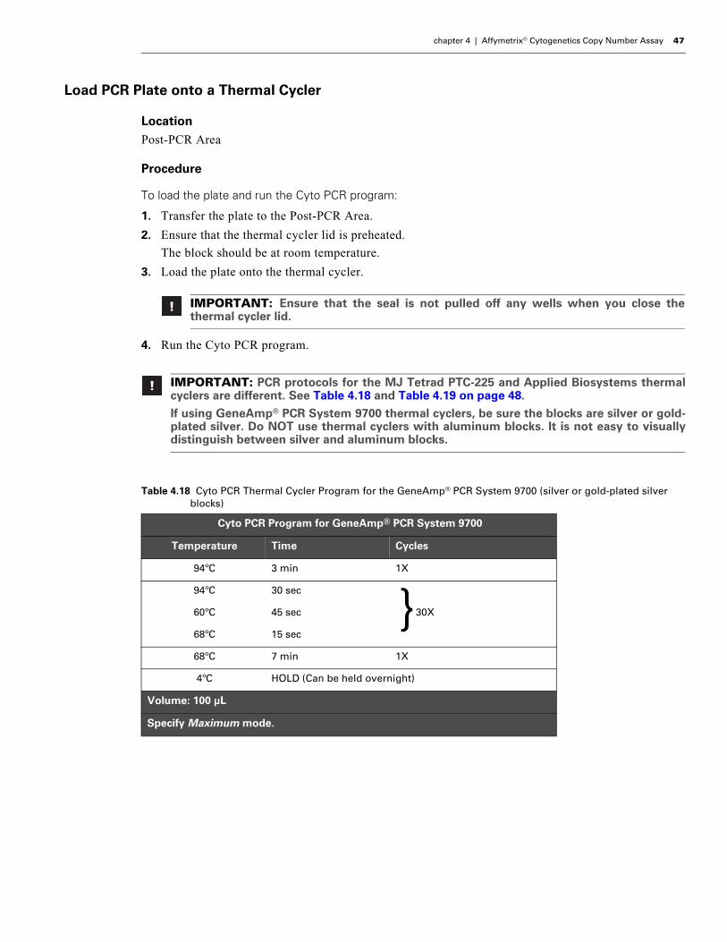
chapter 4 | Affymetrix® Cytogenetics Copy Number Assay 47
Load PCR Plate onto a Thermal Cycler
LocationPost-PCR Area
Procedure
To load the plate and run the Cyto PCR program:
1. Transfer the plate to the Post-PCR Area.
2. Ensure that the thermal cycler lid is preheated.
The block should be at room temperature.
3. Load the plate onto the thermal cycler.
4. Run the Cyto PCR program.
IMPORTANT: Ensure that the seal is not pulled off any wells when you close thethermal cycler lid.
IMPORTANT: PCR protocols for the MJ Tetrad PTC-225 and Applied Biosystems thermalcyclers are different. See Table 4.18 and Table 4.19 on page 48.
If using GeneAmp® PCR System 9700 thermal cyclers, be sure the blocks are silver or gold-plated silver. Do NOT use thermal cyclers with aluminum blocks. It is not easy to visuallydistinguish between silver and aluminum blocks.
Table 4.18 Cyto PCR Thermal Cycler Program for the GeneAmp® PCR System 9700 (silver or gold-plated silver blocks)
Cyto PCR Program for GeneAmp® PCR System 9700
Temperature Time Cycles
94ºC 3 min 1X
94ºC 30 sec
60ºC 45 sec 30X
68ºC 15 sec
68ºC 7 min 1X
4ºC HOLD (Can be held overnight)
Volume: 100 μL
Specify Maximum mode.
}
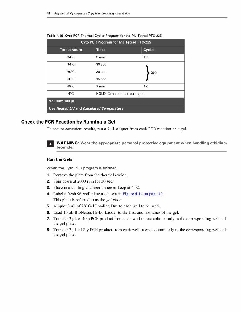
48 Affymetrix® Cytogenetics Copy Number Assay User Guide
Check the PCR Reaction by Running a GelTo ensure consistent results, run a 3 µL aliquot from each PCR reaction on a gel.
Run the Gels
When the Cyto PCR program is finished:
1. Remove the plate from the thermal cycler.
2. Spin down at 2000 rpm for 30 sec.
3. Place in a cooling chamber on ice or keep at 4 °C.
4. Label a fresh 96-well plate as shown in Figure 4.14 on page 49.
This plate is referred to as the gel plate.
5. Aliquot 3 µL of 2X Gel Loading Dye to each well to be used.
6. Load 10 µL BioNexus Hi-Lo Ladder to the first and last lanes of the gel.
7. Transfer 3 µL of Nsp PCR product from each well in one column only to the corresponding wells of the gel plate.
8. Transfer 3 µL of Sty PCR product from each well in one column only to the corresponding wells of the gel plate.
Table 4.19 Cyto PCR Thermal Cycler Program for the MJ Tetrad PTC-225
Cyto PCR Program for MJ Tetrad PTC-225
Temperature Time Cycles
94ºC 3 min 1X
94ºC 30 sec
30X60ºC 30 sec
68ºC 15 sec
68ºC 7 min 1X
4ºC HOLD (Can be held overnight)
Volume: 100 μL
Use Heated Lid and Calculated Temperature
WARNING: Wear the appropriate personal protective equipment when handling ethidiumbromide.
}

chapter 4 | Affymetrix® Cytogenetics Copy Number Assay 49
9. Seal both plates.
10. Vortex the gel plate, then spin them down at 2000 rpm for 30 sec.
11. Load the total volume from each well of the gel plate onto a 2% TBE gel.
12. Run the gel at 120V for 40 min to 1 hr.
13. Verify that the PCR product distribution is between ~250 bp to 1100 bp (Figure 4.15 on page 50).
Figure 4.14 Transferring aliquots of each Nsp and Sty PCR product to the gel plate
SN
GEL
5
+
2
3
4
6
1
—
5
+
2
3
4
6
1
—
SN
PCR
5
+
2
3
4
6
1
—
5
+
2
3
4
6
1
—
5
+
2
3
4
6
1
—
5
+
2
3
4
6
1
—
5
+
2
3
4
6
1
—
5
+
2
3
4
6
1
—
5
+
2
3
4
6
1
—
3 μL 3 μL
PCR Plate
Gel Plate

50 Affymetrix® Cytogenetics Copy Number Assay User Guide
What To Do NextDo one of the following:
• If the PCR has been confirmed, proceed to Stage 4: PCR Product Purification on page 51.
• If not proceeding directly to the next stage, seal the plate with PCR product and store at –20 °C.
Figure 4.15 Example of PCR products run on 2% TBE agarose gel at 120V for 1 hr. Average product distribution is between ~200to 1100 bp.
1 2 3 4 5 6 + —

chapter 4 | Affymetrix® Cytogenetics Copy Number Assay 51
Stage 4: PCR Product Purification
About this StageDuring this stage, you will purify the PCR products by:
• Pooling the Nsp and Sty PCR reactions.
• Adding magnetic beads (Agencourt AMPure®) to each pooled reaction and incubating the mix.
• Adding 75% EtOH to wash DNA.
• Adding Buffer EB to resuspend the beads and elute the DNA.
Location and Duration• Post-PCR Area
• Hands-on time: 1 hr
• DNA binding to magnetic bead: 15 to 20 min
• EtOH wash: approximately 10 to 20 min
• Elution: 15 to 30 min
• Total time for this stage: approximately 1.5 hr
Input Required from Previous StageThe input required from Stage 3: Nsp and Sty PCR is:
Item
Plate of Nsp and Sty PCR products
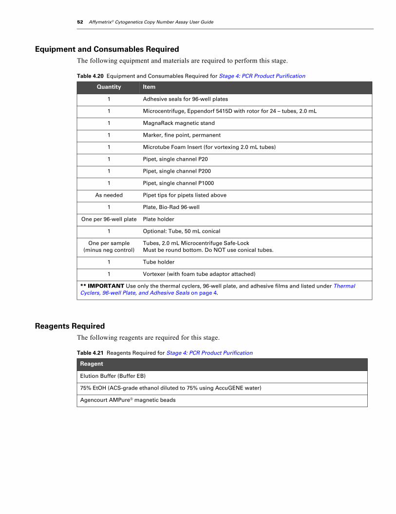
52 Affymetrix® Cytogenetics Copy Number Assay User Guide
Equipment and Consumables RequiredThe following equipment and materials are required to perform this stage.
Reagents RequiredThe following reagents are required for this stage.
Table 4.20 Equipment and Consumables Required for Stage 4: PCR Product Purification
Quantity Item
1 Adhesive seals for 96-well plates
1 Microcentrifuge, Eppendorf 5415D with rotor for 24 – tubes, 2.0 mL
1 MagnaRack magnetic stand
1 Marker, fine point, permanent
1 Microtube Foam Insert (for vortexing 2.0 mL tubes)
1 Pipet, single channel P20
1 Pipet, single channel P200
1 Pipet, single channel P1000
As needed Pipet tips for pipets listed above
1 Plate, Bio-Rad 96-well
One per 96-well plate Plate holder
1 Optional: Tube, 50 mL conical
One per sample (minus neg control)
Tubes, 2.0 mL Microcentrifuge Safe-LockMust be round bottom. Do NOT use conical tubes.
1 Tube holder
1 Vortexer (with foam tube adaptor attached)
** IMPORTANT Use only the thermal cyclers, 96-well plate, and adhesive films and listed under Thermal Cyclers, 96-well Plate, and Adhesive Seals on page 4.
Table 4.21 Reagents Required for Stage 4: PCR Product Purification
Reagent
Elution Buffer (Buffer EB)
75% EtOH (ACS-grade ethanol diluted to 75% using AccuGENE water)
Agencourt AMPure® magnetic beads
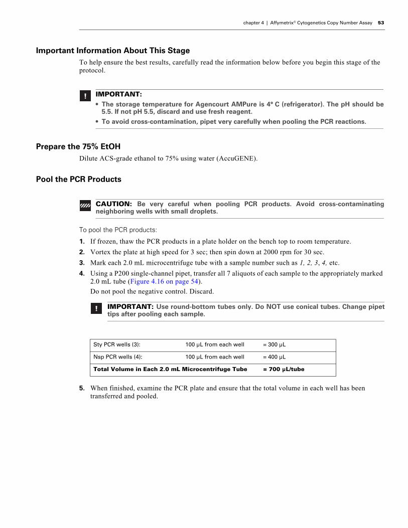
chapter 4 | Affymetrix® Cytogenetics Copy Number Assay 53
Important Information About This StageTo help ensure the best results, carefully read the information below before you begin this stage of the protocol.
Prepare the 75% EtOHDilute ACS-grade ethanol to 75% using water (AccuGENE).
Pool the PCR Products
To pool the PCR products:
1. If frozen, thaw the PCR products in a plate holder on the bench top to room temperature.
2. Vortex the plate at high speed for 3 sec; then spin down at 2000 rpm for 30 sec.
3. Mark each 2.0 mL microcentrifuge tube with a sample number such as 1, 2, 3, 4, etc.
4. Using a P200 single-channel pipet, transfer all 7 aliquots of each sample to the appropriately marked 2.0 mL tube (Figure 4.16 on page 54).
Do not pool the negative control. Discard.
5. When finished, examine the PCR plate and ensure that the total volume in each well has been transferred and pooled.
IMPORTANT:• The storage temperature for Agencourt AMPure is 4° C (refrigerator). The pH should be
5.5. If not pH 5.5, discard and use fresh reagent.
• To avoid cross-contamination, pipet very carefully when pooling the PCR reactions.
CAUTION: Be very careful when pooling PCR products. Avoid cross-contaminatingneighboring wells with small droplets.
IMPORTANT: Use round-bottom tubes only. Do NOT use conical tubes. Change pipettips after pooling each sample.
Sty PCR wells (3): 100 µL from each well = 300 µL
Nsp PCR wells (4): 100 µL from each well = 400 µL
Total Volume in Each 2.0 mL Microcentrifuge Tube = 700 µL/tube
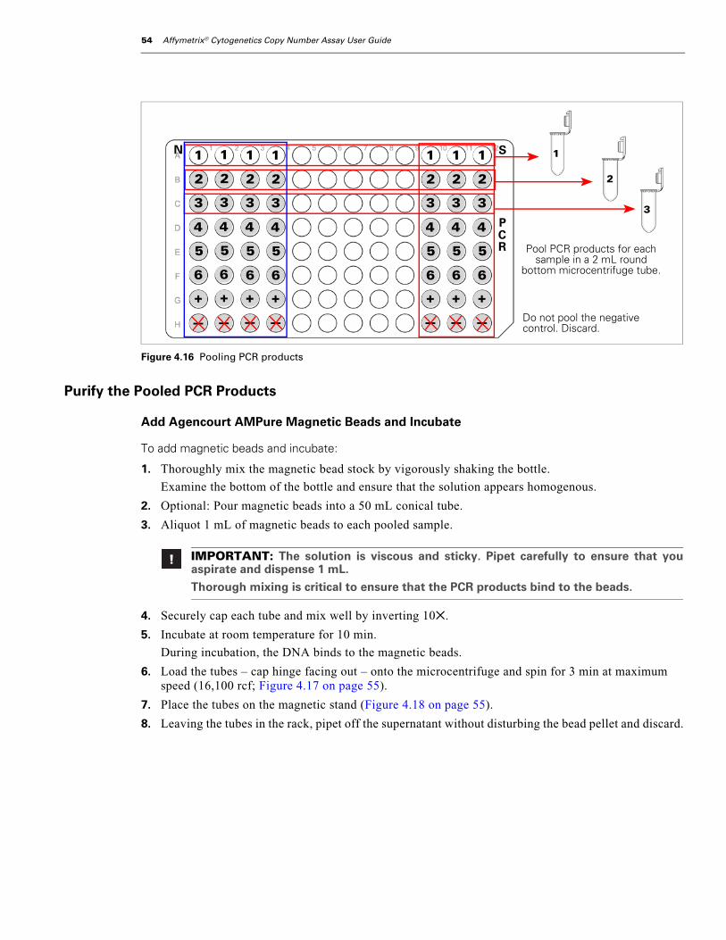
54 Affymetrix® Cytogenetics Copy Number Assay User Guide
Purify the Pooled PCR Products
Add Agencourt AMPure Magnetic Beads and Incubate
To add magnetic beads and incubate:
1. Thoroughly mix the magnetic bead stock by vigorously shaking the bottle.
Examine the bottom of the bottle and ensure that the solution appears homogenous.
2. Optional: Pour magnetic beads into a 50 mL conical tube.
3. Aliquot 1 mL of magnetic beads to each pooled sample.
4. Securely cap each tube and mix well by inverting 10✕.
5. Incubate at room temperature for 10 min.
During incubation, the DNA binds to the magnetic beads.
6. Load the tubes – cap hinge facing out – onto the microcentrifuge and spin for 3 min at maximum speed (16,100 rcf; Figure 4.17 on page 55).
7. Place the tubes on the magnetic stand (Figure 4.18 on page 55).
8. Leaving the tubes in the rack, pipet off the supernatant without disturbing the bead pellet and discard.
Figure 4.16 Pooling PCR products
IMPORTANT: The solution is viscous and sticky. Pipet carefully to ensure that youaspirate and dispense 1 mL.
Thorough mixing is critical to ensure that the PCR products bind to the beads.
SN
PCR5
+
2
3
4
6
1
—
5
+
2
3
4
6
1
—
5
+
2
3
4
6
1
—
5
+
2
3
4
6
1
—
5
+
2
3
4
6
1
—
5
+
2
3
4
6
1
—
5
+
2
3
4
6
1
—
Pool PCR products for each sample in a 2 mL round
bottom microcentrifuge tube.
Do not pool the negative control. Discard.
1
2
3

chapter 4 | Affymetrix® Cytogenetics Copy Number Assay 55
Figure 4.17 Position tubes with cap hinges out
Figure 4.18 Samples with magnetic beads on MagnaRack
Figure 4.19 Bead pulled to back and side of tube in magnetic stand
Position tubes with the cap hinges facing out. Bead pellet will be spun to the bottom and back of the tube.
1 2 3 4 5 6 +
Avoid contact with the bead pellet when pipetting off the supernatant.
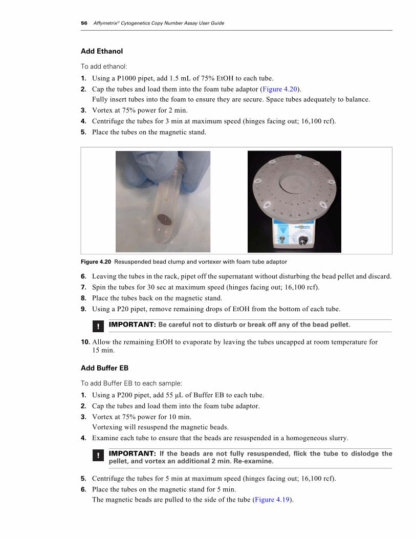
56 Affymetrix® Cytogenetics Copy Number Assay User Guide
Add Ethanol
To add ethanol:
1. Using a P1000 pipet, add 1.5 mL of 75% EtOH to each tube.
2. Cap the tubes and load them into the foam tube adaptor (Figure 4.20).
Fully insert tubes into the foam to ensure they are secure. Space tubes adequately to balance.
3. Vortex at 75% power for 2 min.
4. Centrifuge the tubes for 3 min at maximum speed (hinges facing out; 16,100 rcf).
5. Place the tubes on the magnetic stand.
6. Leaving the tubes in the rack, pipet off the supernatant without disturbing the bead pellet and discard.
7. Spin the tubes for 30 sec at maximum speed (hinges facing out; 16,100 rcf).
8. Place the tubes back on the magnetic stand.
9. Using a P20 pipet, remove remaining drops of EtOH from the bottom of each tube.
10. Allow the remaining EtOH to evaporate by leaving the tubes uncapped at room temperature for 15 min.
Add Buffer EB
To add Buffer EB to each sample:
1. Using a P200 pipet, add 55 µL of Buffer EB to each tube.
2. Cap the tubes and load them into the foam tube adaptor.
3. Vortex at 75% power for 10 min.
Vortexing will resuspend the magnetic beads.
4. Examine each tube to ensure that the beads are resuspended in a homogeneous slurry.
5. Centrifuge the tubes for 5 min at maximum speed (hinges facing out; 16,100 rcf).
6. Place the tubes on the magnetic stand for 5 min.
The magnetic beads are pulled to the side of the tube (Figure 4.19).
Figure 4.20 Resuspended bead clump and vortexer with foam tube adaptor
IMPORTANT: Be careful not to disturb or break off any of the bead pellet.
IMPORTANT: If the beads are not fully resuspended, flick the tube to dislodge thepellet, and vortex an additional 2 min. Re-examine.

chapter 4 | Affymetrix® Cytogenetics Copy Number Assay 57
7. Check that all of the beads have been pulled to the side in each tube.
If all of the beads have not been pulled to the side of the tubes, leave the tubes on the stand an additional 3 min.
8. Transfer 47 µL of eluted sample to the appropriate well on a fresh 96-well plate (Figure 4.21 on page 57).
Brown residue on pipet tips is OK.
9. Tightly seal the plate.
What To Do NextProceed to Stage 5: Quantitation on page 58. Here you will remove 2 µL from each sample for an OD measurement.
NOTE: The eluate will appear yellowish. If you open the cap and look directly into thetube, you will see that the eluate is clear.
Figure 4.21 Transferring each purified sample to a fresh 96-well plate.
1 2 3 4 5 6 +
5
+
2
3
4
6
1
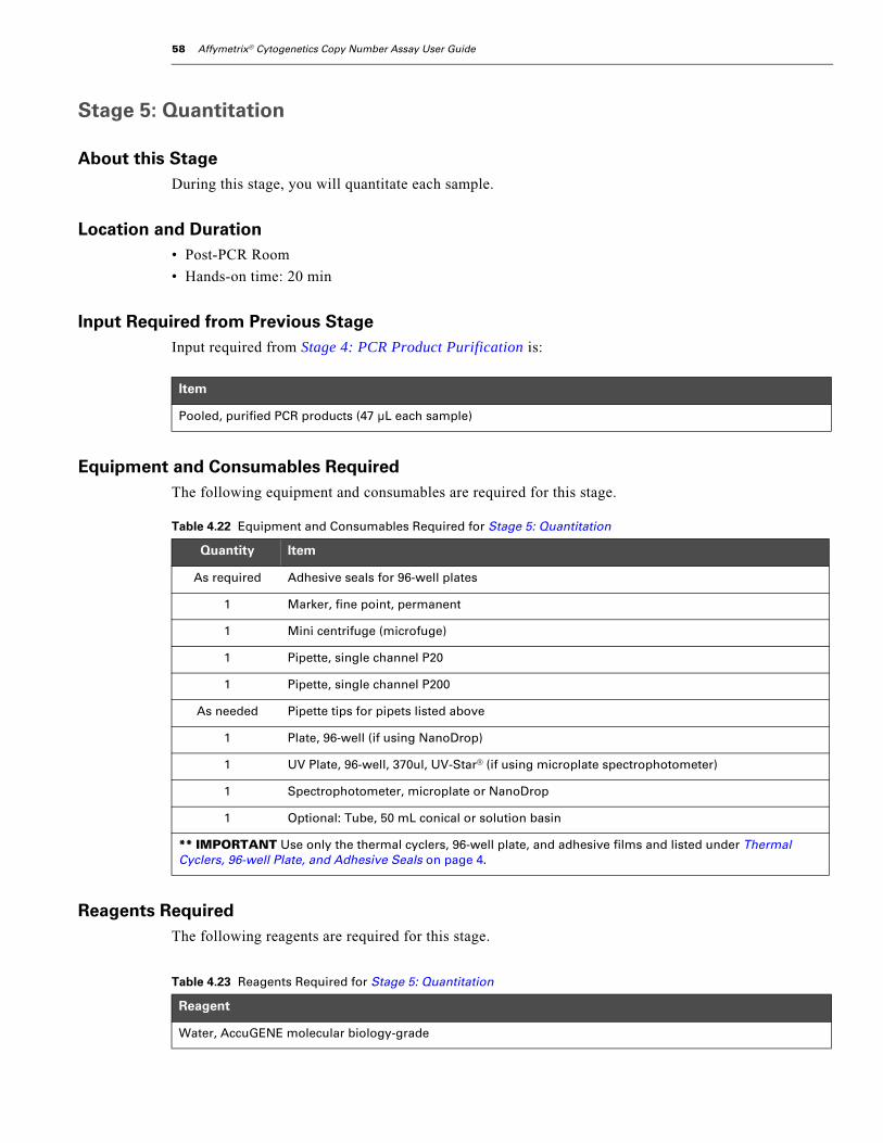
58 Affymetrix® Cytogenetics Copy Number Assay User Guide
Stage 5: Quantitation
About this StageDuring this stage, you will quantitate each sample.
Location and Duration• Post-PCR Room
• Hands-on time: 20 min
Input Required from Previous StageInput required from Stage 4: PCR Product Purification is:
Equipment and Consumables RequiredThe following equipment and consumables are required for this stage.
Reagents RequiredThe following reagents are required for this stage.
Item
Pooled, purified PCR products (47 µL each sample)
Table 4.22 Equipment and Consumables Required for Stage 5: Quantitation
Quantity Item
As required Adhesive seals for 96-well plates
1 Marker, fine point, permanent
1 Mini centrifuge (microfuge)
1 Pipette, single channel P20
1 Pipette, single channel P200
As needed Pipette tips for pipets listed above
1 Plate, 96-well (if using NanoDrop)
1 UV Plate, 96-well, 370ul, UV-Star® (if using microplate spectrophotometer)
1 Spectrophotometer, microplate or NanoDrop
1 Optional: Tube, 50 mL conical or solution basin
** IMPORTANT Use only the thermal cyclers, 96-well plate, and adhesive films and listed under Thermal Cyclers, 96-well Plate, and Adhesive Seals on page 4.
Table 4.23 Reagents Required for Stage 5: Quantitation
Reagent
Water, AccuGENE molecular biology-grade
chapter 4 | Affymetrix® Cytogenetics Copy Number Assay 59
Important Information About This StageTo help ensure the best results, carefully read the information below before you begin this stage of the protocol.
Prepare the Reagents, Equipment and Consumables
Turn on the SpectrophotometerTurn the instrument on and allow it to warm for 10 min before use.
Prepare Your Work Area
To prepare the work area:
1. Place the following on the bench top:
• Optional: conical tube or solution basin
• Water
• UV or 96-well plate
2. Spin down the purified samples at 2000 rpm for 30 sec, and put in a plate holder.
Procedure if Using a Microplate Spectrophotometer
Prepare Diluted Aliquots of Purified Sample
To prepare diluted aliquots of the purified samples:
1. Using a P200 pipet, aliquot 198 µL of water to the corresponding wells of a UV plate.
2. Pipet 200 µL of water into each well of an empty column (Figure 4.22 below).
3. Using a P20 pipet:
A. Transfer 2 µL of each purified sample to the corresponding well of the UV plate.
B. Pipette up and down 2 times to ensure that all of the sample is dispensed.
The result is a 100-fold dilution.
4. Do one of the following to mix the samples:
• Set a P200 pipet to 170 µL and pipet up and down 5 times.
• Seal the plate, vortex, and spin down at 2000 rpm for 30 sec.
IMPORTANT:• The accuracy of the OD measurement is critical. Carefully follow this procedure and be
sure the OD measurement is within the quantitative linear range of the instrument.
• The spectrophotometer should be calibrated regularly to ensure correct readings.
This protocol has been optimized using a UV spectrophotometer for quantitation.
IMPORTANT: The P20 pipet must be accurate to within ± 5%.
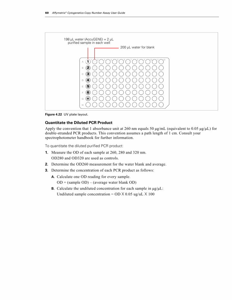
60 Affymetrix® Cytogenetics Copy Number Assay User Guide
Quantitate the Diluted PCR ProductApply the convention that 1 absorbance unit at 260 nm equals 50 µg/mL (equivalent to 0.05 µg/µL) for double-stranded PCR products. This convention assumes a path length of 1 cm. Consult your spectrophotometer handbook for further information.
To quantitate the diluted purified PCR product:
1. Measure the OD of each sample at 260, 280 and 320 nm.
OD280 and OD320 are used as controls.
2. Determine the OD260 measurement for the water blank and average.
3. Determine the concentration of each PCR product as follows:
A. Calculate one OD reading for every sample.
OD = (sample OD) – (average water blank OD)
B. Calculate the undiluted concentration for each sample in µg/µL:
Undiluted sample concentration = OD X 0.05 ug/uL X 100
Figure 4.22 UV plate layout.
5
+
2
3
4
6
1
198 μL water (AccuGENE) + 2 μL purified sample in each well
200 μL water for blank

chapter 4 | Affymetrix® Cytogenetics Copy Number Assay 61
Procedure if Using a NanoDrop
To prepare diluted aliquots of the purified samples:
1. Using a P20 pipet, aliquot 18 µL of water to the corresponding wells of a 96-well plate.
2. Using a P20 pipet:
A. Transfer 2 µL of each purified sample to the corresponding well of the 96-well plate.
B. Pipette up and down 2 times to ensure that all of the sample is dispensed.
The result is a 10-fold dilution.
3. Do one of the following to mix the samples:
• Set a P20 pipet to 17 µL and pipet up and down 5 times.
• Seal the plate, vortex, and spin down at 2000 rpm for 30 sec.
4. Blank the NanoDrop with water.
5. Take 2 µL of the diluted sample and:
A. Measure the OD of each sample at 260, 280 and 320 nm.
OD280 and OD320 are used as controls.
B. Calculate the undiluted concentration for each sample as follows:
Undiluted sample concentration in µg/µL = OD X 10
IMPORTANT: The P20 pipet must be accurate to within ± 5%.
Figure 4.23 96-well plate layout for NanoDrop
5
+
2
3
4
6
118 μL water (AccuGENE) + 2 μLpurified sample in each well
62 Affymetrix® Cytogenetics Copy Number Assay User Guide
Assess the OD Readings
Acceptable OD Range and DNA Yield• An acceptable OD should fall within this range: 0.9 to 1.4.
DNA yield equivalent = 4.5 to 7.0 µg/µL
This OD range is based on the use of a conventional UV spectrophotometer plate reader and assumes a path length of 1 cm.
• The OD260/OD280 ratio should be between 1.8 and 2.0.
Do not proceed if this metric falls outside of this range.
• The OD320 measurement should be very close to zero (≤ 0.1).
- If > 0.2:
A. Centrifuge the sample for 5 min.
B. Place on the MagnaRack and pipet off the eluate.
C. Requantitate the sample.
D. If the OD320 reading is now ≤ 0.1, proceed.If your OD readings are not within the acceptable range, refer to OD Troubleshooting Guidelines on page 99.
What To Do NextDo one of the following:
• Proceed immediately to Stage 6: Fragmentation on page 63.
• If not proceeding immediately to the next step, seal the plate of purified samples, and store at –20 °C.

chapter 4 | Affymetrix® Cytogenetics Copy Number Assay 63
Stage 6: Fragmentation
About this StageDuring this stage the purified samples are fragmented using Fragmentation Reagent (enzyme) by:
• Preparing a Fragmentation Master Mix.
• Quickly adding the mix to each sample.
• Placing the samples onto a thermal cycler and running the Cyto Fragment program.
• Checking each reaction on a gel.
Location and Duration• Post-PCR Area
• Hands-on time: 30 min
• Cyto Fragment thermal cycler program time: 1 hr
Input Required from Previous StageThe input required from Stage 5: Quantitation is:
Item
Plate of purified samples that have been quantitated.

64 Affymetrix® Cytogenetics Copy Number Assay User Guide
Equipment and Consumables RequiredThe following equipment and consumables are required for this stage.
Reagents RequiredThe following reagents are required for this stage.
Table 4.24 Equipment and Consumables Required for Stage 6: Fragmentation
Quantity Item
As required Adhesive seals for 96-well plates
1 Centrifuge, plate
1 Cooler, chilled to –20 °C
1 Cooling chamber, single, chilled to 4 °C on ice (do not freeze)
1 Ice bucket, filled with ice
1 Marker, fine point, permanent
1 Mini centrifuge (microfuge)
1 Pipet, single channel P20
1 Pipet, single channel P100
1 Pipet, single channel P1000
1 Pipet, 8-channel P20 (accurate to within ± 5%)
As needed Pipet tips for pipets listed above
1 Plate, Bio-Rad 96-well
1 Thermal cycler
1 Tube, Eppendorf 1.5 mL
1 Tubes, strip of 8 with caps, 0.2 mL
1 Vortexer
** IMPORTANT Use only the thermal cyclers, 96-well plate, and adhesive films and listed under Thermal Cyclers, 96-well Plate, and Adhesive Seals on page 4.
Table 4.25 Reagents Required for Stage 6: Fragmentation
Reagent
Fragmentation Buffer (10X)
Fragmentation Reagent (enzyme; DNase I)
AccuGENE® water, molecular biology-grade
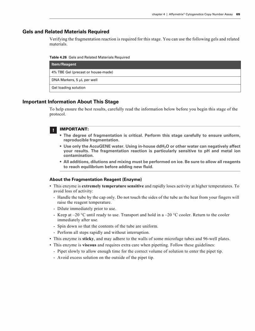
chapter 4 | Affymetrix® Cytogenetics Copy Number Assay 65
Gels and Related Materials RequiredVerifying the fragmentation reaction is required for this stage. You can use the following gels and related materials.
Important Information About This StageTo help ensure the best results, carefully read the information below before you begin this stage of the protocol.
About the Fragmentation Reagent (Enzyme)• This enzyme is extremely temperature sensitive and rapidly loses activity at higher temperatures. To
avoid loss of activity:
- Handle the tube by the cap only. Do not touch the sides of the tube as the heat from your fingers will raise the reagent temperature.
- Dilute immediately prior to use.
- Keep at –20 °C until ready to use. Transport and hold in a –20 °C cooler. Return to the cooler immediately after use.
- Spin down so that the contents of the tube are uniform.
- Perform all steps rapidly and without interruption.
• This enzyme is sticky, and may adhere to the walls of some microfuge tubes and 96-well plates.
• This enzyme is viscous and requires extra care when pipetting. Follow these guidelines:
- Pipet slowly to allow enough time for the correct volume of solution to enter the pipet tip.
- Avoid excess solution on the outside of the pipet tip.
Table 4.26 Gels and Related Materials Required
Item/Reagent
4% TBE Gel (precast or house-made)
DNA Markers, 5 µL per well
Gel loading solution
n
IMPORTANT:• The degree of fragmentation is critical. Perform this stage carefully to ensure uniform,
reproducible fragmentation.
• Use only the AccuGENE water. Using in-house ddH2O or other water can negatively affectyour results. The fragmentation reaction is particularly sensitive to pH and metal ioncontamination.
• All additions, dilutions and mixing must be performed on ice. Be sure to allow all reagentsto reach equilibrium before adding new fluid.

66 Affymetrix® Cytogenetics Copy Number Assay User Guide
Prepare the Reagents, Consumables and Other Components
Thaw ReagentsThaw the Fragmentation Buffer (10X) on ice.
Setup Your Work Area
To setup your work area (Figure 4.24):
1. Place a cooling chamber and the water on ice.
2. Place the plate of purified, quantitated samples in the cooling chamber.
3. Prepare the Fragmentation Buffer as follows:
A. Vortex 3 times, 1 sec each time.
B. Pulse spin for 3 sec.
C. Place in the cooling chamber.
4. Label the 1.5 mL Eppendorf tube Frag and place in the cooling chamber.
Preheat the Thermal Cycler BlockThe block must be heated to 37 °C before samples are loaded.
To preheat the thermal cycler:
1. Power on the thermal cycler and preheat the block to 37 °C.
2. Allow it to heat for 10 min before loading samples.
IMPORTANT: Leave the Fragmentation Reagent at –20 °C until ready to use.
Figure 4.24 Setup for fragmentation.
Purified samples
5
+
2
3
4
6
1
FRAG
Fragmentation Buffer
Water(AccuGENE)
Fragmentation master mix tube

chapter 4 | Affymetrix® Cytogenetics Copy Number Assay 67
Prepare the Samples for Fragmentation
Add Fragmentation Buffer to Samples
To prepare the samples for Fragmentation:
1. Add 5 µL of Fragmentation Buffer to each sample.
2. Check your pipet tips each time to ensure that all of the buffer has been dispensed.
The total volume in each well is now 50 µL.
Prepare the Fragmentation Master Mix
To prepare the Fragmentation Master Mix:
The Fragmentation Reagent must be diluted to 0.1 U/µL in the master mix.
1. Read the Fragmentation Reagent tube label and record the concentration.
2. Based on the number of samples you are processing, determine which Fragmentation Master Mix table to use:
• 4 to 7 samples: Table 4.27 on page 67
• 8 to 16 samples: Table 4.28 on page 68
• 17 to 24 samples: Table 4.29 on page 68
3. Add the appropriate volume of water and Fragmentation Buffer to the Frag tube on ice.
4. Allow to cool on ice for 5 min.
IMPORTANT: All additions in this procedure must be performed on ice.
IMPORTANT: The concentration of stock Fragmentation Reagent (U/μL) may vary fromlot-to-lot. Therefore, read the label on the tube and record the stock concentration beforediluting this reagent.
Table 4.27 Fragmentation Master Mix for 4 to 7 Samples
Reagent Fragmentation Reagent Concentration
2 U/μL 2.25 U/μL 2.5 U/μL 2.75 U/μL 3 U/μL
AccuGENE water 34.00 µL 38.50 µL 43.00 µL 48.50 µL 52.00 µL
10X Fragmentation Buffer 4.00 µL 4.50 µL 5.00 µL 4.50 µL 6.00 µL
Fragmentation Reagent (enzyme) 2.00 µL 2.00 µL 2.00 µL 2.00 µL 2.00 µL
Total 40 µL 45 µL 50 µL 55 µL 60 µL
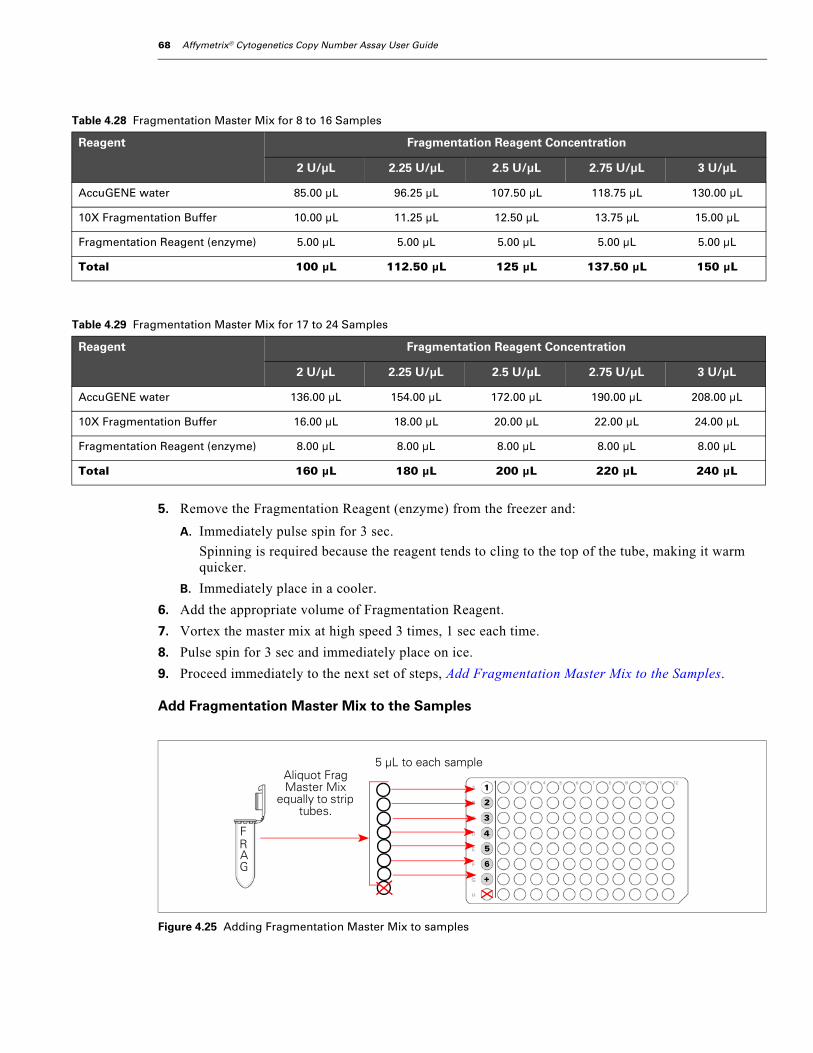
68 Affymetrix® Cytogenetics Copy Number Assay User Guide
5. Remove the Fragmentation Reagent (enzyme) from the freezer and:
A. Immediately pulse spin for 3 sec.
Spinning is required because the reagent tends to cling to the top of the tube, making it warm quicker.
B. Immediately place in a cooler.
6. Add the appropriate volume of Fragmentation Reagent.
7. Vortex the master mix at high speed 3 times, 1 sec each time.
8. Pulse spin for 3 sec and immediately place on ice.
9. Proceed immediately to the next set of steps, Add Fragmentation Master Mix to the Samples.
Add Fragmentation Master Mix to the Samples
Table 4.28 Fragmentation Master Mix for 8 to 16 Samples
Reagent Fragmentation Reagent Concentration
2 U/μL 2.25 U/μL 2.5 U/μL 2.75 U/μL 3 U/μL
AccuGENE water 85.00 µL 96.25 µL 107.50 µL 118.75 µL 130.00 µL
10X Fragmentation Buffer 10.00 µL 11.25 µL 12.50 µL 13.75 µL 15.00 µL
Fragmentation Reagent (enzyme) 5.00 µL 5.00 µL 5.00 µL 5.00 µL 5.00 µL
Total 100 µL 112.50 µL 125 µL 137.50 µL 150 µL
Table 4.29 Fragmentation Master Mix for 17 to 24 Samples
Reagent Fragmentation Reagent Concentration
2 U/μL 2.25 U/μL 2.5 U/μL 2.75 U/μL 3 U/μL
AccuGENE water 136.00 µL 154.00 µL 172.00 µL 190.00 µL 208.00 µL
10X Fragmentation Buffer 16.00 µL 18.00 µL 20.00 µL 22.00 µL 24.00 µL
Fragmentation Reagent (enzyme) 8.00 µL 8.00 µL 8.00 µL 8.00 µL 8.00 µL
Total 160 µL 180 µL 200 µL 220 µL 240 µL
Figure 4.25 Adding Fragmentation Master Mix to samples
5
+
2
3
4
6
1Aliquot Frag Master Mix
equally to strip tubes.
5 μL to each sample
FRAG

chapter 4 | Affymetrix® Cytogenetics Copy Number Assay 69
To add Fragmentation Master Mix to the samples:
1. Quickly and on ice, aliquot out the Fragmentation Master Mix equally to the strip tubes (Figure 4.25).
2. Spin down the strip tubes.
3. Using an 8-channel P20 pipet, transfer 5 µL of Fragmentation Master Mix to each sample — do not pipet up and down.
Avoid introducing air bubbles at the bottom of the tubes to ensure the accurate transfer of 5 µL to each sample.
4. Remove and discard any remaining Fragmentation Master Mix.
Never re-use Fragmentation Master Mix.
5. Tightly seal the plate.
6. Vortex at high speed for 3 sec; then spin down for 30 sec.
7. Immediately load the samples onto the pre-heated block of the thermal cycler (37 °C) and run the Cyto Fragment program (Table 4.30).
What To Do NextProceed directly to the next stage. While the labeling reaction is taking place, check the fragmentation reaction by running gels as described under Check the Fragmentation Reaction by Running a Gel on page 70.
IMPORTANT: To help ensure the same amount of fragmentation for each sample, addthe master mix to the samples as quickly as possible.
Sample with Fragmentation Buffer 50 µL
Fragmentation Master Mix 5 µL
Total 55 µL
IMPORTANT: Ensure that the seal is not pulled off any wells when you close thethermal cycler lid.
Table 4.30 Cyto Fragment Thermal Cycler Program
Cyto Fragment Program
Temperature Time
37ºC 35 min
95ºC 15 min
4ºC Hold
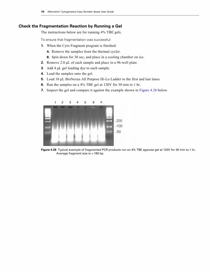
70 Affymetrix® Cytogenetics Copy Number Assay User Guide
Check the Fragmentation Reaction by Running a GelThe instructions below are for running 4% TBE gels.
To ensure that fragmentation was successful:
1. When the Cyto Fragment program is finished:
A. Remove the samples from the thermal cycler.
B. Spin down for 30 sec, and place in a cooling chamber on ice.
2. Remove 2.0 µL of each sample and place in a 96-well plate.
3. Add 4 µL gel loading dye to each sample.
4. Load the samples onto the gel.
5. Load 10 µL BioNexus All Purpose Hi-Lo Ladder to the first and last lanes.
6. Run the samples on a 4% TBE gel at 120V for 30 min to 1 hr.
7. Inspect the gel and compare it against the example shown in Figure 4.26 below.
Figure 4.26 Typical example of fragmented PCR products run on 4% TBE agarose gel at 120V for 30 min to 1 hr. Average fragment size is < 180 bp.
1 2 3 4 5 6 +
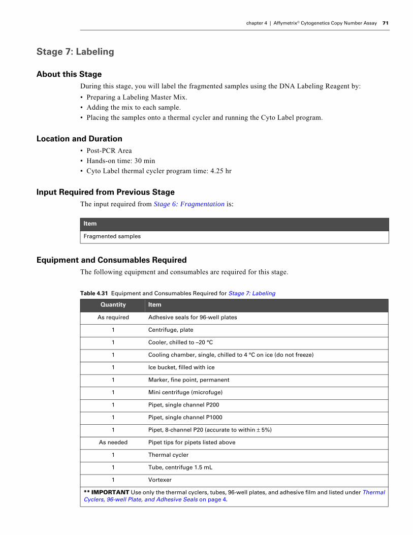
chapter 4 | Affymetrix® Cytogenetics Copy Number Assay 71
Stage 7: Labeling
About this StageDuring this stage, you will label the fragmented samples using the DNA Labeling Reagent by:
• Preparing a Labeling Master Mix.
• Adding the mix to each sample.
• Placing the samples onto a thermal cycler and running the Cyto Label program.
Location and Duration• Post-PCR Area
• Hands-on time: 30 min
• Cyto Label thermal cycler program time: 4.25 hr
Input Required from Previous StageThe input required from Stage 6: Fragmentation is:
Equipment and Consumables RequiredThe following equipment and consumables are required for this stage.
Item
Fragmented samples
Table 4.31 Equipment and Consumables Required for Stage 7: Labeling
Quantity Item
As required Adhesive seals for 96-well plates
1 Centrifuge, plate
1 Cooler, chilled to –20 °C
1 Cooling chamber, single, chilled to 4 °C on ice (do not freeze)
1 Ice bucket, filled with ice
1 Marker, fine point, permanent
1 Mini centrifuge (microfuge)
1 Pipet, single channel P200
1 Pipet, single channel P1000
1 Pipet, 8-channel P20 (accurate to within ± 5%)
As needed Pipet tips for pipets listed above
1 Thermal cycler
1 Tube, centrifuge 1.5 mL
1 Vortexer
** IMPORTANT Use only the thermal cyclers, tubes, 96-well plates, and adhesive film and listed under Thermal Cyclers, 96-well Plate, and Adhesive Seals on page 4.

72 Affymetrix® Cytogenetics Copy Number Assay User Guide
Reagents RequiredThe following reagents are required for this stage.
Prepare the Reagents, Consumables and Other Components
Thaw ReagentsThaw the following reagents on ice:
• 5X TdT Buffer
• DNA Labeling Reagent
Prepare Your Work Area
To prepare the work area:
1. Place a cooling chamber on ice (Figure 4.27 on page 72).
2. Prepare the reagents as follows:
A. Vortex each reagent at high speed 3 times, 1 sec each time.
B. Pulse spin for 3 sec; then place in the cooling chamber.
3. Label the 1.5 mL centrifuge tube LBL, and place in the cooling chamber.
Table 4.32 Reagents Required for Stage 7: Labeling
Reagent
DNA Labeling Reagent (30 mM)
Terminal Deoxynucleotidyl Transferase (TdT; 30 U/µL)
Terminal Deoxynucleotidyl Transferase Buffer (TdT Buffer; 5X)
IMPORTANT: Leave the TdT enzyme at –20 °C until ready to use.
Figure 4.27 Setup for labeling
LBL
DNA Labeling Reagent
TdT Buffer
Label master mix tube
5
+
2
3
4
6
1

chapter 4 | Affymetrix® Cytogenetics Copy Number Assay 73
Preheat the Thermal Cycler Block
The block must be heated to 37 °C before samples are loaded.
To preheat the thermal cycler block:
1. Turn on the thermal cycler and preheat the block to 37 °C.
2. Allow it to heat for 10 min before loading samples.
Prepare the Labeling Master Mix
PreparationKeep all reagents and tubes on ice while preparing the Labeling Master Mix.
To prepare the Labeling Master Mix:
1. Add the following to the 1.5 mL centrifuge tube on ice using the volumes shown in Table 4.33 on page 73:
• 5X TdT Buffer
• DNA Labeling Reagent
2. Remove the TdT enzyme from the freezer and immediately place in the cooler.
3. Pulse spin the enzyme for 3 sec; then immediately place back in the cooler.
4. Add the TdT enzyme to the master mix.
5. Vortex the master mix at high speed 3 times, 1 sec each time.
6. Pulse spin for 3 sec.
7. Immediately proceed to the next set of steps, Add the Labeling Master Mix to the Samples.
Add the Labeling Master Mix to the Samples
To add the Labeling Master Mix to the samples:
Keep samples in the cooling chamber and all tubes on ice when making additions.
1. Optional: If processing 16 or more samples, aliquot the Labeling Master Mix equally into strip tubes.
2. Using a P20 single or 8-channel pipet:
A. Aliquot 20 µL of Labeling Master Mix to each sample.
B. Pipet up and down one time to ensure that all of the mix is added to the samples.
Table 4.33 Labeling Master Mix
Reagent 1 Sample 4 Samples(15% extra)
8 Samples(15% extra)
12 Samples(15% extra)
24 Samples(15% extra)
TdT Buffer (5X) 14.0 µL 64.4 µL 128.8 µL 193.2 µL 386.4 µL
DNA Labeling Reagent (30 mM)
2.0 µL 9.2 µL 18.4 µL 27.6 µL 55.2 µL
TdT enzyme (30 U/µL) 3.5 µL 16.1 µL 32.2 µL 48.3 µL 96.6 µL
Total 19.5 µL 89.5 µL 179.4 µL 269.1 µL 538.2 µL
Fragmented DNA (less 2.0 µL for gel analysis) 53.0 µL
Labeling Mix 19.5 µL
Total 72.5 µL

74 Affymetrix® Cytogenetics Copy Number Assay User Guide
3. Tightly seal the plate.
4. Vortex at high speed for 3 sec; then spin down for 30 sec.
5. Place on the pre-heated thermal cycler block and run the Cyto Label program.
6. When the Cyto Label program is finished, remove the plate from the thermal cycler and spin down for 30 sec.
What To Do NextDo one of the following:
• Proceed to the next stage.
• If not proceeding directly to the next stage, you can:
- Hold at 4 °C on the thermal cycler overnight.
- Freeze the samples at –20 °C.
IMPORTANT: Ensure that the seal is not pulled off any wells when you close thethermal cycler lid.
Table 4.34 Cyto Thermal Cycler Program
Cyto Label Program
Temperature Time
37ºC 4 hr
95ºC 15 min
4ºC Hold(OK to hold overnight)

chapter 4 | Affymetrix® Cytogenetics Copy Number Assay 75
Stage 8: Target Hybridization
About this StageDuring this stage, each sample will be hybridized onto a Genome-Wide Human SNP Array 6.0 by:
• Preparing a Hybridization Master Mix and adding it to each sample.
• Denaturing the samples on a thermal cycler.
• Loading each sample onto a Genome-Wide Human SNP Array 6.0.
• Placing the arrays into a hybridization oven at 50 °C for 16 to 18 hr.
Location and Duration• Post-PCR Area
• Hands-on time: 45 min
• Hybridization time: 16 to 18 hr
Input Required from Previous StageThe input required from Stage 7: Labeling is:
Equipment and Consumables RequiredThe following equipment and consumables are required for this stage.
Item
Labeled samples
IMPORTANT: Increased variability in Cytogenetics Copy Number Assay performance hasbeen observed in GeneChip® Hybridization Oven 640 models (P/N 800138 or 800189)manufactured prior to 2001. Check the serial number of your hybridization oven(s). If theserial numbers are 11214 or lower, contact Affymetrix for an upgrade.
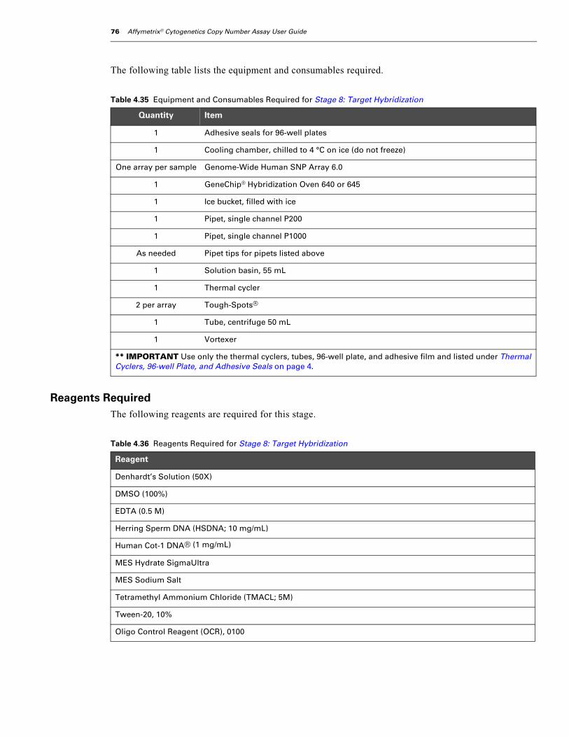
76 Affymetrix® Cytogenetics Copy Number Assay User Guide
The following table lists the equipment and consumables required.
Reagents RequiredThe following reagents are required for this stage.
Table 4.35 Equipment and Consumables Required for Stage 8: Target Hybridization
Quantity Item
1 Adhesive seals for 96-well plates
1 Cooling chamber, chilled to 4 °C on ice (do not freeze)
One array per sample Genome-Wide Human SNP Array 6.0
1 GeneChip® Hybridization Oven 640 or 645
1 Ice bucket, filled with ice
1 Pipet, single channel P200
1 Pipet, single channel P1000
As needed Pipet tips for pipets listed above
1 Solution basin, 55 mL
1 Thermal cycler
2 per array Tough-Spots®
1 Tube, centrifuge 50 mL
1 Vortexer
** IMPORTANT Use only the thermal cyclers, tubes, 96-well plate, and adhesive film and listed under Thermal Cyclers, 96-well Plate, and Adhesive Seals on page 4.
Table 4.36 Reagents Required for Stage 8: Target Hybridization
Reagent
Denhardt’s Solution (50X)
DMSO (100%)
EDTA (0.5 M)
Herring Sperm DNA (HSDNA; 10 mg/mL)
Human Cot-1 DNA® (1 mg/mL)
MES Hydrate SigmaUltra
MES Sodium Salt
Tetramethyl Ammonium Chloride (TMACL; 5M)
Tween-20, 10%
Oligo Control Reagent (OCR), 0100

chapter 4 | Affymetrix® Cytogenetics Copy Number Assay 77
Important Information About This StageTo help ensure the best results, carefully read the information below before you begin this stage of the protocol.
Prepare the Reagents, Consumables and Other Components
Prepare a 12X MES Stock SolutionThe 12X MES stock solution can be prepared in bulk and kept for at least one month if properly stored. Proper storage:
• Protect from light using aluminum foil
• Keep at 4 °C
To prepare 500 mL of 12X MES Stock Solution (1.25 M MES, 0.89 M [Na+]):
1. Combine:
• 35.2 g MES hydrate
• 96.65 g MES sodium salt
• 400 mL AccuGENE® water
2. Mix and adjust volume to 475 mL.
3. Test the pH.
The pH should be between 6.5 and 6.7.
4. Adjust the pH so it falls between 6.5 and 6.7.
5. Adjust the volume to 500 mL.
6. Filter the solution through a 0.2 µm filter.
7. Protect from light using aluminum foil and store at 4 °C.
IMPORTANT:• It is critical that the samples remain on the thermal cycler at 49 °C after denaturation and
while being loaded onto arrays.
• About DMSO:
When adding to the Hybridization Master Mix, pipet DMSO into the middle of the tube.Do not touch the sides of the tube as the DMSO can leach particles out of the plasticwhich, in turn, may cause high background.
DMSO is light sensitive and must be stored in a dark glass bottle. Do not store in a plasticcontainer.
• Be sure to equilibrate the arrays to room temperature; otherwise, the rubber septa maycrack and the array may leak.
• An accurate hybridization temperature is critical for this assay. Therefore, we recommendthat your hybridization ovens be serviced at least once per year to ensure that they areoperating within specifications.
• Gloves, safety glasses, and lab coats must be worn when preparing the HybridizationMaster Mix.
• Consult the appropriate MSDS for reagent storage and handling requirements.
IMPORTANT: Do not autoclave. Store between 2 °C and 8 °C, and shield from light usingaluminum foil. Discard solution if it turns yellow.

78 Affymetrix® Cytogenetics Copy Number Assay User Guide
Preheat the Hybridization Ovens
To preheat the hybridization ovens:
1. Turn on each oven and set the temperature to 50 °C.
2. Set the rpm to 60.
3. Turn the rotation on and allow to preheat for 1 hr before loading arrays.
Prepare the Samples
To prepare the samples:
1. If the labeled samples from the previous stage were frozen. allow them to thaw on the bench top to room temperature.
2. Vortex at high speed for 3 sec; then spin down for 30 sec.
3. Place in a cooling chamber on ice.
Preheat the Thermal Cycler LidPower on the thermal cycler to preheat the lid. Leave the block at room temperature.
Prepare the Arrays
To prepare the arrays:
1. Unwrap the arrays and place on the bench top, septa-side up.
2. Mark the front or back of each array with a designation that will identify which sample is loaded onto each array (Figure 4.28).
3. Allow the arrays to warm to room temperature on the bench top 10 to 15 min.
4. Insert a 200 µL pipet tip into the upper right septum of each array.
IMPORTANT: An accurate hybridization temperature is critical for this assay. Therefore,we recommend that your hybridization ovens be serviced at least once per year to ensurethat they are operating within the manufacturer’s specifications.
IMPORTANT: To ensure that the data collected during scanning is associated with thecorrect sample, mark each array in a meaningful way. It is critical that you know whichsample is loaded onto each array.
Figure 4.28 Arrays prepared for sample loading

chapter 4 | Affymetrix® Cytogenetics Copy Number Assay 79
Prepare the Hybridization Master MixAs an option, you can prepare a larger volume of Hybridization Master Mix than required. The extra mix can be aliquoted and stored at –20 °C for up to one week.
Preparing Fresh Hybridization Master Mix
To prepare the Hybridization Master Mix:
1. To the 50 mL centrifuge tube, add the appropriate volume of each reagent in the order shown in Table 4.37.
DMSO addition: pipet directly into the solution of other reagents. Avoid pipetting along the side of the tube.
2. Mix well.
3. If making a larger volume, aliquot out the volume required, and store the remainder at –20 °C for up to one week.
Using Premixed Hybridization Master MixHybridization Master Mix can be made ahead of time, aliquoted and stored for 1 week at –20 °C.
To prepare stored Hybridization Master Mix:
1. Place the stored Hybridization Master Mix on the bench top, and allow to warm to room temperature.
2. Vortex at high speed until the mixture is homogeneous and without precipitates (may take up to 5 min).
3. Pulse spin for 3 sec.
Table 4.37 Hybridization Master Mix
Reagent 1 Sample 4 Samples(15% extra)
8 Samples(15% extra)
12 Samples(15% extra)
24 Samples(15% extra)
MES (12X; 1.25 M) 12.0 µL 55.2 µL 110.4 µL 165.6 µL 331.2 µL
Denhardt’s Solution (50X) 13.0 µL 59.8 µL 119.6 µL 179.4 µL 358.8 µL
EDTA (0.5 M) 3.0 µL 13.8 µL 27.6 µL 41.4 µL 82.8 µL
HSDNA (10 mg/mL) 3.0 µL 13.8 µL 27.6 µL 41.4 µL 82.8 µL
OCR, 0100 2.0 µL 9.2 µL 18.4 µL 27.6 µL 55.2 µL
Human Cot-1 DNA® (1 mg/mL)
3.0 µL 13.8 µL 27.6 µL 41.4 µL 82.8 µL
Tween-20 (3%) 1.0 µL 4.6 µL 9.2 µL 13.8 µL 27.6 µL
DMSO (100%) 13.0 µL 59.8 µL 119.6 µL 179.4 µL 358.8 µL
TMACL (5 M) 140.0 µL 644.0 µL 1288.0 µL 1932.0 µL 3864.0 µL
Total 190 µL 874 µL 1748 µL 2622 µL 5244 µL
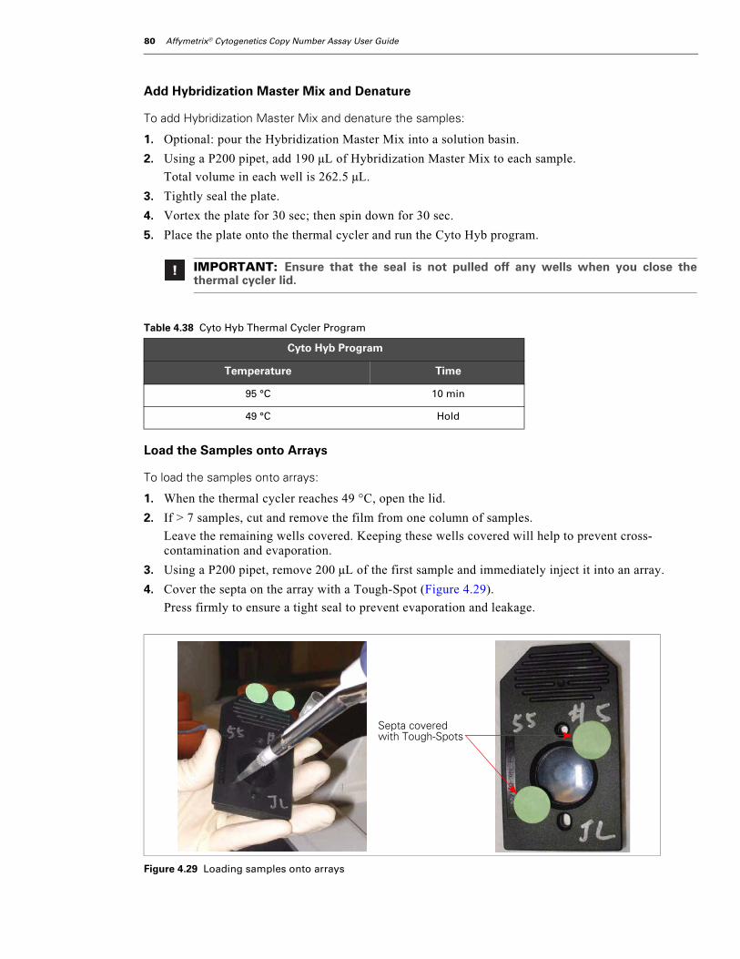
80 Affymetrix® Cytogenetics Copy Number Assay User Guide
Add Hybridization Master Mix and Denature
To add Hybridization Master Mix and denature the samples:
1. Optional: pour the Hybridization Master Mix into a solution basin.
2. Using a P200 pipet, add 190 µL of Hybridization Master Mix to each sample.
Total volume in each well is 262.5 µL.
3. Tightly seal the plate.
4. Vortex the plate for 30 sec; then spin down for 30 sec.
5. Place the plate onto the thermal cycler and run the Cyto Hyb program.
Load the Samples onto Arrays
To load the samples onto arrays:
1. When the thermal cycler reaches 49 °C, open the lid.
2. If > 7 samples, cut and remove the film from one column of samples.
Leave the remaining wells covered. Keeping these wells covered will help to prevent cross-contamination and evaporation.
3. Using a P200 pipet, remove 200 µL of the first sample and immediately inject it into an array.
4. Cover the septa on the array with a Tough-Spot (Figure 4.29).
Press firmly to ensure a tight seal to prevent evaporation and leakage.
IMPORTANT: Ensure that the seal is not pulled off any wells when you close thethermal cycler lid.
Table 4.38 Cyto Hyb Thermal Cycler Program
Cyto Hyb Program
Temperature Time
95 °C 10 min
49 °C Hold
Figure 4.29 Loading samples onto arrays
Septa covered with Tough-Spots

chapter 4 | Affymetrix® Cytogenetics Copy Number Assay 81
5. When 4 arrays are loaded and the septa are covered:
A. Load the arrays into an oven tray evenly spaced.
B. Immediately place the tray into the hybridization oven.
Do not allow loaded arrays to sit at room temperature for more than approximately 1 min. Ensure that the oven is balanced as the trays are loaded, and ensure that the trays are rotating at 60 rpm at all times.
6. Repeat this process until all samples are loaded onto arrays and are placed in a hybridization oven.
All samples should be loaded within 30 min.
7. Allow the arrays to rotate at 50 °C, 60 rpm for 16 to 18 hr.
IMPORTANT: Allow the arrays to rotate in the hybridization ovens for 16 to 18 hr at 50 °Cand 60 rpm. This temperature is optimized for this product, and should be stringentlyfollowed.

82 Affymetrix® Cytogenetics Copy Number Assay User Guide
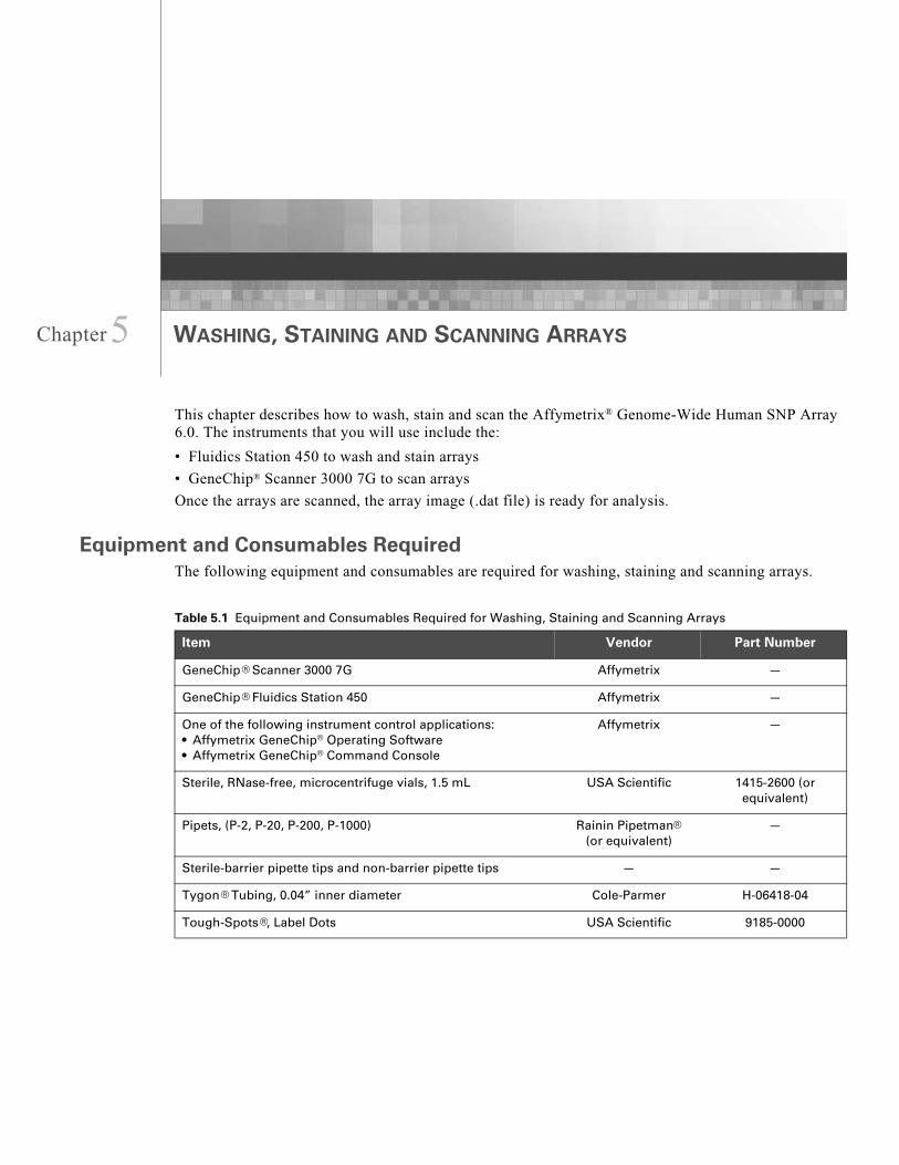
Chapte
r5 WASHING, STAINING AND SCANNING ARRAYSThis chapter describes how to wash, stain and scan the Affymetrix® Genome-Wide Human SNP Array 6.0. The instruments that you will use include the:
• Fluidics Station 450 to wash and stain arrays
• GeneChip® Scanner 3000 7G to scan arrays
Once the arrays are scanned, the array image (.dat file) is ready for analysis.
Equipment and Consumables RequiredThe following equipment and consumables are required for washing, staining and scanning arrays.
Table 5.1 Equipment and Consumables Required for Washing, Staining and Scanning Arrays
Item Vendor Part Number
GeneChip® Scanner 3000 7G Affymetrix —
GeneChip® Fluidics Station 450 Affymetrix —
One of the following instrument control applications: • Affymetrix GeneChip® Operating Software• Affymetrix GeneChip® Command Console
Affymetrix —
Sterile, RNase-free, microcentrifuge vials, 1.5 mL USA Scientific 1415-2600 (or equivalent)
Pipets, (P-2, P-20, P-200, P-1000) Rainin Pipetman®
(or equivalent)—
Sterile-barrier pipette tips and non-barrier pipette tips — —
Tygon® Tubing, 0.04” inner diameter Cole-Parmer H-06418-04
Tough-Spots®, Label Dots USA Scientific 9185-0000
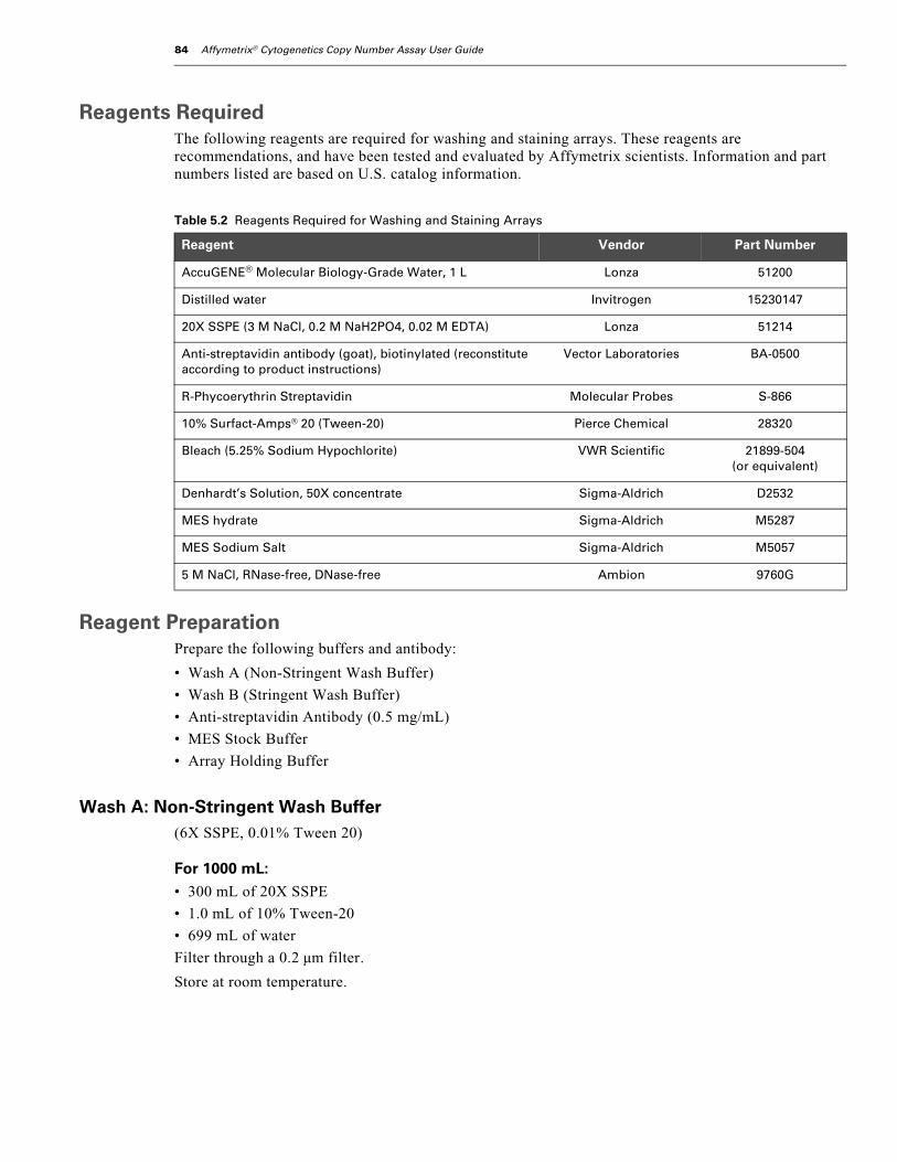
84 Affymetrix® Cytogenetics Copy Number Assay User Guide
Reagents RequiredThe following reagents are required for washing and staining arrays. These reagents are recommendations, and have been tested and evaluated by Affymetrix scientists. Information and part numbers listed are based on U.S. catalog information.
Reagent PreparationPrepare the following buffers and antibody:
• Wash A (Non-Stringent Wash Buffer)
• Wash B (Stringent Wash Buffer)
• Anti-streptavidin Antibody (0.5 mg/mL)
• MES Stock Buffer
• Array Holding Buffer
Wash A: Non-Stringent Wash Buffer(6X SSPE, 0.01% Tween 20)
For 1000 mL:• 300 mL of 20X SSPE
• 1.0 mL of 10% Tween-20
• 699 mL of water
Filter through a 0.2 µm filter.
Store at room temperature.
Table 5.2 Reagents Required for Washing and Staining Arrays
Reagent Vendor Part Number
AccuGENE® Molecular Biology-Grade Water, 1 L Lonza 51200
Distilled water Invitrogen 15230147
20X SSPE (3 M NaCl, 0.2 M NaH2PO4, 0.02 M EDTA) Lonza 51214
Anti-streptavidin antibody (goat), biotinylated (reconstitute according to product instructions)
Vector Laboratories BA-0500
R-Phycoerythrin Streptavidin Molecular Probes S-866
10% Surfact-Amps® 20 (Tween-20) Pierce Chemical 28320
Bleach (5.25% Sodium Hypochlorite) VWR Scientific 21899-504 (or equivalent)
Denhardt’s Solution, 50X concentrate Sigma-Aldrich D2532
MES hydrate Sigma-Aldrich M5287
MES Sodium Salt Sigma-Aldrich M5057
5 M NaCl, RNase-free, DNase-free Ambion 9760G

chapter 5 | Washing, Staining and Scanning Arrays 85
Wash B: Stringent Wash Buffer(0.6X SSPE, 0.01% Tween 20)
For 1000 mL:• 30 mL of 20X SSPE
• 1.0 mL of 10% Tween-20
• 969 mL of water
Filter through a 0.2 µm filter.
Store at room temperature.
The pH should be 8.
0.5 mg/mL Anti-Streptavidin AntibodyResuspend 0.5 mg in 1 mL of water.
Store at 4°C.
12X MES Stock Buffer(1.25 M MES, 0.89 M [Na+])
For 1000 mL:• 70.4g of MES hydrate
• 193.3g of MES Sodium Salt
• 800 mL of Molecular Biology Grade Water
Mix and adjust volume to 1000 mL.
The pH should be between 6.5 and 6.7.
Filter through a 0.2 µm filter.
1X Array Holding Buffer(Final 1X concentration is 100 mM MES, 1M [Na+], 0.01% Tween-20)
For 100 mL:• 8.3 mL of 12X MES Stock Buffer
• 18.5 mL of 5 M NaCl
• 0.1 mL of 10% Tween-20
• 73.1 mL of water
Store at 2°C to 8°C, and shield from light.
IMPORTANT: Prepare Wash B in smaller quantities to avoid long term storage. Tightly sealthe container to avoid changes in salt concentration due to evaporation.
IMPORTANT: Do not autoclave. Store at 2°C to 8°C, and shield from light.
Discard solution if yellow.
86 Affymetrix® Cytogenetics Copy Number Assay User Guide
Fluidics Station and Scanner Control SoftwareYou will use one of the instrument control applications listed below to operate the fluidics station and the scanner. For more information on these applications, refer to the appropriate user’s guide.
• Affymetrix GeneChip® Operating Software (GCOS)
Affymetrix GeneChip® Operating Software User’s Guide
• Affymetrix GeneChip® Command Console (AGCC)
Affymetrix GeneChip® Command Console™ User’s Guide
Register a New Experiment or Sample
To register a new experiment or sample:
• If using GCOS, register a new Experiment.
• If using AGCC, register a new Sample.
Prime the Fluidics StationThe Fluidics Station 450 is used to wash and stain the arrays; it is operated using either GCOS or AGCC software.
To prime the Fluidics Station:
1. Turn on the Fluidics Station.
2. Prime the Fluidics Station.
• Select protocol Prime_450 for each module
• Intake buffer reservoir: use Non-Stringent Wash Buffer
• Intake buffer reservoir B: use Stringent Wash Buffer
About Priming the Fluidics StationPriming ensures the lines of the fluidics station are filled with the appropriate buffers and the fluidics station is ready to run fluidics station protocols.
Priming should be done:
• When the fluidics station is first started
• When wash solutions are changed
• Before washing, if a shutdown has been performed
• If the LCD window instructs the user to prime
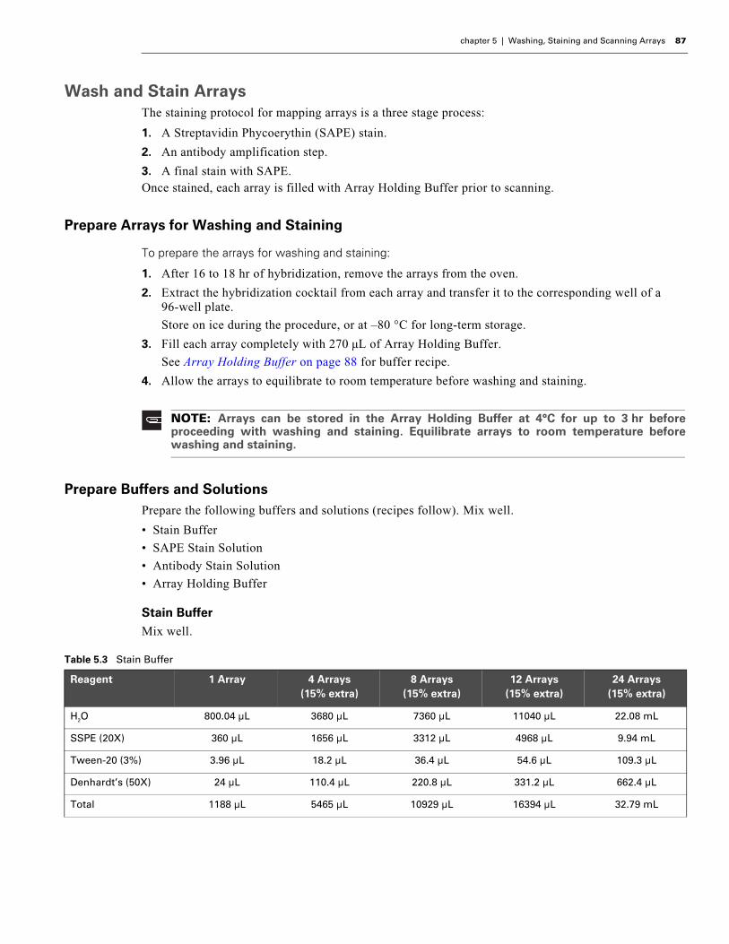
chapter 5 | Washing, Staining and Scanning Arrays 87
Wash and Stain ArraysThe staining protocol for mapping arrays is a three stage process:
1. A Streptavidin Phycoerythin (SAPE) stain.
2. An antibody amplification step.
3. A final stain with SAPE.Once stained, each array is filled with Array Holding Buffer prior to scanning.
Prepare Arrays for Washing and Staining
To prepare the arrays for washing and staining:
1. After 16 to 18 hr of hybridization, remove the arrays from the oven.
2. Extract the hybridization cocktail from each array and transfer it to the corresponding well of a 96-well plate.
Store on ice during the procedure, or at –80 °C for long-term storage.
3. Fill each array completely with 270 µL of Array Holding Buffer.
See Array Holding Buffer on page 88 for buffer recipe.
4. Allow the arrays to equilibrate to room temperature before washing and staining.
Prepare Buffers and SolutionsPrepare the following buffers and solutions (recipes follow). Mix well.
• Stain Buffer
• SAPE Stain Solution
• Antibody Stain Solution
• Array Holding Buffer
Stain BufferMix well.
NOTE: Arrays can be stored in the Array Holding Buffer at 4°C for up to 3 hr beforeproceeding with washing and staining. Equilibrate arrays to room temperature beforewashing and staining.
Table 5.3 Stain Buffer
Reagent 1 Array 4 Arrays (15% extra)
8 Arrays (15% extra)
12 Arrays (15% extra)
24 Arrays (15% extra)
H2O 800.04 µL 3680 µL 7360 µL 11040 µL 22.08 mL
SSPE (20X) 360 µL 1656 µL 3312 µL 4968 µL 9.94 mL
Tween-20 (3%) 3.96 µL 18.2 µL 36.4 µL 54.6 µL 109.3 µL
Denhardt’s (50X) 24 µL 110.4 µL 220.8 µL 331.2 µL 662.4 µL
Total 1188 µL 5465 µL 10929 µL 16394 µL 32.79 mL
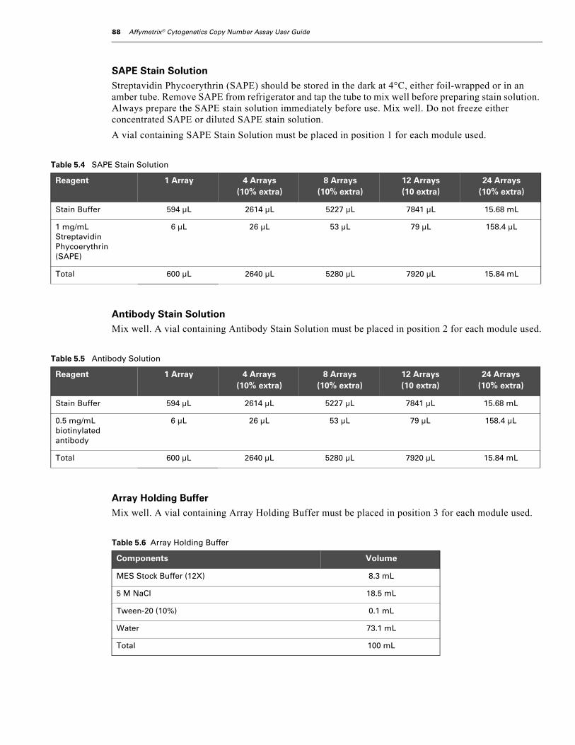
88 Affymetrix® Cytogenetics Copy Number Assay User Guide
SAPE Stain SolutionStreptavidin Phycoerythrin (SAPE) should be stored in the dark at 4°C, either foil-wrapped or in an amber tube. Remove SAPE from refrigerator and tap the tube to mix well before preparing stain solution. Always prepare the SAPE stain solution immediately before use. Mix well. Do not freeze either concentrated SAPE or diluted SAPE stain solution.
A vial containing SAPE Stain Solution must be placed in position 1 for each module used.
Antibody Stain SolutionMix well. A vial containing Antibody Stain Solution must be placed in position 2 for each module used.
Array Holding BufferMix well. A vial containing Array Holding Buffer must be placed in position 3 for each module used.
Table 5.4 SAPE Stain Solution
Reagent 1 Array 4 Arrays (10% extra)
8 Arrays (10% extra)
12 Arrays (10 extra)
24 Arrays (10% extra)
Stain Buffer 594 µL 2614 µL 5227 µL 7841 µL 15.68 mL
1 mg/mL Streptavidin Phycoerythrin (SAPE)
6 µL 26 µL 53 µL 79 µL 158.4 µL
Total 600 µL 2640 µL 5280 µL 7920 µL 15.84 mL
Table 5.5 Antibody Solution
Reagent 1 Array 4 Arrays (10% extra)
8 Arrays (10% extra)
12 Arrays (10 extra)
24 Arrays (10% extra)
Stain Buffer 594 µL 2614 µL 5227 µL 7841 µL 15.68 mL
0.5 mg/mL biotinylated antibody
6 µL 26 µL 53 µL 79 µL 158.4 µL
Total 600 µL 2640 µL 5280 µL 7920 µL 15.84 mL
Table 5.6 Array Holding Buffer
Components Volume
MES Stock Buffer (12X) 8.3 mL
5 M NaCl 18.5 mL
Tween-20 (10%) 0.1 mL
Water 73.1 mL
Total 100 mL
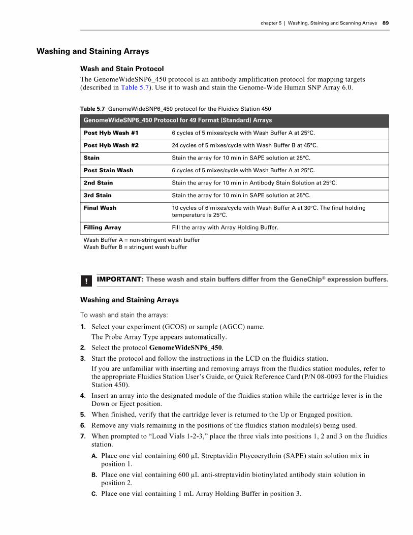
chapter 5 | Washing, Staining and Scanning Arrays 89
Washing and Staining Arrays
Wash and Stain ProtocolThe GenomeWideSNP6_450 protocol is an antibody amplification protocol for mapping targets (described in Table 5.7). Use it to wash and stain the Genome-Wide Human SNP Array 6.0.
Washing and Staining Arrays
To wash and stain the arrays:
1. Select your experiment (GCOS) or sample (AGCC) name.
The Probe Array Type appears automatically.
2. Select the protocol GenomeWideSNP6_450.
3. Start the protocol and follow the instructions in the LCD on the fluidics station.
If you are unfamiliar with inserting and removing arrays from the fluidics station modules, refer to the appropriate Fluidics Station User’s Guide, or Quick Reference Card (P/N 08-0093 for the Fluidics Station 450).
4. Insert an array into the designated module of the fluidics station while the cartridge lever is in the Down or Eject position.
5. When finished, verify that the cartridge lever is returned to the Up or Engaged position.
6. Remove any vials remaining in the positions of the fluidics station module(s) being used.
7. When prompted to “Load Vials 1-2-3,” place the three vials into positions 1, 2 and 3 on the fluidics station.
A. Place one vial containing 600 µL Streptavidin Phycoerythrin (SAPE) stain solution mix in position 1.
B. Place one vial containing 600 µL anti-streptavidin biotinylated antibody stain solution in position 2.
C. Place one vial containing 1 mL Array Holding Buffer in position 3.
Table 5.7 GenomeWideSNP6_450 protocol for the Fluidics Station 450
GenomeWideSNP6_450 Protocol for 49 Format (Standard) Arrays
Post Hyb Wash #1 6 cycles of 5 mixes/cycle with Wash Buffer A at 25°C.
Post Hyb Wash #2 24 cycles of 5 mixes/cycle with Wash Buffer B at 45°C.
Stain Stain the array for 10 min in SAPE solution at 25°C.
Post Stain Wash 6 cycles of 5 mixes/cycle with Wash Buffer A at 25°C.
2nd Stain Stain the array for 10 min in Antibody Stain Solution at 25°C.
3rd Stain Stain the array for 10 min in SAPE solution at 25°C.
Final Wash 10 cycles of 6 mixes/cycle with Wash Buffer A at 30°C. The final holding temperature is 25°C.
Filling Array Fill the array with Array Holding Buffer.
Wash Buffer A = non-stringent wash bufferWash Buffer B = stringent wash buffer
IMPORTANT: These wash and stain buffers differ from the GeneChip® expression buffers.

90 Affymetrix® Cytogenetics Copy Number Assay User Guide
D. Press down on the needle lever to snap needles into position and to start the run.
Once these steps are complete, the fluidics protocol begins. The Fluidics Station dialog box at the workstation terminal and the LCD window displays the status of the washing and staining steps.
8. When staining is finished, remove the microcentrifuge vials containing stain and replace with three empty vials as prompted.
9. Remove the arrays from the fluidics station by first pressing down the cartridge lever to the eject position.
10. Check the array window for large bubbles or air pockets.
If bubbles are present, 1) use a pipette to manually fill the array with Array Holding Buffer, 2) remove one-half of the solution, then 3) manually fill the array with Array Holding Buffer.
11. If the array has no large bubbles, it is ready for scanning. Pull up on the cartridge lever to engage wash block and proceed to Scanning Arrays on page 91.
If the arrays cannot be scanned promptly, store them at 4°C in the dark until ready for scanning. Scan must be performed within 24 hr.
12. When finished washing and staining, shut down the fluidics station following the procedure listed under Shutting Down the Fluidics Station on page 92.
IMPORTANT: If a bubble is present, do not return the array to the array holder. Thearray must be filled manually with Array Holding Buffer.

chapter 5 | Washing, Staining and Scanning Arrays 91
Scanning ArraysThe GeneChip Scanner 3000 7G is controlled by GCOS or AGCC software.
Prepare the ScannerTurn on the scanner at least 10 min before use.
Prepare Arrays for Scanning
To prepare arrays for scanning:
1. If the arrays were stored at 4°C, allow them to warm to room temperature before scanning.
2. If necessary, clean the glass surface of the array with a non-abrasive towel or tissue before scanning.
Do not use alcohol to clean the glass.
3. On the back of the array cartridge, clean excess fluid from around the septa.
4. Carefully cover both septa with Tough Spots (See Figure 5.1 on page 91).
Press to ensure the spots remain flat. If the spots do not apply smoothly (e.g. if you see bumps, bubbles, tears or curled edges) do not attempt to smooth out the spot. Remove the spot and apply a new spot.
5. Insert an array into the scanner and test the autofocus to ensure the spots do not interfere with the focus.
If a focus error message is observed, remove the spot and apply a new spot. Ensure that the spots lie flat.
WARNING: The scanner uses a laser and is equipped with a safety interlock system.Defeating the interlock system may result in exposure to hazardous laser light.
Read and be familiar with the operation of the scanner before attempting to scan an array.Refer to the GeneChip® Scanner 3000 Quick Reference Card (P/N 08-0075).
Figure 5.1 Applying Tough-Spots® to Arrays

92 Affymetrix® Cytogenetics Copy Number Assay User Guide
Scanning the Array
To scan arrays:
1. Select the experiment name (GCOS) or sample name (AGCC) that corresponds to the array being scanned.
2. Following the GCOS or AGCC instructions as appropriate, load the array into the scanner and begin the scan.
Only one scan per array is required. Pixel resolution and wavelength are preset and cannot be changed.
Shutting Down the Fluidics Station
To shut down the Fluidics Station:
1. Gently lift up the cartridge lever to engage (close) the washblock.
After removing an array from the holder, the LCD window displays the message ENGAGE WASHBLOCK. The instrument automatically performs a Cleanout procedure. The LCD window indicates the progress of this procedure.
2. When REMOVE VIALS is displayed in the LCD, remove the vials.
The REMOVE VIALS message indicates the Cleanout procedure is complete.
3. If no other processing is to be performed, place the wash lines into a bottle filled with deionized water.
4. Using GCOS or AGCC, choose the Shutdown_450 protocol for all modules.
5. Run the protocol for all modules.
The Shutdown protocol is critical to instrument reliability. Refer to the instrument User’s Guide for more information.
6. When the protocol is complete, turn the instrument off.
7. Place the wash lines in a different bottle of deionized water than the one used for the shutdown protocol.
NOTE: Customers using the Autoloader should refer to the Autoloader User’s Guide.
WARNING: The scanner door will open and close automatically. Do not attempt tomanually open or close the scanner door as this may damage the instrument.
Do not force the array into the holder.
IMPORTANT: To maintain the cleanliness of the fluidics station and obtain the highestquality image and data possible, a weekly bleach protocol is highly recommended.
chapter 5 | Washing, Staining and Scanning Arrays 93
Data Analysis To analyze the data collected by the scanner, use Affymetrix Genotyping Console™ version 2.1 or later.
Genotyping Console includes copy number and LOH algorithms for the SNP Array 6.0. It also includes the Genotyping Console Browser and Segment Reporting Tool.
Figure 5.2 Use Genotyping Console version 2.1 or later to analyze your data

94 Affymetrix® Cytogenetics Copy Number Assay User Guide
Chapte
r6 TROUBLESHOOTINGGeneral Assay Performance RecommendationsAs with any assay using PCR, the Cytogenetics Copy Number Assay has an inherent risk of contamination with PCR product from previous reactions. In Chapter 2 Laboratory Setup and Recommendations, we strongly recommend two separate work areas be used to minimize the risk of cross contamination during the assay procedure. It is essential to adhere to workflow recommendations. PCR reactions should only be carried out in the Post-PCR Area only. Personnel should not re-enter the Pre-PCR Clean Area once exposed to PCR products without first showering and changing into clean clothes.
Carefully reading and following the protocol as written is essential. The Cytogenetics Copy Number Assay has been validated using the reagents and suppliers listed. Substitution of reagents and taking shortcuts are not recommended as your results could be suboptimal. For example, always use AccuGENE® water from Cambrex, and ligase and restriction enzymes from New England Biolabs.
Additional recommendations are as follows:
• Think ahead to ensure that the reagents and equipment you require, including pipettes, are in the correct work area. Ensuring the proper equipment is available in the proper laboratory areas will make the workflow easier, and will help reduce the risk of sample contamination.
• Pay particular attention to the storage and handling of reagents. Proper storage and handling is particularly important for enzymes such as DNA Ligase and the Fragmentation Reagent (DNase I enzyme). Both of these enzymes are sensitive to temperatures exceeding –20°C.
To prevent loss of enzyme activity:
- Immediately place enzymes in a cooler chilled to –20 °C when removed from the freezer. Immediately return the enzyme to –20 °C after use.
- Take care when pipetting enzymes stored in glycerol, which is viscous. Do not store at –80 °C.
- Because Fragmentation Reagent activity can decline over time after dilution on ice, add it to the samples as quickly as possible.
• Preparing master mixes with a 15% excess ensures consistency in reagent preparation by minimizing pipetting errors and reducing handling time of temperature sensitive reagents. The success of this assay depends on the accurate pipetting and subsequent thorough mixing of small volumes of reagents.
• The PCR reaction for this assay has been validated using the specified thermal cyclers. These thermal cyclers were chosen because of their ramping times. We highly recommend that your PCR thermal cyclers be calibrated regularly. Take care when programming your thermal cycler and use the recommended 96-well plate.
• It is essential to run gels to monitor both the PCR and the fragmentation reactions.
For the PCR reaction, individual PCR products are run on a gel. Product (bands) should be visible in the 200to 1100 bp size range. See Check the PCR Reaction by Running a Gel on page 48 for more information.
96 Affymetrix® Cytogenetics Copy Number Assay User Guide
Following fragmentation, run your samples on a gel. Successful fragmentation is confirmed by the presence of a smear of < 180 bp in size. See Check the Fragmentation Reaction by Running a Gel on page 70 for more information.
• Run controls in parallel with each group of samples.
Substitute water for DNA as a negative control. The absence of bands on your PCR gel for this control confirms no previously amplified PCR product has contaminated your samples. Use Reference Genomic DNA 103 as a positive control (included in the reagent kit). These controls are effective troubleshooting tools that will help you confirm the successful completion of each stage of the assay.
• Oligonucleotide controls are included in the reagent kit. These controls are added to the target samples prior to hybridization and act to confirm successful hybridization, washing, staining, and sensitivity of the array. The oligonucleotide control reagents contain oligo B2 which is used for grid alignment.
• Regularly calibrate all multichannel pipettes.
• Check that your spectrophotometer is accurately calibrated, and be sure the OD measurement is within the quantitative linear range of the instrument (0.2 to 2.0 OD).
• Hybridization ovens should be serviced at least once per year to ensure that they are operating within the manufacturer’s specifications.
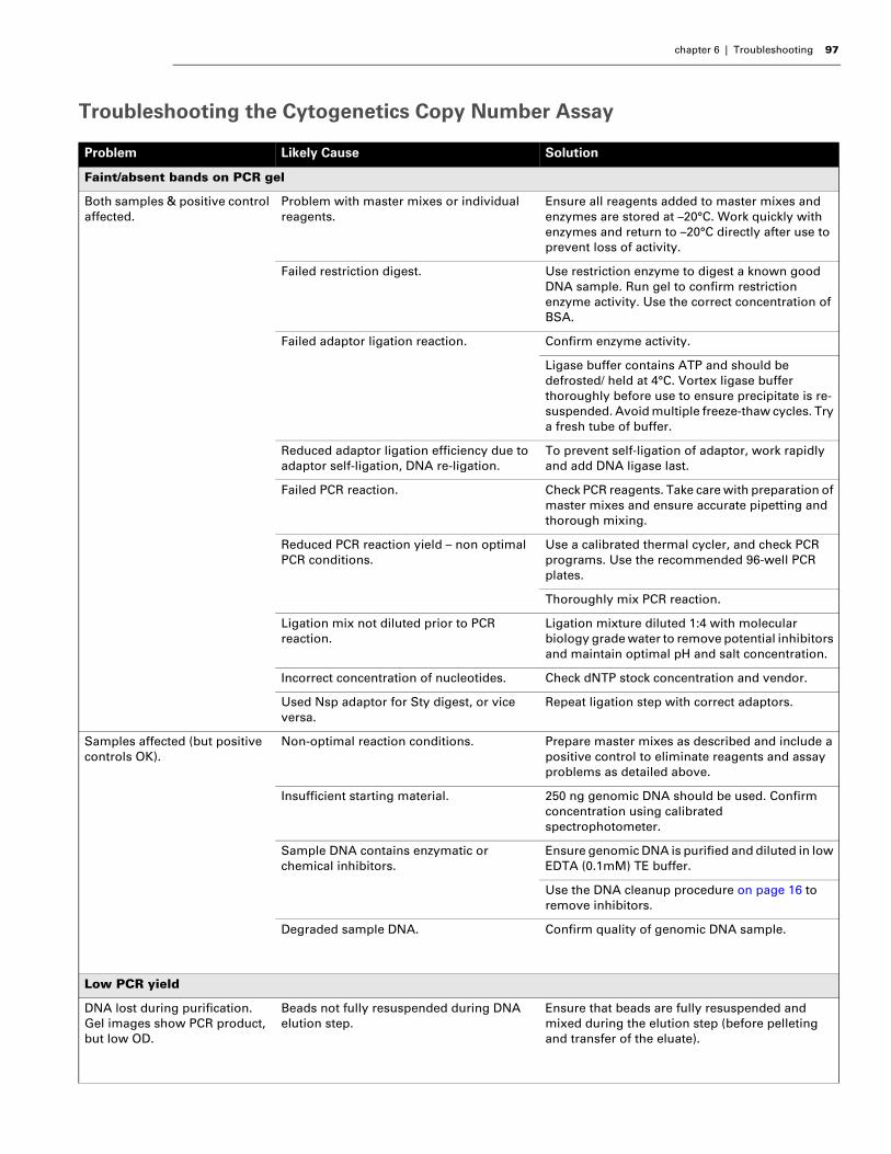
chapter 6 | Troubleshooting 97
Troubleshooting the Cytogenetics Copy Number Assay
Problem Likely Cause Solution
Faint/absent bands on PCR gel
Both samples & positive control affected.
Problem with master mixes or individual reagents.
Ensure all reagents added to master mixes and enzymes are stored at –20°C. Work quickly with enzymes and return to –20°C directly after use to prevent loss of activity.
Failed restriction digest. Use restriction enzyme to digest a known good DNA sample. Run gel to confirm restriction enzyme activity. Use the correct concentration of BSA.
Failed adaptor ligation reaction. Confirm enzyme activity.
Ligase buffer contains ATP and should be defrosted/ held at 4°C. Vortex ligase buffer thoroughly before use to ensure precipitate is re-suspended. Avoid multiple freeze-thaw cycles. Try a fresh tube of buffer.
Reduced adaptor ligation efficiency due to adaptor self-ligation, DNA re-ligation.
To prevent self-ligation of adaptor, work rapidly and add DNA ligase last.
Failed PCR reaction. Check PCR reagents. Take care with preparation of master mixes and ensure accurate pipetting and thorough mixing.
Reduced PCR reaction yield – non optimal PCR conditions.
Use a calibrated thermal cycler, and check PCR programs. Use the recommended 96-well PCR plates.
Thoroughly mix PCR reaction.
Ligation mix not diluted prior to PCR reaction.
Ligation mixture diluted 1:4 with molecular biology grade water to remove potential inhibitors and maintain optimal pH and salt concentration.
Incorrect concentration of nucleotides. Check dNTP stock concentration and vendor.
Used Nsp adaptor for Sty digest, or vice versa.
Repeat ligation step with correct adaptors.
Samples affected (but positive controls OK).
Non-optimal reaction conditions. Prepare master mixes as described and include a positive control to eliminate reagents and assay problems as detailed above.
Insufficient starting material. 250 ng genomic DNA should be used. Confirm concentration using calibrated spectrophotometer.
Sample DNA contains enzymatic or chemical inhibitors.
Ensure genomic DNA is purified and diluted in low EDTA (0.1mM) TE buffer.
Use the DNA cleanup procedure on page 16 to remove inhibitors.
Degraded sample DNA. Confirm quality of genomic DNA sample.
Low PCR yield
DNA lost during purification. Gel images show PCR product, but low OD.
Beads not fully resuspended during DNA elution step.
Ensure that beads are fully resuspended and mixed during the elution step (before pelleting and transfer of the eluate).

98 Affymetrix® Cytogenetics Copy Number Assay User Guide
Insufficient purified PCR product for fragmentation
Volume of eluate for particular samples is < 47 µL.
Do the following in this order:1.Measure the actual volume using a pipettor.2.Add Buffer EB to a final volume of 47 µL.3.Mix by pipetting up and down.4.Transfer 2 µL to the corresponding well(s) in the
OD plate.5.Proceed to fragmentation with 45 µL in each
well.
Fragmented PCR product is not the correct size
PCR product is still visible in 200-1100 bp size region
Failed or incomplete fragmentation due to reduced DNase activity.
Check that you have selected the correct activity of DNase to add to fragmentation reaction. (See Prepare the Fragmentation Master Mix on page 67)
Ensure fragmentation reagent is kept at –20°C. Do not reuse diluted working stock.
.CEL file can not be generated
GCOS or AGCC is unable to align grid.
Unable to place a grid on the .dat file due to the absence of B2 signal.
Hybridization controls including oligo B2 must be added to hybridization cocktail for grid alignment.
.dat image is dim. Insufficient signal intensity or staining failure.
Make fresh stain buffers.
Incorrect wash buffers used on fluidics station.
Prime the fluidics station with the correct buffers prior to running the assay. Incorrect wash buffers will disrupt hybridization of the labeled, fragmented DNA.
High MAPD or Low Contrast QC Values
Gel images and spectrophotometric quantitation indicate successful PCR reaction.
Over fragmentation of DNA sample due to incorrect dilution of Fragmentation Reagent stock.
Check that you have selected the correct activity of DNase to add to fragmentation reaction. (See Prepare the Fragmentation Master Mix on page 67.) Work quickly and on ice; mix thoroughly. Transfer reactions to pre-heated thermal cycler (37°C).
Extremely high MAPD or low Contrast QC values.Sample hybridization is absent on .cel and .dat images but B2 grid is bright.
Labeling reaction suboptimal. Use a new vial of Terminal Dideoxynucleotidyl Transferase. Verify the labeling reagents and repeat labeling.
Positive control has good MAPD and Contrast QC values, but samples do not.
Genomic DNA not optimal. Ensure DNA samples are of high quality (i.e., run in a 1 to 2% gel and compare to Reference 103 DNA control). Use positive control sample as a reference guide for assay procedures. Prepare master mixes for samples and controls.
Very high MAPD or low Contrast QC values.
Mixed up Nsp and Sty enzymes during the digestion or ligation stages.
Repeat the experiment, making sure the correct reagents are used for each digestion and ligation stage.
Problem Likely Cause Solution
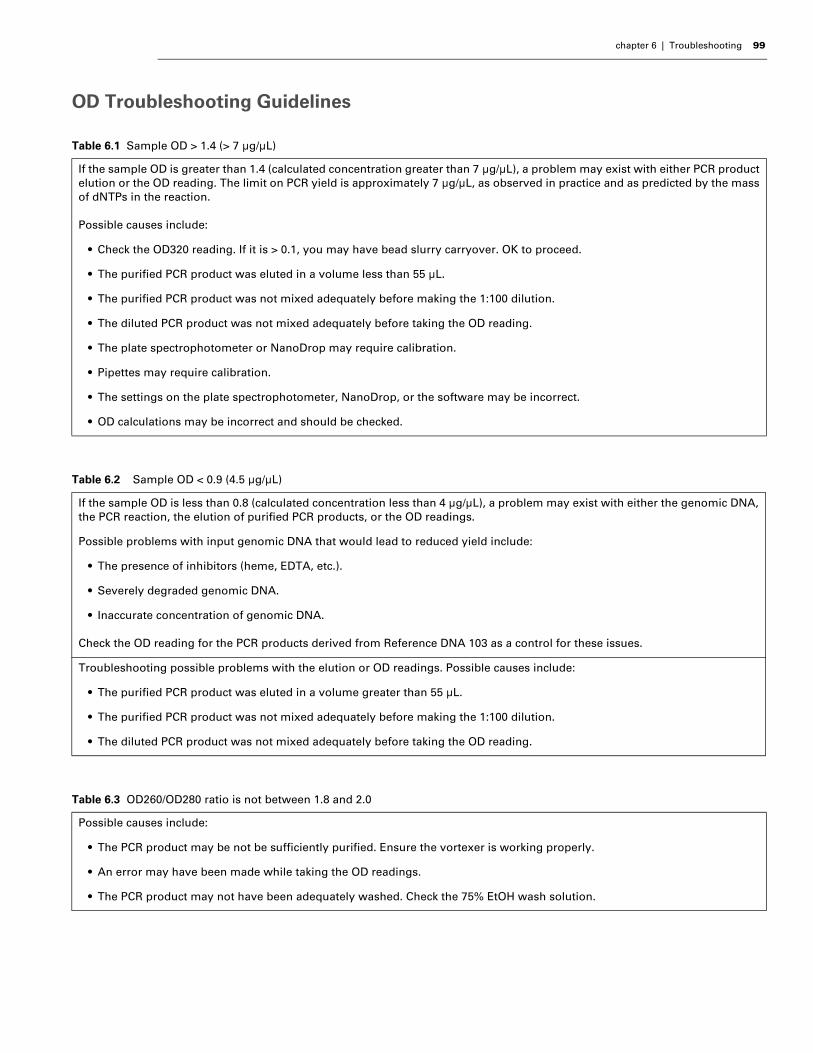
chapter 6 | Troubleshooting 99
OD Troubleshooting Guidelines
Table 6.1 Sample OD > 1.4 (> 7 µg/µL)
If the sample OD is greater than 1.4 (calculated concentration greater than 7 µg/µL), a problem may exist with either PCR product elution or the OD reading. The limit on PCR yield is approximately 7 µg/µL, as observed in practice and as predicted by the mass of dNTPs in the reaction.
Possible causes include:
• Check the OD320 reading. If it is > 0.1, you may have bead slurry carryover. OK to proceed.
• The purified PCR product was eluted in a volume less than 55 µL.
• The purified PCR product was not mixed adequately before making the 1:100 dilution.
• The diluted PCR product was not mixed adequately before taking the OD reading.
• The plate spectrophotometer or NanoDrop may require calibration.
• Pipettes may require calibration.
• The settings on the plate spectrophotometer, NanoDrop, or the software may be incorrect.
• OD calculations may be incorrect and should be checked.
Table 6.2 Sample OD < 0.9 (4.5 µg/µL)
If the sample OD is less than 0.8 (calculated concentration less than 4 µg/µL), a problem may exist with either the genomic DNA, the PCR reaction, the elution of purified PCR products, or the OD readings.
Possible problems with input genomic DNA that would lead to reduced yield include:
• The presence of inhibitors (heme, EDTA, etc.).
• Severely degraded genomic DNA.
• Inaccurate concentration of genomic DNA.
Check the OD reading for the PCR products derived from Reference DNA 103 as a control for these issues.
Troubleshooting possible problems with the elution or OD readings. Possible causes include:
• The purified PCR product was eluted in a volume greater than 55 µL.
• The purified PCR product was not mixed adequately before making the 1:100 dilution.
• The diluted PCR product was not mixed adequately before taking the OD reading.
Table 6.3 OD260/OD280 ratio is not between 1.8 and 2.0
Possible causes include:
• The PCR product may be not be sufficiently purified. Ensure the vortexer is working properly.
• An error may have been made while taking the OD readings.
• The PCR product may not have been adequately washed. Check the 75% EtOH wash solution.

100 Affymetrix® Cytogenetics Copy Number Assay User Guide
Affymetrix InstrumentsUnder any of the following conditions, unplug the instrument from the power source and contact Affymetrix Technical Support:
• When the power cord is damaged or frayed
• If any liquid has penetrated the instrument
• If, after service or calibration, the instrument does not perform to specifications .
Table 6.4 The OD320 measurement is > 0.2
Possible causes include:
• Magnetic beads may have been carried over into purified sample. Action: Spin down the sample for 5 min. Place on the MagnaRack and pipet out the eluate.
• Precipitate may be present in the eluted samples.
• There may be defects in the OD plate.
• Air bubbles in the OD plate or in solutions.
NOTE: Make sure you have the model and serial number available when calling AffymetrixTechnical Support.
Affymetrix, Inc.3420 Central ExpresswaySanta Clara, CA 95051USA
E-mail: [email protected]: 1-888-362-2447 (1-888-DNA-CHIP)Fax: 1-408-731-5441
Affymetrix UK LtdVoyager, Mercury Park,Wycombe Lane, Wooburn Green,High Wycombe HP10 0HHUnited Kingdom
E-mail: [email protected] and Others Tel: +44 (0) 1628 552550France Tel: 0800919505Germany Tel: 01803001334Fax: +44 (0) 1628 552585
Affymetrix Japan, K. K.Mita NN Bldg16 Floor, 4-1-23 Shiba,Minato-ku, Tokyo 108-0014Japan
Tel: (03) 5730-8200Fax: (03) 5730-8201
Chapte
r7 FLUIDICS STATION CARE AND MAINTENANCEGeneral Fluidics Station Care• Use a surge protector on the power line to the fluidics station.
• Always run a Shutdown protocol when the instrument will be off or unused overnight or longer. This will prevent salt crystals from forming within the fluidics system.
• To ensure proper functioning of the instrument, perform periodic maintenance.
• When not using the instrument, leave the sample needles in the lowered position. Each needle should extend into an empty vial. This will protect them from accidental damage.
• Always use deionized water to prevent contamination of the lines. Change buffers with freshly prepared buffer at each system startup.
• The fluidics station should be positioned on a sturdy, level bench away from extremes in temperature and away from moving air.
Fluidics Station Bleach ProtocolAffymetrix recommends a weekly cleaning protocol for the fluidics station. This protocol uses commonly purchased sodium hypochlorite bleach.
This protocol is designed to eliminate any residual SAPE-antibody complex that may be present in the fluidics station tubing and needles. The protocol runs a bleach solution through the system followed by a rinse cycle with deionized (DI) water. This protocol takes approximately one hr and forty minutes to complete. Affymetrix recommends running this protocol weekly, regardless of the frequency of use. The current version of the protocol can be found at:
www.affymetrix.com/support/technical/fluidics_scripts.affx
WARNING: Before performing any maintenance, turn off power to the fluidics station toavoid injury in case of a pump or electrical malfunction.
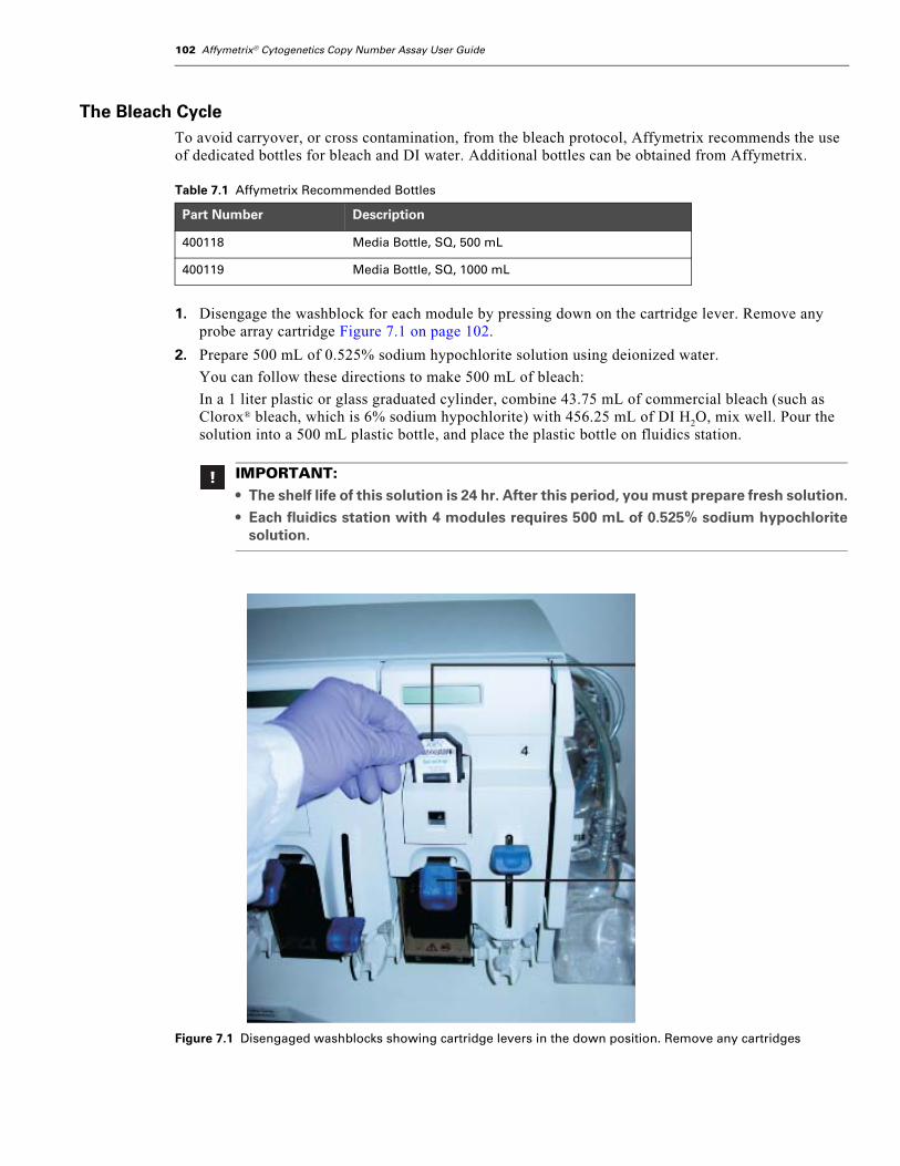
102 Affymetrix® Cytogenetics Copy Number Assay User Guide
The Bleach CycleTo avoid carryover, or cross contamination, from the bleach protocol, Affymetrix recommends the use of dedicated bottles for bleach and DI water. Additional bottles can be obtained from Affymetrix.
1. Disengage the washblock for each module by pressing down on the cartridge lever. Remove any probe array cartridge Figure 7.1 on page 102.
2. Prepare 500 mL of 0.525% sodium hypochlorite solution using deionized water.
You can follow these directions to make 500 mL of bleach:
In a 1 liter plastic or glass graduated cylinder, combine 43.75 mL of commercial bleach (such as Clorox® bleach, which is 6% sodium hypochlorite) with 456.25 mL of DI H2O, mix well. Pour the solution into a 500 mL plastic bottle, and place the plastic bottle on fluidics station.
Table 7.1 Affymetrix Recommended Bottles
Part Number Description
400118 Media Bottle, SQ, 500 mL
400119 Media Bottle, SQ, 1000 mL
IMPORTANT:• The shelf life of this solution is 24 hr. After this period, you must prepare fresh solution.
• Each fluidics station with 4 modules requires 500 mL of 0.525% sodium hypochloritesolution.
Figure 7.1 Disengaged washblocks showing cartridge levers in the down position. Remove any cartridges
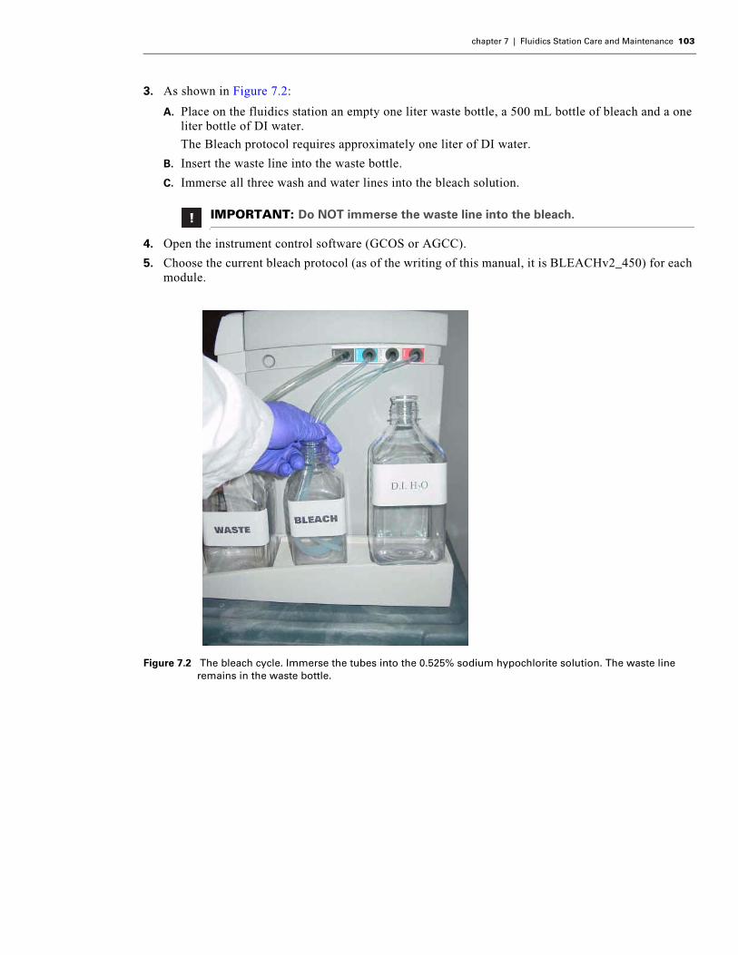
chapter 7 | Fluidics Station Care and Maintenance 103
3. As shown in Figure 7.2:
A. Place on the fluidics station an empty one liter waste bottle, a 500 mL bottle of bleach and a one liter bottle of DI water.
The Bleach protocol requires approximately one liter of DI water.
B. Insert the waste line into the waste bottle.
C. Immerse all three wash and water lines into the bleach solution.
4. Open the instrument control software (GCOS or AGCC).
5. Choose the current bleach protocol (as of the writing of this manual, it is BLEACHv2_450) for each module.
x
IMPORTANT: Do NOT immerse the waste line into the bleach.
Figure 7.2 The bleach cycle. Immerse the tubes into the 0.525% sodium hypochlorite solution. The waste line remains in the waste bottle.
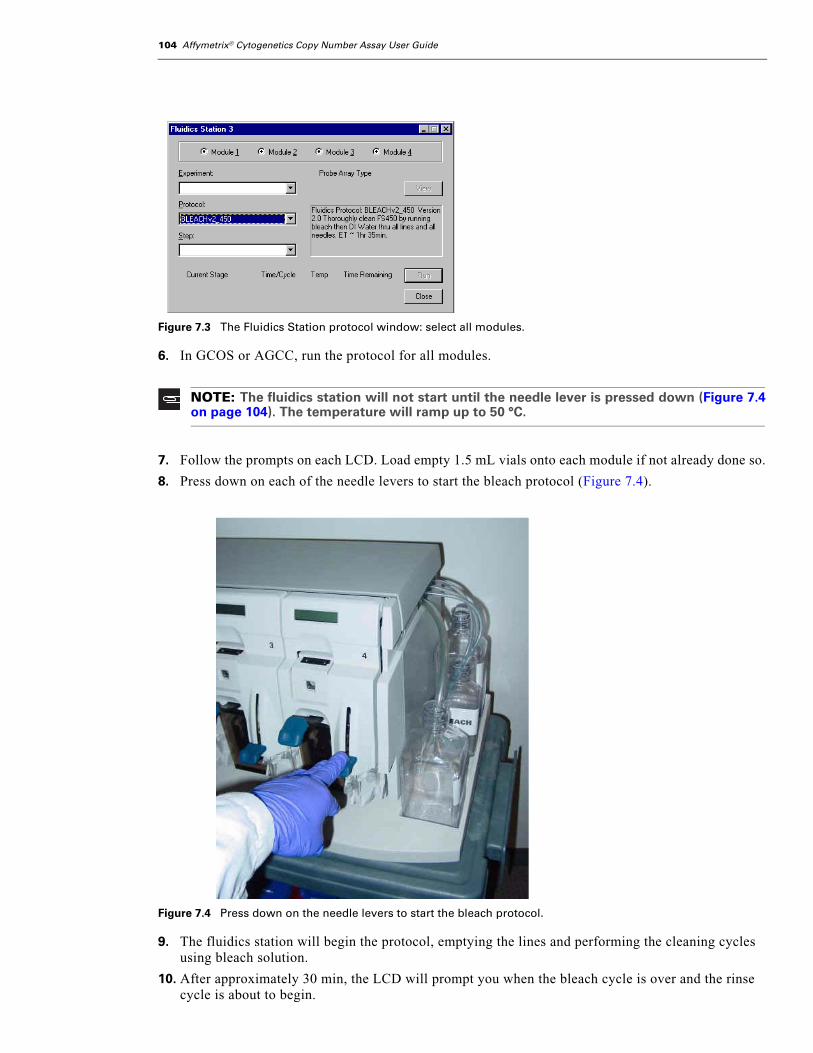
104 Affymetrix® Cytogenetics Copy Number Assay User Guide
6. In GCOS or AGCC, run the protocol for all modules.
7. Follow the prompts on each LCD. Load empty 1.5 mL vials onto each module if not already done so.
8. Press down on each of the needle levers to start the bleach protocol (Figure 7.4).
9. The fluidics station will begin the protocol, emptying the lines and performing the cleaning cycles using bleach solution.
10. After approximately 30 min, the LCD will prompt you when the bleach cycle is over and the rinse cycle is about to begin.
Figure 7.3 The Fluidics Station protocol window: select all modules.
NOTE: The fluidics station will not start until the needle lever is pressed down (Figure 7.4on page 104). The temperature will ramp up to 50 °C.
Figure 7.4 Press down on the needle levers to start the bleach protocol.

chapter 7 | Fluidics Station Care and Maintenance 105
The Rinse CycleOnce the bleach cycle has finished, the second part of the protocol is a rinse step. This step is essential to remove all traces of bleach from the system. Failure to complete this step can result in damaged arrays.
1. Follow the prompts on the LCD for each module. Lift up on the needle levers and remove the bleach vials. Load clean, empty vials onto each module.
2. Remove the three wash and water lines from the bleach bottle and transfer them to the DI water bottle (Figure 7.5).
At this step, there is no need to be concerned about the bleach remaining in the lines.
3. Press down on the needle levers to begin the rinse cycle.
The fluidics station will empty the lines and rinse the needles.
4. When the rinse is completed after approximately one hour, the fluidics station will bring the temperature back to 25°C and drain the lines with air.
The LCD display will read CLEANING DONE.
5. Discard the vials used for the bleach protocol.
6. After completing the bleach protocol, follow the suggestions for storage of the Fluidics Station 450 in Table 7.2 below.
Figure 7.5 Immerse the three wash and water lines in the DI water bottle.
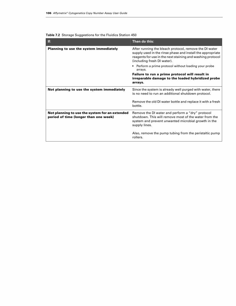
106 Affymetrix® Cytogenetics Copy Number Assay User Guide
Table 7.2 Storage Suggestions for the Fluidics Station 450
If: Then do this:
Planning to use the system immediately After running the bleach protocol, remove the DI water supply used in the rinse phase and install the appropriate reagents for use in the next staining and washing protocol (including fresh DI water).
• Perform a prime protocol without loading your probe arrays.
Failure to run a prime protocol will result in irreparable damage to the loaded hybridized probe arrays.
Not planning to use the system immediately Since the system is already well purged with water, there is no need to run an additional shutdown protocol.
Remove the old DI water bottle and replace it with a fresh bottle.
Not planning to use the system for an extended period of time (longer than one week)
Remove the DI water and perform a “dry” protocol shutdown. This will remove most of the water from the system and prevent unwanted microbial growth in the supply lines.
Also, remove the pump tubing from the peristaltic pump rollers.

Appendix
A GUIDELINES FOR PROCESSING 16 SAMPLESThis appendix illustrates the plate layouts recommended for processing 16 reactions (14 samples plus one positive and one negative control). It also provides a high level overview of the workflow.
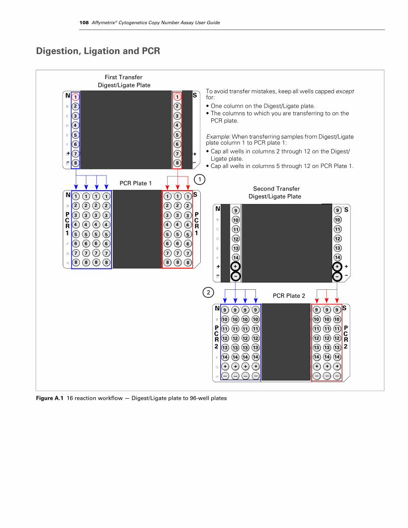
108 Affymetrix® Cytogenetics Copy Number Assay User Guide
Digestion, Ligation and PCR
Figure A.1 16 reaction workflow — Digest/Ligate plate to 96-well plates
N S
+
––
+ +
–
+
–
Digest/Ligate Plate
N S
PCR1
PCR1
N S
PCR2
PCR2
N S
+
––
+ +
–
+
–
Digest/Ligate Plate
1
2
3
4
5
6
7
8
1
2
3
4
5
6
7
8
9
10
11
12
13
14
9
10
11
12
13
14
1
2
3
4
5
6
7
8
1
2
3
4
5
6
7
8
1
2
3
4
5
6
7
8
1
2
3
4
5
6
7
8
1
2
3
4
5
6
7
8
1
2
3
4
5
6
7
8
1
2
3
4
5
6
7
8
1
2
3
4
5
6
7
8
9
10
11
12
13
14
9
10
11
12
13
14
1
2
3
4
5
6
7
8
9
10
11
12
13
14
+
—
9
10
11
12
13
14
+
—
9
10
11
12
13
14
+
—
9
10
11
12
13
14
+
—
9
10
11
12
13
14
+
—
9
10
11
12
13
14
+
—
9
10
11
12
13
14
+
—
PCR Plate 2
PCR Plate 1
To avoid transfer mistakes, keep all wells capped except for:• One column on the Digest/Ligate plate.• The columns to which you are transferring to on the
PCR plate.
Example: When transferring samples from Digest/Ligate plate column 1 to PCR plate 1:• Cap all wells in columns 2 through 12 on the Digest/
Ligate plate.• Cap all wells in columns 5 through 12 on PCR Plate 1.
1
2
Second Transfer
First Transfer
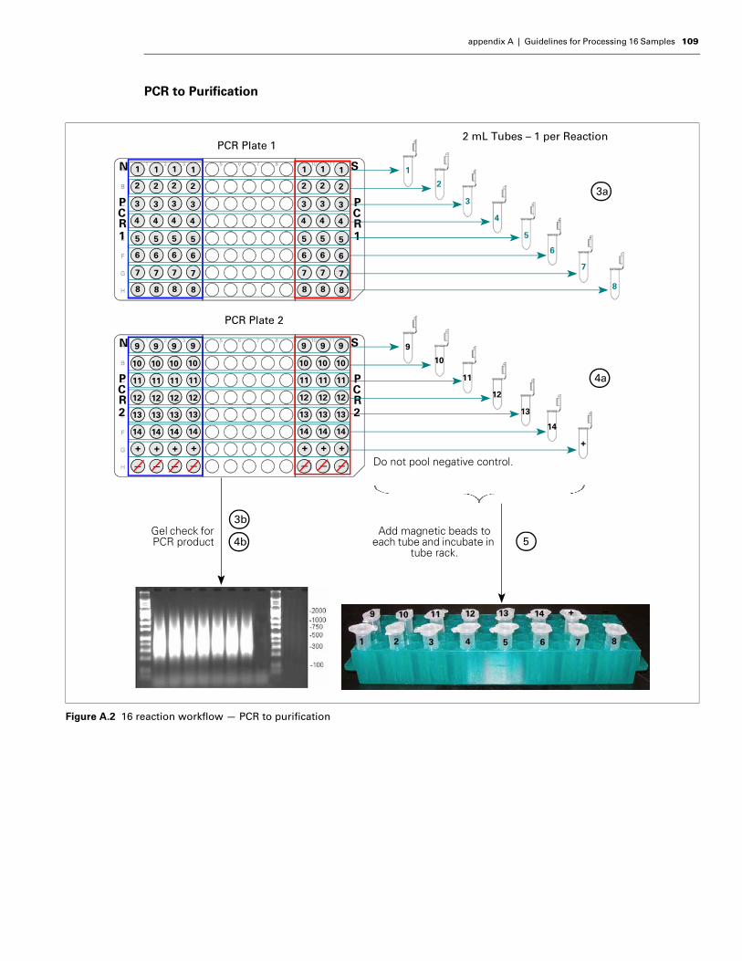
appendix A | Guidelines for Processing 16 Samples 109
PCR to Purification
Figure A.2 16 reaction workflow — PCR to purification
N S
PCR1
PCR1
N S
PCR2
PCR2
1
2
3
4
5
6
7
8
1
2
3
4
5
6
7
8
1
2
3
4
5
6
7
8
1
2
3
4
5
6
7
8
1
2
3
4
5
6
7
8
1
2
3
4
5
6
7
8
1
2
3
4
5
6
7
8
9
10
11
12
13
14
+
—
9
10
11
12
13
14
+
—
9
10
11
12
13
14
+
—
9
10
11
12
13
14
+
—
9
10
11
12
13
14
+
—
9
10
11
12
13
14
+
—
9
10
11
12
13
14
+
—
PCR Plate 2
PCR Plate 12 mL Tubes – 1 per Reaction
9
10
11
12
5
6
7
8
1
2
3
4
13
14
+
Add magnetic beads to each tube and incubate in
tube rack.
Gel check forPCR product
1 2 3 4 5 6 7 8
9 10 11 12 13 14 + –
3a
4a
3b
4b 5
Do not pool negative control.
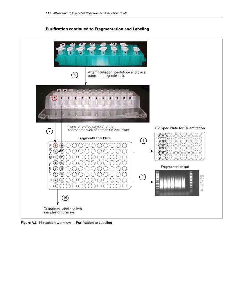
110 Affymetrix® Cytogenetics Copy Number Assay User Guide
Purification continued to Fragmentation and Labeling
Figure A.3 16 reaction workflow — Purification to Labeling
1 2 3 4 5 6 7 8
9 10 11 12 13 14 + –
2 3 4 5 6 7 8 9 10 11 12
After incubation, centrifuge and place tubes on magnetic rack.
+ +
1
2
3
4
5
6
8
9
10
11
12
13
14
FRAG
LBL
Transfer eluted sample to the appropriate well of a fresh 96-well plate
Fragment/Label Plate
1
13 14 +
6
7
8
1
2
3
4
5
6
7
8
9
10
11
12
13
14
+
UV Spec Plate for Quantitation
Quantitate, label and hyb samples onto arrays.
97
Fragmentation gel
10

Appendix
B GUIDELINES FOR PROCESSING 24 SAMPLESThis appendix illustrates the plate layouts recommended for processing 24 reactions (22 samples plus one positive and one negative control). It also provides a high level overview of the workflow.
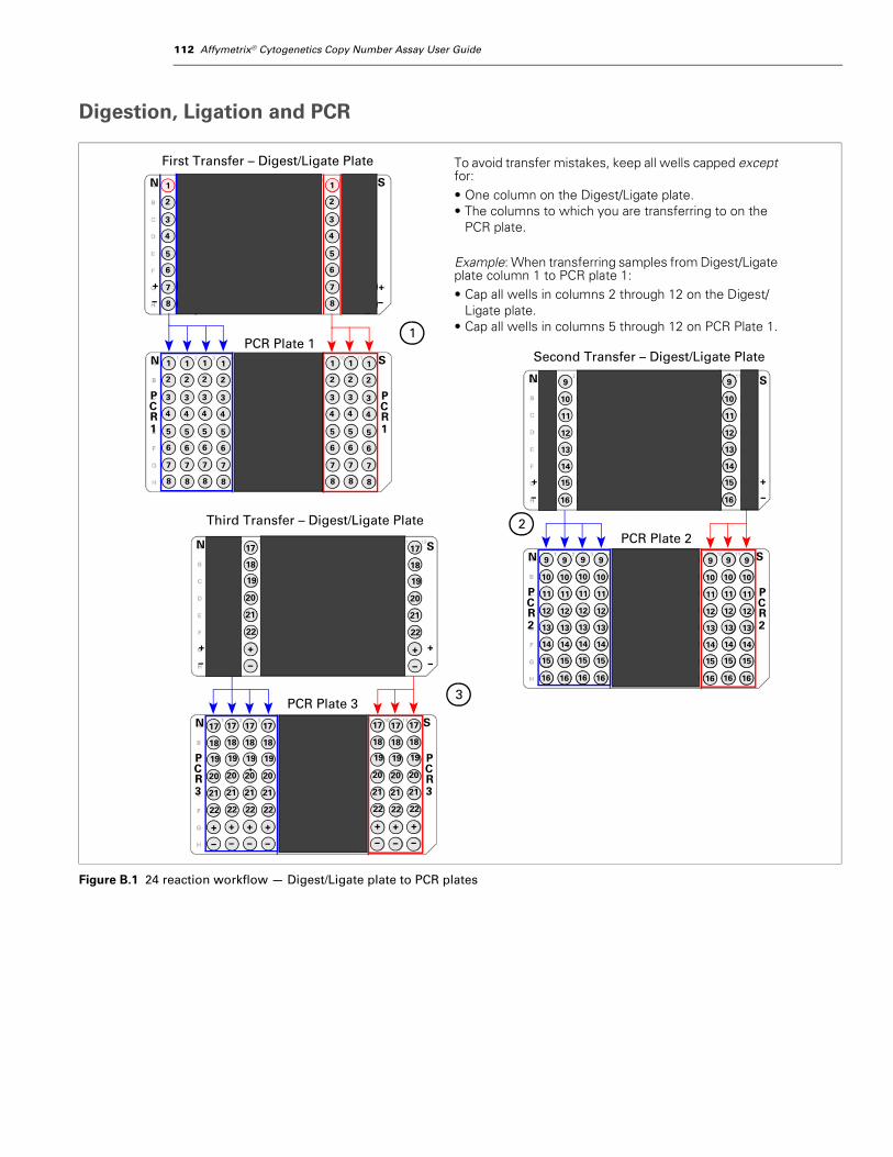
112 Affymetrix® Cytogenetics Copy Number Assay User Guide
Digestion, Ligation and PCR
Figure B.1 24 reaction workflow — Digest/Ligate plate to PCR plates
N S
+
––
+ +
–
+
–
Second Transfer – Digest/Ligate PlateN S
PCR1
PCR1
N S
PCR2
PCR2
N S
+
––
+
First Transfer – Digest/Ligate Plate
1
2
3
4
5
6
7
8
1
2
3
4
5
6
7
8
9
10
11
12
13
14
9
10
11
12
13
14
1
2
3
4
5
6
7
8
1
2
3
4
5
6
7
8
1
2
3
4
5
6
7
8
1
2
3
4
5
6
7
8
1
2
3
4
5
6
7
8
1
2
3
4
5
6
7
8
1
2
3
4
5
6
7
8
1
2
3
4
5
6
7
8
9
10
11
12
13
14
PCR Plate 2
PCR Plate 1
To avoid transfer mistakes, keep all wells capped except for:• One column on the Digest/Ligate plate.• The columns to which you are transferring to on the
PCR plate.
Example: When transferring samples from Digest/Ligate plate column 1 to PCR plate 1:• Cap all wells in columns 2 through 12 on the Digest/
Ligate plate.• Cap all wells in columns 5 through 12 on PCR Plate 1.1
2
N S
PCR3
PCR3
N S
+
––
+ +
–
1
2
3
4
5
6
7
8
Third Transfer – Digest/Ligate Plate
3PCR Plate 3
15
16
9
10
11
12
13
14
15
16
17
18
19
20
21
22
+
–
17
18
19
20
21
22
+
–
17
18
19
20
21
22
+
–
17
18
19
20
21
22
+
–
17
18
19
20
21
22
+
–
17
18
19
20
21
22
+
–
17
18
19
20
21
22
+
–
17
18
19
20
21
22
+
–
17
18
19
20
21
22
9
10
11
12
13
14
15
16
9
10
11
12
13
14
15
16
9
10
11
12
13
14
15
16
9
10
11
12
13
14
15
16
9
10
11
12
13
14
15
16
9
10
11
12
13
14
15
16
9
10
11
12
13
14
15
16
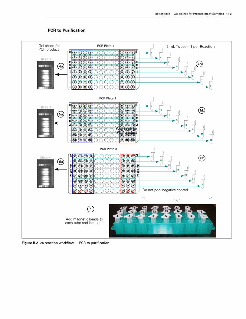
appendix B | Guidelines for Processing 24 Samples 113
PCR to Purification
Figure B.2 24 reaction workflow — PCR to purification
N S
PCR1
PCR1
N S
PCR2
PCR2
1
2
3
4
5
6
7
8
1
2
3
4
5
6
7
8
1
2
3
4
5
6
7
8
1
2
3
4
5
6
7
8
1
2
3
4
5
6
7
8
1
2
3
4
5
6
7
8
1
2
3
4
5
6
7
8
9
10
11
12
13
14
15
9
10
11
12
13
14
9
10
11
12
13
14
9
10
11
12
13
14
9
10
11
12
13
14
9
10
11
12
13
14
9
10
11
12
13
14
PCR Plate 2
PCR Plate 1 2 mL Tubes – 1 per Reaction
9
10
11
12
5
6
7
8
1
2
3
44b
5b
N S
PCR2
PCR2
17
+
—
+
—
+
—
+
—
+
—
+
—
PCR Plate 3
17
18
19
20
21
22
+
Do not pool negative control.
13
14
15
16
15 15 15 15 15 15
16 16 16 16 16 16 16
17 17 17 17 17 17
18 18 18 18 18 18 18
19 19 19 19 19 19 19
20 20 20 20 20 20 20
21
22
21 21 21 21 21 21
22 22 22 22 22 22
+
6b
Gel check forPCR productGel check forPCR product
Gel check for PCR product
4a
5a
6a
1 2 3 4 5 6 7 8
9 10 11 12 13 14
+
–15 16
19 20 21 221817
Add magnetic beads to each tube and incubate.
7
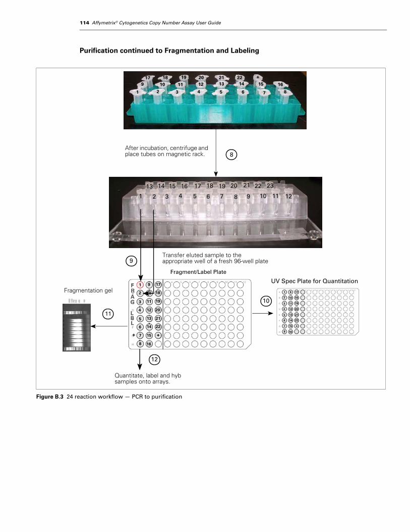
114 Affymetrix® Cytogenetics Copy Number Assay User Guide
Purification continued to Fragmentation and Labeling
Figure B.3 24 reaction workflow — PCR to purification
1 2 3 4 5 6 7 8
9 10 11 12 13 14
+
–15 16
19 20 21 221817
After incubation, centrifuge and place tubes on magnetic rack. 8
1 2 3 4 5 6 7 8 9 10 11 12
13 14 15 16 17 18 19 20 21 22 23
+
1
2
3
4
5
6
8
FRAG
LBL
Transfer eluted sample to the appropriate well of a fresh 96-well plate
Fragment/Label Plate
9
1
2
3
4
5
6
7
8
9
10
11
12
13
14
UV Spec Plate for Quantitation
Quantitate, label and hyb samples onto arrays.
7
10
+15
16
17
18
19
20
21
22
9
10
11
12
13
14
+15
16
17
18
19
20
21
22
11
12
Fragmentation gel

Appendix
C REAGENTS, EQUIPMENT AND CONSUMABLESAbout this AppendixThis appendix includes the vendor and part number information for the reagents, equipment and consumables that have been validated for use with the Affymetrix® Cytogenetics Copy Number Assay.
From Affymetrix
Affymetrix Equipment Required
Affymetrix Software Required
IMPORTANT: This protocol has been optimized using the equipment, consumables andreagents listed in this user guide. For the best results, we strongly recommend that youadhere to the protocol as described. Do not deviate from the protocol; do not substitutereagents.
Table C.1 Affymetrix Equipment Required
Item Part Number
❐ GeneChip® Fluidics Station 450 00-0079
❐ GeneChip® Hybridization Oven 640 or 645 800139 (640)00-0331 (645)
❐ GeneChip® 3000 Scanner with 7G upgrade 901153
Table C.2 Affymetrix Software Required
Item Part Number
❐ One of the following instrument control applications:• GeneChip® Operating Software• GeneChip® Command Console
Latest version
❐ Genotyping Console Latest version
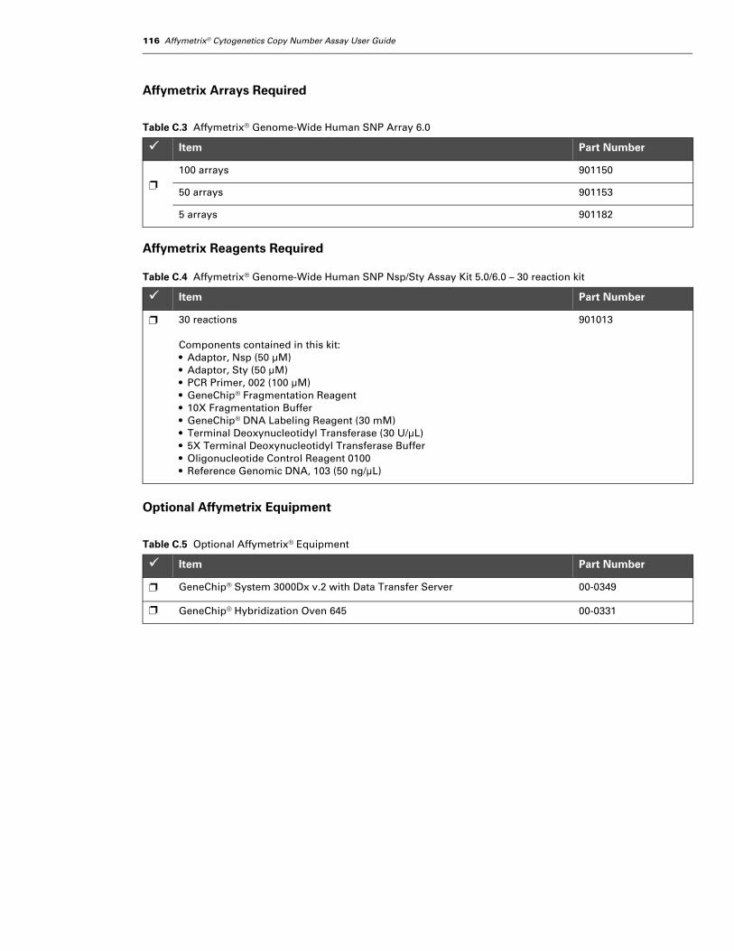
116 Affymetrix® Cytogenetics Copy Number Assay User Guide
Affymetrix Arrays Required
Affymetrix Reagents Required
Optional Affymetrix Equipment
Table C.3 Affymetrix® Genome-Wide Human SNP Array 6.0
Item Part Number
❐
100 arrays 901150
50 arrays 901153
5 arrays 901182
Table C.4 Affymetrix® Genome-Wide Human SNP Nsp/Sty Assay Kit 5.0/6.0 – 30 reaction kit
Item Part Number
❐ 30 reactions
Components contained in this kit:• Adaptor, Nsp (50 µM)• Adaptor, Sty (50 µM)• PCR Primer, 002 (100 µM)• GeneChip® Fragmentation Reagent• 10X Fragmentation Buffer• GeneChip® DNA Labeling Reagent (30 mM)• Terminal Deoxynucleotidyl Transferase (30 U/µL)• 5X Terminal Deoxynucleotidyl Transferase Buffer• Oligonucleotide Control Reagent 0100• Reference Genomic DNA, 103 (50 ng/µL)
901013
Table C.5 Optional Affymetrix® Equipment
Item Part Number
❐ GeneChip® System 3000Dx v.2 with Data Transfer Server 00-0349
❐ GeneChip® Hybridization Oven 645 00-0331

appendix C | Reagents, Equipment and Consumables 117
Equipment Required from Other Suppliers
Pre-PCR Clean Area Equipment RequiredWhen performing the pre-PCR stages of the Cytogenetics Copy Number Assay, great care should be taken to avoid sample contamination with PCR products. If the assay is to be run in a single room, we strongly recommend that the pre-PCR stages be performed in a laminar flow or PCR cabinet.
Table C.6 Pre-PCR Clean Area Equipment Required
Item Vendor Part Number
❐ Recommended if protocol is to be performed in one room only:• Laminar Flow Cabinet, 6 ft (ESCO, SVE-6A)• PCR Cabinet
Laminar Cabinet: ESCO SVE-6A or equivalent
PCR Cabinet: C.B.S. Scientific P-048-02 or equivalent
❐ Benchtop Cooler, –20°C Stratagene 400012
❐ Biocooler aluminum block, 96-wellRequired if processing > 8 samples.(1 for 9 to 16 samples; 2 for 17 to 24 samples)
Bio-Smith 81001
❐ Cooling Chamber, double block Diversified Biotech CHAM-1020
❐ Freezer, –20°C; deep freeze; manual defrost; 17 cu ft Any vendor —
❐ Ice Bucket, 4 to 9 liters (Magic Touch Icewares) Fisher Scientific —
❐ Microfuge (for tubes and strip tubes) Any vendor —
❐ Pipet, single-channel, 2–20 µL Rainin L-20
❐ Pipet, single-channel, 20–200 µL Rainin L-200
❐ Pipet, single-channel, 100–1000 µL Rainin L-1000
❐ Pipet, 8-channel, 2–20 µL Rainin L8-20
❐ Pipet, 8-channel, 20–200 µL Rainin L8-200
❐ Plate centrifuge, multipurpose Eppendorf 5804 or 5810
❐ Vortexer (with plate pad) VWR 58816-1212
❐
Select one of these thermal cyclers:
• GeneAmp PCR System 9700 (gold/silver block) Applied Biosystems 4314878
• 2720 Thermal Cycler Applied Biosystems 4359659
• MJ Tetrad PTC-255 Bio-Rad —
• DNA Engine Tetrad 2 Bio-Rad PTC-0240G

118 Affymetrix® Cytogenetics Copy Number Assay User Guide
Post-PCR Area Equipment Required
Table C.7 Post-PCR Area Equipment Required
Item Vendor Part Number
❐ Benchtop Cooler, –20°C Stratagene 400012
❐ Cooling Chamber, single Diversified Biotech CHAM-1020
❐ Freezer, –20°C; deep freeze; manual defrost; 17 cu ft Any vendor —
❐ Gel box for electrophoresis Any vendor —
❐ Gel imager Any vendor —
❐ Ice Bucket, 4 to 9 liters (Magic Touch Icewares) Fisher Scientific —
❐ MagnaRack, magnetic stand Invitrogen CS15000
❐ Microcentrifuge 5414D or R Eppendorf 022621408
❐ Microcentrifuge Standard Rotor F-45-24-11, 24 bores Eppendorf 022621502
❐ Microfuge (for tubes and strip tubes) Any vendor —
❐ Microtube Foam Insert with stem(Multiple Sample Starter Set, Model H301)(you will attach to one of the vortexers)
Scientific Industries 504-0233-00
❐ Pipet, single-channel, 2–20 µL Rainin L-20
❐ Pipet, single-channel, 20–200 µL Rainin L-200
❐ Pipet, single-channel, 100–1000 µL Rainin L-1000
❐ Pipet, 8-channel, 2–20 µL Rainin L8-20
❐ Pipet, 8-channel, 20–200 µL Rainin L8-200
❐ Pipet, 8-channel, 100–1000 µL Rainin L8-1000
❐ Plate centrifuge, multipurpose Eppendorf 5804 or 5810
❐ Refrigerator, 4°C, 6 cu ft Any vendor —
❐
Spectrophotometer – select one of the following:
• SpectraMax spectrophotometer Molecular Devices Spectramax Plus384
• NanoDrop NanoDrop ND-1000
❐
Select one of these thermal cyclers:(if routinely processing > 8 samples, you may to use additional thermal cyclers for PCR)
• GeneAmp PCR System 9700 (gold/silver block) Applied Biosystems 4314878
• MJ Tetrad PTC-255 Bio-Rad —
• DNA Engine Tetrad 2 Bio-Rad PTC-0240G
❐ Vortexer (2 required)One vortexer must have a plate pad. The Microtube Foam Insert listed above will be attached to the other vortexer.
VWR 58816-1212
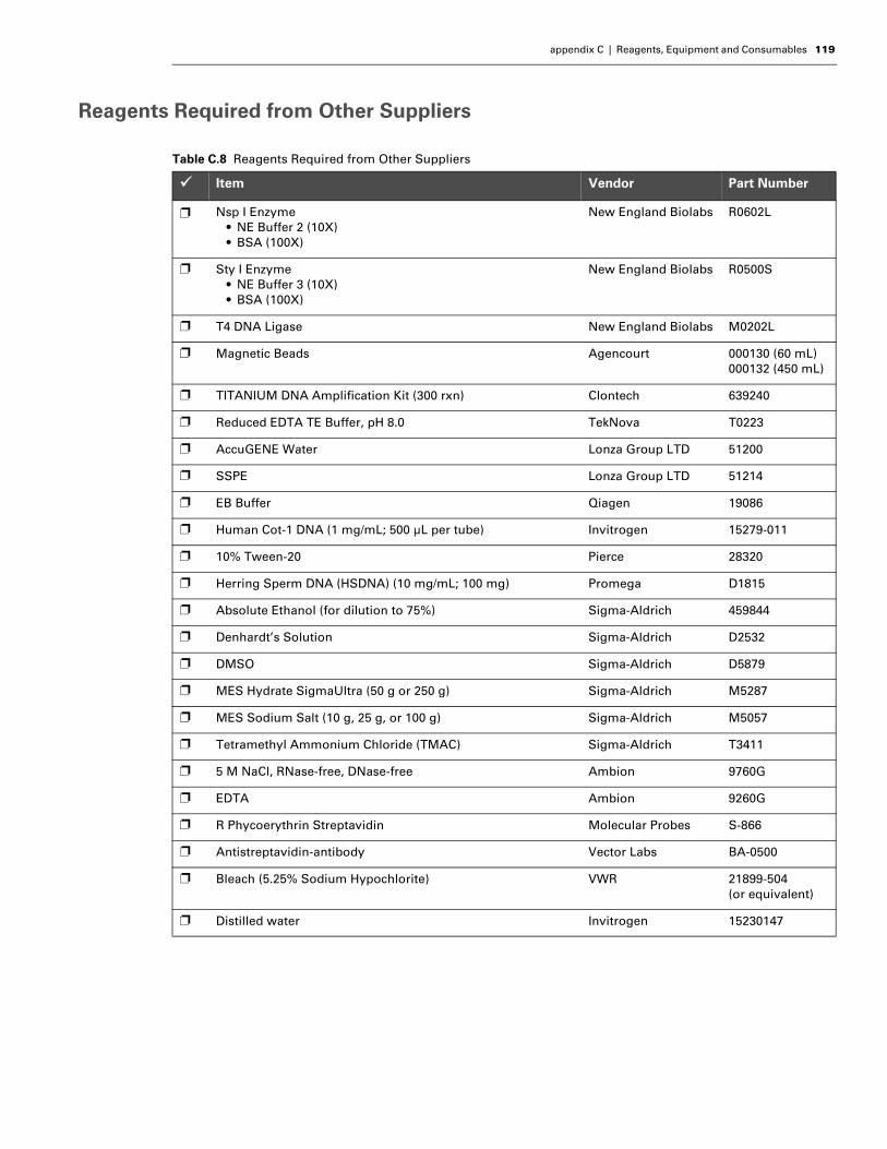
appendix C | Reagents, Equipment and Consumables 119
Reagents Required from Other Suppliers
Table C.8 Reagents Required from Other Suppliers
Item Vendor Part Number
❐ Nsp I Enzyme• NE Buffer 2 (10X)• BSA (100X)
New England Biolabs R0602L
❐ Sty I Enzyme• NE Buffer 3 (10X)• BSA (100X)
New England Biolabs R0500S
❐ T4 DNA Ligase New England Biolabs M0202L
❐ Magnetic Beads Agencourt 000130 (60 mL)000132 (450 mL)
❐ TITANIUM DNA Amplification Kit (300 rxn) Clontech 639240
❐ Reduced EDTA TE Buffer, pH 8.0 TekNova T0223
❐ AccuGENE Water Lonza Group LTD 51200
❐ SSPE Lonza Group LTD 51214
❐ EB Buffer Qiagen 19086
❐ Human Cot-1 DNA (1 mg/mL; 500 µL per tube) Invitrogen 15279-011
❐ 10% Tween-20 Pierce 28320
❐ Herring Sperm DNA (HSDNA) (10 mg/mL; 100 mg) Promega D1815
❐ Absolute Ethanol (for dilution to 75%) Sigma-Aldrich 459844
❐ Denhardt’s Solution Sigma-Aldrich D2532
❐ DMSO Sigma-Aldrich D5879
❐ MES Hydrate SigmaUltra (50 g or 250 g) Sigma-Aldrich M5287
❐ MES Sodium Salt (10 g, 25 g, or 100 g) Sigma-Aldrich M5057
❐ Tetramethyl Ammonium Chloride (TMAC) Sigma-Aldrich T3411
❐ 5 M NaCl, RNase-free, DNase-free Ambion 9760G
❐ EDTA Ambion 9260G
❐ R Phycoerythrin Streptavidin Molecular Probes S-866
❐ Antistreptavidin-antibody Vector Labs BA-0500
❐ Bleach (5.25% Sodium Hypochlorite) VWR 21899-504 (or equivalent)
❐ Distilled water Invitrogen 15230147
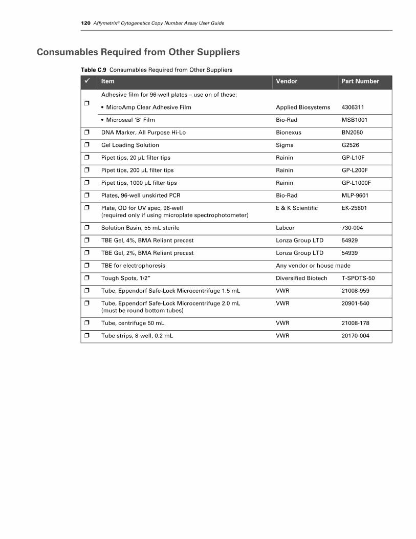
120 Affymetrix® Cytogenetics Copy Number Assay User Guide
Consumables Required from Other Suppliers
Table C.9 Consumables Required from Other Suppliers
Item Vendor Part Number
❐
Adhesive film for 96-well plates – use on of these:
• MicroAmp Clear Adhesive Film Applied Biosystems 4306311
• Microseal 'B' Film Bio-Rad MSB1001
❐ DNA Marker, All Purpose Hi-Lo Bionexus BN2050
❐ Gel Loading Solution Sigma G2526
❐ Pipet tips, 20 µL filter tips Rainin GP-L10F
❐ Pipet tips, 200 µL filter tips Rainin GP-L200F
❐ Pipet tips, 1000 µL filter tips Rainin GP-L1000F
❐ Plates, 96-well unskirted PCR Bio-Rad MLP-9601
❐ Plate, OD for UV spec, 96-well(required only if using microplate spectrophotometer)
E & K Scientific EK-25801
❐ Solution Basin, 55 mL sterile Labcor 730-004
❐ TBE Gel, 4%, BMA Reliant precast Lonza Group LTD 54929
❐ TBE Gel, 2%, BMA Reliant precast Lonza Group LTD 54939
❐ TBE for electrophoresis Any vendor or house made
❐ Tough Spots, 1/2” Diversified Biotech T-SPOTS-50
❐ Tube, Eppendorf Safe-Lock Microcentrifuge 1.5 mL VWR 21008-959
❐ Tube, Eppendorf Safe-Lock Microcentrifuge 2.0 mL(must be round bottom tubes)
VWR 20901-540
❐ Tube, centrifuge 50 mL VWR 21008-178
❐ Tube strips, 8-well, 0.2 mL VWR 20170-004
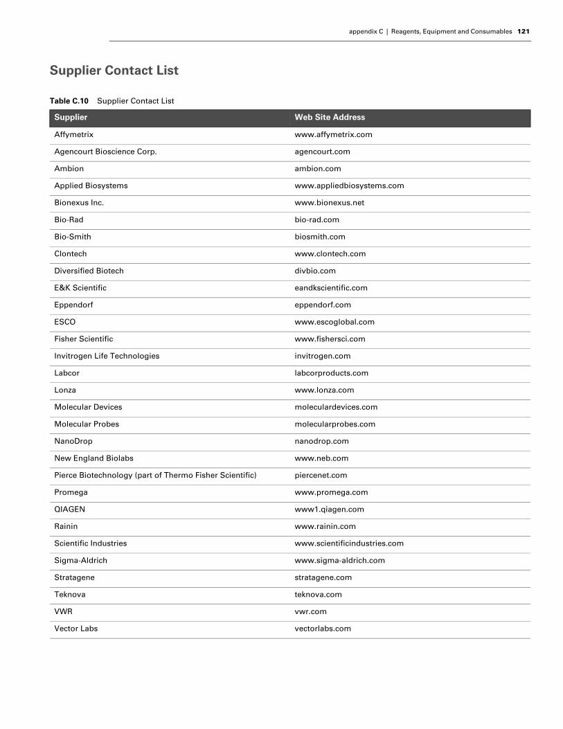
appendix C | Reagents, Equipment and Consumables 121
Supplier Contact List
Table C.10 Supplier Contact List
Supplier Web Site Address
Affymetrix www.affymetrix.com
Agencourt Bioscience Corp. agencourt.com
Ambion ambion.com
Applied Biosystems www.appliedbiosystems.com
Bionexus Inc. www.bionexus.net
Bio-Rad bio-rad.com
Bio-Smith biosmith.com
Clontech www.clontech.com
Diversified Biotech divbio.com
E&K Scientific eandkscientific.com
Eppendorf eppendorf.com
ESCO www.escoglobal.com
Fisher Scientific www.fishersci.com
Invitrogen Life Technologies invitrogen.com
Labcor labcorproducts.com
Lonza www.lonza.com
Molecular Devices moleculardevices.com
Molecular Probes molecularprobes.com
NanoDrop nanodrop.com
New England Biolabs www.neb.com
Pierce Biotechnology (part of Thermo Fisher Scientific) piercenet.com
Promega www.promega.com
QIAGEN www1.qiagen.com
Rainin www.rainin.com
Scientific Industries www.scientificindustries.com
Sigma-Aldrich www.sigma-aldrich.com
Stratagene stratagene.com
Teknova teknova.com
VWR vwr.com
Vector Labs vectorlabs.com

122 Affymetrix® Cytogenetics Copy Number Assay User Guide
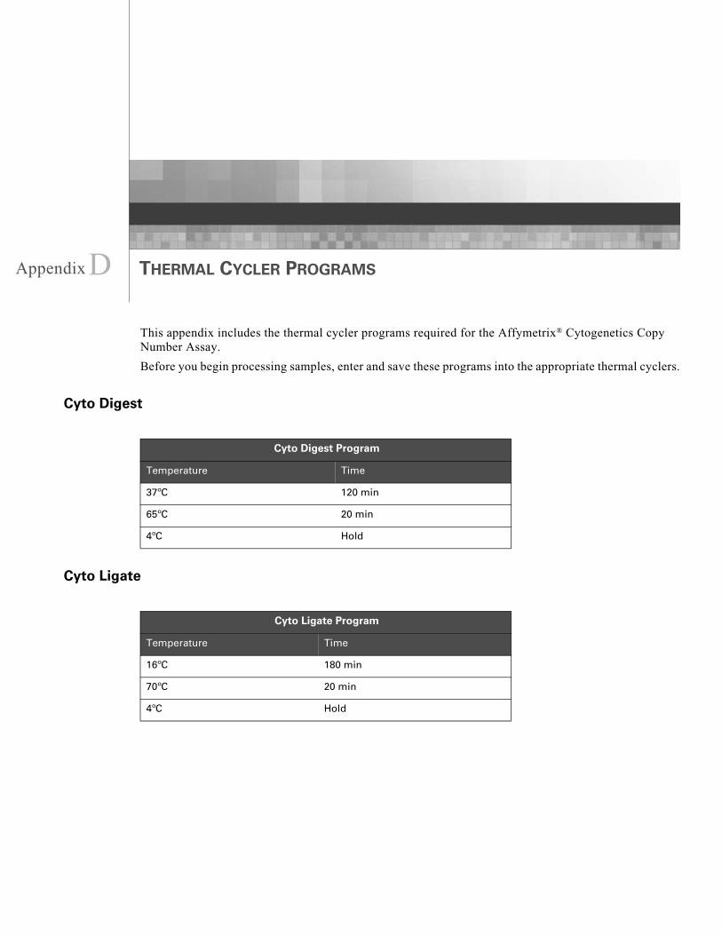
Appendix
D THERMAL CYCLER PROGRAMSThis appendix includes the thermal cycler programs required for the Affymetrix® Cytogenetics Copy Number Assay.
Before you begin processing samples, enter and save these programs into the appropriate thermal cyclers.
Cyto Digest
Cyto Ligate
Cyto Digest Program
Temperature Time
37ºC 120 min
65ºC 20 min
4ºC Hold
Cyto Ligate Program
Temperature Time
16ºC 180 min
70ºC 20 min
4ºC Hold
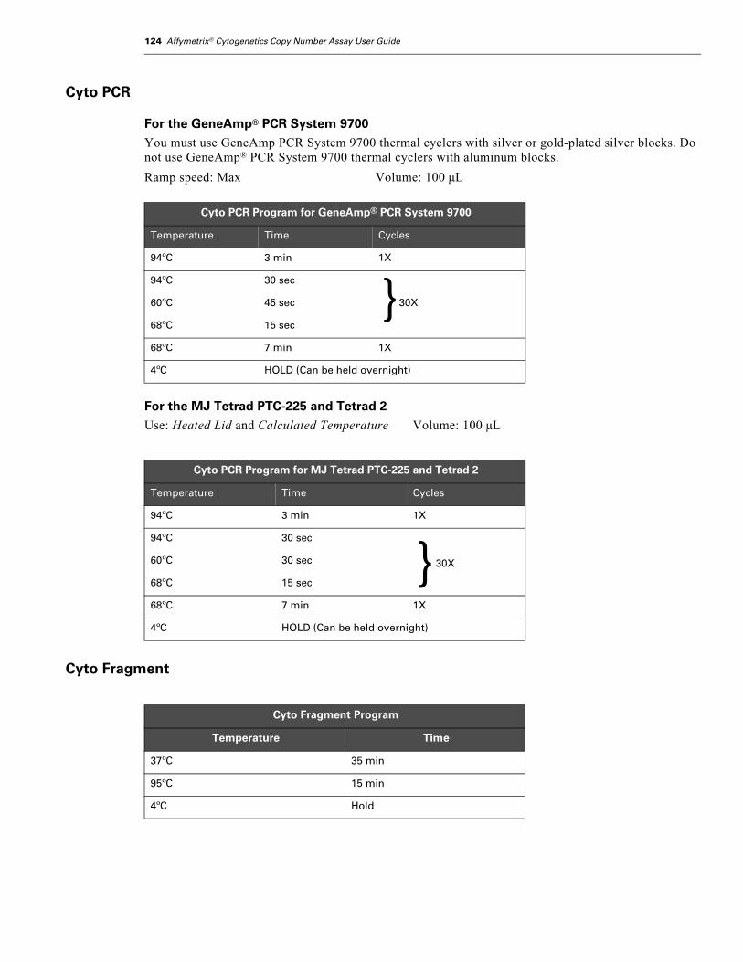
124 Affymetrix® Cytogenetics Copy Number Assay User Guide
Cyto PCR
For the GeneAmp® PCR System 9700You must use GeneAmp PCR System 9700 thermal cyclers with silver or gold-plated silver blocks. Do not use GeneAmp® PCR System 9700 thermal cyclers with aluminum blocks.
Ramp speed: Max Volume: 100 µL
For the MJ Tetrad PTC-225 and Tetrad 2Use: Heated Lid and Calculated Temperature Volume: 100 µL
Cyto Fragment
Cyto PCR Program for GeneAmp® PCR System 9700
Temperature Time Cycles
94ºC 3 min 1X
94ºC 30 sec
60ºC 45 sec 30X
68ºC 15 sec
68ºC 7 min 1X
4ºC HOLD (Can be held overnight)
Cyto PCR Program for MJ Tetrad PTC-225 and Tetrad 2
Temperature Time Cycles
94ºC 3 min 1X
94ºC 30 sec
30X60ºC 30 sec
68ºC 15 sec
68ºC 7 min 1X
4ºC HOLD (Can be held overnight)
Cyto Fragment Program
Temperature Time
37ºC 35 min
95ºC 15 min
4ºC Hold
}
}

appendix D | Thermal Cycler Programs 125
Cyto Label
Cyto Hyb
Cyto Label Program
Temperature Time
37ºC 4 hr
95ºC 15 min
4ºC Hold
Samples can remain at 4°C overnight.
Cyto Hyb Program
Temperature Time
95ºC 10 min
49ºC Hold

126 Affymetrix® Cytogenetics Copy Number Assay User Guide