Embed Size (px)
Citation preview

EDUCATIONAL REVIEW
Urinary schistosomiasis
Oluwatoyin F. Bamgbola
Received: 2 August 2013 /Revised: 22 November 2013 /Accepted: 3 December 2013 /Published online: 28 January 2014# IPNA 2014
Abstract Schistosomiasis is the second most common socio-economically devastating parasitic disease after malaria, affect-ing about 240 million residents of developing countries. InAfrica, it predominantly manifests as urogenital disease, andthe main infective agent is Schistosoma hematobium.Endemicity is propagated by poor socio-economic status andenvironmental degradation due to rapid urbanization.Recreational swimming is a potent medium for the spread ofdisease in children and adolescents. Most affected individualsare asymptomatic. The male and female worms are equippedwith an extraordinary capacity for immune evasion and are ableto co-habit for several decades within the pelvic venous plexus.Eggs deposited in the bladder wall resist elimination by type 1 Tlymphocytes. Instead, they are sustained by pro-fibrogenicencapsulation (as modulated by type 2 helper cells).Progressive bladder disease results in obstructive uropathyand predisposes to (mostly) squamous cell carcinoma.Schistosomal glomerulopathy manifests as a clinical spectrumof asymptomatic proteinuria, nephrosis and/or nephritic syn-drome. Findings on renal biopsy may be influenced by co-morbidity with Salmonella bacteria, amyloidosis and hepatitisC infection. Potentially fatal Katayama fever and spinalradiculopathy may ensue in tourists visiting an endemic zone.Early detection by urine microscopy is hampered by low uri-nary excretion rates of the parasite eggs. Although useful intravelers with newly acquired disease, the results of the sero-logical antibody assay may be false positive in residents of anendemic zone. Cystoscopy, however, may be invaluable. Dueto its safety, effectiveness and once-daily dosing, praziquantel is
the drug of choice. An integrated approach that includes masschemotherapy, environmental health programs and publichealth education is the most cost-effective preventive strategy.
Keywords Urinary schistosomiasis . Schistosomalglomerulopathy . Pediatric tropical disease
Urinary schistosomiasis
Epidemiology
After malaria, schistosomiasis is the second most commonsocio-economically devastating tropical parasitic disease.Parasite infestation has been documented in 78 countries ofAfrica, Asia, the Middle East and South America [1]. Despitethe availability of effective drugs, the annual death rate isaround 200,000 in sub-Sahara Africa alone, making the groupof parasites which cause schistosomiasis the most lethalworms in the world. The majority of human disease is medi-ated by Schistosoma hematobium, S. mansoni andS. japonicum [2]. Each of these species has a tropism fordifferent body organs, with S. hematobium being the maincause of urogenital disease [2, 3]. Poor access to economicopportunity accounts for the uneven distribution of infectionin endemic regions. Subsistence farming, inadequate watersupply, poor public sanitation, rapid urbanization and damconstruction are common predisposing factors [3]. Althoughparasite infection has been reported in early infancy, peakincidence occurs in early adolescence as a result of frequentbathing in contaminated pools of water [4]. Apart from lowerexposure in adults, the capacity to resist new infection byeosinophil secretion of antigen-specific immunoglobulin E(IgE) is age dependent [5]. Children younger than 13 yearshave a higher serum level of IgM, IgG2 and IgG4 isotypeswhich block the protective effect of IgE [6].
O. F. Bamgbola (*)Division of Pediatric Nephrology, Children’s Hospital of NewOrleans, Louisiana State University Health Science Center, 200Henry Clay Avenue, New Orleans, LA 70118, USAe-mail: [email protected]
Pediatr Nephrol (2014) 29:2113–2120DOI 10.1007/s00467-013-2723-1
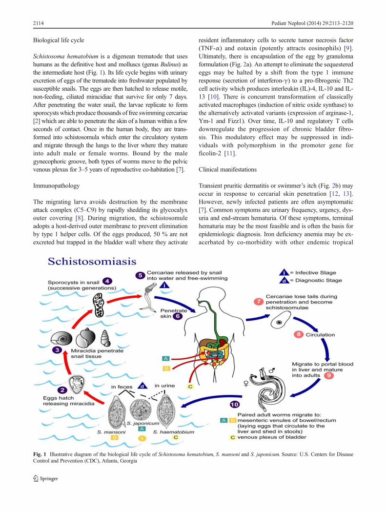
Biological life cycle
Schistosoma hematobium is a digenean trematode that useshumans as the definitive host and molluscs (genus Bulinus) asthe intermediate host (Fig. 1). Its life cycle begins with urinaryexcretion of eggs of the trematode into freshwater populated bysusceptible snails. The eggs are then hatched to release motile,non-feeding, ciliated miracidiae that survive for only 7 days.After penetrating the water snail, the larvae replicate to formsporocysts which produce thousands of free swimming cercariae[2] which are able to penetrate the skin of a human within a fewseconds of contact. Once in the human body, they are trans-formed into schistosomula which enter the circulatory systemand migrate through the lungs to the liver where they matureinto adult male or female worms. Bound by the malegynecophoric groove, both types of worms move to the pelvicvenous plexus for 3–5 years of reproductive co-habitation [7].
Immunopathology
The migrating larva avoids destruction by the membraneattack complex (C5–C9) by rapidly shedding its glycocalyxouter covering [8]. During migration, the schistosomuleadopts a host-derived outer membrane to prevent eliminationby type 1 helper cells. Of the eggs produced, 50 % are notexcreted but trapped in the bladder wall where they activate
resident inflammatory cells to secrete tumor necrosis factor(TNF-α) and eotaxin (potently attracts eosinophils) [9].Ultimately, there is encapsulation of the egg by granulomaformulation (Fig. 2a). An attempt to eliminate the sequesteredeggs may be halted by a shift from the type 1 immuneresponse (secretion of interferon-γ) to a pro-fibrogenic Th2cell activity which produces interleukin (IL)-4, IL-10 and IL-13 [10]. There is concurrent transformation of classicallyactivated macrophages (induction of nitric oxide synthase) tothe alternatively activated variants (expression of arginase-1,Ym-1 and Fizz1). Over time, IL-10 and regulatory T cellsdownregulate the progression of chronic bladder fibro-sis. This modulatory effect may be suppressed in indi-viduals with polymorphism in the promoter gene forficolin-2 [11].
Clinical manifestations
Transient pruritic dermatitis or swimmer’s itch (Fig. 2b) mayoccur in response to cercarial skin penetration [12, 13].However, newly infected patients are often asymptomatic[7]. Common symptoms are urinary frequency, urgency, dys-uria and end-stream hematuria. Of these symptoms, terminalhematuria may be the most feasible and is often the basis forepidemiologic diagnosis. Iron deficiency anemia may be ex-acerbated by co-morbidity with other endemic tropical
Fig. 1 Illustrative diagram of the biological life cycle of Schistosoma hematobium, S. mansoni and S. japonicum. Source: U.S. Centers for DiseaseControl and Prevention (CDC), Atlanta, Georgia
2114 Pediatr Nephrol (2014) 29:2113–2120

diseases, such as malaria and heminthiasis. A substantial amountof iron is sequestered by vitelline cells for the formation of theparasite eggshell [14]. In addition, host iron recycling is disruptedby a pro-inflammatory synthesis of hepcidin, an acute phasereactant [15]. Although structural deficits are sometimes revers-ible by medical treatment, obstructive uropathy may invariablyresult from progressive bladder fibrosis, ureteral dilatation andhydronephrosis, particularly in older patients [16]. Ulcerations ofbladder mucosa, focal bladder wall calcification and renal stoneformation may result. Urological surgery is seldom required forrehabilitation. Genital disease may cause sexual dysfunction,facilitates human immunodeficiency virus transmission and pro-motes infertility in adolescents and young adults [17].
The ease of access to wider geographical regions for tourismhas increased the infection rate among travelers [12]. Suchindividuals lack any acquired immunity and within 6 weeks afterinfection they may experience a severe hypersensitivity reactionin response to the first bout of egg antigen release by adultworms. Katayama fever is characterized by hyperpyrexia, myal-gia, headache, cough, emesis and diarrhea [18]. In addition,paraplegia from transversemyelitismay result fromparavertebralmigration of parasite eggs. Nevertheless complete neurologicalrecovery is feasible with early medical intervention [19].
Urinary tract infection Due to disruption of the mucosalbarrier, there is a high rate of bacterial superinfection, rangingfrom 30 to 80% in endemic communities [20, 21]. Isolates aremostly regular uropathogens, but Salmonella infection is notuncommon [21]. Salmonella evades the host immune
response by attaching itself to the adult worm’s surface recep-tors [22]. Urinary carriers may serve as a source of epidemictyphoid fever. Urine isolation of Salmonella bacteria in en-demic regions should arouse suspicion for a co-morbid schis-tosomiasis [21, 22].
Bladder carcinoma There is a 30-fold higher risk of develop-ing bladder cancer, mostly squamous cell carcinoma (SCC)variant in regions of Egypt with endemic schistosomiasis [23].A fall in the prevalence rate of schistosomiasis from 1980 to2005 due to a public health intervention was followed by asix-fold lower rate of SCC, clearly suggesting a cause andeffect relationship [24].
Schistosomal glomerulopathy Schistosomal glomerulopathy(SGN) occurs in response to infection with bothS. hematobium and S. mansoni. Apparently due to the greateroccurrence of subclinical disease in patients withS. hematobium, a higher prevalence of glomerular disease isoften reported in those infected with S. mansoni (Tables 1, 2)[25–27]. SGN manifests as a clinical, spectrum of asympto-matic, proteinuria, nephrosis and/or nephritic syndrome. Basedon experimental and clinicopathologic data, in 1992 the AfricanAssociation of Nephrology (AFRAN) recognized six categoriesof SGN. The most common category is Class I SGN and it ischaracterized by mesangial proliferative histology. Infected in-dividuals are often asymptomatic (60 %) [26]. Class II SGN isan exudative proliferative lesion that occurs in response toSalmonella bacteria superinfection. Class III SGN has amembrano-proliferative glomerular pattern, Class IV has focalsegmental glomerulosclerosis, Class V is due to secondaryamyloidosis and Class VI is a mixed pathology of proliferative,focal sclerosis, amyloidosis and thrombotic cryoglobulin from aco-morbid hepatitis C infection. Except for the clinical recoveryobserved in patients with Class I and II SGN, patients with theother categories often progress to end-stage kidney disease byadulthood. Data in the 2008 Egyptian Renal Registry showedthat SGN may account for 2.8 % of the 483 adult patients withend stage kidney disease per million of population [www.esnonline.net]. Immune complex deposits (IgM, C3, and C1q)containing adult worm antigens (gut-associated proteoglycan) inthe glomerulus are the early pathological features [27]. Theremay be late deposits of IgG and IgA. Membrano-proliferativeresponse to a direct localization of egg granuloma in kidneytissue is a rare event [25]. Because most immune complexes arenon-nephritogenic, there is no correlation between overt kidneydisease and low level of serum C3 [28].
Diagnosis
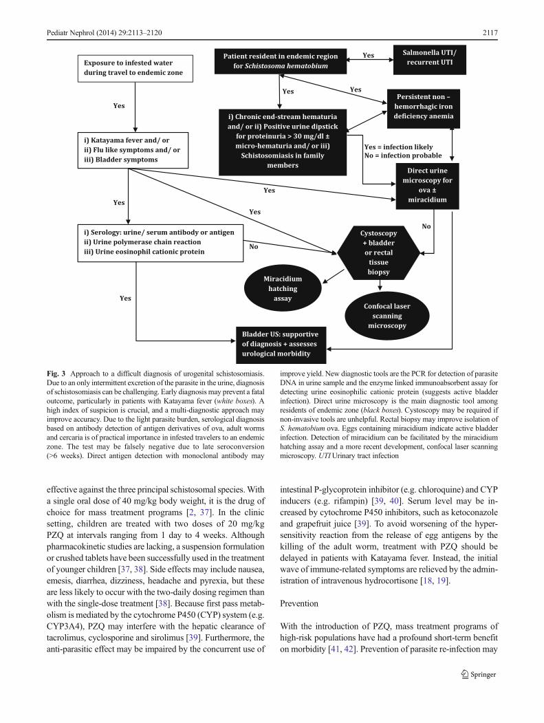
Detection of parasite eggs and motile miracidium by urine mi-croscopy is simple, inexpensive and is considered to be the goldstandard for diagnosis (Fig. 3) [29]. The result may be a false
a
b
Fig. 2 a Granulomatous encapsulation of Schistosoma eggs, b forearmdermatitis from cercaria skin penetration. Source: Centers for DiseaseControl (CDC) Atlanta, Georgia
Pediatr Nephrol (2014) 29:2113–2120 2115

negative in the first 6 weeks prior to full maturity of the wormand due to a lower rate of egg excretion. Collection of three dailyfiltered urine samples after a mid-day session of physical exer-cise, the peak period for egg output, increases the detection rate[2]. Field surveys of end-stream hematuria, urine evaluation forblood and protein and bladder ultrasonography (assesses mor-bidity) are useful screening tools [2, 30, 31]. In addition, theexcretion of eosinophil cationic protein in the urine (as detectedby enzyme-linked immunosorbent assay) may be a biomarker ofinflammatory bladder injury [30]. In cases of diagnostic chal-lenges, cystoscopy may show a typical hemorrhagic mucosa,submucosal nodules and sandy patches (micro-granuloma). Latefindings of cystoscopy are a “cooked rice grain” appearance(macro-granuloma) and erythematous fibrous polyps [31].Bladder biopsy may show granuloma-encased eggs butmay fail to demonstrate the viability of the eggs (Fig. 2a).Direct detection of miracidium-containing eggs usingcystoscopy-aided confocal laser scanning microscopy pro-duces stronger evidence of disease activity [32].
Due to a light parasite burden, serological assays for detec-tion of IgG, IgM or IgE antibody response to antigen derivatives
of parasite are often required for the diagnosis of infectedtravelers to endemic zones [33]. It is less useful diagnostic toolin an endemic community as it fails to discriminate current fromprevious infection, and the parasite antigen may cross-react withthose of other tropical helminths. Due to late seroconversion,these tests may not be positive until a minimum of 6 weeks afterthe primary infection [2]. Although less robust, circulating ca-thodic antigens may be detected in the urine or serum samplesusing labeled monoclonal antibodies [34]. The PCR of parasiteDNA in a urine sample (sensitivity 84 %, specificity 97 %) mayprove useful in post-chemotherapy surveillance [35].
Treatment
In the last four decades, safer and more effective drugs havereplaced the toxic older generation of anti-infective agents[36]. According to the World Health Organization, only11.5 % of the 243 million people in 52 countries who requiredtreatment received pharmacological intervention in 2011 [1].Praziquantel (PZQ), an acylated quinoline–pyrazine com-pound, is regarded as the gold standard of therapy. It is
Table 1 Clinical aspects of schistosomal glomerulopathy as classified by the African Association of Nephrology
AFRAN classification Etiology Clinical syndrome Treatment
I: Mesangio-proliferative SH, SM >50 % asymptomatic; mild proteinuria PZQ, metrifonate
II: Proliferative exudative SH, SM,Salmonella sp.
Rapid onset nephrotic/nephritic syndrome;Salmonella sepsis
PZQ, metrifonate, antibiotics
III: Membrano-proliferative SH, SM Non-Black patient; hepatic cirrhosis; High serumIgA; NS, CGN, ESKD
None effective
IV: Focal segmentalglomerulosclerosis
SM Mostly in Blacks; hepatic cirrhosis; NS, CGN,ESKD
None effective
V: Amyloidosis SH/ SM Systemic amyloidosis; NS, CGN, ESKD None effective
VI: Cryoglobulinemia SM, HCV Purpura, arthralgia, myalgia; low C3, very lowserum C4, NS, CGN, ESKD
IFN-α, ribavirin, immunosuppressiveagent, plasmapheresis
AFRAN, African Association of Nephrology; C3, complement 3, C4, complement 4; CGN, chronic glomerulonephritis; ESKD, end stage kidneydisease; FSGS, focal segmental glomerulosclerosis; HCV, hepatitis C virus; IFN-α, interferon-alpha; NS, nephrotic syndrome; PZQ, praziquantel; SH,Schistosoma hematobium; SM, Schistosoma mansoni.
Table 2 Pathological features of schistosomal glomerulopathy based on the classification of the African Association of Nephrology
AFRAN classification Histology: light microscopy Immunofluorescence/electron microscopy
I: Mesangio-proliferative Mesangial cell proliferation; matrix expansion Mesangial IgM, C3, GASP deposits
II: Proliferative/ exudative Mesangial neutrophil, monocyte & eosinophil; epithelial/endothelial/ mesangial cell proliferation
Sub-endothelial/ mesangial C3, IgG, IgM deposits
III: Membrano-proliferative Mesangial/ endothelial cell proliferation, glomerularbasement membrane thickening
Sub-epithelial & sub-endothelial IgG, IgA, C3, GASPdeposits
IV: Focal segmentalglomerulosclerosis
Focal segmental proliferative or sclerosing lesion Sub-endothelial IgG, IgM, IgA, GASP deposits
V: Amyloidosis Glomerular amyloid; arterial wall involved; interstitialfibrosis, tubular atrophy
Amyloid A deposits in kidney, liver, subcutaneous fat;GASP
VI: Cryoglobulinemia Mesangial/ endothelial cell proliferation, hyaline thrombi;fibrinoid necrosis; fibro-cellular crescents
Mesangial and sub-endothelial IgG, C3, cryoglobulin,fibrin, amyloid A, HCV-RNA deposits
HCV-RNA, HCV ribonuclear acid particles; GASP, gut-associated schistosomal proteoglycan; GN, glomerulonephritis; Ig, immunoglobulin
2116 Pediatr Nephrol (2014) 29:2113–2120
effective against the three principal schistosomal species.Witha single oral dose of 40 mg/kg body weight, it is the drug ofchoice for mass treatment programs [2, 37]. In the clinicsetting, children are treated with two doses of 20 mg/kgPZQ at intervals ranging from 1 day to 4 weeks. Althoughpharmacokinetic studies are lacking, a suspension formulationor crushed tablets have been successfully used in the treatmentof younger children [37, 38]. Side effects may include nausea,emesis, diarrhea, dizziness, headache and pyrexia, but theseare less likely to occur with the two-daily dosing regimen thanwith the single-dose treatment [38]. Because first pass metab-olism is mediated by the cytochrome P450 (CYP) system (e.g.CYP3A4), PZQ may interfere with the hepatic clearance oftacrolimus, cyclosporine and sirolimus [39]. Furthermore, theanti-parasitic effect may be impaired by the concurrent use of
intestinal P-glycoprotein inhibitor (e.g. chloroquine) and CYPinducers (e.g. rifampin) [39, 40]. Serum level may be in-creased by cytochrome P450 inhibitors, such as ketoconazoleand grapefruit juice [39]. To avoid worsening of the hyper-sensitivity reaction from the release of egg antigens by thekilling of the adult worm, treatment with PZQ should bedelayed in patients with Katayama fever. Instead, the initialwave of immune-related symptoms are relieved by the admin-istration of intravenous hydrocortisone [18, 19].
Prevention
With the introduction of PZQ, mass treatment programs ofhigh-risk populations have had a profound short-term benefiton morbidity [41, 42]. Prevention of parasite re-infection may
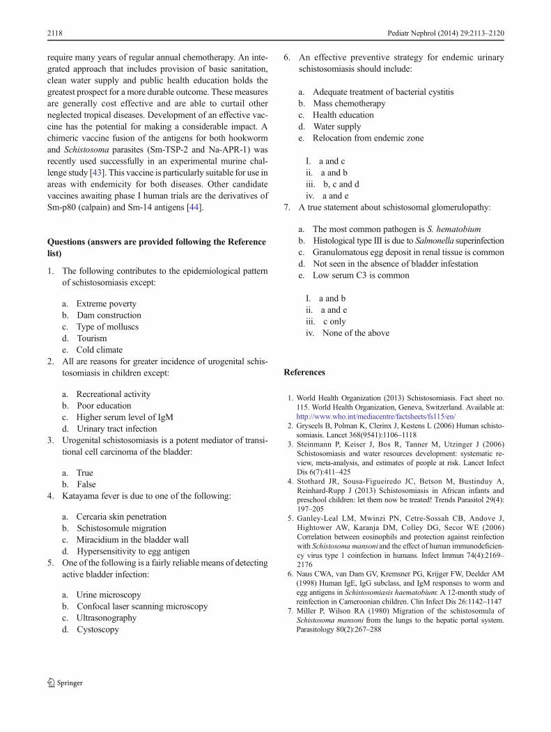
Fig. 3 Approach to a difficult diagnosis of urogenital schistosomiasis.Due to an only intermittent excretion of the parasite in the urine, diagnosisof schistosomiasis can be challenging. Early diagnosis may prevent a fataloutcome, particularly in patients with Katayama fever (white boxes). Ahigh index of suspicion is crucial, and a multi-diagnostic approach mayimprove accuracy. Due to the light parasite burden, serological diagnosisbased on antibody detection of antigen derivatives of ova, adult wormsand cercaria is of practical importance in infested travelers to an endemiczone. The test may be falsely negative due to late seroconversion(>6 weeks). Direct antigen detection with monoclonal antibody may
improve yield. New diagnostic tools are the PCR for detection of parasiteDNA in urine sample and the enzyme linked immunoabsorbent assay fordetecting urine eosinophilic cationic protein (suggests active bladderinfection). Direct urine microscopy is the main diagnostic tool amongresidents of endemic zone (black boxes). Cystoscopy may be required ifnon-invasive tools are unhelpful. Rectal biopsy may improve isolation ofS. hematobium ova. Eggs containing miracidium indicate active bladderinfection. Detection of miracidium can be facilitated by the miracidiumhatching assay and a more recent development, confocal laser scanningmicroscopy. UTIUrinary tract infection
Pediatr Nephrol (2014) 29:2113–2120 2117
require many years of regular annual chemotherapy. An inte-grated approach that includes provision of basic sanitation,clean water supply and public health education holds thegreatest prospect for a more durable outcome. These measuresare generally cost effective and are able to curtail otherneglected tropical diseases. Development of an effective vac-cine has the potential for making a considerable impact. Achimeric vaccine fusion of the antigens for both hookwormand Schistosoma parasites (Sm-TSP-2 and Na-APR-1) wasrecently used successfully in an experimental murine chal-lenge study [43]. This vaccine is particularly suitable for use inareas with endemicity for both diseases. Other candidatevaccines awaiting phase I human trials are the derivatives ofSm-p80 (calpain) and Sm-14 antigens [44].
Questions (answers are provided following the Referencelist)
1. The following contributes to the epidemiological patternof schistosomiasis except:
a. Extreme povertyb. Dam constructionc. Type of molluscsd. Tourisme. Cold climate
2. All are reasons for greater incidence of urogenital schis-tosomiasis in children except:
a. Recreational activityb. Poor educationc. Higher serum level of IgMd. Urinary tract infection
3. Urogenital schistosomiasis is a potent mediator of transi-tional cell carcinoma of the bladder:
a. Trueb. False
4. Katayama fever is due to one of the following:
a. Cercaria skin penetrationb. Schistosomule migrationc. Miracidium in the bladder walld. Hypersensitivity to egg antigen
5. One of the following is a fairly reliable means of detectingactive bladder infection:
a. Urine microscopyb. Confocal laser scanning microscopyc. Ultrasonographyd. Cystoscopy
6. An effective preventive strategy for endemic urinaryschistosomiasis should include:
a. Adequate treatment of bacterial cystitisb. Mass chemotherapyc. Health educationd. Water supplye. Relocation from endemic zone
I. a and cii. a and biii. b, c and div. a and e
7. A true statement about schistosomal glomerulopathy:
a. The most common pathogen is S. hematobiumb. Histological type III is due to Salmonella superinfectionc. Granulomatous egg deposit in renal tissue is commond. Not seen in the absence of bladder infestatione. Low serum C3 is common
I. a and bii. a and eiii. c onlyiv. None of the above
References
1. World Health Organization (2013) Schistosomiasis. Fact sheet no.115. World Health Organization, Geneva, Switzerland. Available at:http://www.who.int/mediacentre/factsheets/fs115/en/
2. Gryseels B, Polman K, Clerinx J, Kestens L (2006) Human schisto-somiasis. Lancet 368(9541):1106–1118
3. Steinmann P, Keiser J, Bos R, Tanner M, Utzinger J (2006)Schistosomiasis and water resources development: systematic re-view, meta-analysis, and estimates of people at risk. Lancet InfectDis 6(7):411–425
4. Stothard JR, Sousa-Figueiredo JC, Betson M, Bustinduy A,Reinhard-Rupp J (2013) Schistosomiasis in African infants andpreschool children: let them now be treated! Trends Parasitol 29(4):197–205
5. Ganley-Leal LM, Mwinzi PN, Cetre-Sossah CB, Andove J,Hightower AW, Karanja DM, Colley DG, Secor WE (2006)Correlation between eosinophils and protection against reinfectionwith Schistosoma mansoni and the effect of human immunodeficien-cy virus type 1 coinfection in humans. Infect Immun 74(4):2169–2176
6. Naus CWA, van Dam GV, Kremsner PG, Krijger FW, Deelder AM(1998) Human IgE, IgG subclass, and IgM responses to worm andegg antigens in Schistosomiasis haematobium: A 12-month study ofreinfection in Cameroonian children. Clin Infect Dis 26:1142–1147
7. Miller P, Wilson RA (1980) Migration of the schistosomula ofSchistosoma mansoni from the lungs to the hepatic portal system.Parasitology 80(2):267–288
2118 Pediatr Nephrol (2014) 29:2113–2120

8. Schroeder H, Skelly P, Zipfel PF, Losson B, Vanderplasschen A(2009) Subversion of complement by hematophagous parasites.Dev Comp Immunol 33(1):5–13
9. Cheever AW, Hoffmann KF, Wynn TA (2000) Immunopathology ofSchistosomiasis mansoni in mice and men. Immunol Today 21:465–466
10. Wilson MS, Mentink-Kane MM, Pesce JT, Ramalingam TR,Thompson R, Wynn TA (2007) Immunopathology of schistosomia-sis. Immunol Cell Biol 85(2):148–154
11. Ouf EA, Ojurongbe O, Akindele AA, Sina-Agbaje OR, Van Tong H,Adeyeba AO, Kremsner PG, Kun JF, Velavan T (2012) Ficolin-2levels and FCN2 genetic polymorphisms as a susceptibility factor inschistosomiasis. J Infect Dis 206(4):562–570
12. Nicolls DJ, Weld LH, Schwartz E, Reed C, von SonnenburgF, Freedman DO, Kozarsky PE (2008) Characteristics ofSchistosomiasis in travelers reported to the GeoSentinelSurveillance Network 1997–2008. Am J Trop Med Hyg79(5):729–734
13. Appleton CC (1984) Schistosome dermatitis: an unrecognized prob-lem in South Africa? S Afr Med J 65:467–469
14. Jones MK, McManus DP, Sivadorai P, Glanfield A, MoertelL, Belli SI, Gobert GN (2007) Tracking the fate of iron inearly development of human blood flukes. Int J Biochem CellBiol 39(9):1646–1658
15. Ayoya MA, Spiekermann-Brouwer GM, Stoltzfus RJ, Nemeth E,Habicht JP, Ganz T, Rawat R, Traoré AK, Garza C (2010) α1-Acidglycoprotein, hepcidin, C-reactive protein, and serum ferritin arecorrelated in anemic schoolchildren with Schistosoma haematobium.Am J Clin Nutr 91(6):1784–1790
16. Khalaf I, Shokeir A, Shalaby M (2012) Urologic complica-tions of genitourinary schistosomiasis. World J Urol 30(1):31–38
17. Hegertun IE, Sulheim Gundersen KM, Kleppa E, Zulu SG,Gundersen SG, Taylor M, Kvalsvig JD, Kjetland EF (2013)S. haematobium as a common cause of genital morbidity in girls: Across-sectional study of children in South Africa. PLoS Negl Trop7(3):e2104
18. Bottieau E, Clerinx J, de Vega MR, Van den Enden E,Colebunders R, Van Esbroeck M, Vervoort T, Van GompelA, Van den Ende J (2006) Imported Katayama fever: clinicaland biological features at presentation and during treatment. JInfect 52:339–345
19. Carod-Artal FJ (2008) Neurological complications of Schistosomainfection. Trans R Soc Trop Med Hyg 102:107–116
20. Dobardzic AM, Dobardzic R (1997) Epidemiological features ofcomplicated UTI in a district hospital of Kuwait. Eur J Epidemiol13(4):465–470
21. Nmorsi OP, Kwandu UN, Ebiaguanye LM (2007) Schistosomahaematobium and urinary tract pathogens co-infections in arural community of Edo State, Nigeria. J Commun Dis 39(2):85–90
22. Barnhill AE, Novozhilova E, Day TA, Carlson SA (2011)Schistosoma-associated Salmonella resist antibiotics via specific fim-brial attachments to the flatworm. Parasit Vectors 4:123
23. Fedewa SA, SolimanAS, Ismail K, Hablas A, Seifeldin IA, RamadanM, Omar HG, Nriagu J, Wilson ML (2009) Incidence analyses ofbladder cancer in the Nile delta region of Egypt. Cancer Epidemiol33(3–4):176–181
24. Felix AS, Soliman AS, Khaled H, Zaghloul MS, Banerjee M, El-Baradie M, El-Kalawy M, Abd-Elsayed AA, Ismail K, Hablas A,Seifeldin IA, Ramadan M,Wilson ML (2008) The changing patternsof bladder cancer in Egypt over the past 26 years. Cancer CausesControl 19(4):421–429
25. Seck SM, Sarr ML, Dial MC, Ka EF (2011) Schistosomahematobium-associated glomerulopathy. Indian J Nephrol 21(3):201–203
26. dos-Santos WL, Sweet GM, Bahiense-Oliveira M, Rocha PN (2011)Schistosomal glomerulopathy and changes in the distribution ofhistological patterns of glomerular diseases in Bahia, Brazil. MemInst Oswaldo Cruz 106(7):901–904
27. Sobh M, Moustafa F, El Arbagy A, El Din MS, Shamaa S, Amer G(1990) Nephropathy in asymptomatic patients with activeSchistosoma mansoni infection. Int Urol Nephrol 22:37–43
28. Madwar MA, O'Shea JM, Skelton JA, Soothill JF (1978)Complement components and immunoglobulins in patients withschistosomiasis. Clin Exp Immunol 34:354–358
29. Feldmeier H, Poggensee G (1993) Diagnostic techniques inSchistosomiasis control: a review. Acta Trop 52:205–220
30. Leutscher PD, Reimert CM, Vennervald BJ, Ravaoalimalala VE,Ramarokoto CE, Serieye J, Raobelison A, Rasendramino M,Christensen NO, Esterre P (2000) Morbidity assessment in urinaryschistosomiasis infection through ultrasonography and measurementof eosinophil cationic protein (ECP) in urine. Trop Med Int Health 5:88–93
31. Bichler KH, Savatovsky I (2006) EAU guidelines for themanagement of urogenital schistosomiasis. Eur Urol 49(6):939–1152
32. Fritzsche C, Stachs O, Holtfreter MC, Nohr-Łuczak C, Guthoff RF,Reisinger EC (2012) Confocal laser scanning microscopy, a newin vivo diagnostic tool for Schistosomiasis. PLoS One 7(4):e34869
33. Kinkel HF, Dittrich S, Bäumer B, Weitzel T (2012) Evaluation ofeight serological tests for diagnosis of imported schistosomiasis. ClinVaccine Immunol 19(6):948–953
34. Tchuem Tchuenté L-A, Kueté Fouodo CJ, Kamwa NgassamRI, Sumo L, Dongmo Noumedem C, Kenfack CM, GipweNF, Nana ED, Stothard JR, Rollinson D (2012) Evaluation ofcirculating cathodic ntigen (CCA) urine—tests for diagnosis ofSchistosoma mansoni infection in Cameroon. PLoS Negl TropDis 6(7):e1758
35. Ibironke OA, Phillips AE, Garba A, Lamine SM, Shiff C (2011)Diagnosis of Schistosoma haematobium by detection of specificDNA fragments from filtered urine samples. Am J Trop Med Hyg84(6):998–1001
36. Fenwick A, Savioli L, Engels D, Robert BN, ToddMH (2003) Drugsfor the control of parasitic diseases: current status and development inschistosomiasis. Trends Parasitol 19:509–515
37. Erko B, Degarege A, Tadesse K, Mathiwos A, Legesse M (2012)Efficacy and side effects of praziquantel in the treatment ofSchistosomiasis mansoni in schoolchildren in Shesha KekeleElementary School, Wondo Genet, Southern Ethiopia. Asian Pac JTrop Biomed 2(3):235–239
38. Stothard JR, Sousa-Figueiredo JC, Betson M, Green HK, Seto EY,Garba A, Sacko M, Mutapi F, Vaz Nery S, Amin MA, Mutumba-Nakalembe M, Navaratnam A, Fenwick A, Kabatereine NB,Gabrielli AF, Montresor A (2011) Closing the praziquantel treatmentgap: new steps in epidemiological monitoring and control of schis-tosomiasis in African infants and preschool-aged children.Parasitology 138(12):1593–1606
39. Castro N, Jung H, Medina R, González-Esquivel D, Lopez M,Sotelo J (2002) Interaction between grapefruit juice andpraziquantel in humans. Antimicrob Agents Chemother 46(5):1614–1616
40. Hayeshi R, Masimirembwa C, Mukanganyama S, Ungell AL (2006)The potential inhibitory effect of antiparasitic drugs and naturalproducts on P-glycoprotein mediated efflux. Eur J Pharm Sci 29(1):70–81
Pediatr Nephrol (2014) 29:2113–2120 2119
41. Prichard RK, Basáñez MG, Boatin BA, McCarthy JS, García HH,Yang GJ, Sripa B, Lustigman S (2012) A research agenda forHelminth diseases of humans: intervention for control and elimina-tion. PLoS Negl Trop Dis 6(4):e1549
42. King CH, Olbrych SK, Soon M, Singer ME, Carter J (2011) ColleyDG (2011) Utility of repeated praziquantel dosing in the treatment ofschistosomiasis in high-risk communities in Africa: a systematicreview. PLoS Negl Trop Dis 5(9):e1321
43. Pearson MS, Pickering DA, McSorley HJ, Bethony JM,Tribolet L, Dougall AM, Hotez PJ, Loukas A (2012) Enhancedprotective efficacy of a chimeric form of the schistosomiasis vaccineantigen Sm-TSP-2. PLoS Negl Trop Dis 6(3):e1564
44. Ahmad G, Zhang W, Torben W, Ahrorov A, Damian RT, WolfRF, White GL, Carey DW, Mwinzi PN, Ganley-Leal L, KennedyRC, Siddiqui AA (2011) Preclinical prophylactic efficacy testingof Sm-p80-based vaccine in a nonhuman primate model of
Schistosoma mansoni infection and immunoglobulin G and Eresponses to Sm-p80 in human serum samples from an areawhere schistosomiasis is endemic. J Infect Dis 204(9):1437–1444
Answers:
1. e2. d3. b4. d5. b6. iii7. iv
2120 Pediatr Nephrol (2014) 29:2113–2120