Embed Size (px)
Citation preview

AAAAllllpppphhhhaaaa PPPPhhhhiiii AAAAllllpppphhhhaaaa FFFFrrrraaaatttteeeerrrrnnnniiiittttyyyy,,,, IIIInnnncccc.... NNNNuuuu MMMMuuuu LLLLaaaammmmbbbbddddaaaa CCCChhhhaaaapppptttteeeerrrr,,,, DDDDeeeeccccaaaattttuuuurrrr,,,, GGGGAAAA
ururururHealthHealthHealthHealth June 2016June 2016June 2016June 2016
2015201520152015----2016 2016 2016 2016 Nu Mu Lambda Nu Mu Lambda Nu Mu Lambda Nu Mu Lambda Wellness Day ChallengeWellness Day ChallengeWellness Day ChallengeWellness Day Challenge
Uterine Uterine Uterine Uterine FibroidsFibroidsFibroidsFibroids

urHealth Nu Mu LambdaNu Mu LambdaNu Mu LambdaNu Mu Lambda............
Alpha Phi Alpha Fraternity, Inc. Nu Mu Lambda Chapter. May not be used, divulged, published, or otherwise disclosed without consent.
1
Health & Wellness CommitteeHealth & Wellness CommitteeHealth & Wellness CommitteeHealth & Wellness Committee
Bro. Chad Pitts, Committee Chair Bro. William J. Bennett, M.D., Ph.D., Medical Editor Bro. Ya’Ron Brown, MFT, LPC, Co-Editor
Bro. Antonio Pruitt, PT, DPT, MPA – Rehabilitation & Wellness Editor Bro. Darius Branch, Life & Style Editor
Bro. Deshea Young Bro. Errlando Mason Bro. Vincent Crawford Bro. Thomas Bonds Bro. Eric Harding
Bro. BJ Jones Bro. Trenton Taylor Bro. Reginald Warren Bro. Kenneth Lively Bro. Detrick Stanford
Bro. Donald Graham Bro. Bryant Williams Bro. Roman Mendes Bro. Deshaun Safford
Email your Editors: Bro. William Bennett: [email protected]
Bro. Antonio Pruitt: [email protected]
Bro. Darius Branch: [email protected].
ururururHealthHealthHealthHealth
ContentsContentsContentsContents
Uterine Fibroids 2 Eye Twitching 8 Major Depression (Clinical Depression) 11 Nail Fungus 14 Thrombocytopenia (low platelet count) 18 What is Groin Pain? 21 COMMIT TO GET FIT:
Cardiovascular Exercise And Aerobic Training 24
Life & Style 26 Clean Eating Improving your life one meal at a time 28 Health & Wellness 29
urHealth Nu Mu LambdaNu Mu LambdaNu Mu LambdaNu Mu Lambda............
Alpha Phi Alpha Fraternity, Inc. Nu Mu Lambda Chapter. May not be used, divulged, published, or otherwise disclosed without consent.
2
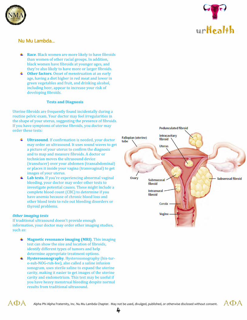
Fibroid location, size and number influence signs and
symptoms:
Submucosal fibroids. Fibroids that grow into the
inner cavity of the uterus (submucosal fibroids) are
more likely to cause prolonged, heavy menstrual
bleeding and are sometimes a problem for women
attempting pregnancy.
Subserosal fibroids. Fibroids that project to the
outside of the uterus (subserosal fibroids) can
sometimes press on your bladder, causing you to
experience urinary symptoms. If fibroids bulge from
the back of your uterus, they occasionally can press
either on your rectum, causing a pressure sensation,
or on your spinal nerves, causing backache.
Intramural fibroids. Some fibroids grow within the
muscular uterine wall (intramural fibroids). If large
enough, they can distort the shape of the uterus and
cause prolonged, heavy periods, as well as pain and
pressure.
Causes
Doctors don't know the cause of uterine fibroids, but research
and clinical experience point to these factors:
Genetic changes. Many fibroids contain changes in
genes that differ from those in normal uterine muscle
cells. There's also some evidence that fibroids run in
families and that identical twins are more likely to
both have fibroids than nonidentical twins.
Hormones. Estrogen and progesterone, two
hormones that stimulate development of the uterine
lining during each menstrual cycle in preparation for
pregnancy, appear to promote the growth of fibroids.
Fibroids contain more estrogen and progesterone
receptors than normal uterine muscle cells do.
Fibroids tend to shrink after menopause due to a
decrease in hormone production.
Other growth factors. Substances that help the body
maintain tissues, such as insulin-like growth factor,
may affect fibroid growth.
Risk Factors
There are few known risk factors for uterine fibroids, other
than being a woman of reproductive age. Other factors that
can have an impact on fibroid development include:
Heredity. If your mother or sister had fibroids,
you're at increased risk of developing them.
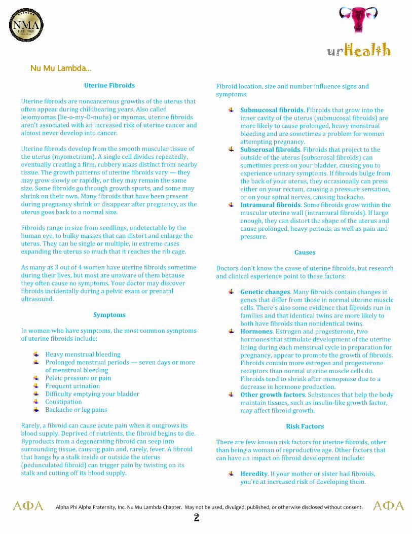
Uterine Fibroids
Uterine fibroids are noncancerous growths of the uterus that
often appear during childbearing years. Also called
leiomyomas (lie-o-my-O-muhs) or myomas, uterine fibroids
aren't associated with an increased risk of uterine cancer and
almost never develop into cancer.
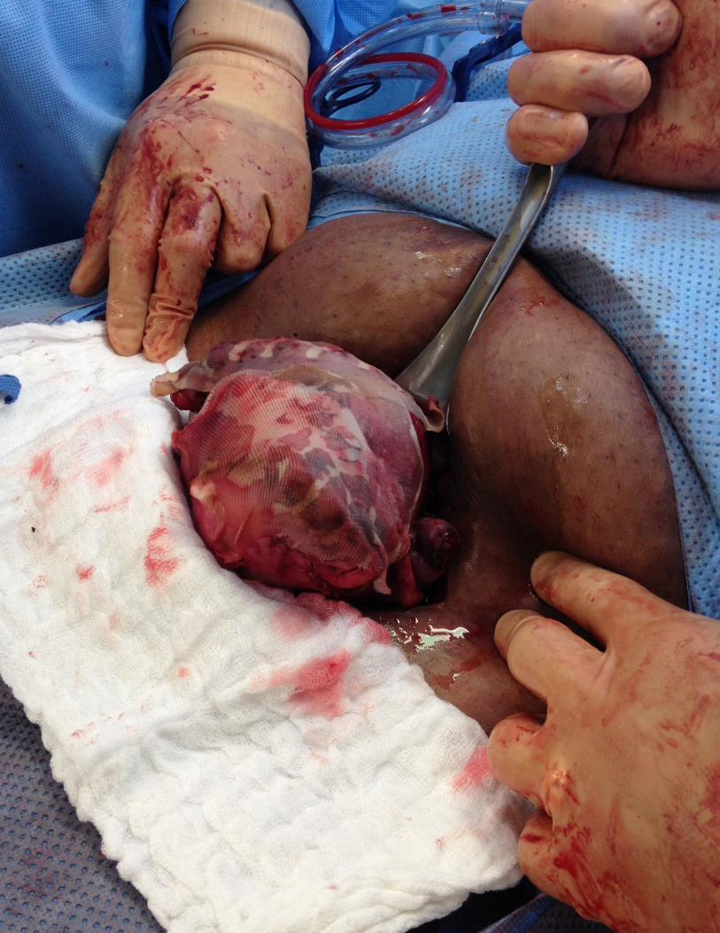
Uterine fibroids develop from the smooth muscular tissue of
the uterus (myometrium). A single cell divides repeatedly,
eventually creating a firm, rubbery mass distinct from nearby
tissue. The growth patterns of uterine fibroids vary — they
may grow slowly or rapidly, or they may remain the same
size. Some fibroids go through growth spurts, and some may
shrink on their own. Many fibroids that have been present
during pregnancy shrink or disappear after pregnancy, as the
uterus goes back to a normal size.
Fibroids range in size from seedlings, undetectable by the
human eye, to bulky masses that can distort and enlarge the
uterus. They can be single or multiple, in extreme cases
expanding the uterus so much that it reaches the rib cage.
As many as 3 out of 4 women have uterine fibroids sometime
during their lives, but most are unaware of them because
they often cause no symptoms. Your doctor may discover
fibroids incidentally during a pelvic exam or prenatal
ultrasound.
Symptoms
In women who have symptoms, the most common symptoms
of uterine fibroids include:
Heavy menstrual bleeding
Prolonged menstrual periods — seven days or more
of menstrual bleeding
Pelvic pressure or pain
Frequent urination
Difficulty emptying your bladder
Constipation
Backache or leg pains
Rarely, a fibroid can cause acute pain when it outgrows its
blood supply. Deprived of nutrients, the fibroid begins to die.
Byproducts from a degenerating fibroid can seep into
surrounding tissue, causing pain and, rarely, fever. A fibroid
that hangs by a stalk inside or outside the uterus
(pedunculated fibroid) can trigger pain by twisting on its
stalk and cutting off its blood supply.

urHealth Nu Mu LambdaNu Mu LambdaNu Mu LambdaNu Mu Lambda............
Alpha Phi Alpha Fraternity, Inc. Nu Mu Lambda Chapter. May not be used, divulged, published, or otherwise disclosed without consent.
3
urHealth Nu Mu LambdaNu Mu LambdaNu Mu LambdaNu Mu Lambda............
Alpha Phi Alpha Fraternity, Inc. Nu Mu Lambda Chapter. May not be used, divulged, published, or otherwise disclosed without consent.
4
Race. Black women are more likely to have fibroids
than women of other racial groups. In addition,
black women have fibroids at younger ages, and
they're also likely to have more or larger fibroids.
Other factors. Onset of menstruation at an early
age, having a diet higher in red meat and lower in
green vegetables and fruit, and drinking alcohol,
including beer, appear to increase your risk of
developing fibroids.
Tests and Diagnosis
Uterine fibroids are frequently found incidentally during a
routine pelvic exam. Your doctor may feel irregularities in
the shape of your uterus, suggesting the presence of fibroids.
If you have symptoms of uterine fibroids, you doctor may
order these tests:
Ultrasound. If confirmation is needed, your doctor
may order an ultrasound. It uses sound waves to get
a picture of your uterus to confirm the diagnosis
and to map and measure fibroids. A doctor or
technician moves the ultrasound device
(transducer) over your abdomen (transabdominal)
or places it inside your vagina (transvaginal) to get
images of your uterus.
Lab tests. If you're experiencing abnormal vaginal
bleeding, your doctor may order other tests to
investigate potential causes. These might include a
complete blood count (CBC) to determine if you
have anemia because of chronic blood loss and
other blood tests to rule out bleeding disorders or
thyroid problems.
Other imaging tests
If traditional ultrasound doesn't provide enough
information, your doctor may order other imaging studies,
such as:
Magnetic resonance imaging (MRI). This imaging
test can show the size and location of fibroids,
identify different types of tumors and help
determine appropriate treatment options.
Hysterosonography. Hysterosonography (his-tur-
o-suh-NOG-ruh-fee), also called a saline infusion
sonogram, uses sterile saline to expand the uterine
cavity, making it easier to get images of the uterine
cavity and endometrium. This test may be useful if
you have heavy menstrual bleeding despite normal
results from traditional ultrasound.
urHealth Nu Mu LambdaNu Mu LambdaNu Mu LambdaNu Mu Lambda............
Alpha Phi Alpha Fraternity, Inc. Nu Mu Lambda Chapter. May not be used, divulged, published, or otherwise disclosed without consent.
5
urHealth Nu Mu LambdaNu Mu LambdaNu Mu LambdaNu Mu Lambda............
Alpha Phi Alpha Fraternity, Inc. Nu Mu Lambda Chapter. May not be used, divulged, published, or otherwise disclosed without consent.
6
Hysterosalpingography. Hysterosalpingography (his-tur-
o-sal-ping-GOG-ruh-fee) uses a dye to highlight the uterine
cavity and fallopian tubes on X-ray images. Your doctor
may recommend it if infertility is a concern. In addition to
revealing fibroids, it can help your doctor determine if your
fallopian tubes are open.
Hysteroscopy. For this, your doctor inserts a small, lighted
telescope called a hysteroscope through your cervix into
your uterus. Your doctor then injects saline into your
uterus, expanding the uterine cavity and allowing your
doctor to examine the walls of your uterus and the
openings of your fallopian tubes.
Treatments and Drugs
There's no single best approach to uterine fibroid treatment —
many treatment options exist. If you have symptoms, talk with your
doctor about options for symptom relief.
Watchful waiting
Many women with uterine fibroids experience no signs or
symptoms, or only mildly annoying signs and symptoms that they
can live with. If that's the case for you, watchful waiting could be the
best option. Fibroids aren't cancerous. They rarely interfere with
pregnancy. They usually grow slowly — or not at all — and tend to
shrink after menopause, when levels of reproductive hormones
drop.
Medications
Medications for uterine fibroids target hormones that regulate your
menstrual cycle, treating symptoms such as heavy menstrual
bleeding and pelvic pressure. They don't eliminate fibroids, but may
shrink them. Medications include:
Gonadotropin-releasing hormone (Gn-RH) agonists.
Medications called Gn-RH agonists (Lupron, Synarel,
others) treat fibroids by blocking the production of
estrogen and progesterone, putting you into a temporary
postmenopausal state. As a result, menstruation stops,
fibroids shrink and anemia often improves. Your doctor
may prescribe a Gn-RH agonist to shrink the size of your
fibroids before a planned surgery. Many women have
significant hot flashes while using Gn-RH agonists. Gn-RH
agonists typically are used for no more than three to six
months because symptoms return when the medication is
stopped and long-term use can cause loss of bone.
Progestin-releasing intrauterine device (IUD). A
progestin-releasing IUD can relieve heavy bleeding caused
by fibroids. A progestin-releasing IUD provides symptom
relief only and doesn't shrink fibroids or make them
disappear.
Other medications. Your doctor might
recommend other medications. For example, oral
contraceptives or progestins can help control
menstrual bleeding, but they don't reduce fibroid
size. Nonsteroidal anti-inflammatory drugs
(NSAIDs), which are not hormonal medications,
may be effective in relieving pain related to
fibroids, but they don't reduce bleeding caused by
fibroids. Your doctor also may suggest that you
take vitamins and iron if you have heavy menstrual
bleeding and anemia.
Noninvasive procedure
MRI-guided focused ultrasound surgery (FUS) is:
A noninvasive treatment option for uterine
fibroids that preserves your uterus, requires no
incision and is done on an outpatient basis.
Performed while you're inside an MRI
scanner equipped with a high-energy ultrasound
transducer for treatment. The images give your
doctor the precise location of the uterine fibroids.
When the location of the fibroid is targeted, the
ultrasound transducer focuses sound waves
(sonications) into the fibroid to heat and destroy
small areas of fibroid tissue.
Newer technology, so researchers are learning
more about the long-term safety and effectiveness.
But so far data collected show that FUS for uterine
fibroids is safe and effective.
Minimally invasive procedures
Certain procedures can destroy uterine fibroids without
actually removing them through surgery. They include:
Uterine artery embolization. Small particles
(embolic agents) are injected into the arteries
supplying the uterus, cutting off blood flow to
fibroids, causing them to shrink and die. This
technique can be effective in shrinking fibroids and
relieving the symptoms they cause. Complications
may occur if the blood supply to your ovaries or
other organs is compromised.
Myolysis. In this laparoscopic procedure, an
electric current or laser destroys the fibroids and
shrinks the blood vessels that feed them. A similar
procedure called cryomyolysis freezes the fibroids.
Myolysis is not used often. Another version of this
procedure, radiofrequency ablation, is being
studied.
urHealth Nu Mu LambdaNu Mu LambdaNu Mu LambdaNu Mu Lambda............
Alpha Phi Alpha Fraternity, Inc. Nu Mu Lambda Chapter. May not be used, divulged, published, or otherwise disclosed without consent.
7
Laparoscopic or robotic myomectomy. In a myomectomy, your surgeon removes the fibroids, leaving the uterus in
place. If the fibroids are small and few in number, you and your doctor may opt for a laparoscopic or robotic procedure,
which uses slender instruments inserted through small incisions in your abdomen to remove the fibroids from your
uterus. Your doctor views your abdominal area on a monitor using a small camera attached to one of the instruments.
Robotic myomectomy gives your surgeon a magnified, 3-D view of your uterus, offering more precision, flexibility and
dexterity than is possible using some other techniques.
Hysteroscopic myomectomy. This procedure may be an option if the fibroids are contained inside the uterus
(submucosal). Your surgeon accesses and removes fibroids using instruments inserted through your vagina and cervix
into your uterus.
Endometrial ablation and resection of submucosal fibroids. This treatment, performed with a specialized
instrument inserted into your uterus, uses heat, microwave energy, hot water or electric current to destroy the lining of
your uterus, either ending menstruation or reducing your menstrual flow. Typically, endometrial ablation is effective in
stopping abnormal bleeding. Submucosal fibroids can be removed at the time of hysteroscopy for endometrial ablation,
but this doesn't affect fibroids outside the interior lining of the uterus.
Traditional surgical procedures
Options for traditional surgical procedures include:
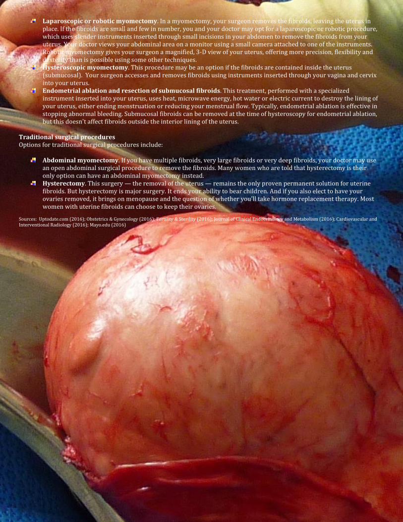
Abdominal myomectomy. If you have multiple fibroids, very large fibroids or very deep fibroids, your doctor may use
an open abdominal surgical procedure to remove the fibroids. Many women who are told that hysterectomy is their
only option can have an abdominal myomectomy instead.
Hysterectomy. This surgery — the removal of the uterus — remains the only proven permanent solution for uterine
fibroids. But hysterectomy is major surgery. It ends your ability to bear children. And if you also elect to have your
ovaries removed, it brings on menopause and the question of whether you'll take hormone replacement therapy. Most
women with uterine fibroids can choose to keep their ovaries.
Sources: Uptodate.com (2016); Obstetrics & Gynecology (2016); Fertility & Sterility (2016); Journal of Clinical Endocrinology and Metabolism (2016); Cardiovascular and
Interventional Radiology (2016); Mayo.edu (2016)
urHealth Nu Mu LambdaNu Mu LambdaNu Mu LambdaNu Mu Lambda............
Alpha Phi Alpha Fraternity, Inc. Nu Mu Lambda Chapter. May not be used, divulged, published, or otherwise disclosed without consent.
8
Eye Twitching
Eye twitching is a repetitive, uncontrollable blinking or spasm of the eyelid, usually the upper lid.
Eye twitching (blepharospasm) often affects the eye muscles of both eyes. If you have eye twitching, you may have an
involuntary movement that recurs every several seconds for a minute or two.
Most people develop a minor eyelid twitch at some point in their lives. Although the cause is generally unknown, it may be
associated with:
Fatigue
Stress
Caffeine
This minor form of twitch is painless and harmless. It usually goes away on its own. But it can be quite annoying. And that's
especially true if the spasms are strong enough to cause the eyelids to completely shut and then reopen.
In some cases, eye twitching is more than a temporary nuisance. Some people have spasms that occur frequently throughout the
day. Symptoms can recur for days, weeks, or even months. That can cause a lot of emotional distress. It can interfere with quality
of life.
In its most serious forms, which are relatively uncommon, eye twitching can become chronic. It can cause persistent winking
and squinting. If it progresses to the point where you have difficulty keeping your eyes open, it can cause
severe vision impairment.
Sometimes, eye twitching can be a sign of eye conditions such as:
Blepharitis (inflammation of the eyelids)
Dry eyes
Light sensitivity
Pink eye
Very rarely, it can be a sign of a brain or nerve disorder, such as:
Bell’s palsy
Dystonia
Parkinson’s disease
Tourette’s syndrome
Eye twitching can also be a side effect of certain medications. The most common offenders include drugs used in the treatment
of psychosis and epilepsy.
Types of Eye Twitching
There are three common types of eye twitch:
Minor eyelid twitch
Benign essential blepharospasm
Hemifacial spasm
Minor eyelid twitch is often associated with lifestyle factors, such as:
Fatigue
urHealth Nu Mu LambdaNu Mu LambdaNu Mu LambdaNu Mu Lambda............
Alpha Phi Alpha Fraternity, Inc. Nu Mu Lambda Chapter. May not be used, divulged, published, or otherwise disclosed without consent.
9
Stress
Irritants such as bright light, wind, or air pollution
As the condition worsens, it may lead to an increased sensitivity
to light, blurry vision, and facial spasms. In severe cases, the
spasms can become so intense that the eyelids stay shut for up
to several hours.
Researchers believe that benign essential blepharospasm may
result from a combination of environmental and genetic factors.
Although the condition is usually random, it sometimes runs in
families.
Hemifacial spasm is quite rare and involves more than just the
eyelid muscles. It also usually involves the muscles around
the mouth. Unlike other types of eyelid twitching, it usually
affects only one side of the face.
In most cases, hemifacial spasm is caused by an artery pressing
on the nerve to the facial muscles.
Diagnosis of Eye Twitching
You should see an eye doctor if you have:
Twitching that persists for more than one week
Twitching that completely closes an eyelid
Spasms that involve other facial muscles
Redness, swelling, or discharge from an eye
A drooping upper eyelid
If your doctor suspects that a brain or nerve disorder is
responsible for eye twitching, he or she will examine you for
other common signs. You may be referred to a neurologist or
other specialist.
Treatment of Eye Twitching
In most cases, minor eyelid twitch will disappear without you
even noticing if you get enough rest and/or reduce or eliminate
your intake of alcohol, tobacco, or caffeine. Blepharospasm does
not occur while sleeping.
If you have dry eyes causing irritation of the cornea or
conjunctiva, treating it with over-the-counter artificial tears will
often relieve minor eyelid twitch.
So far, doctors have not found a successful cure for benign
essential blepharospasm. But several treatment options may
reduce its severity.
The most commonly recommended treatment for benign
essential blepharospasm is botulinum toxin (also known
as Botox, Dysport, or Xeomin). It's approved for this use in both
the U.S. and Canada. Botox is also commonly recommended for
patients with hemifacial spasm.
When injected in very small quantities into the eye muscles,
the drug may relieve spasms for several months. But the effect
gradually wears off. Repeat injections are usually necessary.
In mild cases of benign essential blepharospasm, doctors
sometimes recommend medications such as:
Clonazepam
Lorazepam
Trihexyphenidyl
These usually provide only short-term relief and are rarely
effective.
Alternative treatments for benign essential blepharospasm
include:
Biofeedback
Acupuncture
Hypnosis
Chiropractic
Nutrition therapy
Tinted glasses
But the benefits of these treatments have not been established
by scientific studies.
If other treatments fail, a surgical procedure called a
myectomy is an option. That's a procedure in which some of
the muscles and nerves of the eyelids are removed.
Also generally successful for patients with hemifacial spasm is
a neurosurgical procedure to relieve the pressure of the artery
on the facial nerve. While such surgeries usually produce
permanent results, they can lead to serious complications.
Sources: webmd.com (2016); University of Michigan Kellogg Eye Center: "Eyelid
Spasms (Eye Twitching or Eye Twitch)" and "Understanding Benign Essential
Blepharospasm & Hemifacial Spasm" (2016); National Eye Institute: "Facts About
Blepharospasm" (2016); Dystonia Medical Research Foundation: "Blepharospasm"
(2016); Genetics Home Reference: "Benign Essential Blepharospasm" (2016)
American Academy of Opthalmology: "Blepharospasm" (2016)

urHealth Nu Mu LambdaNu Mu LambdaNu Mu LambdaNu Mu Lambda............
Alpha Phi Alpha Fraternity, Inc. Nu Mu Lambda Chapter. May not be used, divulged, published, or otherwise disclosed without consent.
10
urHealth Nu Mu LambdaNu Mu LambdaNu Mu LambdaNu Mu Lambda............
Alpha Phi Alpha Fraternity, Inc. Nu Mu Lambda Chapter. May not be used, divulged, published, or otherwise disclosed without consent.
11
Major Depression (Clinical Depression)
A constant sense of hopelessness and despair is a sign you
may have major depression, also known as clinical
depression.
With major depression, it may be difficult to work,
study, sleep, eat, and enjoy friends and activities. Some
people have clinical depression only once in their life, while
others have it several times in a lifetime.
Major depression can sometimes occur from one generation
to the next in families, but may affect people with no family
history of the illness.
What Is Major or Clinical Depression?
Most people feel sad or low at some point in their lives. But
clinical depression is marked by a depressed mood most of
the day, sometimes particularly in the morning, and a loss of
interest in normal activities and relationship -- symptoms
that are present every day for at least 2 weeks. In addition,
according to the DSM-5 -- a manual used to diagnose mental
health conditions -- you may have other symptoms with
major depression. Those symptoms might include:
Fatigue or loss of energy almost every day
Feelings of worthlessness or guilt almost every day
Impaired concentration, indecisiveness
Insomnia or hypersomnia (excessive sleeping)
almost every day
Markedly diminished interest or pleasure in almost
all activities nearly every day (called anhedonia, this
symptom can be indicated by reports from
significant others)
Restlessness or feeling slowed down
Recurring thoughts of death or suicide
Significant weight loss or gain (a change of more
than 5% of body weight in a month)
Who Is at Risk for Major Depression?
Major depression affects about 6.7% of the U.S. population
over age 18, according to the National Institute of Mental
Health. Overall, between 20% and 25% of adults may suffer
an episode of major depression at some point during their
lifetime.
Major depression also affects older adults, teens, and
children, but frequently goes undiagnosed and untreated in
these populations.
Are Women at Higher Risk for Major Depression?
Almost twice as many women as men have major or clinical
depression; hormonal changes during
puberty, menstruation, pregnancy, miscarriage, and menopause,
may increase the risk.
Other factors that boost the risk of clinical depression in women
who are biologically vulnerable to it include increased stress at
home or at work, balancing family life with career, and caring for
an aging parent. Raising a child alone will also increase the risk.
What Are the Signs of Major Depression in Men?
Depression in men is significantly underreported. Men who suffer
from clinical depression are less likely to seek help or even talk
about their experience.
Signs of depression in men may include irritability, anger, or drug
and alcohol abuse (substance abuse can also be a cause of
depression rather than the result of it). Suppressing negative
feelings can result in violent behavior directed both inwardly and
outwardly. It can also result in an increase in illness, suicide, and
homicide.
What Triggers Major Depression?
Some common triggers or causes of major depression include:
Loss of a loved one through death, divorce, or separation
Social isolation or feelings of being deprived
Major life changes -- moving, graduation, job change,
retirement
Personal conflicts in relationships, either with a
significant other or a superior
Physical, sexual, or emotional abuse
How Is Major Depression Diagnosed?
A health professional -- such as your primary care doctor or a
psychiatrist -- will perform a thorough medical evaluation. You
might receive a screening for depression at a regular doctor’s visit.
The professional will ask about your personal and family
psychiatric history and ask you questions that screen for the
symptoms of major depression.
There is no blood test, X-ray, or other laboratory test that can be
used to diagnose major depression. However, your doctor may
run blood tests to help detect any other medical problems that
have symptoms similar to those of depression. For
example, hypothyroidism can cause some of the same symptoms
as depression, as can alcohol or drug use and abuse,

urHealth Nu Mu LambdaNu Mu LambdaNu Mu LambdaNu Mu Lambda............
Alpha Phi Alpha Fraternity, Inc. Nu Mu Lambda Chapter. May not be used, divulged, published, or otherwise disclosed without consent.
12

urHealth Nu Mu LambdaNu Mu LambdaNu Mu LambdaNu Mu Lambda............
Alpha Phi Alpha Fraternity, Inc. Nu Mu Lambda Chapter. May not be used, divulged, published, or otherwise disclosed without consent.
13
some medications, and stroke.
How Is Major Depression Treated?
Major or clinical depression is a serious but treatable illness.
Depending on the severity of symptoms, your primary care
doctor or a psychiatrist may recommend treatment with
an antidepressant medication. He or she may also suggest
psychotherapy, or talk therapy, in which you address your
emotional state.
Sometimes, other medications are added to
the antidepressant to boost its effectiveness. Certain
medicines work better for some people. It may be necessary
for your doctor to try different drugs at different doses to
determine which medicine works best for you.
There are other treatment options for clinical depression --
such as electroconvulsive therapy, also called ECT or shock
therapy -- that can be used if drugs prove ineffective or
symptoms are severe.
Can Major Depression Be Prevented?
Once you have had an episode of major depression, you are
at high risk of having another. The best way to prevent
another episode of depression is to be aware of the triggers
or causes of major depression (see above) and to continue
taking the prescribed medication to avoid relapse. It is also
important to know what the symptoms of major depression
are and to talk with your doctor early if you have any of
these symptoms.
Sources: webmd.com (2016); National Institute of Mental Health: "What Is
Depression?" and "What Are the Different Forms of Depression?" (2016); American
Psychiatric Association. Diagnostic and Statistical Manual of Mental Disorders:
DSM-5, American Psychiatric Pub. (2016); The Journal of the American Medical
Association. “Recommendations for Screening Depression in Adults,” (2016)

urHealth Nu Mu LambdaNu Mu LambdaNu Mu LambdaNu Mu Lambda............
Alpha Phi Alpha Fraternity, Inc. Nu Mu Lambda Chapter. May not be used, divulged, published, or otherwise disclosed without consent.
14
Nail Fungus
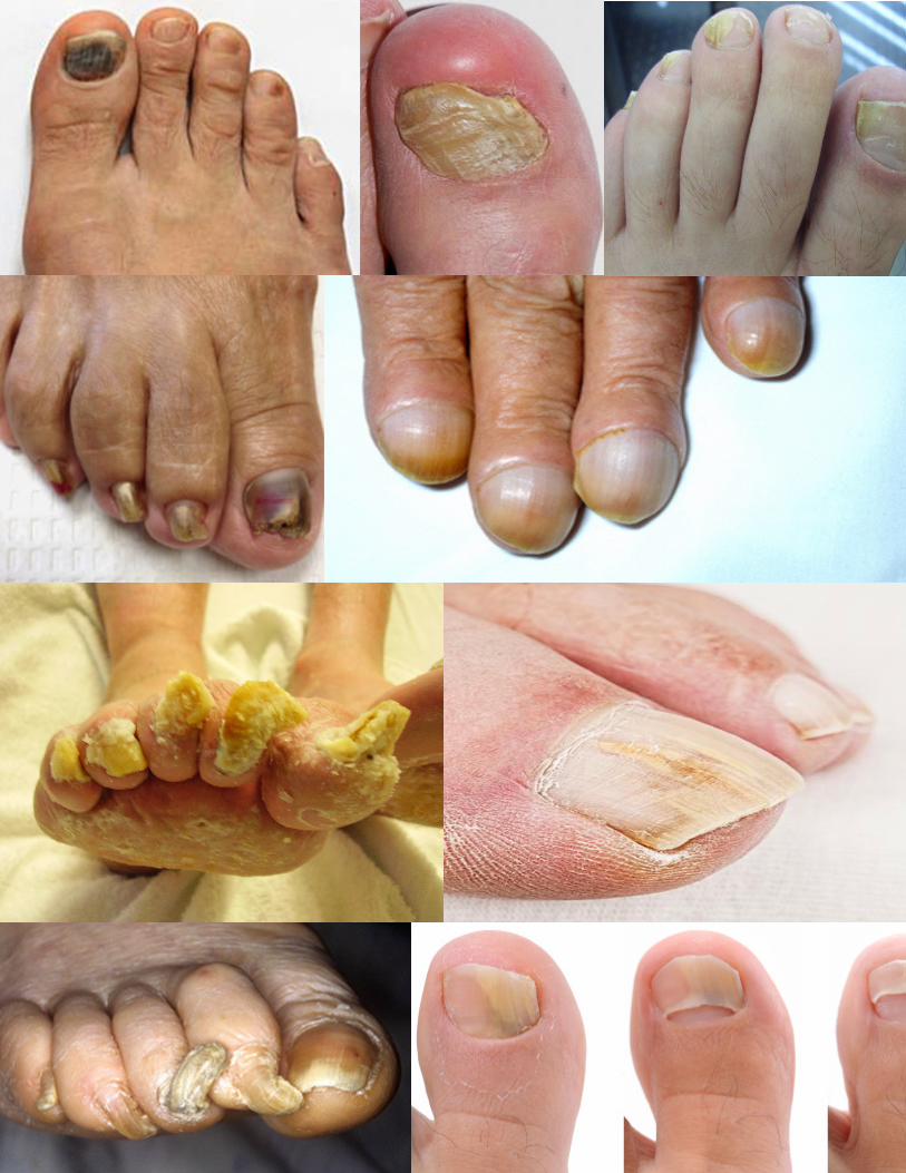
Nail fungus is a common condition that begins as a white or yellow spot under the tip of your fingernail or toenail. As the fungal
infection goes deeper, nail fungus may cause your nail to discolor, thicken and crumble at the edge. It can affect several nails but
usually not all of them.
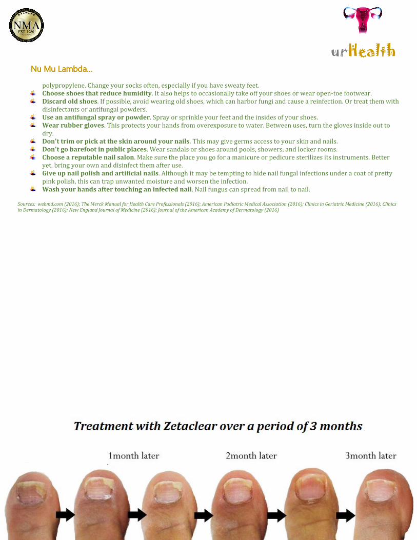
If your condition is mild and not bothering you, you may not need treatment. If your nail fungus is painful and has caused thickened
nails, self-care steps and medications may help. But even if treatment is successful, nail fungus often comes back.
Nail fungus is also called onychomycosis (on-ih-koh-my-KOH-sis) and tinea unguium. When fungus infects the areas between your
toes and the skin of your feet, it's called athlete's foot (tinea pedis).
You may have nail fungus — also called onychomycosis (on-ih-koh-my-KOH-sis) — if one or more of your nails are:
Thickened
Brittle, crumbly or ragged
Distorted in shape
Dull, with no shine
A dark color, caused by debris building up under your nail
Infected nails also may separate from the nail bed, a condition called onycholysis (on-ih-KOL-ih-sis). You may feel pain in your toes
or fingertips and detect a slightly foul odor.
Causes
Nail fungal infections are typically caused by a dermatophyte fungus. Yeasts and molds also can be responsible for nail fungal
infections.
What are fungi?
Fungi are microscopic organisms that don't need sunlight to survive. Some fungi have beneficial uses. Others cause illness and
infection. Fungi:
Live in warm, moist environments, including swimming pools and showers
Can invade your skin through cuts so tiny you can't even see them or through a small separation between your nail and nail
bed
Can cause problems if your nails are often exposed to warm and moist conditions
Toenails vs. fingernails
Nail fungus occurs more often in toenails than in fingernails, partly because:
Toenails often are confined in a dark, warm, moist environment — inside your shoes — where fungi can thrive
Toes usually have less blood flow than do fingers, making it harder for your body's immune system to detect and stop
infection
Risk Factors
Factors that can increase your risk of developing nail fungus include:
Being older, owing to reduced blood flow, more years of exposure to fungi and slower growing nails
Perspiring heavily
urHealth Nu Mu LambdaNu Mu LambdaNu Mu LambdaNu Mu Lambda............
Alpha Phi Alpha Fraternity, Inc. Nu Mu Lambda Chapter. May not be used, divulged, published, or otherwise disclosed without consent.
15

urHealth Nu Mu LambdaNu Mu LambdaNu Mu LambdaNu Mu Lambda............
Alpha Phi Alpha Fraternity, Inc. Nu Mu Lambda Chapter. May not be used, divulged, published, or otherwise disclosed without consent.
16
Being male, especially if you have a family history of
nail fungal infections
Working in a humid or moist environment or in a
job where your hands are often wet, such as
bartending or housekeeping
Wearing socks and shoes that hinder ventilation and
don't absorb perspiration
Living with someone who has nail fungus
Walking barefoot in damp communal areas, such as
swimming pools, gyms and shower rooms
Having athlete's foot
Having a minor skin or nail injury or a skin
condition, such as psoriasis
Having diabetes, circulation problems, a weakened
immune system or, in children, Down syndrome
Tests and Diagnosis
Your doctor will likely examine your nails first. He or she
may scrape some debris from under your nail and send it to
a lab to identify the type of fungus causing the infection.
Other conditions, such as psoriasis, can mimic a fungal
infection of the nail. Microorganisms such as yeast and
bacteria also can infect nails. Knowing the cause of your
infection helps determine the best course of treatment.
If self-care strategies and over-the-counter
(nonprescription) products haven't helped, your doctor may
suggest a combination of prescription drugs and other
approaches. But even if you find relief from your signs and
symptoms, repeat infections are common.
Medications
Oral antifungal drugs. Your doctor may prescribe
an oral antifungal drug. Studies show the most
effective treatments are terbinafine (Lamisil) and
itraconazole (Sporanox). These drugs help a new
nail grow free of infection, slowly replacing the
infected part.
You typically take this type of drug for six to 12
weeks. But you won't see the end result of treatment
until the nail grows back completely. It may take
four months or longer to eliminate an infection.
Treatment success rates with these drugs appear to
be lower in adults over age 65. And treatment
success seems to improve when you combine oral
and topical antifungal therapies.
Oral antifungal drugs may cause side effects ranging
from skin rash to liver damage. You may need
occasional blood tests to check on how you're doing with
these types of drugs. Doctors may not recommend them
for people with liver disease or congestive heart failure
or those taking certain medications.
Medicated nail polish. Your doctor may prescribe an
antifungal nail polish called ciclopirox (Penlac). You
paint it on your infected nails and surrounding skin once
a day. After seven days, you wipe the piled-on layers
clean with alcohol and begin fresh applications. You may
need to use this type of nail polish daily for a year.
Medicated nail cream. Your doctor may prescribe an
antifungal cream, which you rub into your infected nails
after soaking. These creams may work better if you first
thin the nails. This helps the medication get through the
hard nail surface to the underlying fungus.
To thin nails, you apply an over-the-counter
(nonprescription) lotion containing urea. Or your doctor
may thin the surface of the nail (debride) with a file or
other tool.
Surgical or other procedures
Nail removal. If your nail infection is severe or
extremely painful, your doctor may suggest removing
your nail. A new nail will usually grow in its place. But it
will come in slowly and may take as long as a year to
grow back completely. Sometimes surgery is used in
combination with ciclopirox to treat the nail bed.
Laser and light-based therapies. More study is needed,
but these methods — alone or with medications — may
help your nails improve. One study tested the
effectiveness of carbon-dioxide laser therapy combined
with antifungal nail cream. Most of the 24 people in the
study benefited from the treatment.
Laser and light-based therapies are not available
everywhere, are expensive, and often are not covered by
insurance.
Prevention
These habits can help prevent nail fungus or reinfections:
Wash your hands and feet regularly and keep your
nails short and dry. Wash your hands and feet with
soap and water, rinse, and dry thoroughly, including
between the toes. Trim nails straight across and file
down thickened areas.
Wear socks that absorb sweat. Fabrics effective at
wicking away moisture include wool, nylon and
urHealth Nu Mu LambdaNu Mu LambdaNu Mu LambdaNu Mu Lambda............
Alpha Phi Alpha Fraternity, Inc. Nu Mu Lambda Chapter. May not be used, divulged, published, or otherwise disclosed without consent.
17
polypropylene. Change your socks often, especially if you have sweaty feet.
Choose shoes that reduce humidity. It also helps to occasionally take off your shoes or wear open-toe footwear.
Discard old shoes. If possible, avoid wearing old shoes, which can harbor fungi and cause a reinfection. Or treat them with
disinfectants or antifungal powders.
Use an antifungal spray or powder. Spray or sprinkle your feet and the insides of your shoes.
Wear rubber gloves. This protects your hands from overexposure to water. Between uses, turn the gloves inside out to
dry.
Don't trim or pick at the skin around your nails. This may give germs access to your skin and nails.
Don't go barefoot in public places. Wear sandals or shoes around pools, showers, and locker rooms.
Choose a reputable nail salon. Make sure the place you go for a manicure or pedicure sterilizes its instruments. Better
yet, bring your own and disinfect them after use.
Give up nail polish and artificial nails. Although it may be tempting to hide nail fungal infections under a coat of pretty
pink polish, this can trap unwanted moisture and worsen the infection.
Wash your hands after touching an infected nail. Nail fungus can spread from nail to nail. Sources: webmd.com (2016); The Merck Manual for Health Care Professionals (2016); American Podiatric Medical Association (2016); Clinics in Geriatric Medicine (2016); Clinics
in Dermatology (2016); New England Journal of Medicine (2016); Journal of the American Academy of Dermatology (2016)
urHealth Nu Mu LambdaNu Mu LambdaNu Mu LambdaNu Mu Lambda............
Alpha Phi Alpha Fraternity, Inc. Nu Mu Lambda Chapter. May not be used, divulged, published, or otherwise disclosed without consent.
18
Thrombocytopenia (low platelet count)
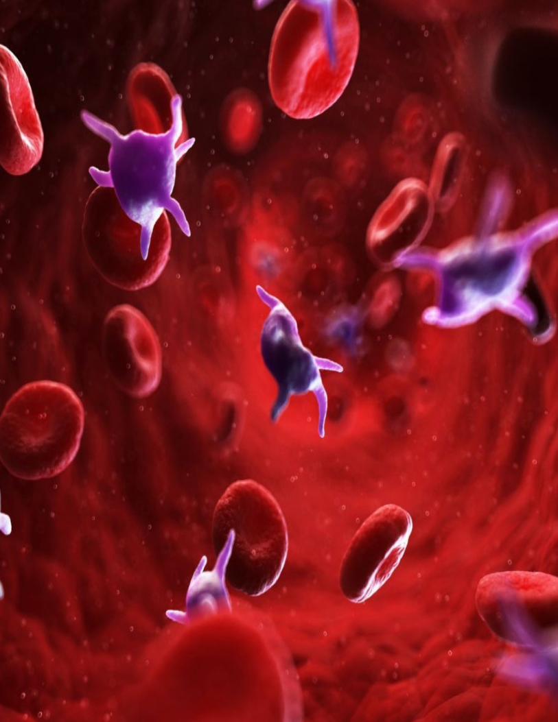
Thrombocytopenia is a condition in which you have a low blood platelet count. Platelets (thrombocytes) are colorless blood cells that
help blood clot. Platelets stop bleeding by clumping and forming plugs in blood vessel injuries.
Thrombocytopenia often occurs as a result of a separate disorder, such as leukemia or an immune system problem. Or it can be a side
effect of taking certain medications. It affects both children and adults.
Thrombocytopenia may be mild and cause few signs or symptoms. In rare cases, the number of platelets may be so low that dangerous
internal bleeding occurs. Treatment options are available.
Symptoms
Thrombocytopenia signs and symptoms may include:
Easy or excessive bruising (purpura)
Superficial bleeding into the skin that appears as a rash of pinpoint-sized reddish-purple spots (petechiae), usually on the
lower legs
Prolonged bleeding from cuts
Bleeding from your gums or nose
Blood in urine or stools
Unusually heavy menstrual flows
Fatigue
Enlarged spleen
Jaundice
Causes
If for any reason your blood platelet count falls below normal, the condition is called thrombocytopenia. Normally, you have anywhere
from 150,000 to 450,000 platelets per microliter of circulating blood. Because each platelet lives only about 10 days, your body
continually renews your platelet supply by producing new platelets in your bone marrow.
Thrombocytopenia can be inherited or it may be caused by a number of medications or conditions. Whatever the cause, circulating
platelets are reduced by one or more of the following processes: trapping of platelets in the spleen, decreased platelet production or
increased destruction of platelets.
Trapped platelets
The spleen is a small organ about the size of your fist located just below your rib cage on the left side of your abdomen. Normally, your
spleen works to fight infection and filter unwanted material from your blood. An enlarged spleen — which can be caused by a number
of disorders — may harbor too many platelets, causing a decrease in the number of platelets in circulation.
Decreased production of platelets
Platelets are produced in your bone marrow. If production is low, you may develop thrombocytopenia. Factors that can decrease
platelet production include:
Leukemia
Some types of anemia
Viral infections, such as hepatitis C or HIV
Chemotherapy drugs
Heavy alcohol consumption
Increased breakdown of platelets
Some conditions can cause your body to use up or destroy platelets more rapidly than they're produced. This leads to a shortage of
urHealth Nu Mu LambdaNu Mu LambdaNu Mu LambdaNu Mu Lambda............
Alpha Phi Alpha Fraternity, Inc. Nu Mu Lambda Chapter. May not be used, divulged, published, or otherwise disclosed without consent.
19
urHealth Nu Mu LambdaNu Mu LambdaNu Mu LambdaNu Mu Lambda............
Alpha Phi Alpha Fraternity, Inc. Nu Mu Lambda Chapter. May not be used, divulged, published, or otherwise disclosed without consent.
20
platelets in your bloodstream. Examples of such conditions include:
Pregnancy. Thrombocytopenia caused by pregnancy is usually mild and improves soon after childbirth.
Immune thrombocytopenia. This type is caused by autoimmune diseases, such as lupus and rheumatoid arthritis. The
body's immune system mistakenly attacks and destroys platelets. If the exact cause of this condition isn't known, it's called
idiopathic thrombocytopenic purpura. This type more often affects children.
Bacteria in the blood. Severe bacterial infections involving the blood (bacteremia) may lead to destruction of platelets.
Thrombotic thrombocytopenic purpura. This is a rare condition that occurs when small blood clots suddenly form
throughout your body, using up large numbers of platelets.
Hemolytic uremic syndrome. This rare disorder causes a sharp drop in platelets, destruction of red blood cells and
impairment of kidney function. Sometimes it can occur in association with a bacterial Escherichia coli (E. coli) infection, such
as may be acquired from eating raw or undercooked meat.
Medications. Certain medications can reduce the number of platelets in your blood. Sometimes a drug confuses the immune
system and causes it to destroy platelets. Examples include heparin, quinine, sulfa-containing antibiotics and anticonvulsants.
Test and Diagnosis
Your doctor may use the following tests and procedures to determine whether you have thrombocytopenia:
Blood test. A complete blood count determines the number of blood cells, including platelets, in a sample of your blood. In
adults, normal platelet count is 150,000 to 450,000 platelets per microliter of blood. If the complete blood count finds you
have fewer than 150,000 platelets, you have thrombocytopenia.
Physical exam, including a complete medical history. Your doctor will look for signs of bleeding under your skin and feel
your abdomen to see if your spleen is enlarged. He or she will also ask you about illnesses you've had and the types of
medications and supplements you've recently taken.
Your doctor may suggest that you undergo other tests and procedures to determine the cause of your condition, depending on your
signs and symptoms.
Treatments and Drugs
People with mild thrombocytopenia may not need treatment. For example, they may not have symptoms or the condition clears up on
its own.
Some people develop severe or long-term (chronic) thrombocytopenia. Depending on what's causing your low platelet count,
treatments may include:
Treating the underlying cause of thrombocytopenia. If your doctor can identify a condition or a medication that's causing
your thrombocytopenia, addressing that cause may clear up your thrombocytopenia
For example, if you have heparin-induced thrombocytopenia, your doctor will direct you to stop using heparin and prescribe a
different blood-thinning drug. Your thrombocytopenia may persist for a week or more despite stopping all heparin therapy.
Blood or platelet transfusions. If your platelet level becomes too low, your doctor can replace lost blood with transfusions of
packed red blood cells or platelets.
Medications. If your condition is related to an immune system problem, your doctor may prescribe drugs to boost your
platelet count. The first-choice drug may be a corticosteroid. If that doesn't work, he or she may try stronger medications to
suppress your immune system.
Surgery. If other treatment options don't help, your doctor may recommend surgery to remove your spleen (splenectomy).
Plasma exchange. Thrombotic thrombocytopenic purpura can result in a medical emergency requiring plasma exchange.
Sources: webmd.com (2016); Thrombocytopenia and platelet dysfunction. The Merck Manual Professional Edition (2016); George JN, et al. Approach to the adult with unexplained
thrombocytopenia (2016); Thrombocytopenia. National Heart, Lung and Blood Institute (2016); E. coli. U.S. Food and Drug Administration (2016); Mayo Foundation for Medical
Education and Research (2016)
urHealth Nu Mu LambdaNu Mu LambdaNu Mu LambdaNu Mu Lambda............
Alpha Phi Alpha Fraternity, Inc. Nu Mu Lambda Chapter. May not be used, divulged, published, or otherwise disclosed without consent.
21
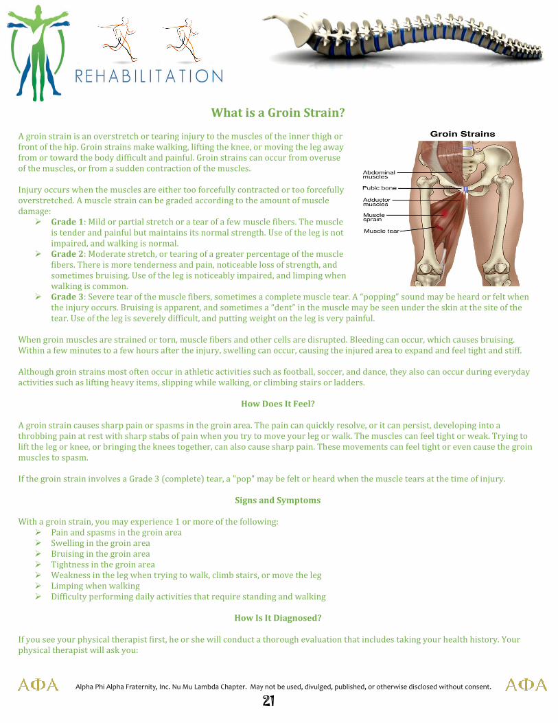
What is a Groin Strain?
A groin strain is an overstretch or tearing injury to the muscles of the inner thigh or
front of the hip. Groin strains make walking, lifting the knee, or moving the leg away
from or toward the body difficult and painful. Groin strains can occur from overuse
of the muscles, or from a sudden contraction of the muscles.
Injury occurs when the muscles are either too forcefully contracted or too forcefully
overstretched. A muscle strain can be graded according to the amount of muscle
damage:
� Grade 1: Mild or partial stretch or a tear of a few muscle fibers. The muscle
is tender and painful but maintains its normal strength. Use of the leg is not
impaired, and walking is normal.
� Grade 2: Moderate stretch, or tearing of a greater percentage of the muscle
fibers. There is more tenderness and pain, noticeable loss of strength, and
sometimes bruising. Use of the leg is noticeably impaired, and limping when
walking is common.
� Grade 3: Severe tear of the muscle fibers, sometimes a complete muscle tear. A “popping” sound may be heard or felt when
the injury occurs. Bruising is apparent, and sometimes a “dent” in the muscle may be seen under the skin at the site of the
tear. Use of the leg is severely difficult, and putting weight on the leg is very painful.
When groin muscles are strained or torn, muscle fibers and other cells are disrupted. Bleeding can occur, which causes bruising.
Within a few minutes to a few hours after the injury, swelling can occur, causing the injured area to expand and feel tight and stiff.
Although groin strains most often occur in athletic activities such as football, soccer, and dance, they also can occur during everyday
activities such as lifting heavy items, slipping while walking, or climbing stairs or ladders.
How Does It Feel?
A groin strain causes sharp pain or spasms in the groin area. The pain can quickly resolve, or it can persist, developing into a
throbbing pain at rest with sharp stabs of pain when you try to move your leg or walk. The muscles can feel tight or weak. Trying to
lift the leg or knee, or bringing the knees together, can also cause sharp pain. These movements can feel tight or even cause the groin
muscles to spasm.
If the groin strain involves a Grade 3 (complete) tear, a "pop" may be felt or heard when the muscle tears at the time of injury.
Signs and Symptoms
With a groin strain, you may experience 1 or more of the following:
� Pain and spasms in the groin area
� Swelling in the groin area
� Bruising in the groin area
� Tightness in the groin area
� Weakness in the leg when trying to walk, climb stairs, or move the leg
� Limping when walking
� Difficulty performing daily activities that require standing and walking
How Is It Diagnosed?
If you see your physical therapist first, he or she will conduct a thorough evaluation that includes taking your health history. Your
physical therapist will ask you:
urHealth Nu Mu LambdaNu Mu LambdaNu Mu LambdaNu Mu Lambda............
Alpha Phi Alpha Fraternity, Inc. Nu Mu Lambda Chapter. May not be used, divulged, published, or otherwise disclosed without consent.
22
� What you were doing when you first felt pain
� Where you felt the pain and if you heard a pop when it occurred
� If you received a direct hit to your leg or groin area
� If you noticed any swelling in the first 2-3 hours following the injury
� If you experience pain when lifting your leg, walking, moving the leg away from you, or drawing your knees together
Your physical therapist will perform special tests to help determine whether you have a groin strain, such as:
� Gently moving your leg away from your body
� Asking you to resist against his or her hand as he or she tries to gently push your leg outward (muscle strength test)
� Gently feeling parts of the muscle to determine the specific location of the injury (palpation)
Your physical therapist may use additional tests to assess possible damage to other parts of your body, such as your hip or lower
back.
To provide a final diagnosis, your physical therapist may collaborate with an orthopedist or other health care provider. The
orthopedist may order further tests—such as an X-ray or magnetic resonance imaging (MRI) —to confirm the diagnosis and to rule
out other potential damage. These tests, however, are not commonly required for groin strain.
How Can a Physical Therapist Help?
Your physical therapist will design a specific treatment program to speed your recovery. This program will include exercises and
treatments you can do at home to help you return to your normal lifestyle and activities.
The First 24-48 Hours
Your physical therapist may advise you to:
� Rest the area by avoiding walking or any activity that causes pain. Crutches may be recommended to reduce further strain
on the muscles when walking.
� Apply ice packs to the area for 15-20 minutes every 2 hours.
� Compress the area with an elastic bandage wrap.
� Consult with another health care provider for further services such as medication or diagnostic tests.
Reduce Pain
Your physical therapist can use different types of treatments and technologies to control and reduce your pain, including ice, heat,
ultrasound, electricity, taping, exercises, and hands-on therapy such as massage.
Improve Motion
Your physical therapist will choose specific activities and treatments to help restore normal movement in the leg and hip. These
might begin with "passive" motions that the therapist performs for you to gently move your leg and hip joint, and progress to active
exercises and stretches that you perform yourself.
Improve Strength
Certain exercises will benefit healing at each stage of recovery; your physical therapist will choose and teach you the appropriate
exercises to steadily restore your strength and agility.
urHealth Nu Mu LambdaNu Mu LambdaNu Mu LambdaNu Mu Lambda............
Alpha Phi Alpha Fraternity, Inc. Nu Mu Lambda Chapter. May not be used, divulged, published, or otherwise disclosed without consent.
23
Improve Strength
Certain exercises will benefit healing at each stage of recovery; your physical therapist will choose and teach you the appropriate
exercises to steadily restore your strength and agility.
Return to Activities
Your physical therapist will collaborate with you to decide on your recovery goals, including your return to work or sport, and will
design your treatment program to help you reach those goals in the safest, fastest, and most effective way possible. Your physical
therapist will apply hands-on therapy such as massage, teach you exercises and work re-training activities. Additionally, if you are
an athlete you will be taught sport-specific techniques and drills to help you achieve your sport-specific goals.
Prevent Future Re-injury
Your physical therapist can recommend a home exercise program to strengthen and stretch the muscles around your hip, upper leg,
and abdomen to help prevent future re-injury of your groin. These may include strength and flexibility exercises for the leg, hip, and
core muscles.
If Surgery Is Necessary
Surgery is rarely necessary in the case of groin strain, but if a groin muscle fully tears and requires surgical repair, your physical
therapist will help you minimize pain, restore motion and strength, and return to normal activities in the speediest manner possible
after surgery.
Sources: Sportsinjuryclinic.net (2016); moveforwardpt.com (2016); Webmd.com (2016)
urHealth Nu Mu LambdaNu Mu LambdaNu Mu LambdaNu Mu Lambda............
Alpha Phi Alpha Fraternity, Inc. Nu Mu Lambda Chapter. May not be used, divulged, published, or otherwise disclosed without consent.
24
Cardiovascular Exercise and Aerobic Training
Aerobic exercise and cardiovascular exercise are the same thing. Both types of exercise achieve the same results: improved
fitness by increasing both your oxygen intake and heart rate.
You cannot do one without the other, as there is no way you can increase your respiratory rate without also making your heart
pump harder and vice versa. Perhaps a less confusing blanket term for both is endurance exercise, as both aerobic and cardio
exercises increase your endurance level.
There's no 'right' cardio exercise and the best choice is the one you enjoy and the one you'll work hardest at, but below are some
of the best cardio workouts you can do to burn calories and fat.
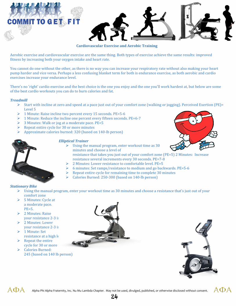
Treadmill
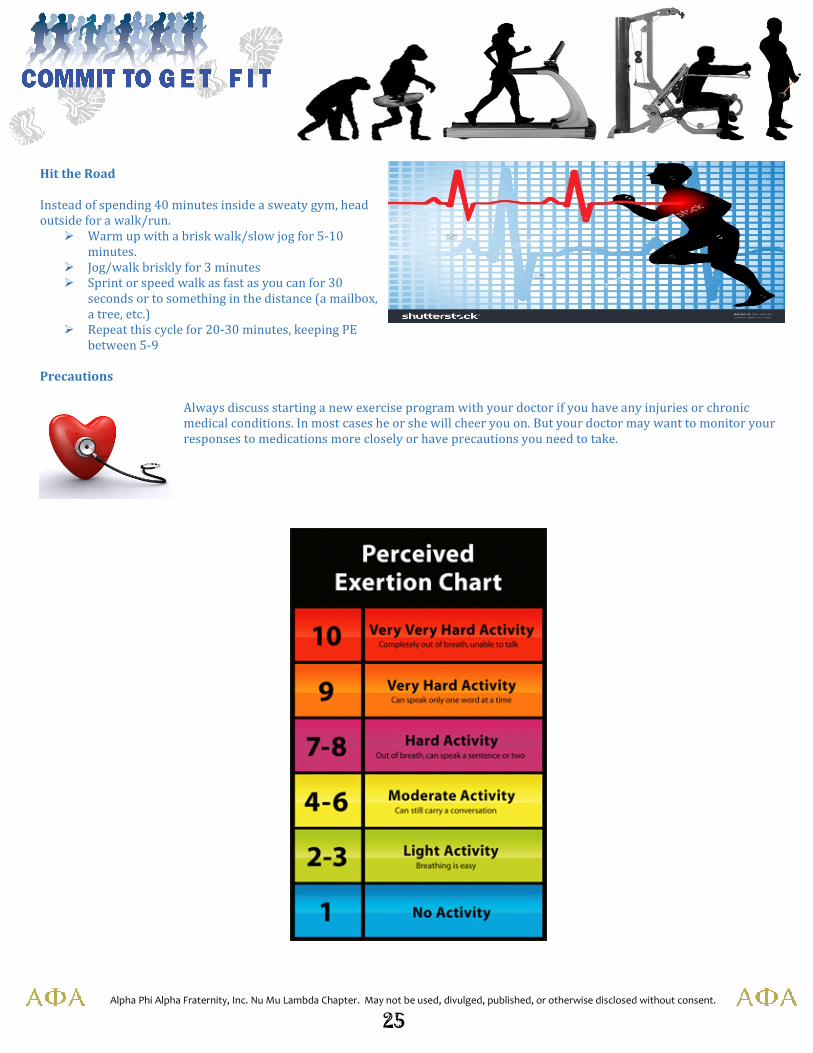
� Start with incline at zero and speed at a pace just out of your comfort zone (walking or jogging). Perceived Exertion (PE)=
Level 5
� 1 Minute: Raise incline two percent every 15 seconds. PE=5-6
� 1 Minute: Reduce the incline one percent every fifteen seconds. PE=6-7
� 3 Minutes: Walk or jog at a moderate pace. PE=5
� Repeat entire cycle for 30 or more minutes
� Approximate calories burned: 320 (based on 140-lb person)
Elliptical Trainer
� Using the manual program, enter workout time as 30
minutes and choose a level of
resistance that takes you just out of your comfort zone (PE=5) 2 Minutes: Increase
resistance several increments every 30 seconds. PE=7-8
� 2 Minutes: Lower resistance to comfortable level. PE=5
� 6 minutes: Set ramps/resistance to medium and go backwards. PE=5-6
� Repeat entire cycle for remaining time to complete 30 minutes
� Calories Burned: 250-300 (based on 140-lb person)
Stationary Bike
� Using the manual program, enter your workout time as 30 minutes and choose a resistance that's just out of your
comfort zone
� 5 Minutes: Cycle at
a moderate pace.
PE=5.
� 2 Minutes: Raise
your resistance 2-3 increments every 30 seconds. PE=6-8
� 2 Minutes: Lower
your resistance 2-3 increments every 30 seconds. PE=6-8.
� 1 Minute: Set
resistance at a high level and cycle as fast as you can. PE=8-9
� Repeat the entire
cycle for 30 or more minutes
� Calories Burned:
245 (based on 140 lb person)
urHealth Nu Mu LambdaNu Mu LambdaNu Mu LambdaNu Mu Lambda............
Alpha Phi Alpha Fraternity, Inc. Nu Mu Lambda Chapter. May not be used, divulged, published, or otherwise disclosed without consent.
25
Hit the Road
Instead of spending 40 minutes inside a sweaty gym, head
outside for a walk/run.
� Warm up with a brisk walk/slow jog for 5-10
minutes.
� Jog/walk briskly for 3 minutes
� Sprint or speed walk as fast as you can for 30
seconds or to something in the distance (a mailbox,
a tree, etc.)
� Repeat this cycle for 20-30 minutes, keeping PE
between 5-9
Precautions
Always discuss starting a new exercise program with your doctor if you have any injuries or chronic
medical conditions. In most cases he or she will cheer you on. But your doctor may want to monitor your
responses to medications more closely or have precautions you need to take.

urHealth Nu Mu LambdaNu Mu LambdaNu Mu LambdaNu Mu Lambda............
Alpha Phi Alpha Fraternity, Inc. Nu Mu Lambda Chapter. May not be used, divulged, published, or otherwise disclosed without consent.
26
Did you know…Did you know…Did you know…Did you know…
…If muscle isn’t stimulated, your body senses that you don’t need it. I’m
sure you’ve heard the saying, “If you don’t use it, you’ll lose it.”
Well, use your muscles! …all of them.
…Type 2 diabetes (the most common kind) occurs when
the body becomes insensitive to insulin in the blood, which
causes blood sugar levels to rise. Exercise reverses the
damage by increasing insulin sensitivity.
…Numerous studies support the link between exercise and increased
mental sharpness.
…Even those who already have heart disease can lower their risk of dying by
beginning an exercise program.
…People who are active are 25% less likely to have a stroke than those who
are inactive. Exercise lowers blood pressure, raises HDL (good) cholesterol,
and reduces the risk of blood clots.
urHealth Nu Mu LambdaNu Mu LambdaNu Mu LambdaNu Mu Lambda............
Alpha Phi Alpha Fraternity, Inc. Nu Mu Lambda Chapter. May not be used, divulged, published, or otherwise disclosed without consent.
27
Did you know…Did you know…Did you know…Did you know…
…Exercise can increase your sexual stamina,
performance, and desire.
…Intensity is more important than the number of sets when trying to
get bigger and stronger. A good rule of thumb is to use a weight that
allows you to do at least 8 bur no more than 15 reps.
…Doing a 5-10-minute pre-workout warm-up using cardio activity will
increase your muscular and joint flexibility and extensibility.
…It’s never too late to start strength
training. Elderly people should start
with light weights, then gradually
progress to heavier weights as they get stronger.
…Without exercise, men start to lose
muscle at about age 50. For every
decade after age 50, men lose about
6% of muscle mass and 10-15% of
their strength.

urHealth Nu Mu LambdaNu Mu LambdaNu Mu LambdaNu Mu Lambda............
Alpha Phi Alpha Fraternity, Inc. Nu Mu Lambda Chapter. May not be used, divulged, published, or otherwise disclosed without consent.
28
Braised Paprika Chicken
INGREDIENTS
� 3-3 ½ lbs bone-in chicken pieces, (thighs, drumsticks and/or breasts), skin removed, trimmed
� ¾ teaspoon coarse salt, divided
� ½ teaspoon freshly ground pepper
� 2 tablespoons canola oil
� 1 tablespoon butter
� 4 cups finely diced onions
� Pinch of sugar
� 1 cup diced red bell pepper
� ½ cup diced green bell pepper
� 2 tablespoons tomato paste
� 2 tablespoons sweet paprika
� 1 teaspoon crushed red pepper
� 1 teaspoon dried marjoram
� 1 cup reduced-sodium chicken broth
� ½ cup reduced-fat sour cream
� 1 tablespoon all-purpose flour
� 2 tablespoons finely minced fresh parsley, dill and/or chives
PREPARATION
1. Pat chicken pieces dry with paper towels and season with ½ teaspoon salt and pepper.
2. Heat oil and butter in a large heavy casserole or Dutch oven over medium heat. Add onions and sprinkle with sugar.
3. Cook, stirring frequently, until the onions are very soft and light brown, 10 to 15 minutes.
4. Stir in bell peppers, tomato paste, paprika and crushed red pepper.
5. Add the chicken and stir it gently into the onion mixture.
6. Sprinkle with marjoram and add broth.
7. Cover the pot with a tight-fitting lid and simmer over medium-low heat until the chicken is very tender, about 50
minutes.
8. Just before the chicken is done, whisk sour cream, flour and the remaining ¼ teaspoon salt in a small bowl until
smooth.
9. When the chicken is done, remove it to a plate.
10. Stir the sour cream mixture into the sauce; return to a simmer and cook, stirring, until the sauce coats the spoon.
11. Reduce heat to low, return the chicken to the sauce and reheat, about 1 minute.
12. Serve garnished with parsley, dill and/or chives, if desired.
NUTRITION
1. 331 calories
2. 14 g fat (4 g sat, 5 g mono)
3. 115 mg cholesterol
4. 17 g carbohydrates
5. 35 g protein
6. 4 g fiber
7. 384 mg sodium
8. 705 mg potassium
Carbohydrate Servings: 1
Exchanges: 2 vegetables, 4 lean meat, 1 fat
Courtesy: eatingwell.com (2016)

urHealth Nu Mu LambdaNu Mu LambdaNu Mu LambdaNu Mu Lambda............
Alpha Phi Alpha Fraternity, Inc. Nu Mu Lambda Chapter. May not be used, divulged, published, or otherwise disclosed without consent.
29
2015201520152015----2016 2016 2016 2016 Nu Mu Lambda Nu Mu Lambda Nu Mu Lambda Nu Mu Lambda Wellness Day ChallengeWellness Day ChallengeWellness Day ChallengeWellness Day Challenge