Embed Size (px)
Citation preview
Acc
epte
d A
rtic
le
This article has been accepted for publication and undergone full peer review but has not been
through the copyediting, typesetting, pagination and proofreading process, which may lead to
differences between this version and the Version of Record. Please cite this article as doi:
10.1111/tpj.13761
This article is protected by copyright. All rights reserved.
DR HIROAKI FUJII (Orcid ID : 0000-0002-0013-5891)
Article type : Original Article
Upstream kinases of plant SnRKs are involved in salt stress
tolerance
Juan de Dios Barajas-Lopeza, Jose Ramon Morenob, Francisco M. Gamez-Arjonab, Jose M. Pardoc,
Matleena Punkkinen a, Jian-Kang Zhud, e, Francisco J. Quinteroc, Hiroaki Fujiia, 1
a Molecular Plant Biology Unit, Department of Biochemistry, University of Turku, 20014, Turku,
Finland
b Instituto de Recursos Naturales y Agrobiología de Sevilla, Consejo Superior de Investigaciones
Cientificas, 41012 Sevilla, Spain;
c Instituto de Bioquímica Vegetal y Fotosíntesis, Consejo Superior de Investigaciones Cientificas,
41092 Sevilla, Spain;
d Department of Horticulture and Landscape Architecture, Purdue University, West Lafayette, IN,
USA.
e Shanghai Center for Plant Stress Biology, Shanghai Institutes for Biological Sciences, Chinese
Academy of Sciences, Shanghai 200032, China
1. To whom correspondence should be addressed;
Acc
epte
d A
rtic
le
This article is protected by copyright. All rights reserved.
Hiroaki Fujii: ORCID: 0000-0002-0013-5891
Molecular Plant Biology Unit, Department of Biochemistry, University of Turku,
Tykistökatu 6A 6thFloor, Turku, FI-20520, Finland, Email: [email protected]
Phone: +358-2-333-7915, Fax: +358-29-450-5040 (main for the university),
Email addresses of other authors
Juan de Dios Barajas-Lopez: [email protected]
Jose Ramon Moreno: [email protected]
Francisco M. Gamez-Arjona: [email protected]
Jose M. Pardo: [email protected]
Matleena Punkkinen: [email protected]
Jian-Kang Zhu: [email protected]
Francisco J. Quintero: [email protected]
Running title Upstream kinases of SnRKs in salt stress tolerance
Key words GRIKs, SnRKs, SOS2, upstream kinases, salinity, sugar, phosphorylation, stress,
Arabidopsis thaliana
Acc
epte
d A
rtic
le
This article is protected by copyright. All rights reserved.
Summary
Sucrose-Non-Fermenting1-related protein kinases (SnRKs) are important for plant growth and stress
responses. This family has three clades: SnRK1, SnRK2, and SnRK3. Although plant SnRKs are thought
to be activated by upstream kinases, the overall mechanism remains obscure. Geminivirus
Rep-Interacting Kinase (GRIK)1 and GRIK2 phosphorylate SnRK1s, which are involved in sugar/energy
sensing, and the grik1-1 grik2-1 double mutant shows growth retardation under regular growth
conditions. In this study, we established another Arabidopsis mutant line harbouring a different
allele of gene GRIK1 (grik1-2 grik2-1) that grows similarly to the wild type, enabling us to evaluate
the function of GRIKs under stress conditions. In the grik1-2 grik2-1 double mutant, phosphorylation
of SnRK1.1 was reduced, but not eliminated, suggesting that the grik1-2 mutation is a weak allele. In
addition to high sensitivity to glucose, the grik1-2 grik2-1 mutant was sensitive to high salt,
indicating that GRIKs are also involved in salinity signalling pathways. Salt Overly Sensitive (SOS)2, a
member of the SnRK3 subfamily, is a critical mediator of the response to salinity. GRIK1
phosphorylated SOS2 in vitro, resulting in elevated kinase activity of SOS2. The salt tolerance of sos2
was restored to normal levels by wild-type SOS2, but not by a mutated form of SOS2 lacking the
T168 residue phosphorylated by GRIK1. Activation of SOS2 by GRIK1 was also demonstrated in a
reconstituted system in yeast. Our results indicate that GRIKs phosphorylate and activate SnRK1 and
other members of the SnRK3 family and that they play important roles in multiple signalling
pathways in vivo.
Introduction
Reversible protein phosphorylation is one of the most important mechanisms by which cell signalling
responds to environmental changes. Sucrose non-fermenting 1 (SNF1)/AMP-activated protein
kinases (AMPKs) are involved in the responses to energy depletion in yeasts and mammals (Kemp et
Acc
epte
d A
rtic
le
This article is protected by copyright. All rights reserved.
al., 2003, Hardie, 2004). The plant homologs of SNF1, the SNF1-related protein kinases (SnRK),
comprise three subfamilies: SnRK1, SnRK2 and SnRK3. SnRK1 proteins, including SnRK1.1 and 1.2
(also known as KIN10 and 11) of Arabidopsis thaliana, have the highest similarity to SNF1. As
expected based on homology, SnRK1s play important roles in energy signalling (Baena-González et
al., 2007). On the other hand, SnRK2 and SnRK3 proteins are plant specific subfamilies consisting of
10 and 25 members, respectively, in Arabidopsis, and are involved in diverse signalling pathways
(Hrabak et al., 2003, Umezawa et al., 2010, Luan, 2009). One of the best characterized SnRK3s (a.k.a.
calcineurin B-like proteins-interacting protein kinases, CIPKs), SOS2 (Salt Overly Sensitive
2/SnRK3.11/CIPK24), is a Ser/Thr protein kinase acting in the SOS pathway that is required for salt
tolerance (Zhu et al., 1998, Liu et al., 2000). In the SOS pathway, SOS2 activates the plasma
membrane Na+/H+ antiporter SOS1 (Shi et al., 2000; Quan et al., 2007; Quintero et al., 2011) and
vacuolar targets such as the tonoplast K+(Na+)/H+ exchangers NHXs (Qiu et al., 2004) and the H+/Ca2+
antiporter CAX1 (Cheng et al., 2004). SnRK3s bind to the calcium-binding proteins, SCaBPs (SOS3-like
calcium-binding protein, a.k.a. Calcineurin B-like/CBL), with varying degrees of specificity and
combinatorial diversity (Halfter et al., 2000, Kim et al., 2000). SOS2 is activated by SOS3 or
SCaBP8/CBL10 after binding to the FISL motif (a.k.a. NAF domain) of SOS2, which is thought to
comprise an autoinhibitory domain, in a Ca2+-dependent manner (Halfter et al., 2000, Quan et al.,
2007).
In addition to interactions with binding proteins, other regulatory mechanisms of the SnRK family
have been proposed. Phosphorylation in the activation-loop (a.k.a. T-loop) is an important
regulatory mechanism of SnRK proteins (Hanks and Hunter 1995, Cutler et al., 2010). In yeast, three
kinases, SAK1/PAK1, TOS3 and ELM1, phosphorylate SNF1 in vitro, and the triple deletion of genes
encoding these upstream kinases abolishes the catalytic activity of SNF1 and causes a phenotype
similar to that of snf1 (Hong et al., 2003, Sutherland et al., 2003). In Arabidopsis, two kinases have
been identified as homologs of yeast SAK1, TOS3 and ELM1. These proteins were named Geminivirus
Rep-Interacting Kinases (GRIK) 1 and GRIK2 because their expression is induced by geminivirus
Acc
epte
d A
rtic
le
This article is protected by copyright. All rights reserved.
infection and they bind to geminivirus replication protein AL1 (Kong and Hanley-Bowdoin 2002, Shen
and Hanley-Bowdoin, 2006). GRIK1 or GRIK2 (a.k.a. SnRK1 activating kinase, Hey et al., 2007) can
functionally complement the yeast elm1 sak1 tos3 triple mutant (Shen and Hanley-Bowdoin, 2006).
Extracts from yeast cells expressing GRIK1 or GRIK2 phosphorylate a peptide corresponding to the
activation-loop of the SnRK1 clade (Hey et al., 2007). In addition, recombinant GRIK1 and GRIK2
phosphorylate the activation-loop of SnRK1.1 and 1.2, activating them in vitro (Shen et al., 2009,
Crozet et al. 2010). A large-scale study of double mutants suggested that the grik1-1 grik2-1 double
mutant was embryonic lethal (Bolle et al. 2013), but a recent report showed that the grik1-1 grik2-1
double mutant could be rescued on sugar-supplemented medium, albeit plants were small and did
not produce seeds (Glab et al., 2017). Phosphorylation of SnRK1s was reduced in the double mutant
(Glab et al., 2017). Thus, GRIK1 and GRIK2 play essential roles in sugar/energy signalling.
Resembling the activation of SnRK1 proteins, phosphorylation-mimicking mutations in the
activation-loop make SOS2 constitutively active (Guo et al., 2001). Three candidate phosphorylation
sites in the activation-loop are conserved among the SnRK3 family (Gong et al., 2002b,
Chaves-Sanjuan et al 2014). Replacement of the Ser-156, Thr-168 or Tyr-175 residue in the
activation-loop of SOS2 with an Asp residue to mimic phosphorylation significantly increases the
activity of the enzyme in vitro (Gong et al., 2002b). Transgenic plants overexpressing SOS2 with the
Thr168→Asp mutation are more tolerant to salt (Guo et al., 2004), and similar effects have been
observed on other SnRK3s (Gong et al., 2002a, 2002c). These results strongly suggest that
phosphorylation in the activation-loop activates SOS2, raising the question of the identity of the
upstream kinases involved.
To investigate the possible role of GRIK1 and GRIK2 as upstream regulators of SnRK3s, we have
inspected the phenotype of Arabidopsis plants bearing mutations in genes GRIK1 and GRIK2.
Because the strong phenotype of the previously characterized grik1-1 grik2-1 double mutant could
Acc
epte
d A
rtic
le
This article is protected by copyright. All rights reserved.
mask the responses to environmental stress conditions, we used a different combination of mutant
alleles. The grik1-2 grik2-1 double mutant used here grew normally under regular conditions but was
salt sensitive. We show that GRIK1 and GRIK2 phosphorylate and activate SOS2 to increase salt
tolerance. Thus, GRIK1 and GRIK2 coordinate the responses to metabolic and environmental stresses
in plants.
Results
GRIK1 and 2 affect the amount and phosphorylation state of SnRK1.1.
To investigate the function of GRIK1 and GRIK2 in vivo, we analysed T-DNA insertion lines in which
either the GRIK1 or GRIK2 genes was disrupted (Salk_142938 for grik1 and Salk_015230 for grik2;
Fig. 1a). For this study, we will denote these mutant alleles as grik2-1 since this is the only GRIK2
knock-out line characterized so far, and grik1-2 because this is a grik1 allele different to the
GABI-713C09 mutant reported previously (Bolle et al., 2013; Glab et al., 2017). Because we observed
no significant differences between the wild-type, grik1-2 and grik2-1 plants, we crossed the two
mutant lines and identified a grik1-2 grik2-1 double mutant in the F2 generation. We verified that
the mutant lines grik1-2 and grik2-1 used in this study harbour T-DNA insertions in the first intron
and tenth exon, respectively, and confirmed by RT-PCR that no full-length mRNA for either GRIK1 or
GRIK2 is expressed in the double mutant (Fig. 1b). A large-scale study of double mutants of
paralogous genes suggested that the grik1-1 grik2-1 double mutant was embryonic lethal (Bolle et
al. 2013). Recently, Glab et al (2017) have shown that the grik1-1 grik2-1 mutant could be rescued by
sugar supplementation, yet the mature plant was stunted and sterile. The grik1-2 grik2-1 double
mutant used here grew and reproduced normally under our growth-room conditions (Fig. 1c). Thus,
it was possible that grik1-2 is a weak allele and not a complete loss-of-function. To investigate the
possibility that the T-DNA insertion in the first intron yielded a truncated form of GRIK1 in the
Acc
epte
d A
rtic
le
This article is protected by copyright. All rights reserved.
grik1-2 mutant, northern blotting was performed using as probe the middle region of GRIK1,
downstream of the T-DNA insertion. In the grik1-2 mutant, a hybridizing band stronger than the
endogenous GRIK1 transcript in the wild type was detected, while no band was detected with similar
size to the wild type in the grik1-1 mutant (Fig. 1d). Sequencing the of the RT-PCR amplicon
pertaining to the grik1-2 allele revealed that there was an mRNA containing the T-DNA left border
fused to the first intron and the second exon of GRIK1 in the 5´end region of grik1-2 mRNA. Because
the second and third introns were correctly spliced out, the remaining mRNA may have the same
sequence as the wild type in the downstream 3´end region. While several short ORFs could possibly
encode peptides 7-, 11-, and 59-residues long, the longest ORF in the grik1-2 transcript encoded an
N-terminal truncated GRIK1 protein starting at Met-156 in the wild-type protein, (Fig. 1e). These
results and the phenotype of the grik1-2 mutant suggest that this truncated GRIK1 expressed in the
grik1-2 mutant could provide residual GRIK1 function. Importantly, the robust growth of the grik1-2
grik2-1 double mutant allowed the inspection of stress related responses, which could be separated
from the role of GRIKs in plant development.
SnRK1s are the primary targets of GRIKs in Arabidopsis (Shen et al., 2009). In the grik1-1 grik2-1
mutant the phosphorylation in the activation-loop of SnRK1.1 was almost eliminated (Glab et al.,
2017). To evaluate the phosphorylation rate in the grik1-2 grik2-1 mutant, we used an antibody
against phosphorylated AMPK protein that recognizes the Arabidopsis SnRK1 proteins
(Baena-González et al., 2007, Cho et al., 2016). Results showed that the amounts of phosphorylated
SnRK1.1 and SnRK1.2 were lower in the grik1-2 grik2-1 mutant compared to WT on Murashige and
Skoog medium (MS) plates with 1% sucrose, although phosphorylation was not entirely eliminated
(Fig. 1f). Western blotting with the anti-SnRK1.1 antibody showed that the total amount of SnRK1.1
was also reduced in the grik1-2 grik2-1 double mutant (Fig. 1f). These results suggest that the
phosphorylation and amount of SnRK1 proteins are reduced in the grik1-2 grik2-1 mutant.
Acc
epte
d A
rtic
le
This article is protected by copyright. All rights reserved.
The grik1-2 grik2-1 double mutant is sensitive to glucose during post-germination growth
Next, we examined the phenotype of the grik1-2 grik2-1 double mutant under low energy
conditions, in which SnRK1s play important roles (Baena-González et al., 2007). Four-week-old plants
were kept in the dark or submerged in water for 60h, conditions in which mutants with altered
SnRK1.1 activity manifested phenotypic differences with the wild type (Cho et al., 2016). We
detected no significant growth difference between the wild type and grik1-2 grik2-1 double mutant
in the dark (Supplemental Fig. S1a) or under submergence (Supplemental Fig. S1b), suggesting that
the residual phosphorylation of SnRK1s is sufficient to mediate physiologically important functions
under the tested conditions. By contrast, the grik1-2 grik2-1 double mutant had a glucose-sensitive
phenotype. On MS plates with 1% sucrose, grik1-2 grik2-1 showed post-germination growth similar
to the wild type. On the other hand, growth of the double mutant was arrested before greening on
MS plates with 1% sucrose and supplemented with 3% glucose (Supplemental Fig. S1c). By contrast,
on plates with 3% additional sucrose (total 4%), we observed no difference in greening between the
wild type and the double mutant, although the wild type tended to grow slightly faster
(Supplemental Fig. S1c). On 3% sorbitol plates, in which the osmolarity was identical to that of the
glucose and sucrose supplemented plates, we observed no difference in growth between the wild
type and the double mutant (Supplemental Fig. S1c). These results indicate that GRIK1 and GRIK2
specifically function in high-glucose conditions, which is consistent with the reported phenotype of
the grik1-1 grik2-1 mutant (Glab et al, 2017).
The grik1-2 grik2-1 double mutant is sensitive to high concentrations of NaCl.
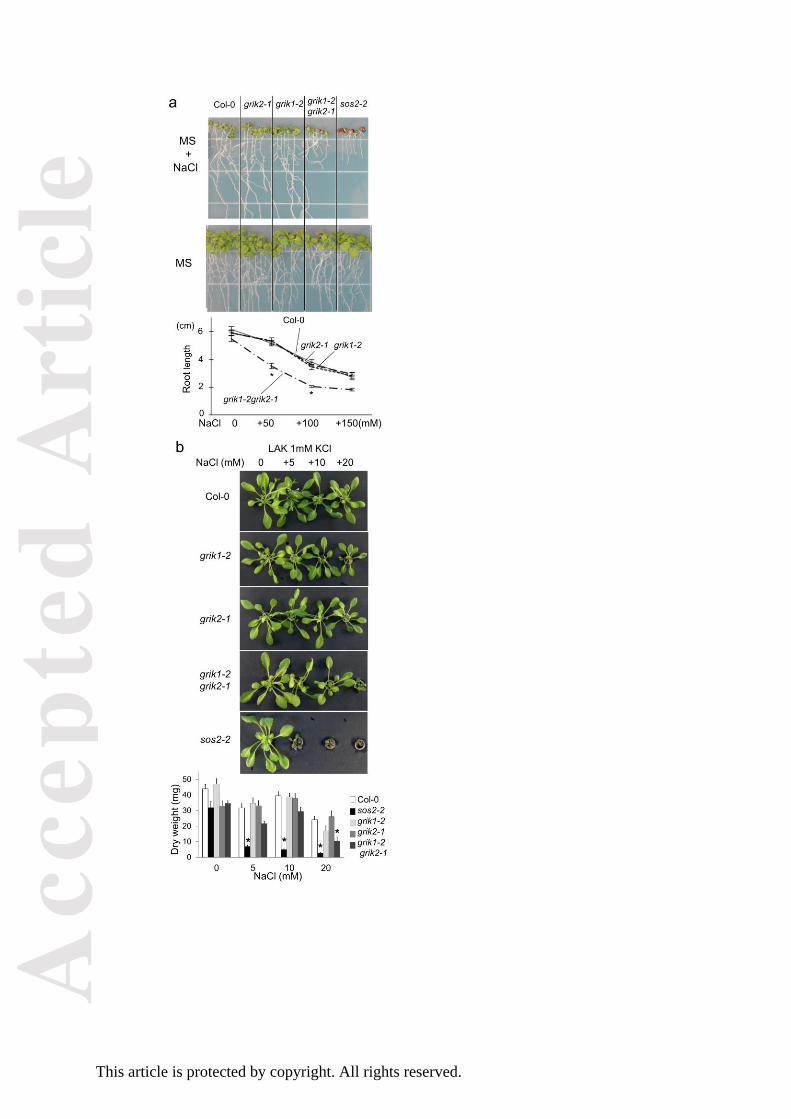
Next, we assessed the sensitivity of the double mutant to environmental stresses. When
similarly-sized 3–to-4-day-old seedlings were transferred to MS agar plates supplemented with 50
Acc
epte
d A
rtic
le
This article is protected by copyright. All rights reserved.
mM or 100 mM NaCl, the roots of the double mutant were shorter than those of the wild type,
grik1-2 or grik2-1 (Fig. 2a); on regular MS plates, the double mutant had slightly shorter roots. The
grik1-2 grik2-1 double mutant was not as sensitive to NaCl as the sos2-2 mutant, which could not
survive on 100 mM NaCl plates (Fig. 2a). On the other hand, when seedlings were transferred to MS
plates supplemented with 200 mM or 300 mM mannitol, we detected no significant differences in
root length among any of the strains tested, in keeping with the similar sensitivity of the double
mutant and wild-type plants to 3% sorbitol (165 mM; Supplemental Fig. S1d). These results indicate
that GRIK1 and GRIK2 are instrumental in mounting tolerance to the ionic component of salinity and
are likely to function in concert with the SOS pathway. Hence, we also evaluated the contribution of
GRIK1 and GRIK2 to the salt tolerance of Arabidopsis in hydroponic culture with LAK medium, which
maximises ionic-sensitive phenotypes due to the enhanced sodium uptake linked to
evapotranspiration (Barragan et al., 2012). The grik1-2 grik2-1 double mutant had a significant
salt-sensitive phenotype at 20 mM NaCl, but was still less sensitive than the sos2-2 mutant (Fig. 2b).
Together, these results indicate that GRIK1 and GRIK2 play redundant yet important roles in the
sodium tolerance of Arabidopsis.
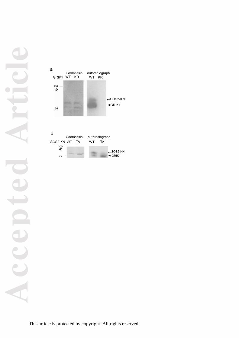
GRIK1 phosphorylates Thr168 in the activation-loop of SOS2.
SOS2, which belongs to the SnRK3 subfamily and plays an important role in salt tolerance, is
phylogenetically related to SnRK1s. Hence, we investigated whether GRIKs could also phosphorylate
SOS2. For these experiments, we produced glutathione S-transferase (GST)-fused GRIK1
recombinant proteins in E. coli. A kinase-dead type mutant protein of GRIK1 (GRIK1-KR), in which the
ATP-binding residue Lys137 was changed to Arg, was also produced as a negative control. In vitro
kinase assays revealed that GRIK1 had autophosphorylation activity, whereas GRIK1-KR did not (Fig.
3a). A kinase-dead type of SOS2, in which Lys40 was mutated to Gln (SOS2-KN; Gong et al. 2002b),
Acc
epte
d A
rtic
le
This article is protected by copyright. All rights reserved.
was phosphorylated by GRIK1, but not by GRIK1-KR (Fig. 3a, Supplemental Fig. S2). These results
indicate that GRIK1 phosphorylates SOS2 in vitro.
To identify the GRIK1 phosphorylation site in SOS2, we introduced mutations in the activation-loop
at the three candidate sites (Ser-156, Thr-168 and Tyr-175), because they are fully conserved in all
the members of the Arabidopsis SnRK3 subfamily and replacement of these residues with Asp to
mimic phosphorylation significantly increased the activity of SOS2 in vitro (Gong et al., 2002b,
Chaves-Sanjuan et al 2014). We generated SOS2 mutants, in which one or all three of the putative
phosphorylated residues were changed to Ala (single mutants SOS2-S156A, SOS2-T168A and
SOS2-Y175A; and triple mutant SOS2-AAA), and used them as GRIK1 substrates in an in vitro kinase
assay. These mutations of SOS2 were combined with the K40N mutation to abrogate
auto-phosphorylation. Phosphorylation of SOS2-T168A and SOS2-AAA by GRIK1 was not detected,
whereas SOS2-S156A and SOS2-Y175A were still phosphorylated by GRIK1 (Fig. 3b, Supplemental
Fig.S2). These results suggest that GRIK1 phosphorylates Thr168 in the activation-loop of SOS2 in
vitro.
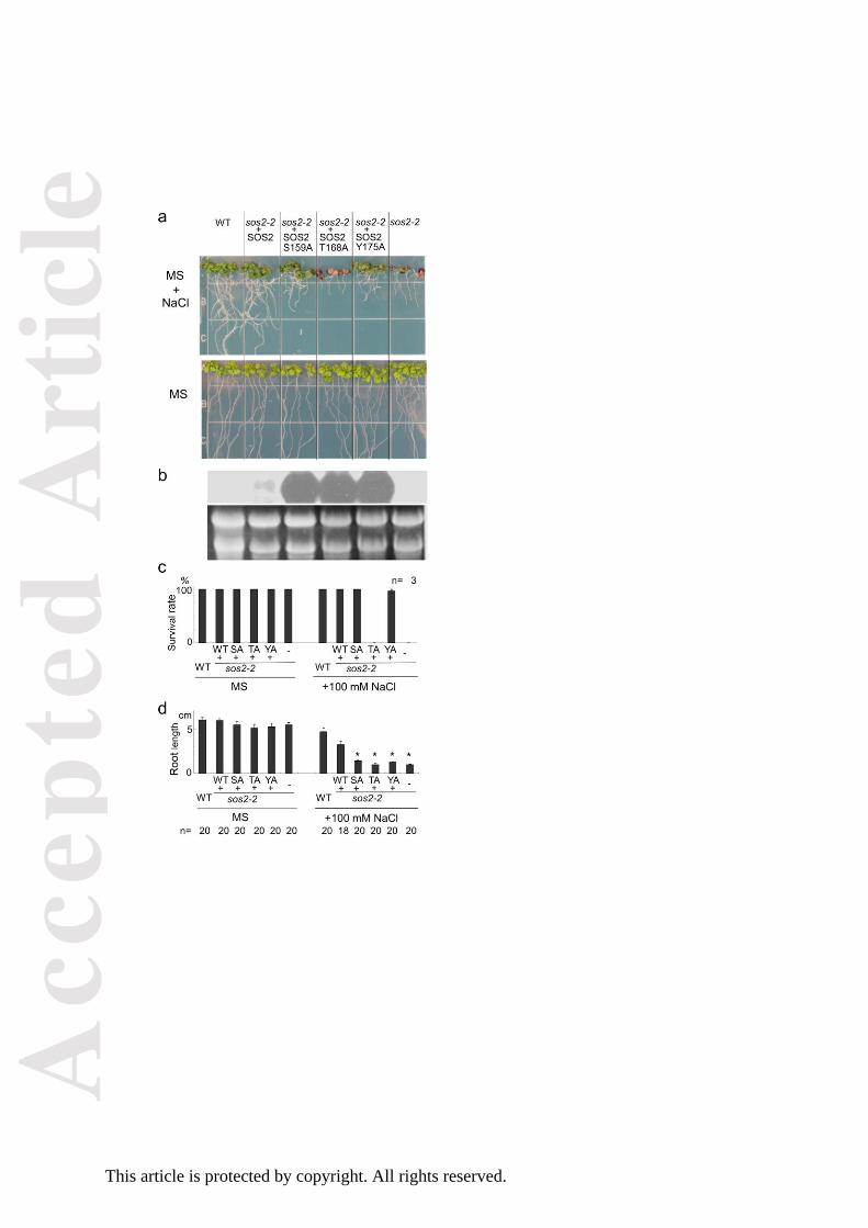
The T168A mutant of SOS2 cannot rescue the salt sensitive phenotype of sos2-2.
We next examined whether the phosphorylation site in the activation-loop is essential for the
activity of SOS2 under salt stress in vivo. For this purpose, phosphorylation site-mutated forms of
SOS2 were expressed in the sos2-2 background under the control of the 35S promoter of Cauliflower
Mosaic Virus (35S promoter). Transgenic plants expressing wild-type SOS2 in the sos2-2 background
could grow well under high-salt conditions (Fig. 4). Transgenic plants expressing SOS2-S156A or
SOS2-Y175A in the sos2-2 background could survive on 100 mM NaCl plates, albeit their roots were
shorter than those of the plants expressing wild-type SOS2. The lines presenting the best resistance
to the NaCl plates among more than 10 independent lines for each transgene were selected for
Acc
epte
d A
rtic
le
This article is protected by copyright. All rights reserved.
these experiments in Fig. 4. On the other hand, all the 12 lines tested of T2 transgenic plants
expressing SOS2-T168A in the sos2-2 background died on the 100 mM NaCl plates (Fig. 4). Northern
blot analysis revealed that transcripts of SOS2-S156A, SOS2-T168A and SOS2-Y175A in each
transgenic line were substantially more abundant than those of wild-type SOS2 in the transgenic line
expressing wild-type or endogenous SOS2 (Fig. 4b). The reason for this disproportionate
accumulation of SOS2-S156A, SOS2-T168A and SOS2-Y175A transcripts is unclear, but could be due
to the known upregulation of the SOS2 transcript by salt stress and that transgenic plants expressing
inactive SOS2 mutants would suffer from acute salinity stress compared to the wild-type and sos2-2
transgenic plants complemented by the wild-type SOS2 gene. In any case, transgenic lines expressing
the SOS2-S156A, SOS2-T168A and SOS2-Y175A transcripts at levels commensurate with the
wild-type SOS2 transgene showed similar complementation results (Supplemental Fig. S3),
demonstrating that differences in transgene expression were not the reason for root growth
retardation in plants expressing SOS2-S156A or SOS2-Y175A. Together, these results indicate that
Ser156, Thr168 and Tyr175 of SOS2 are involved in the function of SOS2 under salt stress and that
Thr168 in particular is essential for survival under salt stress.
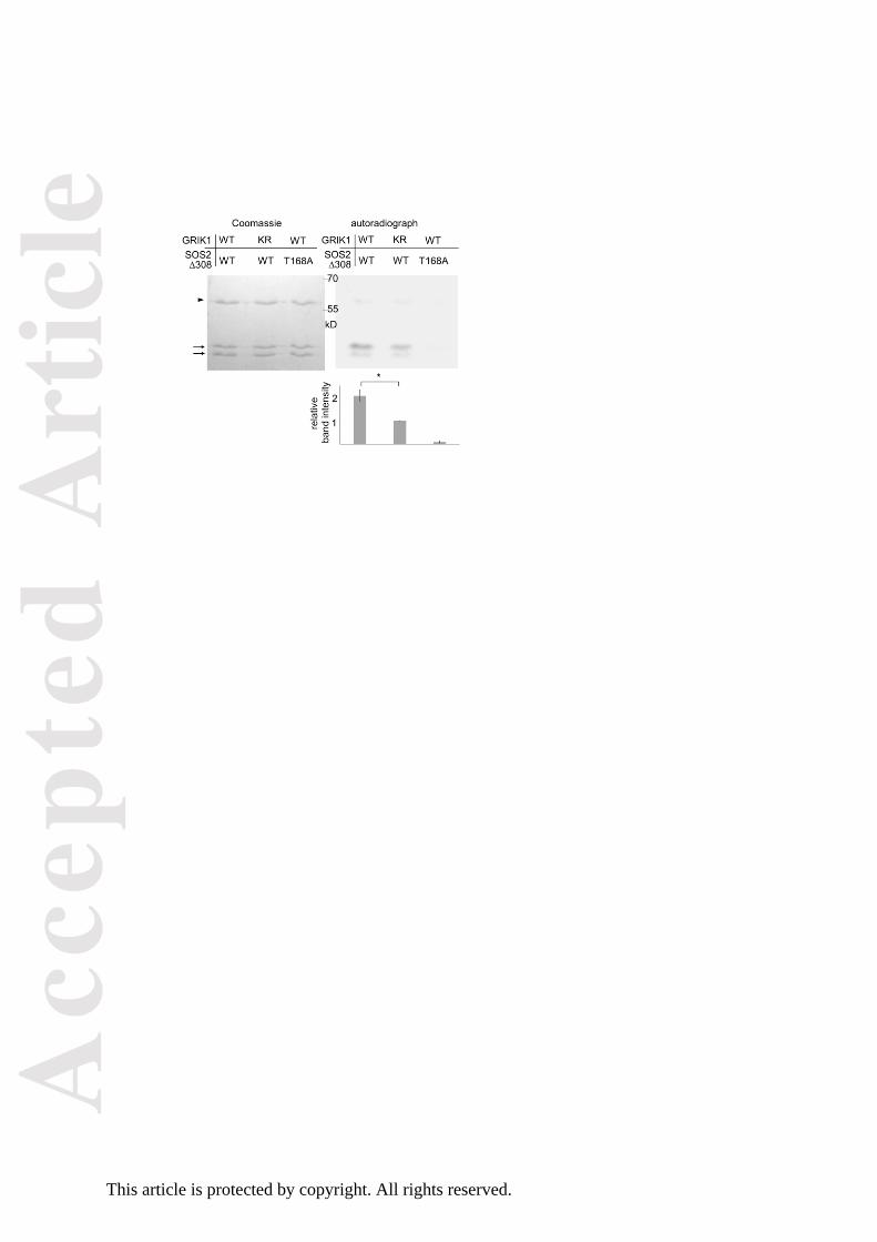
In vitro activation of SOS2 by GRIK1
Next, we asked whether phosphorylation by GRIK1 activates SOS2 in vitro. Deletion of the C-terminal
138 amino acids of SOS2 (SOS2-∆308) removes the autoinhibitory domain of SOS2, yielding a
SOS3-independent form of the kinase (Guo et al., 2001). The T168A-mutated form of SOS2-∆308
(SOS2-∆308/T168A) was used as a negative control. The GST-fused C-terminal 148 amino acids of
SOS1 (SOS1-CT, Fujii and Zhu, 2009) were used as a substrate for SOS2. SOS1-CT was phosphorylated
by SOS2-∆308 to a greater degree in the presence of GRIK1 than in the presence of GRIK1-KR (Fig. 5,
Supplemental Fig. S2). Little phosphorylation of SOS1 was observed when GRIK1 was combined with
Acc
epte
d A
rtic
le
This article is protected by copyright. All rights reserved.
the SOS2-∆308/T168A protein. These results indicate that phosphorylation of Thr168 by GRIK1 can
activate SOS2 in vitro.
To confirm the physical interaction of GRIK1 and SOS2 in planta, we conducted Bimolecular
Fluorescence Complementation (BiFC) experiments in N. benthamiana. The results demonstrated
that SOS2 and GRIK1 interact at least in the cytosol (Fig 6). BiFC fluorescence was visible in the
cytoplasmic rims around nuclei, in cytosol-filled transvacuolar strands, and in cytoplasmic pockets.
Although SOS2 shows a nucleo-cytoplasmic distribution when expressed alone (Kim et al., 2007), no
GRIK1-SOS2 complex was detected inside the nuclei.
GRIK1 activates SOS2 in yeast
The yeast SNF1 kinase is essential for carbon utilization, as evidenced by the inability of yeast to
grow in non-fermentable carbon sources if SNF1 is inactive. To test the ability of GRIKs to activate
SNF1, Arabidopsis GRIK1 was expressed in the yeast strain YPDahl55 (sak1Δ::KanMX elm1Δ::KanMX
tos3Δ::TRP1), which lacks all three upstream regulatory kinases of SNF1 (Ye et al., 2008). Restoration
of growth in media with the non-fermentable carbon sources glycerol and ethanol by the wild-type
GRIK1 but not by the dead-kinase mutant bearing the mutation K137R demonstrated that GRIK1 was
fully active in the yeast cell (Supplemental Fig. S4).
The SOS pathway, comprising the Na/H exchanger SOS1, SOS2 and SOS3, can be reconstituted in
yeast strains that lack all major sodium efflux transporters and are exceedingly sensitive to sodic
stress (Quintero et al., 2002, 2011). We used this system to analyse the in vivo activation of SOS2 by
GRIK1. For this study, we used strain YP890, a derivative of AXT3K (∆ena1::HIS3::ena4, nha1::LEU2,
nhx1::KanMX) (Quintero et al., 2002) in which a PGK1prom:SOS1:CYC1ter expression cassette was
inserted chromosomally to provide moderate and constitutive expression of the SOS1 protein (Guo
et al., 2004). This strain expresses the endogenous SNF1-upstream kinases SAK1/PAK1, ELM1 and
Acc
epte
d A
rtic
le
This article is protected by copyright. All rights reserved.
TOS3, which are the fungal homologues of GRIKs (Shen et al., 2006). For reconstitution of the
GRIK1/SOS2/SOS1 phosphorylation cascade, we first tested the non-phosphorylatable mutant
SOS2-AAA in strain YP890. SOS2-AAA was unable to activate SOS1 in this system, irrespective of the
removal of the autoinhibitory domain of SOS2 (SOS2-AAAΔ308, Fig. 7a) or the co-expression of SOS3
(Fig. 7b). These results indicate that, in contrast to wild-type SOS2, the SOS2-AAA mutant cannot be
activated by endogenous upstream kinases.
Next, we generated strain Δ3K4E, in which the genes encoding the three fungal upstream kinases
SAK1/PAK1, ELM1 and TOS3, and the ENA1-4 sodium pumps that are the major determinants of Na+
tolerance in S. cerevisiae, had been deleted. Transformation of Δ3K4E cells with the core
components of the Arabidopsis SOS pathway (SOS1, SOS2 and SOS3) failed to complement the
salt-sensitive phenotype (Fig. 7c). Expression of GRIK1 in Δ3K4E cells improved growth in
glucose-supplemented AP medium due to the complementation of sak1 elm1 tos3 mutations. At low
NaCl concentrations (100 mM NaCl), the residual salt tolerance imparted by the full complement of
SOS proteins was identical to that conferred by SOS1 alone, indicating that the SOS2-SOS3 regulatory
module was essentially inactive in Δ3K4E (Fig. 7c). However, co-expression of GRIK1 increased the
capacity of the SOS proteins to confer salt-tolerance to Δ3K4E cells. These results demonstrate that
GRIK1 can activate SOS2 in yeast.
Discussion
In this study, we successfully produced a grik1-2 grik2-1 double mutant with near wild-type
vegetative growth. Previous data had indicated that the grik1-1 grik2-1 double mutant was lethal
before germination (Bolle et al., 2013) or needed supplementation of sugar to grow beyond the
cotyledon-stage (Glab et al., 2017). Whereas the grik2-1 mutant line used here (Salk_015230) is
identical to that of previous reports, the mutant lines harbouring T-DNA insertions in GRIK1 were
Acc
epte
d A
rtic
le
This article is protected by copyright. All rights reserved.
different. Previous studies used the GABI line 713C09 with the T-DNA insertion in the 8th intron, and
we have used line Salk_142938 bearing the T-DNA insertion at the 1st intron (Fig. 1a). Northern
blotting and RT-PCR data (Fig. 1d,e) suggest that in grik1-2 the T-DNA insertion disrupts the
full-length GRIK1 transcript but allows the expression of a mRNA encoding a truncated form of GRIK1
starting at methionine 156. Our results showing that phosphorylation of SnRK1s at the
activation-loop was reduced but not eliminated in the grik1-2 grik2-1 double mutant (Fig. 1f) suggest
that the putative protein encoded by the grik1-2 allele retains some level of activity. The remaining
phosphorylation of SnRK1s may be also sufficient to maintain SnRK1-mediated pathways, as the
grik1-2 grik2-1 double mutant did not exhibit any defects under dark or submergence conditions
(Supplemental Fig. S1a and b), in which SnRK1s play important roles (Baena-González et al., 2007,
Cho et al., 2016). On the other hand, the grik1-2 grik2-1 double mutant was sensitive to high glucose
(Supplemental Fig. S1c) as previously shown for the grik1-1 grik2-1 double mutant (Glab et al.,
2017), indicating that the truncated protein encoded by the grik1-2 allele cannot fully replace the
intact GRIK1. Lack of GRIKs might have multiple effects on SnRK1-mediated pathways, including
decrease in the amount of SnRK1.1 in the grik1-2 grik2-1 double mutant (Fig. 1f). In the grik1-1
grik2-1 double mutant, the amount of SnRK1.1 was similar to that of the wild type (Glab et al., 2017).
Since SnRK1.1 degradation is strictly dependent on its activity and inactive SnRK1.1 variants are
disproportionally stable (Crozet et al., 2016), it is likely that the synthesis of SnRK1.1 is somehow
compromised in both double mutants compared to the wild type, whereas higher degradation of
SnRK1.1 would occur in grik1-2 grik2-1 compared to the grik1-1 grik2-1 mutant due to the residual
activity of SnRK1.1 in the grik1-2 background.
Acc
epte
d A
rtic
le
This article is protected by copyright. All rights reserved.
The grik1-2 grik2-1 double mutant was sensitive to high NaCl (Fig. 2). The effects on salt sensitivity
indicated that GRIKs play important roles in several signalling pathways and suggested that they
might have substrates other than SnRK1s. Shen et al. (2009) concluded that GRIK1 and GRIK2 do not
phosphorylate SOS2. However, their in-vitro kinase assay detected bands possibly corresponding to
phosphorylated SOS2, but none corresponding to phosphorylated SnRK2.4. In our experiments,
phosphorylation of SOS2 by GRIK1 was strong enough to be detectable (Fig. 3a). Based on the
observation that phosphorylation intensity of the mutated form of SOS2 (T168A, T169A) was similar
to that of the wild-type SOS2, Shen et al. also concluded that GRIK1 and GRIK2 could not
phosphorylate the activation-loop of SOS2. We observed that the phosphorylated SOS2 band
disappeared in SOS2-T168A and SOS2-AAA (Fig. 3b, Supplemental Fig. S2c), indicating that GRIK1
indeed phosphorylates SOS2 at T168. The reason for the discrepancy between our results and those
of Shen et al. remains unclear. Under their conditions, SOS2 (T168A, T169A) may have been
phosphorylated on other sites to a sufficient degree to mask the reduction of phosphorylation on
T168/T169; alternatively, the difference may be due to unintended effects of the fused tags. The
T168 residue of SOS2 is conserved among SnRK3s (Gong et al., 2002b), whereas this residue is a Ser
in SnRK2s (with the exception of SnRK2.8). This suggests that SnRK3s, but not SnRK2s, may be
targeted by GRIKs. In our hands, GRIK1 phosphorylated and activated SOS2-∆308 in vitro (Fig. 4).
GRIK1 and SOS2 physically interact in N. benthamiana (Fig. 6). In addition, GRIK1 could activate the
SOS pathway in a yeast-reconstituted system (Fig. 7). Taken together, our results indicate that GRIK1
has the ability to phosphorylate the activation-loop of SOS2 and to activate this kinase, suggesting
that the roles of GRIKs are not limited to upstream regulation of SnRK1s.
Our data show that T168 in the activation-loop of SOS2 is important for function of SOS2 under salt
stress in vivo (Fig. 5). Mutations at S159 and Y175 also affected salt tolerance, as reflected by root
growth, although these residues were not essential for plant survival under acute salt stress (Fig. 5).
Acc
epte
d A
rtic
le
This article is protected by copyright. All rights reserved.
These two sites might be important for the local molecular conformation of SOS2, thereby exerting
an effect on phosphorylation on T168, or for another unknown mechanism involving the
activation-loop such as phosphorylation by additional kinases contributing to the full activation of
SOS2 under salinity. In any case, our data demonstrate that phosphorylation of T168 by GRIK1 is
important for SOS2 function in vitro and in the yeast system. The grik1-2 grik2-1 double mutant,
however, was less sensitive to salt than the sos2-2 mutant. If GRIKs are the upstream kinases of
SOS2 in vivo, then either the putative protein encoded by the grik1-2 allele works to some extent or
there exist parallel mechanisms responsible for salt resistance, e.g. another upstream kinase(s)
contributing to the full activation of SOS2, perhaps by phosphorylating residues S159 and/or Y175.
SOS2 is also activated by SOS3 and SCaBP8 in a Ca2+-dependent manner (Halfter et al., 2000, Quan et
al., 2007). GRIK-mediated phosphorylation may affect the competence of SOS2 for Ca2+-dependent
activation. In that case, when salt stress induces Ca2+ signalling, SOS3 or SCaBP8 may activate SOS2
depending upon the sugar/energy information gating by GRIKs (Fig.8).
In summary, GRIKs can phosphorylate and activate members of the SnRK3 family in addition to their
well-known targets SnRK1s, and they play important roles not only in the sugar signalling pathway
but also in the SOS pathway for salt tolerance in vivo.
Experimental procedures
Construction and site directed mutagenesis
Total RNA prepared from 10 days seedling of Arabidopsis thaliana Columbia-0 ecotype was primed
with oligo(dT) and reverse-transcribed with the Superscript II RT (Invitrogen). GRIK1 cDNA was
amplified by polymerase chain reaction (PCR) with KOD polymerase (Takara) under the following
conditions: 1 x 95 °C for 5 min, 30x (95 °C for 20 s, 55 °C 20 s, 70 °C 2 min). Primers used to obtain a
Acc
epte
d A
rtic
le
This article is protected by copyright. All rights reserved.
1207 bp fragment encoding the GRIK1 are in Supplemental Table 1. The PCR product was subcloned
into pGEX4T1 vector between EcoRI and XhoI sites. Inserted fragments of all constructs were
sequenced.
GRIK1 cDNA was inserted into the vector p425GPD (Mumberg et al., 1995) as BamHI/EcoRI for
expression in yeast. Plasmids for the expression in yeast of core components of the SOS pathway
have been described elsewhere (Quintero et al., 2002; 2011).
To introduce the point mutations into SOS2-pGEX4T1 (Guo et al., 2001), primer pairs in
Supplemental Table 1 were used for the first PCR: 1 x 95 °C for 5 min, 20x (95 °C for 20 s, 51 °C 20 s,
70 °C 90 s). Using the PCR products as templates, second amplification was performed under the
following conditions; 1 x 95 °C for 5 min, 30x (95 °C for 20 s, 51 °C 20 s, 70 °C 90 s). The final product
was cloned into pGEX4T1 (BamHI-EcoRI sites for SOS2). For expression in A. thaliana, fragments
were cloned into a binary vector (pCAMBIA1200) under the control of the CaMV 35S promoter
derived from pRT105. For expression in yeast, the full length mutant allele SOS2-AAA and the
truncated version SOS2-AAA∆308 were subcloned as BamHI/EcoRI fragments in p414GPD (Mumberg
et al., 1995).
Arabidopsis T-DNA insertion lines
The seeds of T-DNA insertion lines (Salk_142938 and Salk_015230) were obtained from Arabidopsis
Biological Resource Center (Alonso et al., 2003). Homozygous insertion lines were identified with
PCR following the instructions (http://signal.salk.edu/cgi-bin/tdnaexpress). The following conditions
were used: 1 x 95 °C for 5 min, 35x (95 °C for 20 s, 55 °C 20 s, 70 °C 1 min) with primers described in
Supplemental Table 1. The insertion sites were identified by sequencing the amplicons.
cDNAs purified from 10-day-old seedlings of Col-0 plants and T-DNA insertion lines as mentioned
above were used as templates for RT-PCR. The following conditions were used: 1 x 95 °C for 5 min,
Acc
epte
d A
rtic
le
This article is protected by copyright. All rights reserved.
35x (95 °C for 20 s, 58 °C 20 s for GRIK1 and GRIK2 or 54 °C 20 s for tubulin, respectively, 70 °C 45 s)
with primers in Supplemental Table 1.
Northern blot analysis was performed as described in Fujii and Zhu (2009). For GRIK1, the cDNA
fragment digested with PstI (248-889 of ORF) was used as a probe. The mRNA of grik1-2 was
amplified by RT-PCR with primers given in Supplemental Table 1, followed by sequencing.
Hydroponic culture with LAK medium
For salt treatment in hydroponic culture, seeds were directly grown on hydroponics as described by
Barragan et al. (2012). A modified Long Ashton mineral solution with 1 mM K+ and nominally free of
Na+ and NH4+ (LAK medium) was used for hydroponic cultures. This medium was designed to
maximize the toxicity of Na+ ions, while minimizing the osmotic effects of supplemental NaCl. The
final composition of the LAK base solution was as follows: 1 mM KH2PO4, 2 mM Ca(NO3)2, 1 mM
MgSO4, 30 mM H3BO3, 10 mM MnSO4, 1 mM ZnSO4, 1 mM CuSO4, 0.03 mM (NH4)6Mo7O24, and 100
mM Fe2+ as Sequestrene 138- Fe, pH 5.3. Seedlings were grown in LAK medium for one week
followed by salt treatment at the indicated NaCl concentrations.
Expression and Purification of GST Fusion Proteins in E. coli
The constructs encoding GST-fused proteins were transformed into E. coli Rosetta cells (Novagen).
Single colonies were grown overnight at 37ºC, transferred to fresh 20x volume of Luria-Bertani
media, and further cultured for 1 hour. Recombinant protein expression was induced by 0.2 mM
isopropyl beta-D-thiogalactopyranoside for 4h at 37ºC. The cells were harvested by centrifugation
(5,000 x g, 5 min, 4ºC), and the pellets were resuspended in pre-chilled lysis buffer (10 mM Tris
pH8.0, 150 mM NaCl, 1 mM EDTA and 100 μg/ml lysozyme), incubated on ice for 15 min. After
Acc
epte
d A
rtic
le
This article is protected by copyright. All rights reserved.
dithiothreitol (50 mM), phenylmethanesulfonyl fluoride (1 mM) and Triton X-100 (1.5%) were added,
the suspension was centrifuged at 30,000 x g for 5 min at 4ºC. Then, glutathione-agarose beads
(Sigma) were added to the supernatant and the mixture were incubated with gentle agitation for at
least 1 hour at 4ºC. The beads were washed six times with pre-chilled buffer (10 mM Tris pH8.0, 150
mM NaCl, 1 mM EDTA).
Western Blotting
Two-week-old Col-0 and grik1-2 grik2-1 seedlings, grown on MS media with 1% sucrose, were
collected and ground in liquid nitrogen. The tissue samples were added to Laemmli sample buffer
(Laemmli, 1970) and heat-treated (10 min at 65 °C). Solid material was removed by centrifugation,
and samples were run in SDS-polyacrylamide gel electrophoresis followed by western blot. The blots
were incubated with either anti-phospho-T172-AMPK-α antibody (Cell Signaling, MS, USA) or
anti-SnRK1.1 antibody (Agrisera, Sweden) over-night at 4 °C with slow agitation. All blots were then
incubated with HRP-conjugated anti-rabbit antibody (GE Healthcare, UK) for 2 h at room
temperature with slow agitation. All antibody dilutions were made in TTBS-buffer (20 mM Tris-HCl
pH 7.5, 150 mM NaCl, and 0.05% Tween-20). Signals from the blots were detected using
Westernbright ECL (Advansta, CA, USA), and band strengths were evaluated with LiCor Image Studio
program (LiCor, UK). Experiments were replicated with five biological samples.
In vitro Kinase Assays
In vitro phosphorylation assays were performed as described previously (Fujii and Zhu, 2009) with
some modification. Twenty microliters of the reaction mixture contained 20 mM Tris (pH 7.2), 10
mM or 40 mM MgCl2, 10 μM ATP, 5 μCi [γ-32P] ATP and 2 mM dithiothreitol. Reaction mixtures were
Acc
epte
d A
rtic
le
This article is protected by copyright. All rights reserved.
incubated at 30°C for 40 min. The reaction was stopped by the addition of Laemmli’s sample buffer,
followed by SDS-polyacrylamide gel electrophoresis.
Yeast strains and media.
Yeast strain YP890 (∆ena1::HIS3::ena4, nha1::LEU2, nhx1::KanMX, PGK1prom::AtSOS1::CYC1ter) (Guo et
al., 2004) was used to test the function of wild-type and mutant SOS2. The yeast strain YPDahl55
(sak1Δ::KanMX elm1Δ::KanMX tos3Δ::TRP1) lacking all three upstream regulatory kinases of SNF1
(Ye et al., 2008) was used as the starting biological material to produce a strain suitable to test
activation of SOS2 by GRIK1. The ENA1-ENA4 gene tandem array encoding Na+-ATPases was
disrupted by transformation with an ena1::hisG::URA3::hisG::ena4 gene replacement cassette,
followed by selection of uracil prototrophs and sodium-sensitive transformants. Gene replacement
was confirmed by diagnostic PCR. Next, a loss-of-function mutant of the TRP1 gene marker in
tos3Δ::TRP1 was isolated by counter-selection with 5-fluoroanthranilic acid (Toyn et al., 2000). The
resulting strain was denoted Δ3K4E. Transformation of S. cerevisiae was performed using a standard
lithium acetate–polyethylene glycol method. Yeast cells were propagated in rich YPD medium (1%
yeast extract, 2% peptone, 2% glucose). To test growth in non-fermentable carbon sources, glucose
was substituted by 3% ethanol 2% glycerol. The ability of yeast cells to grow in salt was tested on AP
medium (Rodriguez-Navarro and Ramos 1984). Strains were cultured overnight in liquid AP medium
supplemented with 1 mM KCl. After harvest, cells were resuspended and diluted decimally in
distilled water. Five-μL aliquots were spotted onto AP plates supplemented with 1 mM KCl and
various concentrations of NaCl, and grown for 3 to 4-d at 28°C.
Acc
epte
d A
rtic
le
This article is protected by copyright. All rights reserved.
BiFC experiments in Nicotiana benthamiana.
For BiFC assays, the full-length cDNA of GRIK1 was transferred to the pSPYCE(M) vector (Waadt et al
2008) using the XbaI and SmaI sites to create a C-terminal translational fusion of GRIK1 to the
C-terminal moiety of YFP. The N-terminal fusion of SOS2 to the N-terminal moiety of YFP in
pSPYNE(R)173 has been described before (Waadt et al 2008). All the YFP fusions were expressed
under the control of the 35S promoter. The resulting plasmids were electroporated into
Agrobacterium tumefaciens (strain GV3101). The bacterial cultures were grown at 28ºC overnight
and centrifuged at 15000 x g for 10 min. Pellets were resuspended with infiltration buffer (10 mM
MES pH 5.6, 10 mM MgCl2, 0.1 mM acetosyringone) and kept at RT for 5 h. Cell cultures were
adjusted to a final OD600nm of 0.2. Appropriate combinations of cultures were mixed with equal
amounts of an Agrobacterium suspension carrying the p19 suppressor of post-transcriptional gene
silencing (Silhavy et al., 2002). The Agrobacterium suspensions were then infiltrated into the leaves
of 3- to 4-week-old N. benthamiana plants as described (Marillonnet et al., 2005). The infiltrated
plants were kept in a controlled growth chamber (16 h day/8 h night, 25/22ºC, 60-70% relative
humidity, 150 μmol m-2 s-1 PAR) for 3 days until analysis by confocal microscopy. Images were taken
with a FluoView FV1000 Confocal Microscope (Olympus) using a 488-nm Ar/ArKr laser and 60x
objective. The Olympus FluoView 4.2 software was used to analyse the images.
Statistics
Student T-test and one-way ANOVA followed by Tukey's multiple comparison test was performed for
single and multiple comparison, respectively. For normalized values, non-parametric binomial test
was adopted with 0.5 as a priori probability of >1.
Acc
epte
d A
rtic
le
This article is protected by copyright. All rights reserved.
Accession numbers
GRIK1: AT3G45240, GRIK2: AT5G60550, SOS2: AT5G35410, SOS1: AT2G01980, SOS3: AT5G24270,
grik1-2: Salk_142938, grik2-1: Salk_015230
Acknowledgement
We thank the Arabidopsis Biological Resource Center for providing the T-DNA insertion mutants. This
work was supported by the Turku Collegium for Science and Medicine and by the Academy of
Finland (Projects number 259169, 263853, 271832, 292763, 307335) to HF, by National Institutes of
Health (Grant R01GM059138) to JZ and by grants BFU2015-64671 to JMP and BIO2015-70946-R to
FJQ from the Spanish Ministry of Economy and Competitiveness, co-financed by FEDER, and with
additional support from the SSAC grant PJ01105105 from the Rural Development Administration,
Republic of Korea.
Conflict of interest
The authors declare that there is no conflict of interest.
Short Supporting Information Legends
Supplemental Figure 1. Other phenotypes of grik1-2 grik2-1 lines.
Supplemental Figure 2. Control experiments for the in vitro kinase assays.
Supplemental Figure 3. Transgenic lines with similar expression levels of SOS2-S159A (SA), T168A
(TA), and Y175A (YA) in the sos2-2.
Acc
epte
d A
rtic
le
This article is protected by copyright. All rights reserved.
Supplemental Figure 4. The complementation of the SNF1 upstream kinase by GRIK regarding
carbon source use.
Supplemental Table 1. Primer sequences.
References
Alonso, J.M., Stepanova, A.N., Leisse, T.J., Kim, C. J., Chen, H., Shinn, P., Stevenson, D. K.,
Zimmerman, J., Barajas, P., Cheuk, R., Gadrinab, C., Heller, C., Jeske, A., Koesema, E., Meyers,
C. C., Parker, H., Prednis, L., Ansari, Y., Choy, N., Deen, H., Geralt, M., Hazari, N., Hom, E.,
Karnes, M., Mulholland, C., Ndubaku, R., Schmidt, I., Guzman, P., Aguilar-Henonin, L.,
Schmid, M., Weigel, D., Carter, D. E., Marchand, T., Risseeuw, E., Brogden, D., Zeko, A.,
Crosby, W. L., Berry, C. C. and Ecker, J. R. (2003) Genome-wide insertional mutagenesis of
Arabidopsis thaliana. Science, 301, 653–7.
Baena-González, E., Rolland, F., Thevelein, J.M. and Sheen, J. (2007) A central integrator of
transcription networks in plant stress and energy signalling. Nature, 448, 938–42.
Barragan, V., Leidi, E.O., Andres, Z., Rubio, L., Luca, A. De, Fernandez, J.A., Cubero, B. and Pardo,
J.M. (2012) Ion Exchangers NHX1 and NHX2 Mediate Active Potassium Uptake into Vacuoles to
Regulate Cell Turgor and Stomatal Function in Arabidopsis. Plant Cell, 24, 1127–1142.
Bolle, C., Huep, G., Kleinbölting, N., Haberer, G., Mayer, K., Leister, D. and Weisshaar, B. (2013)
GABI-DUPLO: a collection of double mutants to overcome genetic redundancy in Arabidopsis
thaliana. Plant J., 75, 157–171.
Acc
epte
d A
rtic
le
This article is protected by copyright. All rights reserved.
Carvalho, R.F., Szakonyi, D., Simpson, C.G., Barbosa, I.C.R., Brown, J.W.S., Baena-González, E. and
Duque, P. (2016) The Arabidopsis SR45 Splicing Factor, a Negative Regulator of Sugar Signaling,
Modulates SNF1-Related Protein Kinase 1 Stability. Plant Cell, 28, 1910–1925.
Chaves-Sanjuan, A., Sanchez-Barrena, M.J., Gonzalez-Rubio, J.M., Moreno, M., Ragel, P., Jimenez,
M., Pardo, J. M., Martinez-Ripoll, M., Quintero, F. J. and Albert, A. (2014) Structural basis of
the regulatory mechanism of the plant CIPK family of protein kinases controlling ion
homeostasis and abiotic stress. Proc. Natl. Acad. Sci. U. S. A., 111, E4532-41.
Cheng, N.H., Pittman, J.K., Zhu, J.K. and Hirschi, K.D. (2004) The Protein Kinase SOS2 Activates the
Arabidopsis H+/Ca2+ Antiporter CAX1 to Integrate Calcium Transport and Salt Tolerance. J. Biol.
Chem., 279, 2922–2926.
Cho, H.-Y., Wen, T.-N., Wang, Y.-T. and Shih, M.-C. (2016) Quantitative phosphoproteomics of
protein kinase SnRK1 regulated protein phosphorylation in Arabidopsis under submergence. J.
Exp. Bot., 67, 2745–2760.
Crozet, P., Jammes, F., Valot, B., Ambard-Bretteville, F., Nessler, S., Hodges, M., Vidal, J. and
Thomas, M. (2010) Cross-phosphorylation between Arabidopsis thaliana Sucrose
Nonfermenting 1-related Protein Kinase 1 (AtSnRK1) and Its Activating Kinase (AtSnAK)
Determines Their Catalytic Activities. J. Biol. Chem., 285, 12071–12077.
Crozet, P., Margalha, L., Butowt, R., Fernandes, N., Elias, C. A., Orosa, B., Tomanov, K., Teige, M.,
Bachmair, A., Sadanandom, A. and Baena-González, E. (2016) SUMOylation represses SnRK1
signaling in Arabidopsis. Plant J., 85, 120–133.
Cutler, S.R., Rodriguez, P.L., Finkelstein, R.R. and Abrams, S.R. (2010) Abscisic Acid: Emergence of a
Core Signaling Network. Annu. Rev. Plant Biol., 61, 651–679.
Acc
epte
d A
rtic
le
This article is protected by copyright. All rights reserved.
Fujii, H. and Zhu, J.-K. (2009) An autophosphorylation site of the protein kinase SOS2 is important
for salt tolerance in Arabidopsis. Mol. Plant, 2, 183–90.
Glab, N., Oury, C., Guérinier, T., Domenichini, S., Crozet, P., Thomas, M., Vidal, J., Hodges, M.
(2017) The impact of Arabidopsis thaliana SNF1-Related-Kinase1 (SnRK1)-Activating Kinase
(SnAK) 1 and 2 on SnRK1 phosphorylation status: Characterisation of a SnAK double mutant.
Plant J., 89, 1031-41..
Gong, D., Gong, Z., Guo, Y., Chen, X. and Zhu, J.-K. (2002a) Biochemical and functional
characterization of PKS11, a novel Arabidopsis protein kinase. J. Biol. Chem., 277, 28340–50.
Gong, D., Guo, Y., Jagendorf, A.T. and Zhu, J.-K. (2002b) Biochemical characterization of the
Arabidopsis protein kinase SOS2 that functions in salt tolerance. Plant Physiol., 130, 256–264.
Gong, D., Zhang, C., Chen, X., Gong, Z. and Zhu, J.-K. (2002c) Constitutive Activation and Transgenic
Evaluation of the Function of an Arabidopsis PKS Protein Kinase. J. Biol. Chem., 277,
42088–42096.
Guo, Y., Halfter, U., Ishitani, M. and Zhu, J.K. (2001) Molecular characterization of functional
domains in the protein kinase SOS2 that is required for plant salt tolerance. Plant Cell, 13,
1383–400.
Guo, Y., Qiu, Q.-S., Quintero, F.J., Pardo, J.M., Ohta, M., Zhang, C., Schumaker, K.S. and Zhu, J.-K.
(2004) Transgenic evaluation of activated mutant alleles of SOS2 reveals a critical requirement
for its kinase activity and C-terminal regulatory domain for salt tolerance in Arabidopsis
thaliana. Plant Cell, 16, 435–49.
Acc
epte
d A
rtic
le
This article is protected by copyright. All rights reserved.
Halfter, U. (2000) The Arabidopsis SOS2 protein kinase physically interacts with and is activated by
the calcium-binding protein SOS3. Proc. Natl. Acad. Sci., 97, 3735–3740.
Hanks, S.K. and Hunter, T. (1995) Protein kinases 6. The eukaryotic protein kinase superfamily:
kinase (catalytic) domain structure and classification. FASEB J., 9, 576–96.
Hardie, D.G. (2004) The AMP-activated protein kinase pathway - new players upstream and
downstream. J. Cell Sci., 117, 5479–5487.
Hawley, S.A., Pan, D.A., Mustard, K.J., Ross, L., Bain, J., Edelman, A.M., Frenguelli, B.G. and Hardie,
D.G. (2005) Calmodulin-dependent protein kinase kinase-β is an alternative upstream kinase
for AMP-activated protein kinase. Cell Metab., 2, 9–19.
Hey, S., Mayerhofer, H., Halford, N.G. and Dickinson, J.R. (2007) DNA sequences from Arabidopsis,
which encode protein kinases and function as upstream regulators of Snf1 in yeast. J. Biol.
Chem., 282, 10472–9.
Hong, S.-P., Leiper, F.C., Woods, A., Carling, D. and Carlson, M. (2003) Activation of yeast Snf1 and
mammalian AMP-activated protein kinase by upstream kinases. Proc. Natl. Acad. Sci., 100,
8839–8843.
Hrabak, E.M., Chan, C.W.M., Gribskov, M., Harper, J. F., Choi, J. H., Halford, N., Luan, S., Nimmo, H.
G., Sussman, M. R., Thomas, M., Walker-simmons, K., Zhu, J., Harmon, A. C., Kudla, J., Luan,
S., Nimmo, H. G., Sussman, M. R., Thomas, M., Walker-simmons, K., Zhu, J. and Harmon, A. C.
(2003) The Arabidopsis CDPK-SnRK Superfamily of Protein Kinases. Plant Physiol., 132,
666–680.
Acc
epte
d A
rtic
le
This article is protected by copyright. All rights reserved.
Kemp, B.E., Stapleton, D., Campbell, D.J., Chen, Z.-P., Murthy, S., Walter, M., Gupta, A., Adams, J.
J., Katsis, F., van Denderen, B., Jennings, I. G., Iseli, T., Michell, B. J. and Witters, L. A. (2003)
AMP-activated protein kinase, super metabolic regulator. Biochem. Soc. Trans., 31, 162–8.
Kim, K.N., Cheong, Y.H., Gupta, R. and Luan, S. (2000) Interaction specificity of Arabidopsis
calcineurin B-like calcium sensors and their target kinases. Plant Physiol., 124, 1844–53.
Kim, B.-G., Waadt, R., Cheong, Y.H., Pandey, G.K., Dominguez-Solis, J.R., Schültke, S., Lee, S.C.,
Kudla, J. and Luan, S. (2007) The calcium sensor CBL10 mediates salt tolerance by regulating
ion homeostasis in Arabidopsis. Plant J., 52, 473–84.
Kong, L.-J. and Hanley-Bowdoin, L. (2002) A geminivirus replication protein interacts with a protein
kinase and a motor protein that display different expression patterns during plant development
and infection. Plant Cell, 14, 1817–32.
Laemmli, U.K. (1970) Cleavage of structural proteins during the assembly of the head of
bacteriophage T4. Nature, 227, 680–5.
Liu, J., Ishitani, M., Halfter, U., Kim, C.S. and Zhu, J.K. (2000) The Arabidopsis thaliana SOS2 gene
encodes a protein kinase that is required for salt tolerance. Proc Natl Acad Sci U S A, 97,
3730–3734.
Luan, S. (2009) The CBL-CIPK network in plant calcium signaling. Trends Plant Sci., 14, 37–42.
Marillonnet, S., Thoeringer, C., Kandzia, R., Klimyuk, V. and Gleba, Y. (2005) Systemic
Agrobacterium tumefaciens-mediated transfection of viral replicons for efficient transient
expression in plants. Nat. Biotechnol., 23, 718–23.
Mumberg, D., Müller, R. and Funk, M. (1995) Yeast vectors for the controlled expression of
heterologous proteins in different genetic backgrounds. Gene 156: 119-22.
Acc
epte
d A
rtic
le
This article is protected by copyright. All rights reserved.
Qiu, Q.S., Guo, Y., Quintero, F.J., Pardo, J.M., Schumaker, K.S. and Zhu, J.K. (2004) Regulation of
Vacuolar Na+/H+ Exchange in Arabidopsis thaliana by the Salt-Overly-Sensitive (SOS) Pathway. J.
Biol. Chem., 279, 207–215.
Quan, R., Lin, H., Mendoza, I., Zhang, Y., Cao, W., Yang, Y., Shang, M., Chen, S., Pardo, J. M. and
Guo, Y. (2007) SCABP8/CBL10, a Putative Calcium Sensor, Interacts with the Protein Kinase
SOS2 to Protect Arabidopsis Shoots from Salt Stress. Plant Cell, 19, 1415–1431.
Quintero, F.J., Ohta, M., Shi, H., Zhu, J.-K. and Pardo, J.M. (2002) Reconstitution in yeast of the
Arabidopsis SOS signaling pathway for Na+ homeostasis. Proc. Natl. Acad. Sci., 99, 9061–9066.
Quintero, F.J., Martinez-Atienza, J., Villalta, I., Jiang, X., Kim, W.-Y., Ali, Z., Fujii, H., Mendoza, I.,
Yun, D.-J., Zhu, J.-K. and Pardo, J. M. (2011) Activation of the plasma membrane Na/H
antiporter Salt-Overly-Sensitive 1 (SOS1) by phosphorylation of an auto-inhibitory C-terminal
domain. Proc. Natl. Acad. Sci. U. S. A., 108, 2611–6.
Rodríguez-Navarro, A. and Ramos, J. (1984) Dual system for potassium transport in Saccharomyces
cerevisiae. J. Bacteriol., 159, 940–5.
Rolland, F., Baena-Gonzalez, E. and Sheen, J. (2006) SUGAR SENSING AND SIGNALING IN PLANTS:
Conserved and Novel Mechanisms. Annu. Rev. Plant Biol., 57, 675–709.
Shen, W. and Hanley-Bowdoin, L. (2006) Geminivirus infection up-regulates the expression of two
Arabidopsis protein kinases related to yeast SNF1- and mammalian AMPK-activating kinases.
Plant Physiol., 142, 1642–55.
Shen, W., Reyes, M.I. and Hanley-Bowdoin, L. (2009) Arabidopsis protein kinases GRIK1 and GRIK2
specifically activate SnRK1 by phosphorylating its activation loop. Plant Physiol., 150, 996–1005.
Acc
epte
d A
rtic
le
This article is protected by copyright. All rights reserved.
Shi, H., Ishitani, M., Kim, C. and Zhu, J.K. (2000) The Arabidopsis thaliana salt tolerance gene SOS1
encodes a putative Na+/H+ antiporter. Proc. Natl. Acad. Sci. U. S. A., 97, 6896–6901.
Silhavy, D., Molnár, A., Lucioli, A., Szittya, G., Hornyik, C., Tavazza, M. and Burgyán, J. (2002) A viral
protein suppresses RNA silencing and binds silencing-generated, 21- to 25-nucleotide
double-stranded RNAs. EMBO J., 21, 3070–80.
Sutherland, C.M., Hawley, S.A., McCartney, R.R., Leech, A., Stark, M.J.R., Schmidt, M.C. and
Hardie, D.G. (2003) Elm1p is one of three upstream kinases for the Saccharomyces cerevisiae
SNF1 complex. Curr. Biol., 13, 1299–305.
Toyn, J.H., Gunyuzlu, P.L., Hunter White, W., Thompson, L.A. and Hollis, G.F. (2000) A
counterselection for the tryptophan pathway in yeast: 5-fluoroanthranilic acid resistance.
Yeast, 16, 553–560.
Umezawa, T., Nakashima, K., Miyakawa, T., Kuromori, T., Tanokura, M., Shinozaki, K. and
Yamaguchi-Shinozaki, K. (2010) Molecular basis of the core regulatory network in ABA
responses: Sensing, signaling and transport. Plant Cell Physiol., 51, 1821–1839.
Waadt, R., Schmidt, L.K., Lohse, M., Hashimoto, K., Bock, R. and Kudla, J. (2008) Multicolor
bimolecular fluorescence complementation reveals simultaneous formation of alternative
CBL/CIPK complexes in planta. Plant J., 56, 505–16.
Ye, T., Elbing, K. and Hohmann, S. (2008) The pathway by which the yeast protein kinase Snf1p
controls acquisition of sodium tolerance is different from that mediating glucose regulation.
Microbiology, 154, 2814–2826.
Zhu, J.K., Liu, J. and Xiong, L. (1998) Genetic analysis of salt tolerance in Arabidopsis. Evidence for a
critical role of potassium nutrition. Plant Cell, 10, 1181–1191.
Acc
epte
d A
rtic
le
This article is protected by copyright. All rights reserved.
Figure legends
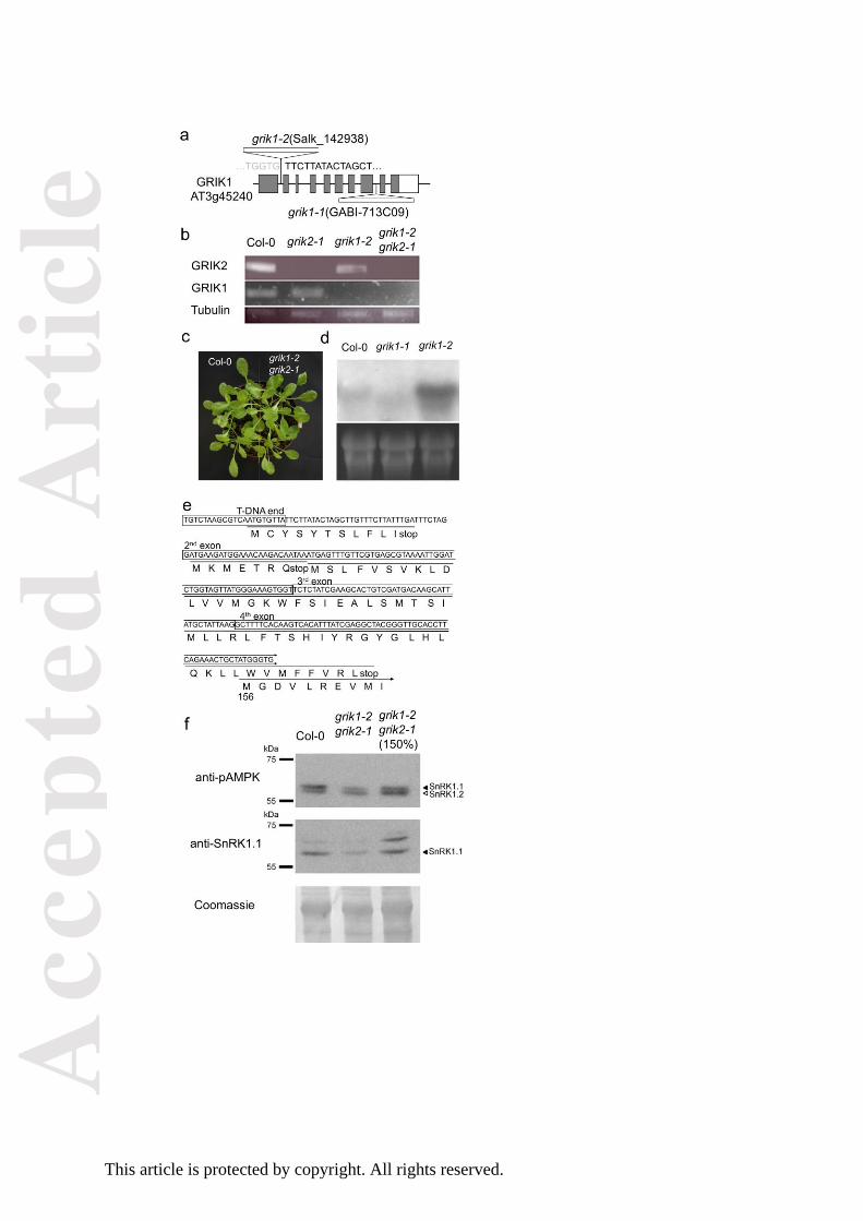
Figure 1. Characterization of the grik1-2 grik2-1 mutant line.
(a) Schematic diagram of grik1 T-DNA insertion lines.
(b) RT-PCR of full length of GRIK1, GRIK2 and tubulin8 in the wild type Col-0, grik1-2, grik2-1 and
grik1-2 grik2-1.
(c) Phenotype of the wild type and grik1-2 grik2-1 under short-day condition for 4 weeks
(d) Northern blotting with the middle region of GRIK1 as probe in the wild type, grik1-1, and grik1-2.
(e) cDNA sequence of the 5´end region of grik1-2 and potential ORFs. The longest ORF corresponded
an N-terminal truncated GRIK1 protein starting at Met-156 of the wild-type protein.
(f) Amount and phosphorylation status of SnRK1s in the wild type and grik1-2 grik2-1.
Western blot with anti-pAMPK antibody (upper panel) and anti-SnRK1.1 antibody (middle panel).
Coomassie staining was used to show protein amounts (lower panel). A greater amount of proteins
from grik1-2 grik2-1 (150%) was also loaded to compare the phosphorylation ratio in equivalent
amounts of SnRK1.1.
Figure 2. Salt-sensitive phenotype of grik1-2 grik2-1 double mutant.
(a) Seedlings of the wild type (Col-0), grik1-2, grik2-1, grik1-2 grik2-1 and sos2-2 mutants grown on
MS agar plate were transferred to fresh plates with or without 100 mM NaCl. Photographs were
taken 12d after transfer. The line graph represents primary root length of seedlings 12d after transfer
to MS agar plates with indicated concentration of NaCl (mean ± S.E., n=20). Asterisks indicate
significant differences from wild type (P<0.05, in one-way ANOVA followed by Tukey's multiple
comparison test).
Acc
epte
d A
rtic
le
This article is protected by copyright. All rights reserved.
(b) Hydroponic culture of Col-0, grik1-2, grik2-1, grik1-2 grik2-1 and sos2-2 mutants in LAK medium
supplemented with NaCl as indicated. Plants were grown for 4-weeks. The bar graph represents the
dry weight of plants (mean ± S.E., n=7) at the end of the experiment. Asterisks indicate significant
differences from wild type (P<0.01, in one-way ANOVA followed by Tukey's multiple comparison
test)
Figure 3. In vitro phosphorylation of SOS2 by GRIK1.
(a) A kinase-dead SOS2 fused to GST (SOS2-KN, arrow) was incubated with GST-fused GRIK1 or the
kinase-dead (K137R) of GRIK1 (GRIK1-KR) also fused to GST (arrowhead). Proteins were separated in
SDS-PAGE followed by Coomassie staining (left) and autoradiograph (right).
(b) GST-fused mutant T168A of SOS2 was incubated with GST-fused GRIK1. Proteins were separated
in SDS-PAGE followed by Coomassie staining (left) and autoradiograph (right).
Figure 4. Expression of SOS2 mutants S159A (SA) and Y175A (YA), but not T168A (TA), partially
rescued the sos2-2 under salt stress condition.
(a) Seedlings grown on MS agar medium were transferred to fresh plates with or without 100 mM
NaCl. Photographs were taken 12 d after transfer. The lines presenting the best resistance to the
NaCl plates were selected for this test.
(b) Northern blot for SOS2 transcript was performed with total RNA extracted from wild type (WT),
sos2-2 transgenic plants expressing the S159A (SA), T168A (TA) and Y175A (YA) mutant forms of
SOS2, and non-transformed sos2-2. Total RNA was purified 12h after transfer to MS agar plates with
100 mM NaCl. rRNA (ethidium bromide stained) was used as a loading control.
Acc
epte
d A
rtic
le
This article is protected by copyright. All rights reserved.
(c) Survival rates of the seedlings on MS agar plates with or without 100 mM NaCl. The experiment
was repeated three times and mean values (± S.E.) are shown.
(d) Primary root length of seedlings 12d after transfer to MS agar plates with or without 100 mM NaCl
(mean ± S.E.). Asterisks indicate significant differences from sos2-2 transformed with the wild type
SOS2 (P<0.05, in one-way ANOVA followed by Tukey's multiple comparison test).
Figure 5. In vitro activation of SOS2-∆308 by GRIK1.
The GST-fused SOS3-independent form of SOS2 (SOS2-∆308, arrowhead), or the combined T168A
mutation in SOS2-∆308 were incubated with GST-fused GRIK1 or GST-fused kinase-dead (K137R)
version of GRIK1 (GRIK1-KR). The SOS1 C-terminal fragment (SOS1-CT, arrows) was added as a
phosphorylation substrate of SOS2. Proteins were separated in SDS-PAGE followed by Coomassie
staining (left) and autoradiograph (right). Note that the small amount of GRIK1 used was enough to
achieve the activation of SOS2, even though GRIK1 bands were not observed. The relative band
intensity of SOS1-CT bands was normalized to the signal from the SOS2-∆308WT/GRIK1-KR lane
(mean ± S.E., n=6). Asterisks indicate significant differences (P<0.05, binomial test).
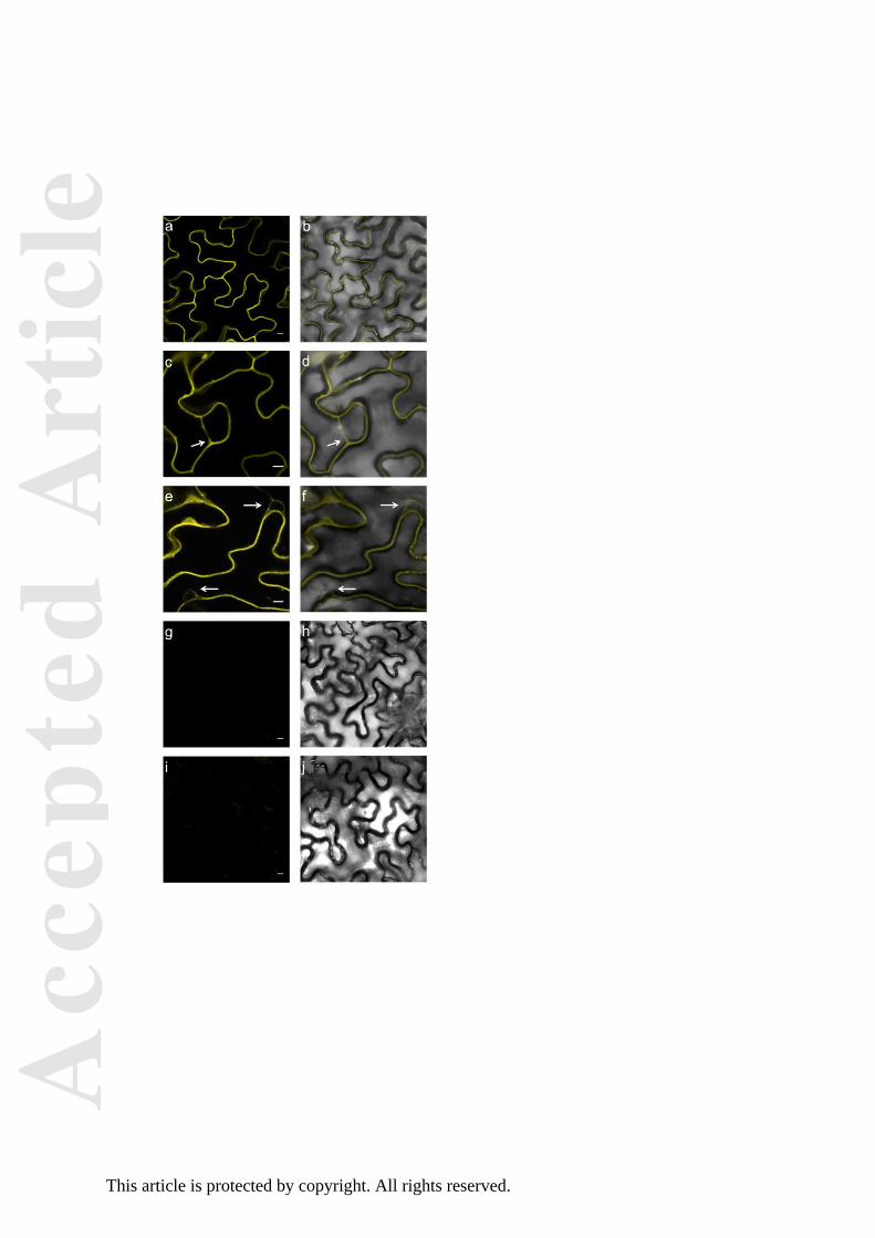
Figure 6. SOS2-GRIK1 interaction visualised by BiFC.
Panels from (a) to (f) show several images of reconstituted BiFC by GRIK1-SOS2 interaction (left:
fluorescence images; right: overlay with transmitted light). Arrows indicate the presence of a
transvacuolar strand in panels (c) and (d) and nuclei in panels (e) and (f). Panels (g) and (h) show a
negative control with GRIK1 in pSPYCE(M) and the empty vector pSPYNE(R)173. Panels (i) and (j)
show co-transformation with SOS2 in pSPYNE(R)173 and the empty vector pSPYCE(M). Scale bar: 10
μm.

Acc
epte
d A
rtic
le
This article is protected by copyright. All rights reserved.
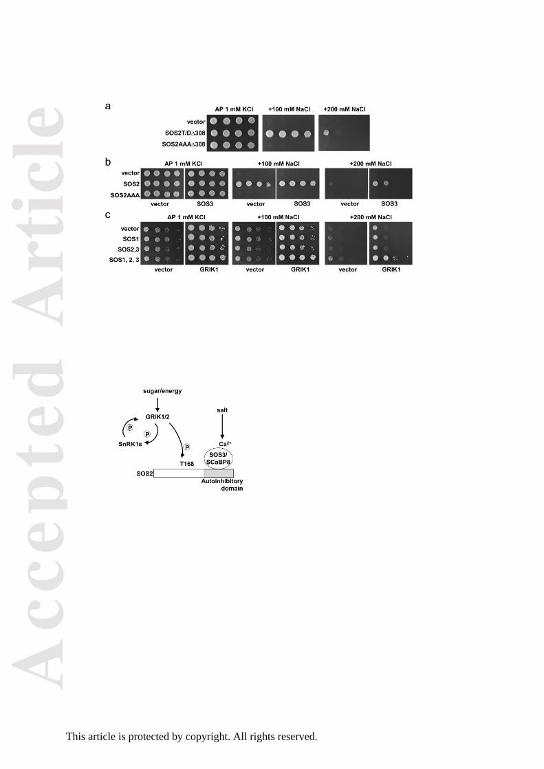
Figure 7. Activation of SOS2 by GRIK1 in yeast.
(a) The constitutively active form of SOS2 lacking the C-terminal autoinhibitory domain and with
either the phosphomimic T168D mutation in the activation-loop (SOS2-T/D∆308) or with the three
putative phosphorylation sites by GRIK kinases (S156/T168/Y175) converted to alanine residues
(SOS2-AAA∆308) were expressed in the yeast strain YP890 bearing mutations ena1-4 nha1 nhx1 and
expressing SOS1 from a chromosomal integration. The salt sensitivity of all transformants was
analysed by spotting decimal dilutions of starting cultures in AP plates supplemented with the
indicated amounts of NaCl. Growth in NaCl-supplemented media reported the activation of SOS1 by
SOS2.
(b) The full-length SOS2 protein and the triple mutant S156A/T168A/Y175A (SOS2-AAA) were
expressed in the yeast strain YP890 harbouring a chromosomal integration of SOS1. When indicated,
SOS3 was also co-expressed. The salt tolerance of the transformants was analysed in AP medium
with increasing concentration of NaCl as described above. Failure to convey salt tolerance indicated
that SOS2AAA is unable to form a productive complex with SOS3 to activate SOS1.
(c) SOS1, SOS2 and SOS3 were expressed, in various combination as indicated, in strain 3K4E, which
lacks the three SNF1-activating kinases SAK1, ELM1 and TOS3 in addition to the Na+ pumps ENA1-4.
When indicated, GRIK1 was also co-expressed to test for the ability to complement the sak1 elm1
tos3 mutations with regard to SOS2 activation. Results indicated that only the full complement of
GRIK1, SOS2, SOS3 and SOS1 was able to restore salt tolerance.
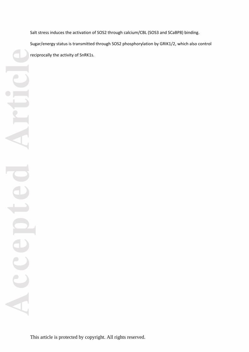
Figure 8. Model of the placement of GRIK kinases at the interface between sensing of the energy
status and salinity stress signalling.
Acc
epte
d A
rtic
le
This article is protected by copyright. All rights reserved.
Salt stress induces the activation of SOS2 through calcium/CBL (SOS3 and SCaBP8) binding.
Sugar/energy status is transmitted through SOS2 phosphorylation by GRIK1/2, which also control
reciprocally the activity of SnRK1s.
Acc
epte
d A
rtic
le
This article is protected by copyright. All rights reserved.
Acc
epte
d A
rtic
le
This article is protected by copyright. All rights reserved.
Acc
epte
d A
rtic
le
This article is protected by copyright. All rights reserved.
Acc
epte
d A
rtic
le
This article is protected by copyright. All rights reserved.
Acc
epte
d A
rtic
le
This article is protected by copyright. All rights reserved.
Acc
epte
d A
rtic
le
This article is protected by copyright. All rights reserved.
Acc
epte
d A
rtic
le
This article is protected by copyright. All rights reserved.

![Receptor-Like Kinases Sustain Symbiotic Scrutiny1[OPEN]...Update on Receptor-Like Kinases in Symbiosis Receptor-Like Kinases Sustain Symbiotic Scrutiny1[OPEN] Chai Hao Chiu,2 and Uta](https://img.dokumen.tips/doc/110x75/60aa214268722c0ce00ae5e7/receptor-like-kinases-sustain-symbiotic-scrutiny1open-update-on-receptor-like.jpg)