Embed Size (px)
Citation preview
Pathologie Helmut Hopfer
Update: Renal cell carcinoma
Agenda
• clinical context
• what‘s new?
• prognostic markers
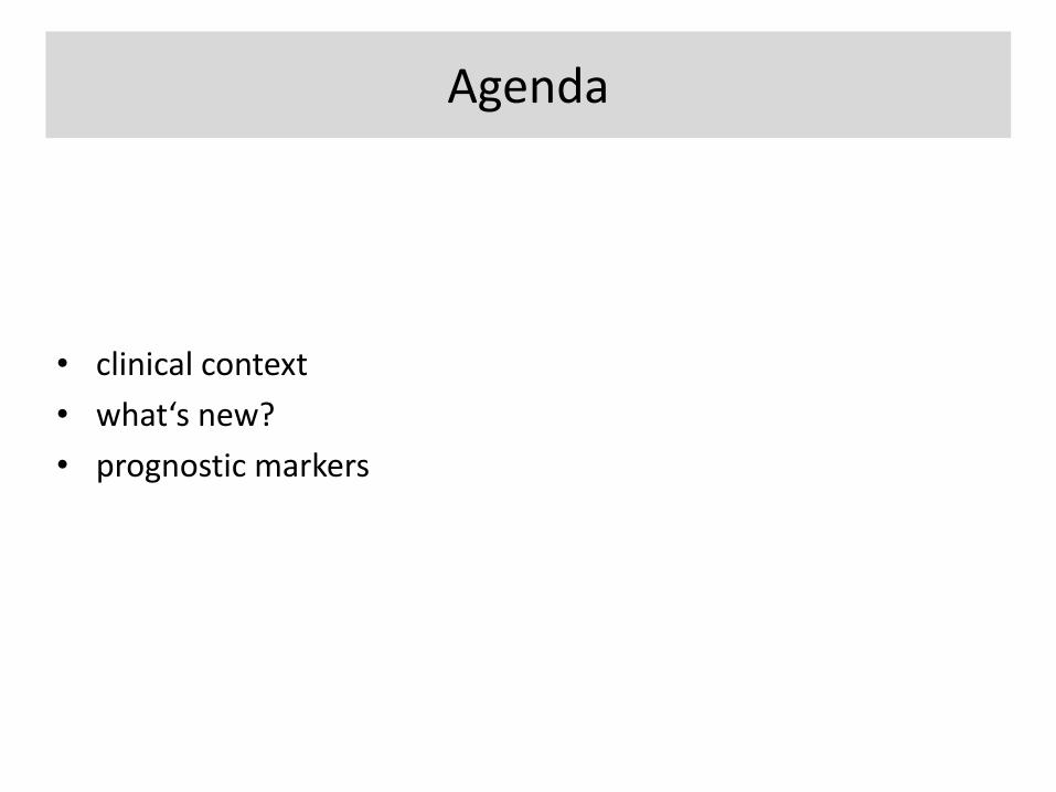
RCC at diagnosis
extent of disease
localized
regional
metastatic
unstaged
Stage 1 T1 N0 M0
Stage 2 T2 N0 M0
Stage 3 T3 T1, T2, T3
N0 N1
M0 M0
Stage 4 T4 any T
any N any N
M0 M1
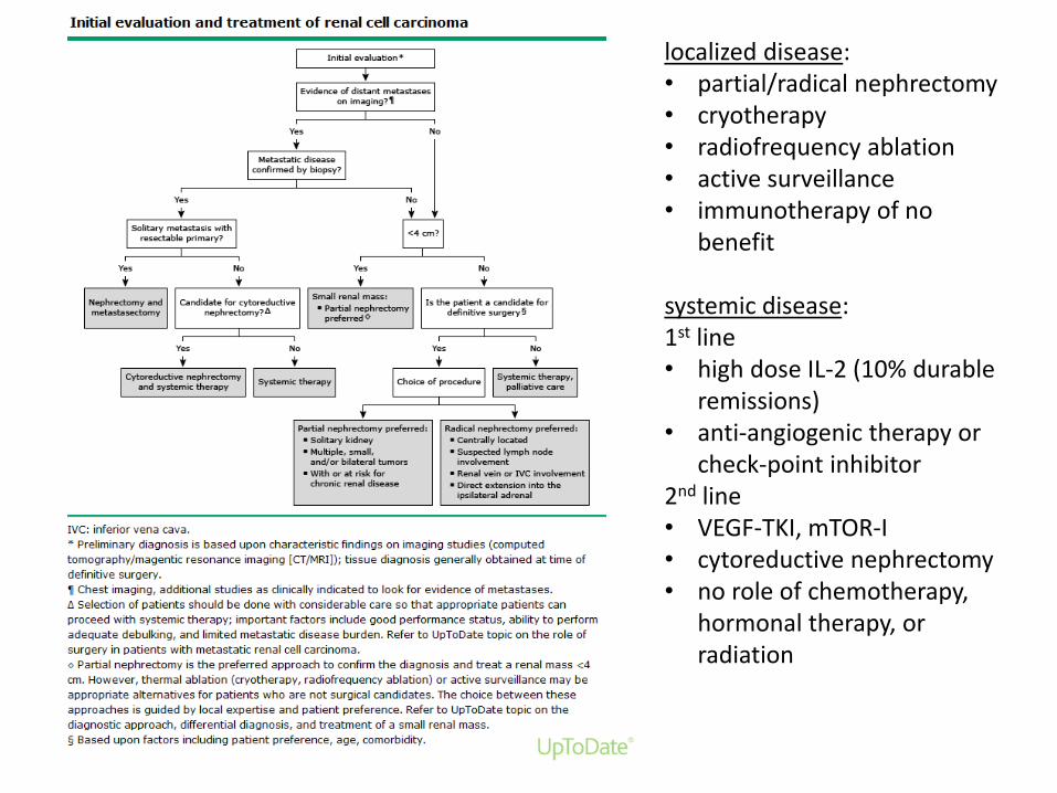
localized disease: • partial/radical nephrectomy • cryotherapy • radiofrequency ablation • active surveillance • immunotherapy of no
benefit
systemic disease: 1st line • high dose IL-2 (10% durable
remissions) • anti-angiogenic therapy or
check-point inhibitor 2nd line • VEGF-TKI, mTOR-I • cytoreductive nephrectomy • no role of chemotherapy,
hormonal therapy, or radiation
Agenda
• clinical context
• what‘s new? – Vancouver classification and
beyond
– new entities, names and codes
– molecular characterization
• prognostic markers
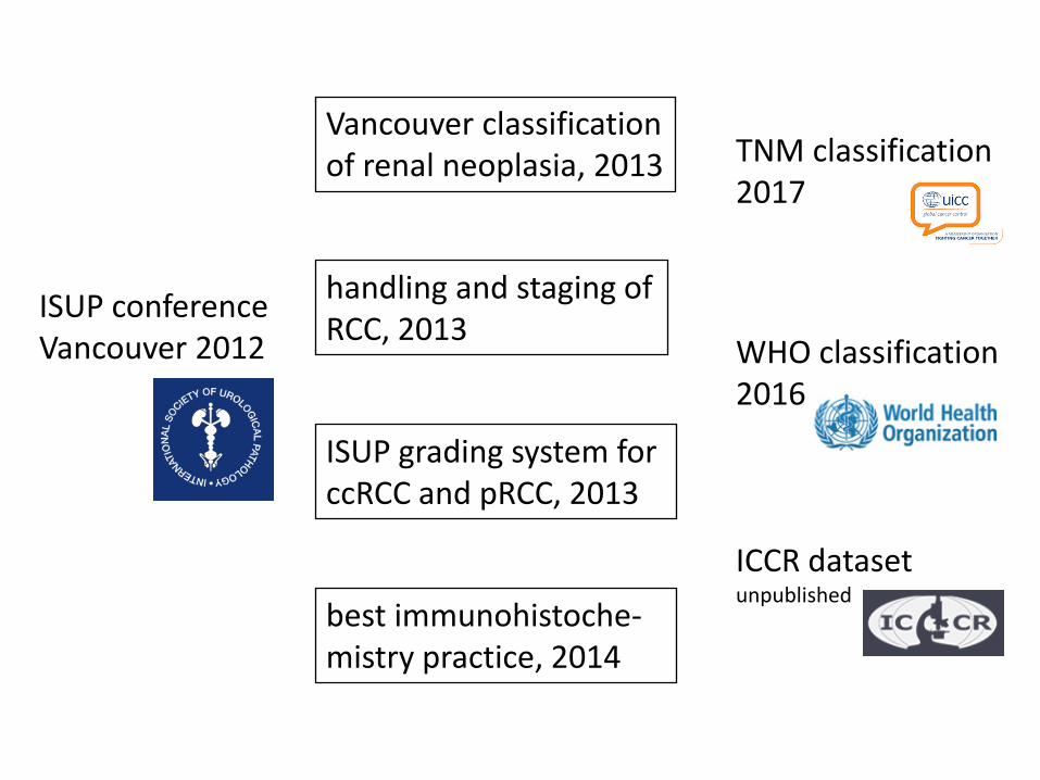
ISUP conference Vancouver 2012
TNM classification 2017
WHO classification 2016
ICCR dataset unpublished
Vancouver classification of renal neoplasia, 2013
handling and staging of RCC, 2013
best immunohistoche-mistry practice, 2014
ISUP grading system for ccRCC and pRCC, 2013
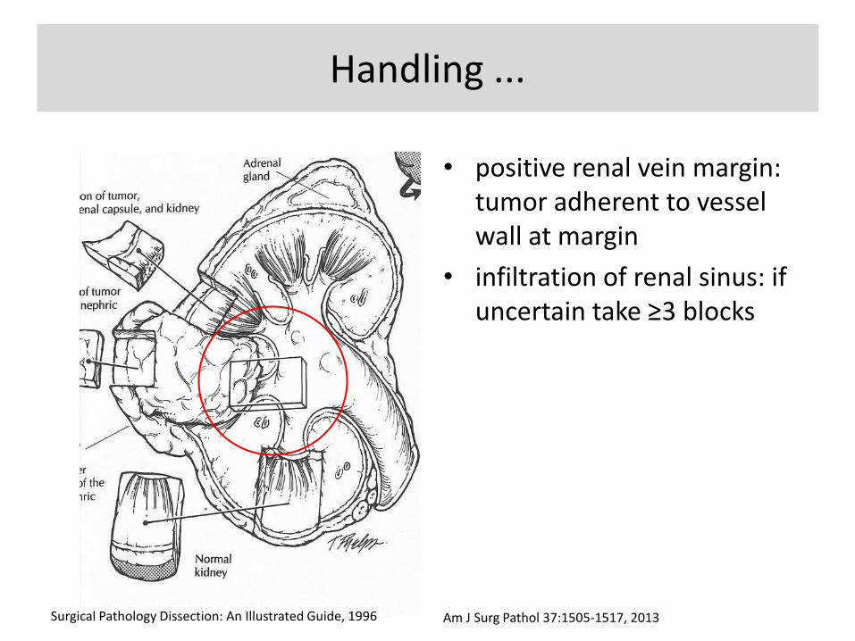
• positive renal vein margin: tumor adherent to vessel wall at margin
• infiltration of renal sinus: if uncertain take ≥3 blocks
Handling ...
Surgical Pathology Dissection: An Illustrated Guide, 1996 Am J Surg Pathol 37:1505-1517, 2013
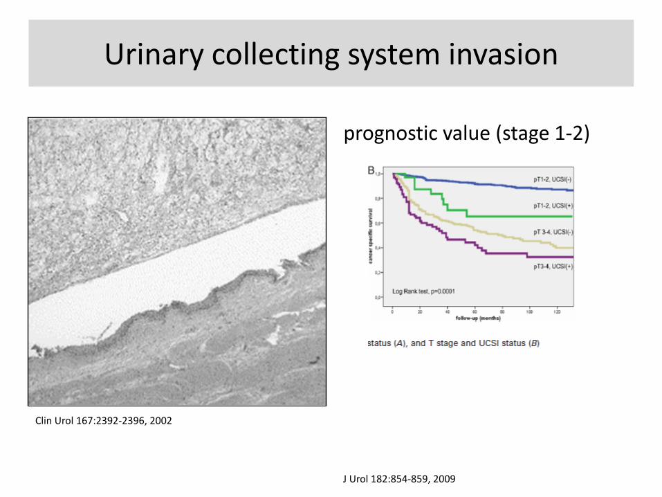
Urinary collecting system invasion
prognostic value (stage 1-2)
Clin Urol 167:2392-2396, 2002
J Urol 182:854-859, 2009
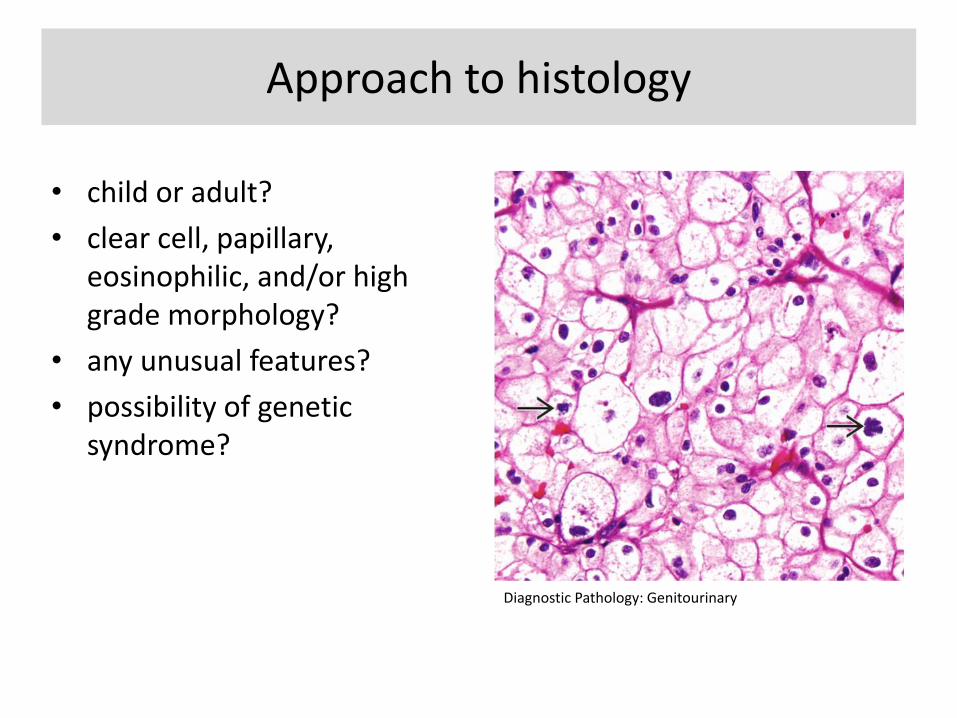
Approach to histology
• child or adult?
• clear cell, papillary, eosinophilic, and/or high grade morphology?
• any unusual features?
• possibility of genetic syndrome?
Diagnostic Pathology: Genitourinary
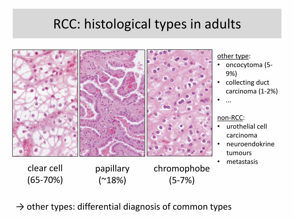
RCC: histological types in adults
clear cell (65-70%)
papillary (~18%)
chromophobe (5-7%)
→ other types: differential diagnosis of common types
other type: • oncocytoma (5-
9%) • collecting duct
carcinoma (1-2%) • ... non-RCC: • urothelial cell
carcinoma • neuroendokrine
tumours • metastasis
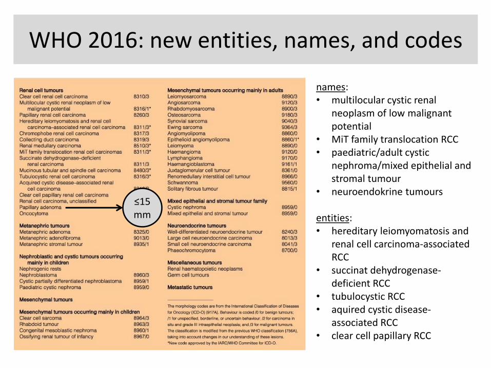
WHO 2016: new entities, names, and codes
names: • multilocular cystic renal
neoplasm of low malignant potential
• MiT family translocation RCC • paediatric/adult cystic
nephroma/mixed epithelial and stromal tumour
• neuroendokrine tumours
entities: • hereditary leiomyomatosis and
renal cell carcinoma-associated RCC
• succinat dehydrogenase-deficient RCC
• tubulocystic RCC • aquired cystic disease-
associated RCC • clear cell papillary RCC
≤15 mm
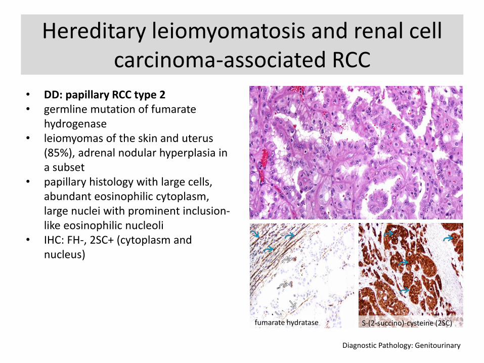
Hereditary leiomyomatosis and renal cell carcinoma-associated RCC
• DD: papillary RCC type 2 • germline mutation of fumarate
hydrogenase • leiomyomas of the skin and uterus
(85%), adrenal nodular hyperplasia in a subset
• papillary histology with large cells, abundant eosinophilic cytoplasm, large nuclei with prominent inclusion-like eosinophilic nucleoli
• IHC: FH-, 2SC+ (cytoplasm and nucleus)
Diagnostic Pathology: Genitourinary
fumarate hydratase S-(2-succino)-cysteine (2SC)
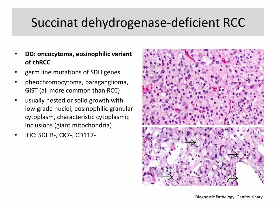
Succinat dehydrogenase-deficient RCC
• DD: oncocytoma, eosinophilic variant of chRCC
• germ line mutations of SDH genes
• pheochromocytoma, paraganglioma, GIST (all more common than RCC)
• usually nested or solid growth with low grade nuclei, eosinophilic granular cytoplasm, characteristic cytoplasmic inclusions (giant mitochondria)
• IHC: SDHB-, CK7-, CD117-
Diagnostic Pathology: Genitourinary
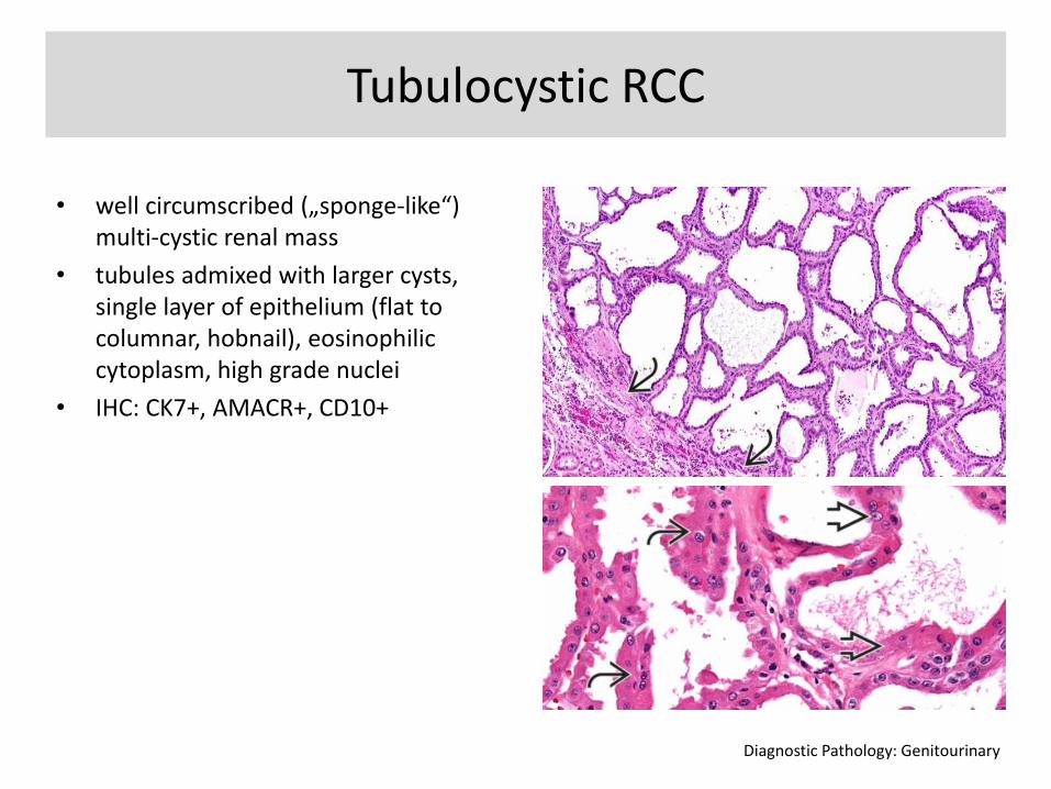
Tubulocystic RCC
• well circumscribed („sponge-like“) multi-cystic renal mass
• tubules admixed with larger cysts, single layer of epithelium (flat to columnar, hobnail), eosinophilic cytoplasm, high grade nuclei
• IHC: CK7+, AMACR+, CD10+
Diagnostic Pathology: Genitourinary
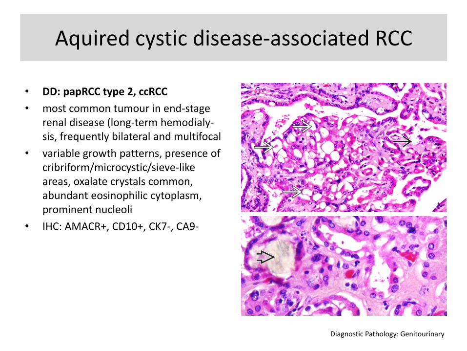
Aquired cystic disease-associated RCC
• DD: papRCC type 2, ccRCC
• most common tumour in end-stage renal disease (long-term hemodialy-sis, frequently bilateral and multifocal
• variable growth patterns, presence of cribriform/microcystic/sieve-like areas, oxalate crystals common, abundant eosinophilic cytoplasm, prominent nucleoli
• IHC: AMACR+, CD10+, CK7-, CA9-
Diagnostic Pathology: Genitourinary
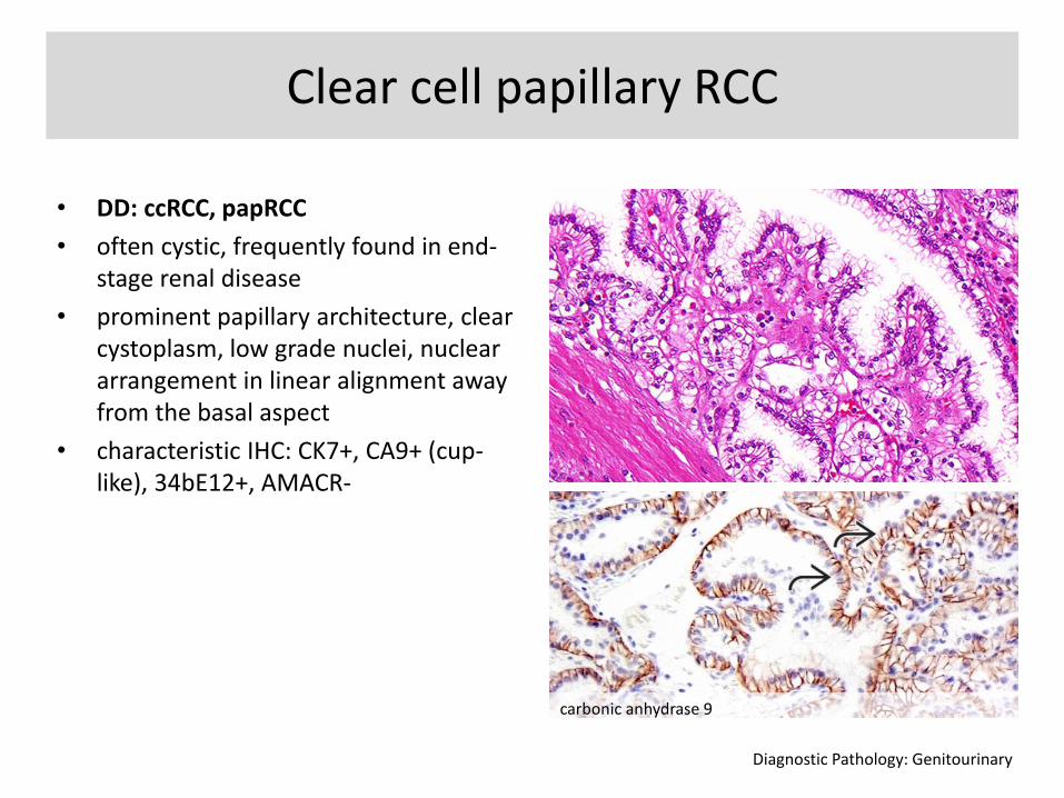
Clear cell papillary RCC
• DD: ccRCC, papRCC
• often cystic, frequently found in end-stage renal disease
• prominent papillary architecture, clear cystoplasm, low grade nuclei, nuclear arrangement in linear alignment away from the basal aspect
• characteristic IHC: CK7+, CA9+ (cup-like), 34bE12+, AMACR-
Diagnostic Pathology: Genitourinary
carbonic anhydrase 9
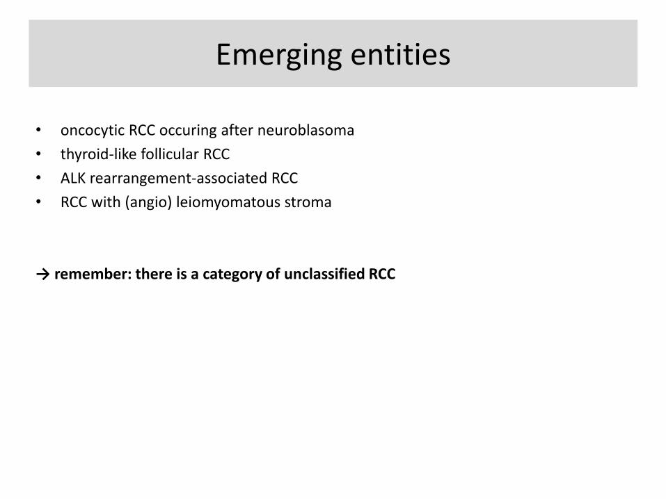
Emerging entities
• oncocytic RCC occuring after neuroblasoma
• thyroid-like follicular RCC
• ALK rearrangement-associated RCC
• RCC with (angio) leiomyomatous stroma
→ remember: there is a category of unclassified RCC
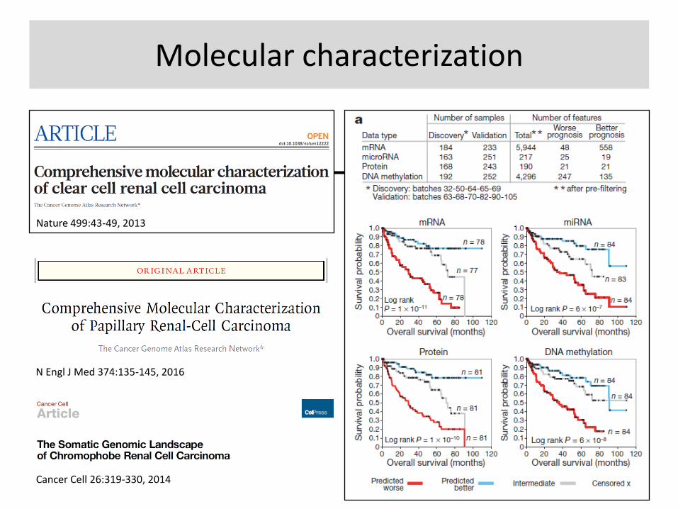
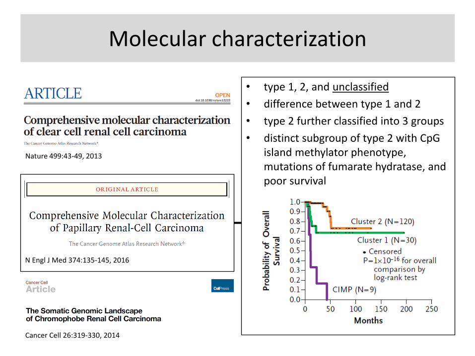
Molecular characterization
Nature 499:43-49, 2013
N Engl J Med 374:135-145, 2016
Cancer Cell 26:319-330, 2014
Molecular characterization
Nature 499:43-49, 2013
N Engl J Med 374:135-145, 2016
Cancer Cell 26:319-330, 2014
• type 1, 2, and unclassified
• difference between type 1 and 2
• type 2 further classified into 3 groups
• distinct subgroup of type 2 with CpG island methylator phenotype, mutations of fumarate hydratase, and poor survival
Agenda
• clinical context
• what‘s new?
• prognostic markers
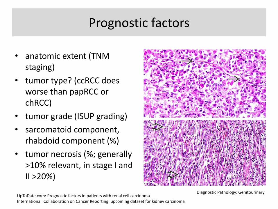
Prognostic factors
• anatomic extent (TNM staging)
• tumor type? (ccRCC does worse than papRCC or chRCC)
• tumor grade (ISUP grading)
• sarcomatoid component, rhabdoid component (%)
• tumor necrosis (%; generally >10% relevant, in stage I and II >20%)
UpToDate.com: Prognostic factors in patients with renal cell carcinoma International Collaboration on Cancer Reporting: upcoming dataset for kidney carcinoma
Diagnostic Pathology: Genitourinary
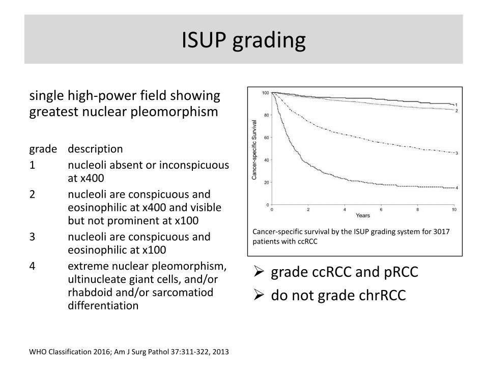
ISUP grading
single high-power field showing greatest nuclear pleomorphism
grade description
1 nucleoli absent or inconspicuous at x400
2 nucleoli are conspicuous and eosinophilic at x400 and visible but not prominent at x100
3 nucleoli are conspicuous and eosinophilic at x100
4 extreme nuclear pleomorphism, ultinucleate giant cells, and/or rhabdoid and/or sarcomatiod differentiation
WHO Classification 2016; Am J Surg Pathol 37:311-322, 2013
Cancer-specific survival by the ISUP grading system for 3017 patients with ccRCC
grade ccRCC and pRCC
do not grade chrRCC
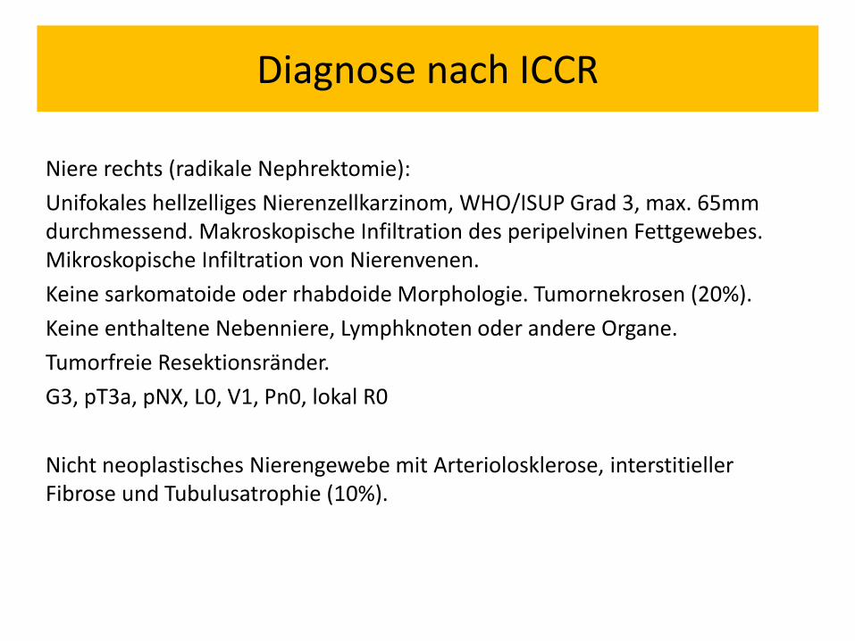
Diagnose nach ICCR
Niere rechts (radikale Nephrektomie):
Unifokales hellzelliges Nierenzellkarzinom, WHO/ISUP Grad 3, max. 65mm durchmessend. Makroskopische Infiltration des peripelvinen Fettgewebes. Mikroskopische Infiltration von Nierenvenen.
Keine sarkomatoide oder rhabdoide Morphologie. Tumornekrosen (20%).
Keine enthaltene Nebenniere, Lymphknoten oder andere Organe.
Tumorfreie Resektionsränder.
G3, pT3a, pNX, L0, V1, Pn0, lokal R0
Nicht neoplastisches Nierengewebe mit Arteriolosklerose, interstitieller Fibrose und Tubulusatrophie (10%).