Embed Size (px)
Citation preview
UNIVERSIDADE FEDERAL DE PELOTAS Programa de Pós-Graduação em Biotecnologia
Dissertação
Avaliação do efeito antitumoral in vitro de nanocápsulas de
núcleo lipídico de tretinoína sobre células de adenocarcinoma de pulmão, linhagem A549
Eduarda Schultze
Pelotas, 2013
EDUARDA SCHULTZE
Avaliação do efeito antitumoral in vitro de nanocápsulas de núcleo
lipídico de tretinoína sobre células de adenocarcinoma de pulmão,
linhagem A549
Dissertação apresentada ao Programa de Pós- Graduação em Biotecnologia
da Universidade Federal de Pelotas, como requisito parcial à obtenção do
título de Mestre em Biotecnologia
Orientador: Prof. Dr. Tiago Collares
Co-Orientador (es): Prof. Dra. Fabiana Seixas
Prof. Dr. Vinicius Farias Campos
Pelotas, 2013
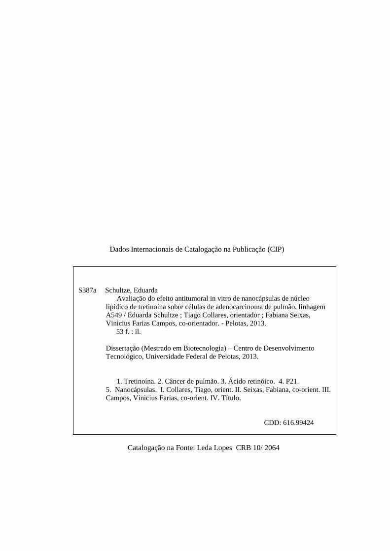
S387a Schultze, Eduarda
Avaliação do efeito antitumoral in vitro de nanocápsulas de núcleo
lipídico de tretinoína sobre células de adenocarcinoma de pulmão, linhagem
A549 / Eduarda Schultze ; Tiago Collares, orientador ; Fabiana Seixas,
Vinicius Farias Campos, co-orientador. - Pelotas, 2013.
53 f. : il.
Dissertação (Mestrado em Biotecnologia) – Centro de Desenvolvimento
Tecnológico, Universidade Federal de Pelotas, 2013.
1. Tretinoína. 2. Câncer de pulmão. 3. Ácido retinóico. 4. P21.
5. Nanocápsulas. I. Collares, Tiago, orient. II. Seixas, Fabiana, co-orient. III.
Campos, Vinicius Farias, co-orient. IV. Título.
CDD: 616.99424
Dados Internacionais de Catalogação na Publicação (CIP)
Catalogação na Fonte: Leda Lopes CRB 10/ 2064

Dedico este trabalho àquelas que foram incansáveis
em estar ao meu lado, trabalhando, discutindo
(temas científicos e não) e fazendo tudo ficar mais fácil,
Karine Rech Begnini e Virginia Yurgel.
Meus sinceros agradecimentos à Aline Ourique, sem a qual este trabalho não
existiria, e à equipe da Faculdade de Farmácia da UFRGS, Professoras Dras. Silvia
Guterres e Adriana Pohllmann e Professor Dr. Ruy Bech.
Agradeço ao meu orientador Professor Dr. Tiago Collares por ter aceitado me
orientar ao longo deste trabalho e por ter me proporcionar o retorno à vida
acadêmica.
Agradeço a minha co-orientadora Professora Dra. Fabiana Seixas pelas orientações
recebidas ao longo deste tempo e pelo apoio que recebi em decisões difíceis e ao
co-orientador Professor Dr. Vinicius Farias Campos pelo auxilio técnico-científico.
Agradeço também aos meus colegas de laboratório por todos os momentos de
convívio harmonioso, discussões filosóficas e científicas e pelo sagrado momento do
café.
Por fim e não menos importante, agradeço a todos os meus amigos, familiares e
pessoas que de alguma forma ouviram minhas reclamações, vibraram com minhas
vitórias e torceram pelo meu sucesso.

Banca examinadora:
Profa. Dra. Sibele Borsuk, Ufpel
Prof. Dr. Alan McBride, Ufpel
Profa. Dra. Isabel Oliveira de Oliveira, Ufpel
RESUMO
Schultze, Eduarda. Avaliação do efeito antitumoral in vitro de nanocápsulas de
núcleo lipídico de tretinoína sobre células de adenocarcinoma de pulmão,
linhagem A549. Dissertação (Mestrado) - Programa de Pós-Graduação em
Biotecnologia. Universidade Federal de Pelotas, Pelotas
Derivados e análogos retinóides têm sido largamente estudados como agentes
antitumorais devido a seus efeitos sobre a proliferação e diferenciação celular.
Tretinoína (TT), também conhecida como ácido retinóico é um derivado retinóide
que tem sido usado como adjuvante no tratamento de leucemia promielocítica aguda
com excelentes índices de remissão da doença. Este composto exerce atividade
antiproliferativa em diversos tipos de tumores. Entretanto, células de
adenocarcinoma de pulmão humano em geral exibem uma forte resistência aos
efeitos da tretinoína, a qual pode estar relacionada com a deficiência na
internalização celular de tretinoína nesse tipo de célula. Uma estratégia para
aumentar a atividade antiproliferativa de tretinoína é aumentar a captação celular do
composto através de carreadores como lipossomas ou outras vesículas como
nanocápsulas ou nanoesferas. Neste trabalho nanocápsulas de núcleo lipídico
contendo tretinoína (TT-LCNC) foram avaliadas quanto ao seu potencial de inibir o
crescimento, induzir a apoptose e interferir com o ciclo celular de células de
adenocarcinoma de pulmão, linhagem A549, resistentes ao tratamento com TT livre.
Os resultados demonstraram que TT-LCNC foi capaz de superar a resistência
celular ao tratamento com TT, reduzindo a viabilidade celular e induzindo apoptose,
superexpressão de P21 e parada do ciclo celular em G1.
Palavras-chave: Tretinoína, ácido retinóico, nanocápsulas, câncer de pulmão, P21
ABSTRACT
Schultze, Eduarda. In Vitro Antitumor Activity of Tretinoin-Loaded Lipid-Core
Nanocapsules on human lung adenocarcinoma cell line (A549). Dissertação
(Mestrado) - Programa de Pós-Graduação em Biotecnologia. Universidade Federal
de Pelotas, Pelotas.
Retinoid derivatives and analogs have been widely studied as antitumor agents due
to their effects on cell proliferation and differentiation. Tretinoin (TT), also known as
retinoic acid is a retinoid derivative that has been used as an adjuvant in the
treatment of acute promyelocytic leukemia with excellent rates of remission. This
compound has antiproliferative activity in various tumor types. However, non small
cell lung cancer in general exhibit strong resistance to the effects of TT, which may
be related to the deficiency in the cellular up-take of TT in that cell type. A strategy to
enhance the antiproliferative activity of TT is to increase the cellular internalization of
the compound through carriers such as liposomes or other vesicles or nanospheres
or nanocapsules. Here we evaluated TT lipid-core nanocapsules (TT-LCNC) for their
power to inhibit growth, induce apoptosis and interfere with the cell cycle of lung
adenocarcinoma, A549 cell line, which is resistant to treatment with TT. The results
showed that TT-LCNC was able to overcome the cellular resistance to treatment with
TT, reducing cell viability and inducing apoptosis, upregulation of P21 and cell cycle
arrest in G1 phase.
Keywords: tretinoin, retinoic acid, nanocapsules, lung cancer, P21.
Lista de Figuras
Resumo gráfico.................................................................................... 42
Figura 1 Efeito das TT-LCNC sobre a proliferação das células de
linhagem A549 ……………............................................................ 43
Figura 2 Análise morfológica das células de linhagem A549 após 72h de
incubação com 20 μM de TT, TT-LCNC e LCNC .......……......…. 44
Figura 3 Tratamento com TT-LCNC aumenta a taxa de morte celular
sobre células de linhagem A549 ………………............................. 45
Figura 4 Indução de apoptose por TT-LCNC em células de linhagem
A549 ............................................................................................. 46
Figura 5 Perfil de expressão gênica de células A549 tratadas com TT,
TT-LCNC e LCNC ...............……… …………………...………....... 47
Figura 6 Indução de parada do ciclo celular por TT-LCNC ........................ 48

Lista de Tabelas
Tabela 1 Primers utilizados para reação de PCR em tempo real …………… 41
Lista de abreviações:
Câncer de pulmão de células não-pequenas - NSCLC
Elementos responsivos ao ácido retinóico – RARE
Elementos responsivos a retinóides X - RXRE
Fator de ativação de apoptose 1 – Apaf-1
Fator de necrose tumoral - TNF
Nanocápsulas de núcleo lipídico - LCNC
Nanocápsulas de núcleo lipídico contendo tretinoína – TT-LCNC
Receptor de ácido retinóico – RAR
Receptor retinóide X – RXR
Tretinoína - TT
Sumário
1. INTRODUÇÃO ............................................................................................................................... 11
1.1. Câncer ...................................................................................................................................... 11
1.2. Terapias antitumorais ............................................................................................................. 12
1.3. Apoptose .................................................................................................................................. 12
1.4. Parada do ciclo celular ........................................................................................................... 14
1.5. Tretinoína ................................................................................................................................. 15
1.6. Câncer de Pulmão .................................................................................................................. 17
1.7. Nanocarreadores .................................................................................................................... 18
2. OBJETIVOS: ................................................................................................................................... 21
3. ARTIGO ........................................................................................................................................... 22
ABSTRACT ..................................................................................................................................... 24
1. INTRODUCTION ........................................................................................................................ 25
2. MATERIALS AND METHODS ................................................................................................ 27
2.1 Preparation of nanocapsules ............................................................................................. 27
2.2 Characterization of nanocapsules ..................................................................................... 27
2.3.Cell Culture ........................................................................................................................... 28
2.4. Determination of cytotoxicity by MTT assay ................................................................... 28
2.5 Viability assessment and LIVE/DEAD assay ................................................................... 29
2.6. Measurement of apoptosis by Annexin V staining ......................................................... 30
2.7. Cell cycle analyses ............................................................................................................. 30
2.8. RNA extraction, cDNA synthesis and Real-Time PCR ................................................. 30
2.9. Data analysis ....................................................................................................................... 31
3. RESULTS .................................................................................................................................... 32
3.1. Production and characterization of lipid-core nanocapsules ....................................... 32
3.2. TTN-LNC inhibited cell proliferation of A549 cells ......................................................... 32
3.3. TTN-LNC alters morphology of A549 cells ..................................................................... 32
3.4. TTN-LNC reduces the cell viability ................................................................................... 33
3.5 Apoptosis induction by TTN-LNC on A549 cells ............................................................. 33

3.6 Analysis of gene expression .............................................................................................. 33
3.7. TTN-LNC-induced cell cycle arrest .................................................................................. 34
4. DISCUSSION ............................................................................................................................. 34
5. CONCLUSION ........................................................................................................................... 37
ACKNOWLEDGMENTS ................................................................................................................ 37
REFERENCES ............................................................................................................................... 38
4. CONCLUSÃO ................................................................................................................................. 49
5. REFERÊNCIAS .............................................................................................................................. 50
11
1. INTRODUÇÃO
1.1. Câncer
Câncer é o termo utilizado para um conjunto de doenças em que as células se
dividem de maneira anormal, descontrolada e com capacidade de invadir outros
tecidos que não o seu de origem (INSTITUTO NACIONAL DE CÂNCER, 2013).
Dados da Organização Mundial da Saúde indicam que são esperados para 2030, 27
milhões de casos incidentes de câncer, 17 milhões de mortes por câncer e 75
milhões de pessoas vivas, anualmente, com câncer. No Brasil, as estimativas mais
recentes, de 2012, que valerão também para o ano de 2013, apontam a ocorrência
de cerca de 518.000 casos novos de câncer. Dentre os tipos de câncer mais
incidentes estão os cânceres de pele não melanoma, próstata, pulmão, cólon/reto e
estômago para o sexo masculino; e os cânceres de pele não melanoma, mama, colo
do útero, cólon e reto e glândula tireóide para o sexo feminino (INSTITUTO
NACIONAL DE CÂNCER JOSÉ ALENCAR GOMES DA SILVA, 2013).
A carcinogênese envolve alteração no DNA de uma ou mais células normais,
geralmente em genes de controle do crescimento, levando a um crescimento celular
autônomo e descontrolado. Durante o desenvolvimento do câncer, as células
adquirirem algumas características biológicas que sustentam esse processo. Estas
características incluem sinalização proliferativa sustentada, evasão de supressores
de crescimento, resistência à morte celular, indução de angiogênese, imortalidade
replicativa e capacidade de invasão e metástase. Esses processos são
acompanhados por uma instabilidade genômica e pela presença de inflamação no
local, o que culmina por acelerar e agravar o desenvolvimento da neoplasia
(HANAHAN; WEINBERG, 2000; HANAHAN; WEINBERG, 2011). Os autores citam
ainda duas outras características importantes na manutenção da carcinogênese: a
capacidade de reprogramação do mecanismo energético, o que permite a
sobrevivência de células mais internas de tumores sólidos, onde há acesso limitado
aos nutrientes, e a capacidade de evasão do sistema imune. Qualquer uma dessas
características pode ser alvo de terapias antitumorais (HANAHAN; WEINBERG,
2011).
12
1.2. Terapias antitumorais
O que se espera de uma terapia antitumoral é uma alta capacidade de inibir a
proliferação das células alteradas, um eficiente índice de remissão do tumor e uma
baixa incidência de efeitos colaterais. Duas abordagens que vem sendo muito
descritas na literatura para o tratamento de diversos tipos de câncer são: a indução
de diferenciação celular, o que leva as células a retomarem o seu estado de
crescimento controlado e a reduzirem sua taxa de proliferação (BROWN; HUGHES,
2012) (NIU et al., 2010); e a indução de parada permanente do ciclo celular,
chamada senescência celular. Entretanto, a ação mais comum de um agente
terapêutico antitumoral é a indução de apoptose.
1.3. Apoptose
Apoptose é um mecanismo de morte celular controlado geneticamente, que
está envolvido na regulação da homeostase tecidual (VANGESTEL et al., 2009). A
apoptose pode ser desencadeada por diferentes estímulos como estresse celular e
outras formas de dano, sinalização mediada por outras células ou privação de
fatores de crescimento (ELMORE, 2007). Existem duas rotas apoptóticas principais:
uma via extrínseca, da qual faz parte a ativação de receptores como o Fas ou o
receptor de TNF, e a via intrínseca, que está relacionada com danos internos à
célula, como danos ao DNA e ao citoesqueleto. A via extrínseca é iniciada pela
ativação de receptores de morte na superfície celular, os quais desencadeiam uma
cascata de sinalização que envolve a ativação da caspase 8 (VANGESTEL et al.,
2009).
As caspases (cysteine-aspartic proteases) são uma família de cisteíno-
proteases que são sintetizadas na forma inativa de pró-caspases e são ativadas
quando interagem com proteínas específicas. Estas proteases são responsáveis por
clivar outras proteínas em domínios específicos após resíduo de ácido aspártico
(LAMKANFI et al., 2007). Quando recrutada pela cascata da via extrínseca, a
caspase 8 ativa a caspase 3. A ativação da caspase 3 leva a degradação de
13
proteínas celulares necessárias para a manutenção da sobrevivência e integridade
celular (ELMORE, 2007). A via intrínseca de ativação das capases acontece quando
uma série de estímulos pró-apoptóticos culminam com a liberação do citocromo c
pela mitocôndria, independentemente da ativação da caspase 8. O Citocromo c
interage com as proteínas Apaf-1 e Caspase 9 para promover ativação da caspase
3. As caspases 3, 6 e 7 são caspases efetoras que clivam substratos protéicos,
incluindo lâmina nuclear e moléculas de actina do citoesqueleto, ativando os
processos apoptóticos (ADAMS; CORY, 2007).
A célula que entra na rota apoptótica tem as seguintes características:
condensação do núcleo e do citoplasma, sem alterações das organelas até quase o
final do processo; formação de circunvoluções na membrana; formação de corpos
apoptóticos, não ocorrendo extravasamento do material citoplasmático; e a célula
morta normalmente é encontrada entre células vivas e é removida do tecido por
células vizinhas ou por fagócitos especializados, frente à exposição de sinais de
reconhecimento em sua superfície (VANGESTEL et al., 2009). Entre os sinais de
reconhecimento pelos fagócitos está a externalização do fosfolipídio aniônico,
fosfatidilserina, a qual é muito utilizada como marcador de apoptose em testes in
vitro. (WLODKOWIC; SKOMMER; DARZYNKIEWICZ, 2012).
Muitos agentes antitumorais agem induzindo apoptose (VANGESTEL et al.,
2009). Entretanto, muitas células tumorais desenvolveram mecanismos de
resistência à morte celular e, portanto, ao tratamento com agentes indutores de
apoptose. Um dos mecanismos tumorais mais comuns de evasão da apoptose é a
perda da função do gene supressor tumoral TP53 (HANAHAN; WEINBERG, 2011).
Este gene codifica a proteína P53, que desencadeia a rota apoptótica em resposta a
danos no DNA. Com essa proteína afuncional, a célula perde um importante
mecanismo de checagem de dano celular. As células cancerígenas podem, ainda,
aumentar a expressão de genes anti-apoptóticos, como o Bcl-2, e fatores de
crescimento, através da regulação negativa de agentes pró-apoptóticos, como o
Bax.
Outras formas de morte celular programada têm sido amplamente descritas
(MADDIKA et al., 2007) (LEIST; JAATTELA, 2001). A chamada morte celular
semelhante à apoptose ocorre sem a ativação de caspases efetoras e possui
14
características semelhentes às células apoptóticas como condensação da cromatina
e presença de moléculas de reconhecimento para fagócitos. Existe ainda outra
forma de morte independente de caspases, que cursa sem a condensação da
cromatina, mas com outras características apoptóticas, como a redução do potencial
transmembrana mitocondrial e a externalização de fosfatidilserina (MATEO et al.,
1999). Considerando que muitas células cancerígenas apresentam deficiências nas
rotas apoptóticas comuns, drogas potenciais que agem em outras rotas de morte
estão emergindo (LEIST; JAATTELA, 2001).
1.4. Parada do ciclo celular
Ainda que a maioria das terapias anticancerígenas vise matar as células
alteradas, uma opção terapêutica interessante é a indução de parada do ciclo
celular, fazendo com que as células não se dupliquem (JAIN et al., 2013). O ciclo
celular é um processo altamente controlado pelo qual a célula passa a fim de fazer a
duplicação do DNA e subsequente divisão celular. Este processo pode ser divido em
interfase e fase M (de mitose). A interfase pode ser dividida ainda em quatro
períodos principais: G0, G1, S e G2. Chama-se G0 o período em que a célula não
recebe estímulos de divisão, podendo permanecer nesta fase por horas, dias ou por
uma vida inteira, como as células neuronais. G1 é a fase em que a célula está se
preparando para duplicar seu DNA. Nesta fase, a célula responde a estímulos
positivos ou negativos, sendo levada ao crescimento, à diferenciação, à
multiplicação ou à apoptose, bem como à produção de enzimas e outras moléculas
necessárias para a próxima fase do ciclo, a fase de síntese. Na fase S ocorre a
duplicação do DNA. Na fase G2 ocorre a síntese de RNA, de proteínas e de outras
estruturas necessárias para o início da divisão celular (VERMEULEN; VAN
BOCKSTAELE; BERNEMAN, 2003) (MADDIKA et al., 2007).
Na maioria das células, há vários pontos no ciclo celular, chamados pontos de
checagem, em que o ciclo pode ser detido ou atrasado se eventos anteriores não
foram concluídos. Por exemplo, a entrada em mitose é impedida se a replicação do
DNA não for completa, e a separação dos cromossomos na mitose pode ser
atrasada se alguns cromossomos não estiverem devidamente ligados ao fuso
15
mitótico. A progressão através de G1 e G2 é atrasada por mecanismos de bloqueio
se o DNA nos cromossomos estiver danificado por radiação ou compostos químicos.
Atrasos nesses pontos de verificação de danos ao DNA proporcionam tempo para
que o DNA danificado possa ser reparado. Após a liberação do mecanismo de trava
do ciclo celular, a progressão ao longo da fase continua. Estes pontos de checagem
evitam que a célula duplique descontroladamente, acumulando danos que podem
transformar a célula numa célula tumoral (VERMEULEN; VAN BOCKSTAELE;
BERNEMAN, 2003).
Os pontos de verificação são ainda importantes por outros fatores. É através
deles que o sistema de controle pode ser regulado por sinais extracelulares de
outras células. Esses sinais, que podem promover ou inibir a proliferação celular
tendem a agir regulando a progressão através de um ponto de controle em G1. Além
disso, a maioria das mudanças genéticas que promove a tumorigênese envolve uma
desregulação da progressão do ciclo celular em G1 (FOSTER et al., 2010).
Muitos agentes terapêuticos que induzem parada irreversível do ciclo celular,
chamada de senescência celular têm sido estudados (VANGESTEL et al., 2009).
Um exemplo é a Tretinoína, ou ácido retinóico, um derivado de vitamina A que,
dentre outras ações, induz parada do ciclo celular em G1 (SOPRANO; QIN;
SOPRANO, 2004).
1.5. Tretinoína
Tretinoína (TT), ou ácido retinóico, é um metabólito ativo de vitamina A
(retinol) que desempenha um papel importante na mediação do crescimento e
diferenciação de células normais e transformadas. Esse composto é essencial para
diversas funções biológicas como crescimento, visão, reprodução, desenvolvimento
embrionário, diferenciação de tecidos epiteliais, e respostas imunológicas
(SOPRANO; QIN; SOPRANO, 2004). Tretinoína, bem como muitos de seus análogos
e derivados (chamados de retinóides), tem sido estudada através de ensaios clínicos,
testes em animais de laboratórios, modelos celulares e através de abordagens
epidemiológicas, apresentando um bom valor terapêutico em oncologia devido à sua
ação antiproliferativa e de indução de diferenciação (ORLANDI et al., 2003).
16
Tretinoína tem a capacidade de induzir completa remissão na maioria dos
pacientes com leucemia promielocítica aguda, através da indução de diferenciação
dos blastos (REGO et al., 2013). Além disto, este composto tem se mostrado eficaz,
sozinho ou por efeito sinérgico com outros agentes, contra diversos tipos de câncer,
como câncer de cabeça e pescoço, câncer de células escamosas de boca e câncer
de pulmão (HIGUCHI et al., 2003) (XU et al., 2008) (ARRIETA et al., 2010).
A ação da tretinoína se dá por modulação da transcrição gênica através da
interação com receptores de ácido retinóico (RAR, , ), convertendo-os de
repressores a ativadores da transcrição (ALTUCCI; GRONEMEYER, 2001). Estes
receptores nucleares são codificados por genes distintos dentro do genoma e são
membros da superfamília de receptores para hormônios esteróides e da tireóide.
RAR são fatores de transcrição que atuam como heterodímeros com receptores
retinoides X (RXR, , ) RAR-RXR, os quais podem ainda atuar como
homodímeros RXR-RXR, e se ligam ao DNA em seqüências chamadas de
elementos responsivos ao ácido retinóico (RAREs) ou elementos responsivos a
retinóides X (RXREs), localizados na região promotora de genes alvo (SOPRANO;
QIN; SOPRANO, 2004). RARs podem ser ativados tanto por tretinoína como por
ácido 9-cis-retinóico, enquanto que os RXRs são exclusivamente ativados pelo ácido
9-cis-retinóico (BUSHUE; WAN, 2010). Três receptores (RAR, RXR e RXR) são
largamente expressos, enquanto os outros (RAR, RAR e RXR) mostram um
padrão de expressão mais complexo, tecido-específico. Assim, a maioria dos tecidos
são alvos potenciais da ação dos retinóides (RHINN; DOLLE, 2012).
Muitos estudos têm demonstrado que o nível de expressão de RAR está
reduzido em muitos tipos celulares de câncer e também em muitas amostras
tumorais, incluindo câncer de pulmão (SOPRANO; QIN; SOPRANO, 2004) (XU,
2007). Este nível reduzido de RAR está relacionado com a tumorigênese e a
resistência ao tratamento com retinóides. De fato, tem sido descrito que câncer de
pulmão de células não pequenas, o qual apresenta resistência ao tratamento com
ácido retinóico, apresenta também expressão reduzida de RAR (GERADTS et al.,
1993) (INUI et al., 2003). Entretanto, a expressão deste receptor pode ser induzida
por tretinoína (ALTUCCI et al., 2007) (RHINN; DOLLE, 2012).
17
1.6. Câncer de Pulmão
O câncer de pulmão é a principal causa mundial de mortes por câncer
(BUNN, Jr., 2012). No Brasil, estimativas do Instituto Nacional do Câncer para 2012
prevêem 17.210 casos novos de câncer de pulmão em homens e 10.110 em
mulheres. O tabagismo é a principal causa de câncer de pulmão e a cessação do
tabagismo é o principal método de prevenção da mortalidade por esse tipo de
neoplasia (BUNN, Jr., 2012). O câncer de pulmão é uma doença heterogênea com
dois subtipos principais: o de células não pequenas (NSCLC), mais comum, sendo
cerca de 85% dos casos, e o de células pequenas (SCLC). NSCLC compreende
dois tipos: adenocarcinoma, mais comum em mulheres, especialmente fumantes, e
carcinoma de células escamosas, mais frequente em homens e em idosos de ambos
os sexos. Apesar dos avanços recentes feitos em oncologia clínica e experimental, o
prognóstico de câncer de pulmão ainda é desfavorável, com uma taxa de sobrevida
global em 5 anos de apenas cerca de 11% (ZHANG et al., 2012).
Vários fatores prognósticos independentes para a sobrevivência de pacientes
com NSCLC foram identificados, como estado físico, estágio da doença, idade, sexo
e quantidade de peso perdido, dos quais o mais importante é o estágio do tumor.
Segundo a Aliança Global Contra o Câncer de Pulmão, o estadiamento do câncer de
pulmão é definido como: estágio I – o câncer está presente somente em uma parte
do pulmão; estágio II: a doença se disseminou para os gânglios linfáticos ou tecidos
próximos; estágio III: o câncer se disseminou de forma mais extensa dentro do tórax
e, em geral, para os gânglios linfáticos maiores; e estágio IV: o câncer se
disseminou para outras partes do corpo, por exemplo, para o fígado ou ossos. Em
estágio inicial NSCLC é tratado principalmente por ressecção cirúrgica, com
quimioterapia adjuvante para pacientes em estágio IB, II e III da doença (GADGEEL;
RAMALINGAM; KALEMKERIAN, 2012). No entanto, mesmo na fase inicial da
doença, cerca de 30% dos pacientes sofrem de recidiva e morrem dentro de 5 anos
de cirurgia. Pacientes com NSCLC localmente avançado de estágio III são
normalmente tratados com terapia combinada de diferentes modalidades. NSCLC
avançado ainda é uma doença incurável.
18
O câncer de pulmão é um tipo de neoplasia para a qual está sendo estudada a
utilização de TT. Em ensaio clínico de fase II em pacientes com NSCLC foi
demonstrado que o tratamento com TT aumentou a taxa de resposta, bem como
prolongou o tempo de sobrevida livre de progressão (ARRIETA et al., 2010).
Entretanto, muitas células tumorais pulmonares exibem resistência ao tratamento
com TT (GERADTS et al., 1993), a qual pode estar relacionada com a redução da
expressão de RAR e (INUI et al., 2003) e com a deficiência na internalização
celular deste composto (KAWAKAMI et al., 2006). Tem-se proposto que a superação
da resistência celular possa ser alcançada através do uso de sistemas eficientes de
entrega de drogas, como lipossomas ou nanocápsulas (GAO; ZHANG; SUN, 2012).
1.7. Nanocarreadores
Uma abordagem pela qual o interesse dos pesquisadores da área da
oncologia celular vem aumentando é o uso de carreadores de escala nanométrica.
Dentre vários benefícios que esses carreadores podem apresentar, destacam-se o
aumento da seletividade de um agente terapêutico, a melhora na sua solubilidade e
estabilidade e a redução de seus efeitos colaterais (SULTANA et al., 2012). Vários
pesquisadores vêm estudando a associação de tretinoína (TT) a nanocarreadores a
fim de aprimorar suas atividades antitumorais (OURIQUE et al., 2010) (CHANSRI et
al., 2008) (TRAPASSO et al., 2009), bem como superar a resistência que algumas
células apresentam ao tratamento (KAWAKAMI et al., 2006). Além disso, tretinoína é
um composto que isomeriza dependendo das condições do meio e que apresenta
uma fotosensibilidade elevada, características que podem ser atenuadas por
sistemas de entrega de droga que estabilizam o composto (BUSHUE; WAN, 2010)
(TRAPASSO et al., 2009).
Kawakami et al. (2006) demonstraram que a incorporação de tretinoína em
lipossomas catiônicos compostos por DOTAP/colesterol foi capaz de superar a
resistência apresentada pelas células de linhagem de adenocarcinoma de pulmão
(A549) aos efeitos inibidores de crescimento de tretinoína livre (KAWAKAMI et al.,
2006). Os autores discutiram que esta superação da resistência se deveu ao
aumento da internalização celular do composto promovido pela incorporação aos
19
lipossomas. Entretanto, a instabilidade inerente dos carreadores a base de lípidos na
presença de componentes do soro resulta na libertação rápida e instantânea das
drogas quimioterápicas, o que tem limitado a sua utilização como sistema de entrega
de agentes terapêuticos para tratamento do câncer (JOO et al., 2013). Além disso,
ainda que lipossomas catiônicos proporcionem uma maior taxa de absorção celular
do que os aniônicos e neutros, aqueles são removidos mais rapidamente pelos rins
do que estes e apresentam uma interação eletrostática forte com proteínas
plasmáticas, o que pode resultar em agregação (GAO; ZHANG; SUN, 2012). Dessa
forma, outros nanocarreadores tem sido estudados para servirem de sistema de
entrega de tretinoína, bem como outros retinóides.
Nanoesferas e nanocápsulas tem sido largamente estudadas para o uso
como carreadores de agentes terapêuticos. Nanoesferas são definidas como uma
estrutura polimérica matricial onde drogas podem ser fisicamente aprisionadas ou
dispersas. Nanocápsulas, entretanto, são caracterizadas por um núcleo lipofílico
envolvido por uma camada polimérica, nas quais as drogas podem estar dissolvidas
no núcleo lipofílico, dispersos no interior da partícula ou adsorvidos na interface
partícula/àgua.
Em 2008, nanocápsulas de tretinoína foram produzidas a fim de avaliar o
benefício desta formulação na fotoestabilidade do composto (OURIQUE et al.,
2008). No entanto, esta formulação apresentou uma estabilidade limitada na
armazenagem. Foram produzidas então, em 2010, nanocápsulas de nucleo-lipídico
contendo tretinoína (TT-LCNC), e sua estabilidade físico-química, bem como efeitos
fotoprotetores frente à exposição da droga à radiação UV foram avaliados
(OURIQUE et al., 2010). Esta formulação apresentou uma alta estabilidade físico-
química, característica aquosa e fotoestabilidade adequada sobre radiação UVA e
UVB. Além disso, TT-LCNC se mostrou mais eficiente a longo prazo quando
comparada com tretinoína livre em tratamento de linhagem celular de células de
leucemia promielocítica aguda. Os autores consideraram TT-LCNC como um
inovador e promissor sistema terapêutico nanométrico para a administração
parenteral de tretinoína no tratamento de pacientes com leucemia pró-mielocítica
aguda.

20
Os benefícios apresentados pela nanoencapsulação de TT em nanocápsulas
de núcleo lipídico frente a sua estabilidade e ação antitumoral em células
leucêmicas, bem como os benefícios apresentados por outras nanoformulações de
TT frente a sua ação antitumoral em células resistentes, justificam o uso da
formulação de TT-LCNC para avaliação do efeito antitumoral sobre células de
adenocarcinoma de pulmão resistentes ao tratamento com TT.
21
2. OBJETIVOS:
Geral:
O presente trabalho teve como objetivo avaliar a superação da resistência de células
de adenocarcinoma de pulmão (A549) ao efeito antitumoral de TT in vitro, através da
utilização de nanocápsulas de núcleo lipídico (TT-LCNC).
Específico:
Utilizando-se células de adenocarcinoma de pulmão, linhagem A549, buscou-se
avaliar:
1- A capacidade antiproliferativa in vitro de uma formulação já caracterizada de
tretinoína nanoencapsulada;
2 - A capacidade da nanoformulação de induzir apoptose nestas células;
3 – A capacidade desta formulação de interferir com o ciclo celular e;
3 – A indução ou repressão da transcrição de genes relacionados à ativação ou
inibição das rotas apoptóticas e vias de controle do ciclo celular.

22
3. ARTIGO
O presente artigo foi formatado com as normas do periódico
European Journal of Pharmaceutics and Biopharmaceutics
23
Encapsulation in lipid-core nanocapsules overcomes lung cancer cell
resistance to tretinoin
Eduarda Schultzea,#, Aline Ouriqueb,#, Virginia Campello Yurgela, Karine Rech
Begninia, Helena Thurowa, Priscila Marques Moura de Leona, Vinicius Farias
Camposa, Odir Antônio Dellagostina, Silvia R. Guterresb, Adriana R. Pohlmannb,c,
Fabiana Kömmling Seixasa, Ruy Carlos Ruver Beckb,*, Tiago Collaresa,*
a Programa de Pós-Graduação em Biotecnologia (PPGB), Grupo de Pesquisa em
Oncologia Celular e Molecular, Biotecnologia/Centro de Desenvolvimento
Tecnológico, Universidade Federal de Pelotas, Pelotas, RS, Brazil
b Faculdade de Farmácia, Universidade Federal do Rio Grande do Sul, Av. Ipiranga,
2752, Porto Alegre, 90610-000, RS, Brazil
c Departamento de Química Orgânica, Instituto de Química, Universidade Federal do
Rio Grande do Sul, PBox 15003, Porto Alegre, 91501-970, RS, Brazil
* Corresponding authors:
Tiago Collares, Universidade Federal de Pelotas, Campus Universitário s/n, Capão
do Leão, RS, Brazil, Cep: 96010-900. RS, Brazil; E-mail: [email protected];
Phone number: +555332757350; Ruy Carlos Ruver Beck, Faculdade de Farmácia,
Universidade Federal do Rio Grande do Sul, Av. Ipiranga, 2752, Porto Alegre,
90610-000, RS, Brazil; Email: [email protected]
Word Count: 5395
Number of tables: 1
Number of Figures: 6
1
1 Abbreviations: LNC – lipid-core nanocapsules; TTN – tretinoin; TTN-LNC – tretinoin-loaded lipid-core
nanocapsules
24
ABSTRACT
Tretinoin is a retinoid derivative that has antiproliferative effect on several
kinds of tumors. Human lung adenocarcinoma epithelial cell line (A549) exhibit a
profound resistance to the effects of tretinoin. Nanocarriers seem to be a good
alternative to overcome cellular resistance to drugs. The aim of this study was to test
whether tretinoin-loaded lipid core nanocapsules exert an antitumor effect
on A549 cells. A549 cells were incubated with free tretinoin (TTN), blank
nanocapsules (LNC) and tretinoin-loaded lipid-core nanocapsules (TTN-LNC). Data
from evaluation of DNA content and Anexin V binding assay by flow cytometry
showed that TTN-LNC induced apoptosis and cell cycle arrest at the G1-phase while
TTN did not. TTN-LNC showed higher cytotoxic effects than TTN on A549 cells
evaluated by MTT and Live/Dead cell viability assay. Gene expression profiling
identified up regulated expression of gene P21 by TTN-LNC, supporting the cell cycle
arrest effect. These results showed for the first time that TTN-LNC are able to
overcome the resistance of adenocarcinoma cell line A549 to treatment with TTN by
inducing apoptosis and cell cycle arrest, providing support for its use in applications
on lung cancer therapy.
Keywords: tretinoin; apoptosis, nanocapsules; P21; antitumor activity.
25
1. INTRODUCTION
Retinoids are metabolites of vitamin A that play many roles in the body.
Retinoids analogs and derivatives have been extensively investigated as promising
anticancer agents due to their antiproliferative and prodifferentiation effects [1] [2] [3]
[4]. Tretinoin (TTN) also known as all-trans retinoic acid is a naturally occurring
retinoid that plays an essential role in regulation of differentiation, growth, and
development of normal and malignant epithelial cells in various tissues [5]. TTN and
its derivatives have been recognized as a group of cancer chemopreventive and
therapeutic agents [6] [6]. It have been used as effective agent to induce remission in
patients with acute promyelocytic leukemia [7] [7]. This compound also has effect in
other types of tumor as solid tumor [7], brain tumor stem cells [8] and squamous cells
carcinoma [9]. The mechanism of action is mainly through regulation of gene
expression by nuclear receptors, known as retinoic acid receptors (RAR), which have
three subunits called RAR, and [4]. RARs form heterodimers with another type
of nuclear receptors, retinoid X receptors (RXRs) and bind to retinoic acid response
elements (RAREs) in the promoter region of target genes [6]. Tretinoin binds to RAR
subunit of heterodimer RAR-RXR and induces transcription. These receptors can
also interact with other pathways independently of interaction with RAREs [10].
Lung cancer is a heterogeneous disease with two main subtypes: non-small
cell lung cancer (NSCLC) and small cell lung cancer (SCLC). NSCLC can be
subdivided in adenocarcinoma, squamous-cell lung carcinoma, and large-cell lung
carcinoma. The treatment for this type of cancer depends on the stage of disease
and includes surgery, radiotherapy and chemotherapy. TTN has aroused a deep
interest as a potential therapeutic agent for lung cancer [11] [12]. However many
26
NSCLC exhibited a profound resistance to the effects of tretinoin [13]. The
intracellular delivery of tretinoin may be involved in this resistance [14]. One strategy
to overcome tretinoin resistance could be to enhance cellular uptake of this
compound by its nanoencapsulation [14] [15].
TTN incorporated in DOTAP/cholesterol liposomes showed a potent cytotoxic
effect on A549 cells, which are insensitive to the effects of free TTN [14]. This result
was assigned to increased internalization of TTN promoted by the cationic
liposomes. Further studies have investigated the benefits provided by incorporation
into liposomes or other drug delivery systems compared to the action of free TTN
[16]. However there are no reports on the use of tretinoin lipid-core nanocapsules
TTN as a strategy to improve the antitumor effect on lung cancer cells.
Tretinoin loaded nanocapsules were reported in 2008 as having a significant
protection of the drug against UVC radiation. However, the formulation showed a
poor stability during storage, dropping the drug content below 90% after 1 month of
storage [17]. To improve the physicochemical stability of tretinoin-loaded polymeric
nanocarriers our group changed the type of nanocapsules, preparing a lipid-core
nanocapsules containing TTN. Lipid-core nanocapsules are polymeric nanocapsules
present a core composed of a dispersion of a liquid lipid, capric/caprylic triglyceride,
and a solid lipid, sorbitan monostearate. TTN-LNC was described as having high
physicochemical stability and others important characteristics such as aqueous
characteristics, which is desirable for a formulation for parenteral use [18]. Besides
that TTN-LNC showed an adequate photostability under UVA and UVC exposition
protecting the drug during the manipulation and/or the administration procedure.
These are relevant characteristics considering the TTN instability against UV
radiation and its high hydrophobicity and low aqueous solubility.
27
Taking all our previous results on the feasibility to encapsulate tretinoin in
aqueous dispersions by its nanoencapsulation into account, the aim of this study was
to test if tretinoin-loaded lipid-core nanocapsules would be able to overcome the
tretinoin resistance of human lung adenocarcinoma epithelial cell line, A549 to TTN
treatment.
2. MATERIALS AND METHODS
2.1 Preparation of nanocapsules
TTN-LNC were prepared as described before [18] by interfacial deposition of
polymer, using poly(-caprolactone) at 1% (w/v) as a biodegradable polymer. Two
hundred fifty mg of polymer, 0.77% (w/v) of sorbitan monostearate, 3.3% (v/v) of
caprylic/capric triglyceride mixture and 0.05% (w/v) of tretinoin were dissolved in 67
mL of acetone. This organic solution was added to 134 mL of an aqueous phase
containing 0.77% (w/v) of polysorbate 80 under moderate magnetic stirring during 10
minutes. Acetone was removed and the aqueous phase concentrated by evaporation
at 40ºC under reduced pressure to obtain 25 mL. The concentration of tretinoin was
set to 0.5 mg.mL−1. Blank lipid-core nanocapsules (LNC) were prepared in a similar
way, but without the addition of the drug into the organic phase. All preparations were
kept protected from the light during all the time.
2.2 Characterization of nanocapsules
Characterization of nanocapsules was previously described for three
independent batches [18]. In the present study we confirm the following
characteristics: particle size, polydispersity index, zeta potential and drug content.
Particle size as well as the polydispersity index (PDI) were analyzed by photon
28
correlation spectroscopy (PCS) (Zetasizer® Nanoseries ZEN3600, Malvern
Instruments, Worcestershire, England) after adequate dilution (1:500, v/v) of an
aliquot of the suspension in filtered water (0.45 µm, Millipore, Bedford, USA). The
zeta potential (ζ-potential) (Zetasizer® Nanoseries ZEN3600, Malvern Instruments,
Worcestershire, England) measurements were performed at 25°C according to the
electrophoretic mobility principle after diluting (1:500, v/v) the samples with a filtered
(0.45 µm, Millipore, Bedford, USA) 10 mM NaCl aqueous solution. Drug content was
evaluated by HPLC according to the method previously validated [20].
2.3.Cell Culture
The human lung adenocarcinoma cells (A549) were obtained from the Rio de
Janeiro Cell Bank (PABCAM, Federal University of Rio de Janeiro, RJ, Brazil). They
were cultured in Dulbecco’s modified Eagle’s medium (DMEM), supplemented with
10% fetal bovine serum (FBS), purchased, respectively from Vitrocell Embriolife
(Campinas, Brazil) and Gibco (Grand Island, NY, USA). Cells were grown at 37°C in
an atmosphere of 95% humidified air and 5% CO2. The experiments were performed
with cell in the logarithmic phase of growth.
2.4. Determination of cytotoxicity by MTT assay
The viability of the A549 cell line was determined by measuring the reduction
of soluble MTT [3-(4,5-dimethylthiazol-2-yl)-2,5-diphe-nyltetrazolium bromide] to
water insoluble formazan [19]. A549 cells were seeded on a 96-well cluster dish at a
density of 1x10⁴ cells per well and grown at 37°C in a 5 % CO2 atmosphere. Twenty-
four hours later, cells were incubated with medium containing tretinoin (TTN), blank
lipid-core nanocapsules (LNC) or tretinoin-loaded lipid-core nanocapsule (TTN-LNC)
29
at various concentrations (1, 5, 10 and 20 M) for 24, 48 and 72 h at 37 ºC. After
these periods cells were washed twice with phosphate-buffered saline (PBS; Gibco®,
Carlsbad, USA); five mg/mL of MTT solution was added to each well and cells were
incubated for 3 h at 37 ºC in 5% CO2. The medium was removed and then 200 μL of
DMSO was added to each well, in order to dissolve formazan crystals using a shaker
for 20 min at 150 rpm. The absorbance of each well was read on a microplate reader at
a wavelength of 492 nm. The inhibition (%) of cell proliferation was determined as
follows: growth inhibition rate (%) = [1- (Abs492 treated cells/Abs492 control cells)] x 100
[19]. Obtained results are a media of three independent experiments in triplicates for
each experiment.
2.5 Viability assessment and LIVE/DEAD assay
The LIVE/DEAD cell viability assay (Invitrogen™, Carlsbad, USA) was
conducted following the manufacturer’s instructions. Live cells were able to take up
calcein and could be analyzed by green fluorescent light emission (488 nm).
Ethidium bromide homodimer diffuses through the now permeable membrane of
dead cells and binds to DNA, which was detected by the red fluorescent signal (546
nm). The LIVE/DEAD assay was analyzed with a fluorescence microscope Olympus
IX71 (Olympus Optical Co., Tokyo, Japan) by multicolour imaging. After excitation at
480 nm and emission at 510 nm the fluorescent images were stored as TIFF files
using a digital camera (Olympus, Tokyo, Japan) attached to a fluorescence
microscope (DP 12; IX 71; Olympus, Tokyo, Japan). The recorded images was
analyzed using Cell^F software (Cell^F, Olympus, Tokyo, Japan). The data were
expressed as the mean±SEM of percentage of dead cell, based 3 different fields of
view, with 100 cells in each field.
30
2.6. Measurement of apoptosis by Annexin V staining
The A549 cells were seeded on a 6-well cluster dish at a density of 1x10⁵ cells
per well. Twenty-four hours later, the medium containing 5 or 20 M of TTN, LNC, or
TTN-LNC was added to the plates. After 72h of incubation, cells were trypsinized,
collected and centrifuged. After washing in phosphate-buffered saline, the viable cell
number in each well was counted using the Guava ViaCount Assay (Guava
Technologies, Industrial Boulevard Hayward, USA). Apoptosis was assessed by
using the Guava Nexin kit and the Guava TUNEL assay (Guava Technologies,
Industrial Boulevard Hayward, USA).
2.7. Cell cycle analyses
Cells (2x10⁵ cells per well) were cultured in triplicate in 6-well plates by 24
hours and synchronized by growing without serum for additional 24 hours. Cells were
pre-incubated with 5 and 20 µM of TTN and TTN-LNC, as well as the relative
amounts of LNC for 24h. After this, for flow cytometry analysis using Guava Cell
Cycle Reagent (EMD Millipore Corporation, Billerica, USA), supernatant and cells
were harvested, fixed with 70% ethanol and stained according to manufactures. Data
acquisition and analysis were performed on flow cytometer (guava easyCyte™ Plus,
EMD Millipore Corporation, Billerica, USA).
2.8. RNA extraction, cDNA synthesis and Real-Time PCR
To evaluate the gene expression profile of cell cycle-related, apoptotic, and
oxidative stress-related genes, total RNA extraction and cDNA was synthesized as
previously described [20]. Cells were seeded on a 6-well cluster dish at a density of
31
1x10⁵ cells per well and grown at 37°C in a 5% CO2 atmosphere. Cells were then
incubated for 72h with 5 and 20 M of TTN, TTN-LNC or LNC. After the incubation
period, RNA samples were isolated using TRIzol® Reagent (Invitrogen™, Carlsbad,
USA) and samples were DNase-treated with a DNA-free® kit (Ambion®, Carlsbad,
USA). First-strand cDNA synthesis was performed with 2 µg of RNA using High
Capacity cDNA Reverse Transcription kit (Applied Biosystems®, Carlsbad, USA). All
steps described were performed according to the manufacturer’s protocol.
Real-Time PCR reactions were run on a Stratagene® Mx3005P™ Real-Time
PCR System (Agilent Technologies, Santa Clara, CA, USA) using SYBR® Green
PCR Master Mix (Applied Biosystems™, UK) using primers described in Table 1.
Initial validation experiments were conducted to ensure that all primer pairs had
equivalent PCR efficiencies. Amplification was carried out at cycling conditions of 95
ºC for 2 min, followed by 40 cycles of at 95 ºC for 15 sec, 60ºC for 60 sec followed by
conditions to calculate the melting curve. The real-time PCR data were analyzed
using the 2–ΔΔCt method, according to [21].
2.9. Data analysis
Data sets were analyzed using a factorial ANOVA followed by a Tukey test for
multiple comparisons. Three treatments were considered: free tretinoin, blank
nanocapsules and tretinoin-loaded lipid-core nanocapsules. Chi-square was used to
analyze the cell cycle data. Significance was considered at P value < 0.05 in all
analyses. All data were expressed as mean ± SEM.
32
3. RESULTS
3.1. Production and characterization of lipid-core nanocapsules
Formulations of TTN-LNC were previously described [18]. Each preparation
used in the present study was evaluated regarding their drug content, showing
values between 0.45 and 0.55 mg.mL−1, in accordance with the theoretical value (0.5
mg.mL−1). Encapsulation efficiency of tretinoin in lipid-core nanocapsules was
previously reported as higher than 99.9% [18]. Mean particle sizes were below 250
nm (225 ± 2 nm and 208 ± 2 nm for TTN-LNC and LNC, respectively). Low
polydispersity indices (0.08 ± 0.01 and 0.15 ± 0.01 for TTN-LNC and LNC) showed
an adequate homogeneity of the particle sizes. Zeta potential were negative,
regardless the presence of the drug (-12.7 ± 0.9 mV and -11.0 ± 0.6 mV for TTN-LNC
and LNC)
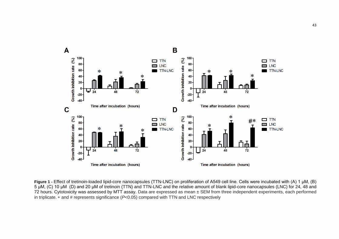
3.2. TTN-LNC inhibited cell proliferation of A549 cells
The results from MTT assay showed that incubation with TTN by 24h, 48h or
72h did not induce cytotoxicity on A549 cells, confirming the resistance of these cells
to treatment. However TTN-LNC showed a dose-dependent cytotoxicity with an
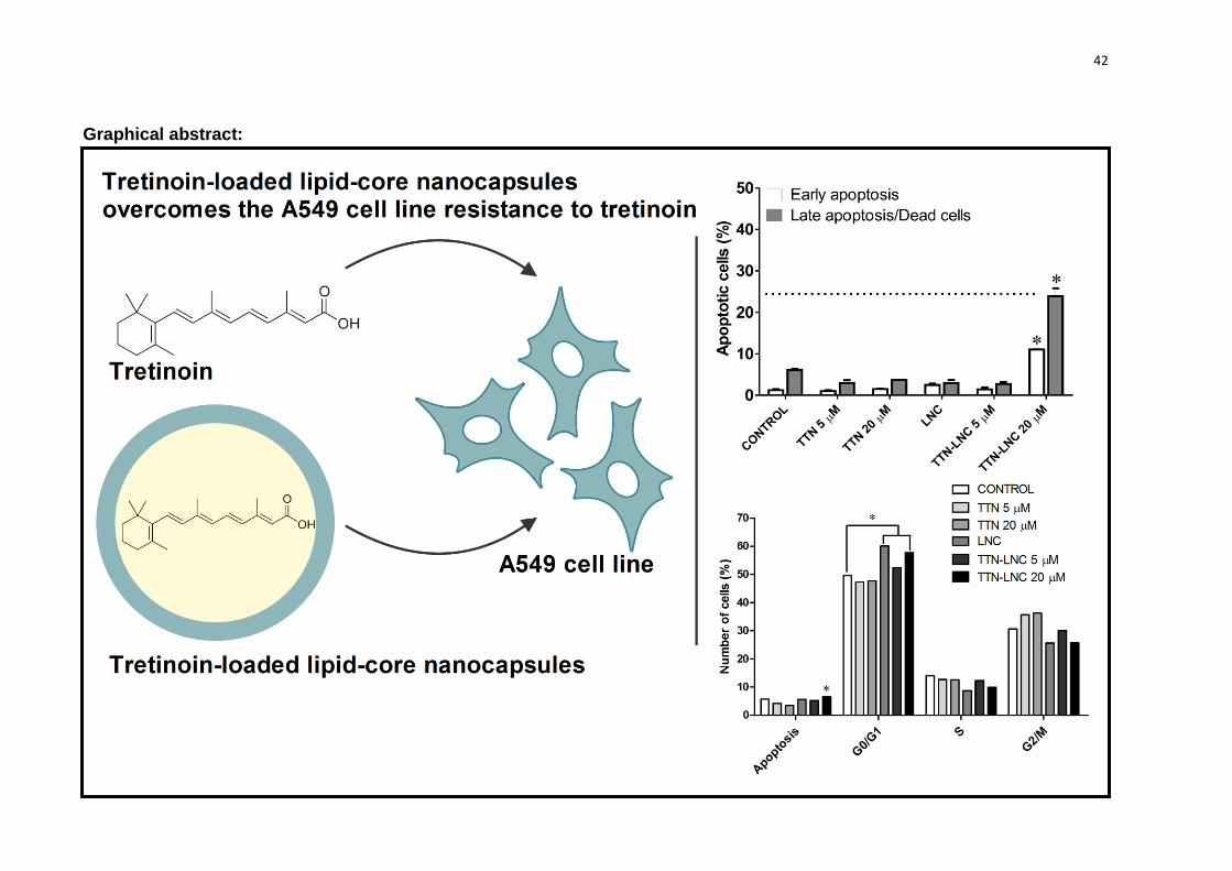
inhibition rate upper to 50% with concentration of 20 µM. At 72h the inhibition rate of
20 µM of TTN and TTN-LNC as well as the equivalent volume of LNC was
respectively 14.0%, 64.2% and 11.2% (Fig. 1).
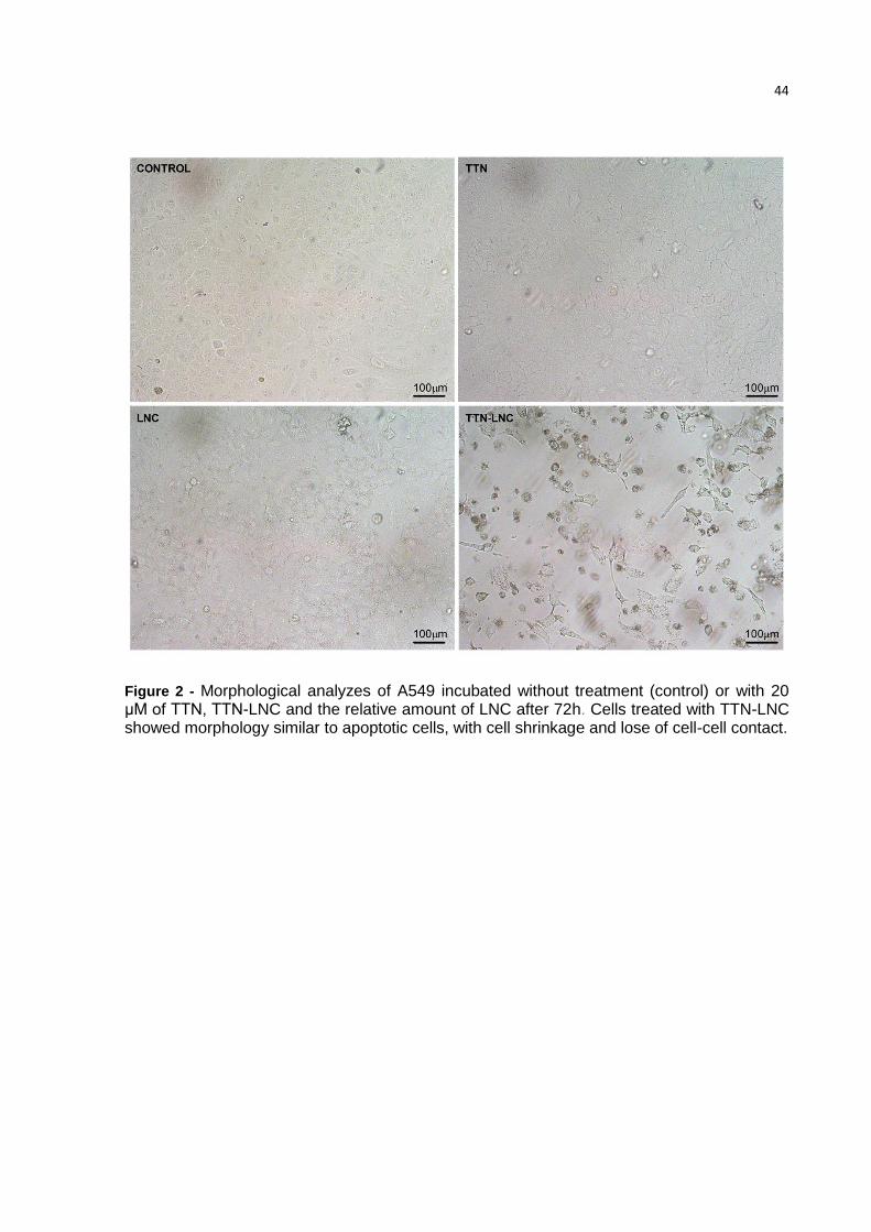
3.3. TTN-LNC alters morphology of A549 cells
A549 cells are human alveolar basal epithelial cells, squamous in nature and
grow adherently, as a monolayer, in vitro. Cells treated with TTN-LNC showed
33
apoptotic morphology such as loss of attachments to other cells and extracellular
matrix and rounding up. Cells incubated with TTN or LNC were similar to control (Fig.
2).
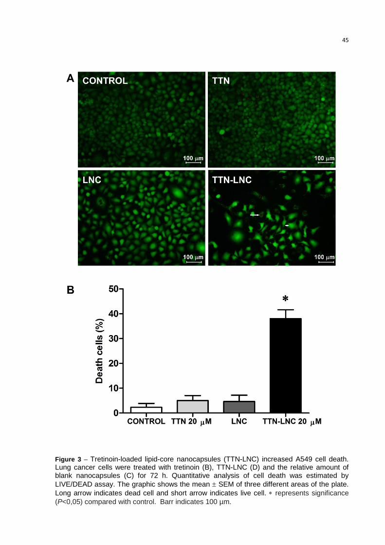
3.4. TTN-LNC reduces the cell viability
The Figure 3 top panel shows that TTN-LNC reduces significantly the cell
viability, while TTN and LNC did not affect the cell viability. Fig.3 bottom panel shows
the mean of dead cells calculated from three different areas of the plate. Samples
without treatment (A), with TTN (B) and with LNC (C) showed no more than 5% of
dead cells, while sample incubated with TTN-LNC (D) showed an average of 38% of
dead cells.
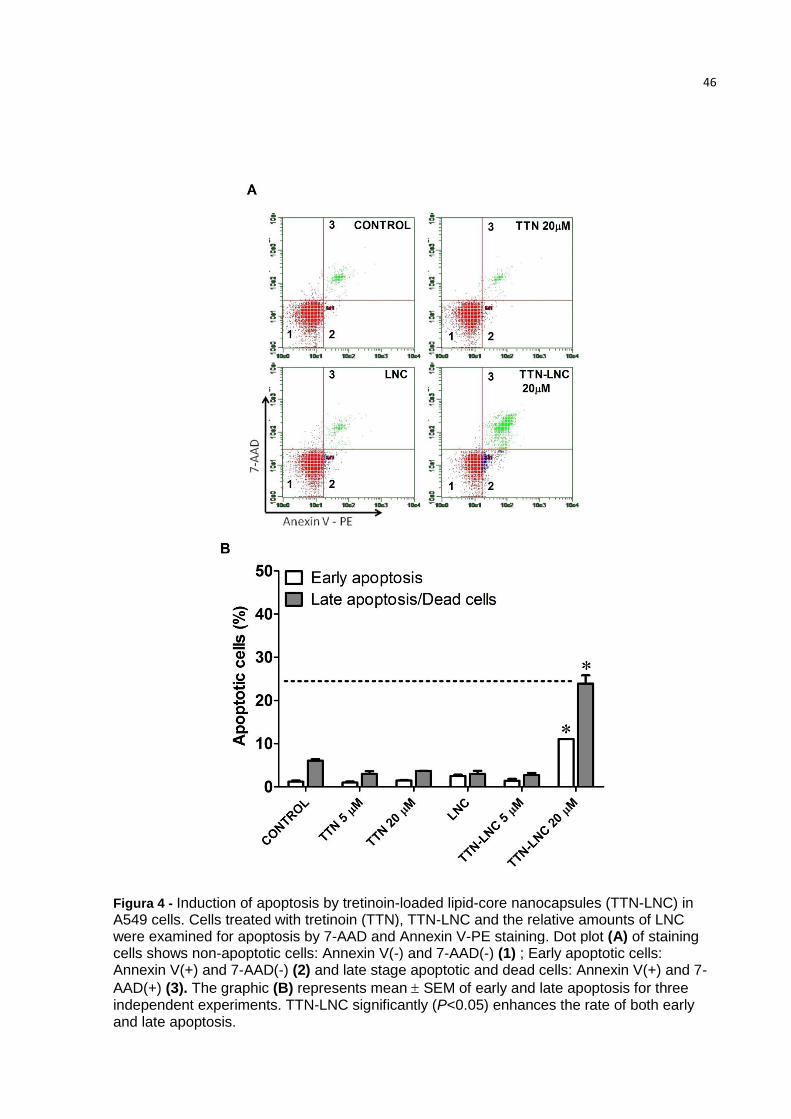
3.5 Apoptosis induction by TTN-LNC on A549 cells
In order to analyze the apoptotic induction by TTN, LNC and TTN-LNC, A549
cells were assessed by flow cytometry with annexin V-PE/7-AAD staining. Figure 4
shows that cells treated with TTN-LNC showed a rate of 11% of early apoptosis and
24.8% of late apoptosis/dead cells. Cells treated with TTN and LNC showed a rate of
apoptosis similar to control.
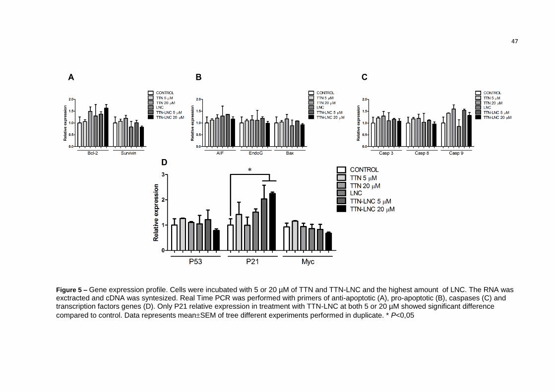
3.6 Analysis of gene expression
An evaluation of gene expression profile was performed to assess which
pathway was involved in TTN-LNC cytotoxic effect. Surprisingly, none of the
apoptotic genes tested was significantly induced by TTN-LNC, except from P21
(Figure 5, D). Transcripts of P21 of cells treated with both concentration of 5 µM and
20 µM of TTN-LNC were increased more than 2-fold related to control (Figure 5).
34
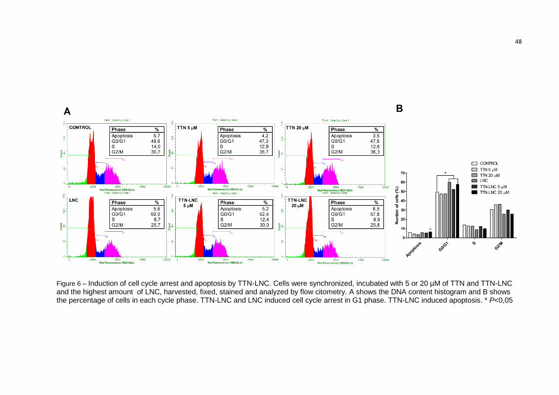
3.7. TTN-LNC-induced cell cycle arrest
Data from flow cytometry of cells stained with propidium iodide showed that
the population with the amount of DNA related to the G0/G1 phase of cell cycle was
significantly increase in cells treated with TTN-LNC (Figure 5). Cells without
treatment showed a rate of 49.6% of cells in G0/G1 phase, while cells incubated for
24 h with TTN-LNC showed a rate of 57.8% of cells in G0/G1 phase. Consequently,
the rate of cells in S and G2/M phase decreased related to control (9.93% and
25.73% from TTN-LNC treatment versus 13.98% and 30.68% from control).
Unexpectedly, the treatment with LNC also increased significantly the rate of cells in
G0/G1 phase (60.5%), which needs to be better understood. Additionally the
presence of Sub-G1 population is representative of apoptotic cells. Wells incubated
with TTN-LNC showed significantly more apoptotic cells.
4. DISCUSSION
A large number of studies have investigated tretinoin therapy in combination
with chemotherapy on different kinds of tumors showing a promising therapeutic
value [12] [22] [23] [24]. However many lung cancer cells exhibit resistance to TTN,
which may explain why some patients do not respond well to treatment [13] [25]. In
this study we demonstrate by the first time that a formulation of tretinoin-loaded lipid
core nanocapsules may overcome the cell resistance showed by human
adenocarcinoma epithelial cell line (A549) to TTN anticancer effects. The formulation
evaluated in this study was previously developed [18]. However, we confirm some of
their important properties before use in the present study. Formulations showed
feasible properties, as particle size below 250 nm, low polydispersity indices (< 0.25)
35
and negative zeta potential, regardless the presence of the drug. These
characteristics are according to our previous study [18] and allow us to investigate
their properties against lung cancer cells.
The MTT assay was performed to detect cytotoxic effect of free tretinoin
(TTN), TTN-LNC and unloaded-nanocapsules (LNC). TTN did not show inhibition of
cell growth at all tested concentrations, confirming the cell resistance for this
compound. TTN-LNC was able to increase significantly the rate of growth inhibition
even with lowest concentrations. However, the treatment with LNC also showed a
reduction in the number of viable cells, which was not sustained over the time. This
result may indicate an interference of nanocapsules in cell metabolism or even
interference in cellular MTT uptake. Nevertheless, TTN-LNC showed significantly
higher growth inhibition than LNC, which reflect an additional activity. Furthermore, in
treatment with LNC the percentage of non viable cells evaluated by Live/Dead assay
was similar to control, while in treatment with TTN-LNC this percentage was
significantly higher, showing that LNC did not affect cell viability.
TTN exerts anti-cancer activities mainly because of its differential effects and
control of cell proliferation. However, a large number of studies have demonstrated
that TTN induces apoptosis of cancer cells [7] [26]. Here we demonstrated that TTN
did not induce apoptosis on A549 cells at tested concentrations, but analyses of
Annexin-V/7-AAD staining as well as DNA fragmentation by flow cytometry showed
that TTN-LNC increases significantly this rate. These results supported the benefits
of TTN encapsulation on its anti-tumor effect.
Apoptosis is one of the programmed cell death process targeted by the
antitumor agents. Many therapies as chemotherapy and radiotherapy induce cellular
stress and DNA damage, triggering the intrinsic apoptotic pathway [27]. This pathway
36
is mediated by the mitochondria and controlled by the balance and interactions
between pro- and antiapoptotic members of the Bcl-2 family proteins, such as Bax
and Bcl-2 [28]. The results of expression analyses showed that the transcripts of anti-
and pro-apoptotic genes, Bcl-2 and Bax were not significantly altered by the
treatment, suggesting that the intrinsic apoptotic pathway was not activated.
Caspases are intracellular cysteine proteases which are cleaved and
activated, causing most of the biochemical changes observed in apoptotic cells [29].
Apoptotic extrinsic pathway activates caspase 8 and apoptotic intrinsic pathway
activates caspase 9. Both caspase 8 and 9 can activate effectors caspases, like
caspase 3. Ours data showed that TTN-LNC does not induced up-regulation of
caspase 3, 8 or 9 what suggest that the anticancer activity of this compound may not
be through apoptosis dependent of Bcl-2 or caspases expression. Some reports
have been showing other types of programmed cell death that may present apoptosis
features by caspase-independent pathways [27] [30].Transcriptional products
of apoptosis induction factor (AIF) and Endonuclease G (Endo G), which
are responsible for DNA degradation in caspase-independent apoptotic
pathway, were also not increased by the treatments tested in this study.
Some studies have related the induction of p21 by retinoic acid [31]. Here we
demonstrated that TTN-LNC up-regulated the p21 transcription and promoted cell
cycle arrest at G1 phase of A549 cell line. p21 protein is a cyclin dependent kinase
inhibitor that is able to inactivate cyclin dependent kinase 2 (CDK2), as well as
interact with other proteins implicated in various cellular processes [32]. p21 blocks
the expression of S phase specific genes through regulation of cyclin E CDK2 and
arrest the cell cycle in G1 phase. This protein can also suppress the cell proliferation
by other pathways such as interaction with DNA-polymerase subunit [32]. The
37
promoter region of p21 gene contains many regulatory sequences. These sites
provide a potential regulation of this gene by a large amount of inducers or
repressors related to cell cycle and differentiation and stress, including retinoic acid.
We suggest that TTN-LNC was able to enhance the internalization of TTN in A549
cells and that TTN induced expression of P21 by interacting with its receptor in the
promoter region of the gene. p21 expression is also regulated by the protein p53,
however, the p53 gene was not up-regulated by TTN-LNC. Induction of p21 by TTN
without induction of p53 and its association with differentiation, senescence and
inhibition of cell proliferation has been reported [33] [34] [35].
Our results showed for the first time the ability of TTN-LNC overcome lung
cancer cell resistance to TTN, providing support to future applications on lung cancer
therapy. Due to complex activity of tretinoin on cancer cells, more studies are
necessary to elucidate the exact mechanisms by which TTN-LNC was efficient to
induce cell growth inhibition, decrease in cell viability and cell cycle arrest.
5. CONCLUSION
We conclude that TTN-LNC was able to overcome de resistance of adenocarcinoma
cell line A549 to tretinoin, inducing apoptosis, reducing the cell viability and causing
cell cycle arrest in G1 phase, by P21 up-regulation.
ACKNOWLEDGMENTS
This work was supported by the Brazilian funding agencies CAPES, CNPq
and FAPERGS.
Competing interests: The authors have no conflict of interest.
38
REFERENCES
[1] C. Chomienne, N. Balitrand, P. Ballerini, S. Castaigne, T.H. de, and L. Degos, All-trans retinoic acid modulates the retinoic acid receptor-alpha in promyelocytic cells, J. Clin. Invest, 88 (1991) 2150-2154.
[2] M. Orlandi, B. Mantovani, K. Ammar, E. Avitabile, M.P. Dal, and G. Bartolini, Retinoids and cancer: antitumoral effects of ATRA, 9-cis RA and the new retinoid IIF on the HL-60 leukemic cell line, Med. Princ. Pract., 12 (2003) 164-169.
[3] L. Altucci and H. Gronemeyer, The promise of retinoids to fight against cancer, Nat. Rev. Cancer, 1 (2001) 181-193.
[4] D.R. Soprano, P. Qin, and K.J. Soprano, Retinoic acid receptors and cancers, Annu. Rev. Nutr., 24 (2004) 201-221.
[5] M. Rhinn and P. Dolle, Retinoic acid signalling during development, Development, 139 (2012) 843-858.
[6] N. Bushue and Y.J. Wan, Retinoid pathway and cancer therapeutics, Adv. Drug Deliv. Rev., 62 (2010) 1285-1298.
[7] M.C. Chen, C.Y. Huang, S.L. Hsu, E. Lin, C.T. Ku, H. Lin, and C.M. Chen, Retinoic Acid Induces Apoptosis of Prostate Cancer DU145 Cells through Cdk5 Overactivation, Evid. Based. Complement Alternat. Med., 2012 (2012) 580736.
[8] C.S. Niu, M.W. Li, Y.F. Ni, J.M. Chen, J.M. Mei, J. Li, and X.M. Fu, Effect of all-trans retinoic acid on the proliferation and differentiation of brain tumor stem cells, J. Exp. Clin. Cancer Res., 29 (2010) 113.
[9] Q. Xu, Z. Zhang, P. Zhang, and W. Chen, Antisense oligonucleotides and all-trans retinoic acid have a synergistic anti-tumor effect on oral squamous cell carcinoma, BMC. Cancer, 8 (2008) 159.
[10] L.M. Austenaa, H. Carlsen, A. Ertesvag, G. Alexander, H.K. Blomhoff, and R. Blomhoff, Vitamin A status significantly alters nuclear factor-kappaB activity assessed by in vivo imaging, FASEB J., 18 (2004) 1255-1257.
[11] R. Connolly, N.K. Nguyen, and S. Sukumar, Molecular Pathways: Current Role and Future Directions of the Retinoic Acid Pathway In Cancer Prevention and Treatment, Clin. Cancer Res., (2013).
[12] O. Arrieta, Gonzalez-De la Rosa CH, E. Arechaga-Ocampo, G. Villanueva-Rodriguez, T.L. Ceron-Lizarraga, L. Martinez-Barrera, M.E. Vazquez-Manriquez, M.A. Rios-Trejo, M.A. Alvarez-Avitia, N. Hernandez-Pedro, C. Rojas-Marin, and J. De la Garza, Randomized phase II trial of All-trans-retinoic acid with chemotherapy based on paclitaxel and cisplatin as first-line
39
treatment in patients with advanced non-small-cell lung cancer, J. Clin. Oncol., 28 (2010) 3463-3471.
[13] J. Geradts, J.Y. Chen, E.K. Russell, J.R. Yankaskas, L. Nieves, and J.D. Minna, Human lung cancer cell lines exhibit resistance to retinoic acid treatment, Cell Growth Differ., 4 (1993) 799-809.
[14] S. Kawakami, S. Suzuki, F. Yamashita, and M. Hashida, Induction of apoptosis in A549 human lung cancer cells by all-trans retinoic acid incorporated in DOTAP/cholesterol liposomes, J. Control Release, 110 (2006) 514-521.
[15] Z. Gao, L. Zhang, and Y. Sun, Nanotechnology applied to overcome tumor drug resistance, J. Control Release, 162 (2012) 45-55.
[16] E. Trapasso, D. Cosco, C. Celia, M. Fresta, and D. Paolino, Retinoids: new use by innovative drug-delivery systems, Expert. Opin. Drug Deliv., 6 (2009) 465-483.
[17] A.F. Ourique, A.R. Pohlmann, S.S. Guterres, and R.C. Beck, Tretinoin-loaded nanocapsules: Preparation, physicochemical characterization, and photostability study, Int. J. Pharm., 352 (2008) 1-4.
[18] A.F. Ourique, S. Azoubel, C.V. Ferreira, C.B. Silva, M.C. Marchiori, A.R. Pohlmann, S.S. Guterres, and R.C. Beck, Lipid-core nanocapsules as a nanomedicine for parenteral administration of tretinoin: development and in vitro antitumor activity on human myeloid leukaemia cells, J. Biomed. Nanotechnol., 6 (2010) 214-223.
[19] K.R. Begnini, C. Rizzi, V.F. Campos, S. Borsuk, E. Schultze, V.C. Yurgel, F. Nedel, O.A. Dellagostin, T. Collares, and F.K. Seixas, Auxotrophic recombinant Mycobacterium bovis BCG overexpressing Ag85B enhances cytotoxicity on superficial bladder cancer cells in vitro, Appl. Microbiol. Biotechnol., (2012).
[20] F. Nedel, V.F. Campos, D. Alves, A.J. McBride, O.A. Dellagostin, T. Collares, L. Savegnago, and F.K. Seixas, Substituted diaryl diselenides: cytotoxic and apoptotic effect in human colon adenocarcinoma cells, Life Sci., 91 (2012) 345-352.
[21] M.W. Pfaffl, A new mathematical model for relative quantification in real-time RT-PCR, Nucleic Acids Res., 29 (2001) e45.
[22] M. Bryan, E.D. Pulte, K.C. Toomey, L. Pliner, A.C. Pavlick, T. Saunders, and R. Wieder, A pilot phase II trial of all-trans retinoic acid (Vesanoid) and paclitaxel (Taxol) in patients with recurrent or metastatic breast cancer, Invest New Drugs, 29 (2011) 1482-1487.
[23] K.A. David, N.P. Mongan, C. Smith, L.J. Gudas, and D.M. Nanus, Phase I trial of ATRA-IV and Depakote in patients with advanced solid tumor malignancies, Cancer Biol. Ther., 9 (2010) 678-684.
40
[24] S.H. Park, W.C. Gray, I. Hernandez, M. Jacobs, R.A. Ord, M. Sutharalingam, R.G. Smith, D.A. Van Echo, S. Wu, and B.A. Conley, Phase I trial of all-trans retinoic acid in patients with treated head and neck squamous carcinoma, Clin. Cancer Res., 6 (2000) 847-854.
[25] A. Raif, G.M. Marshall, J.L. Bell, J. Koach, O. Tan, C. D'andreti, W. Thomas, E. Sekyere, M. Norris, M. Haber, M. Kavallaris, and B.B. Cheung, The estrogen-responsive B box protein (EBBP) restores retinoid sensitivity in retinoid-resistant cancer cells via effects on histone acetylation, Cancer Lett., 277 (2009) 82-90.
[26] L. Dhandapani, P. Yue, S.S. Ramalingam, F.R. Khuri, and S.Y. Sun, Retinoic acid enhances TRAIL-induced apoptosis in cancer cells by upregulating TRAIL receptor 1 expression, Cancer Res., 71 (2011) 5245-5254.
[27] C. Vangestel, C. Van de Wiele, G. Mees, and M. Peeters, Forcing cancer cells to commit suicide, Cancer Biother. Radiopharm., 24 (2009) 395-407.
[28] J.M. Adams and S. Cory, The Bcl-2 apoptotic switch in cancer development and therapy, Oncogene, 26 (2007) 1324-1337.
[29] M. Lamkanfi, N. Festjens, W. Declercq, T. Vanden Berghe, and P. Vandenabeele, Caspases in cell survival, proliferation and differentiation, Cell Death. Differ., 14 (2007) 44-55.
[30] M. Leist and M. Jaattela, Four deaths and a funeral: from caspases to alternative mechanisms, Nat. Rev. Mol. Cell Biol., 2 (2001) 589-598.
[31] T. Otsuki, H. Sakaguchi, T. Hatayama, P. Wu, A. Takata, and F. Hyodoh, Effects of all-trans retinoic acid (ATRA) on human myeloma cells, Leuk. Lymphoma, 44 (2003) 1651-1656.
[32] V.S. Romanov, V.A. Pospelov, and T.V. Pospelova, Cyclin-dependent kinase inhibitor p21(Waf1): contemporary view on its role in senescence and oncogenesis, Biochemistry (Mosc. ), 77 (2012) 575-584.
[33] O. Clark, S. Daga, and A.W. Stoker, Tyrosine phosphatase inhibitors combined with retinoic acid can enhance differentiation of neuroblastoma cells and trigger ERK- and AKT-dependent, p53-independent senescence, Cancer Lett., 328 (2013) 44-54.
[34] B. Su, X. Chen, C. Zhong, N. Guo, J. He, and Y. Fan, All-trans retinoic acid inhibits mesangial cell proliferation by up-regulating p21Waf1/Cip1 and p27Kip1 and down-regulating Skp2, J. Nephrol., 25 (2012) 1031-1040.
[35] Q. Wu, Z. Chen, and W. Su, Growth inhibition of gastric cancer cells by all-trans retinoic acid through arresting cell cycle progression, Chin Med. J. (Engl. ), 114 (2001) 958-961.

41
Table 1. Primers sequences used in this study.
Primers Sequence 5’→ 3’
p53 For AGCGAGCACTGCCCAACA
p53 Rev CACGCCCACGGATCTGAA
Bcl-2 For GTGTGGAGAGCGTCAACC
Bcl-2 Rev CTTCAGAGACAGCCAGGAG
Bax For ATGCGTCCACCAAGAAGC
Bax Rev ACGGCGGCAATCATCCTC
KRAS For TTATAAGGCCTGCTGAAAATGACTGAA
KRAS Rev TGAATTAGCTGTATCGTCAAGGCACT
Casp9 For CCAGAGATTCGCAAACCAGAGG
Casp9 Rev GAGCACCGACATCACCAAATCC
Survivin For CTGTGGGCCCCTTAGCAAT
Survivin Rev TAAGCCCGGGAATCAAAACA
p21 For CCTAATCCGCCCACAGGAA
p21 Rev ACCTCCGGGAGAGAGGAAAA
MYC For TCAGCAACAACCGAAAATGC
MYC Rev TTCCGTAGCTGTTCAAGTTTGTG
GAPDH For GGATTTGGTCGTATTGGG
GAPDH Rev TCGCTCCTGGAAGATGG
42
Graphical abstract:
43
Figure 1 - Effect of tretinoin-loaded lipid-core nanocapsules (TTN-LNC) on proliferation of A549 cell line. Cells were incubated with (A) 1 μM, (B) 5 μM, (C) 10 μM (D) and 20 μM of tretinoin (TTN) and TTN-LNC and the relative amount of blank lipid-core nanocapsules (LNC) for 24, 48 and 72 hours. Cytotoxicity was assessed by MTT assay. Data are expressed as mean ± SEM from three independent experiments, each performed
in triplicate. and # represents significance (P<0.05) compared with TTN and LNC respectively
44
Figure 2 - Morphological analyzes of A549 incubated without treatment (control) or with 20 μM of TTN, TTN-LNC and the relative amount of LNC after 72h. Cells treated with TTN-LNC showed morphology similar to apoptotic cells, with cell shrinkage and lose of cell-cell contact.
45
Figure 3 – Tretinoin-loaded lipid-core nanocapsules (TTN-LNC) increased A549 cell death. Lung cancer cells were treated with tretinoin (B), TTN-LNC (D) and the relative amount of blank nanocapsules (C) for 72 h. Quantitative analysis of cell death was estimated by
LIVE/DEAD assay. The graphic shows the mean SEM of three different areas of the plate.
Long arrow indicates dead cell and short arrow indicates live cell. represents significance
(P<0,05) compared with control. Barr indicates 100 µm.
46
Figura 4 - Induction of apoptosis by tretinoin-loaded lipid-core nanocapsules (TTN-LNC) in A549 cells. Cells treated with tretinoin (TTN), TTN-LNC and the relative amounts of LNC were examined for apoptosis by 7-AAD and Annexin V-PE staining. Dot plot (A) of staining cells shows non-apoptotic cells: Annexin V(-) and 7-AAD(-) (1) ; Early apoptotic cells: Annexin V(+) and 7-AAD(-) (2) and late stage apoptotic and dead cells: Annexin V(+) and 7-
AAD(+) (3). The graphic (B) represents mean SEM of early and late apoptosis for three independent experiments. TTN-LNC significantly (P<0.05) enhances the rate of both early and late apoptosis.
47
Figure 5 – Gene expression profile. Cells were incubated with 5 or 20 µM of TTN and TTN-LNC and the highest amount of LNC. The RNA was exctracted and cDNA was syntesized. Real Time PCR was performed with primers of anti-apoptotic (A), pro-apoptotic (B), caspases (C) and transcription factors genes (D). Only P21 relative expression in treatment with TTN-LNC at both 5 or 20 µM showed significant difference
compared to control. Data represents meanSEM of tree different experiments performed in duplicate. * P<0,05
48
Figure 6 – Induction of cell cycle arrest and apoptosis by TTN-LNC. Cells were synchronized, incubated with 5 or 20 µM of TTN and TTN-LNC and the highest amount of LNC, harvested, fixed, stained and analyzed by flow citometry. A shows the DNA content histogram and B shows the percentage of cells in each cycle phase. TTN-LNC and LNC induced cell cycle arrest in G1 phase. TTN-LNC induced apoptosis. * P<0,05
49
4. CONCLUSÃO
Neste trabalho demonstramos que nanocápsulas de núcleo lipídico de tretinoína
(TT-LCNC) foram capazes de superar a resistência que as células de
adenocarcinoma de pulmão humano, linhagem A549, apresentam aos efeitos
antitumorais de TT. Esta superação foi demonstrada a partir da indução de
citotoxicidade, medida pelo ensaio de Live/Dead e MTT; apoptose, medida por
citometria de fluxo através da avaliação da externalização de fosfatidilserina, bem
como fragmentação de DNA; e parada do ciclo celular, medida também por
citometria de fluxo através da avaliação de conteúdo de DNA corado com iodeto de
propideo. Além do mais, pode se demonstrar que esse efeito está ocorrendo via
regulação da expressão de P21, uma proteína inibidora de ciclinas que controla,
dentre outros mecanismos, a passagem do ciclo celular de G1 para S. Os resultados
encontrados estão de acordo com os efeitos de TT descritos na literatura. Com isso
o trabalho evidenciou um possível potencial terapêutico dessa formulação para o
tratamento de câncer de pulmão resistente a TT.
50
5. REFERÊNCIAS
ADAMS, J. M.;CORY, S. The Bcl-2 apoptotic switch in cancer development and therapy. Oncogene, v.26, p.1324-1337, 2007.
ALTUCCI, L.;GRONEMEYER, H. The promise of retinoids to fight against cancer. Nat. Rev. Cancer, v.1, p.181-193, 2001.
ALTUCCI, L.; LEIBOWITZ, M. D.; OGILVIE, K. M.; DE LERA, A. R.; GRONEMEYER, H. RAR and RXR modulation in cancer and metabolic disease. Nat. Rev. Drug Discov., v.6, p.793-810, 2007.
ARRIETA, O.; GONZALEZ-DE LA ROSA CH; ARECHAGA-OCAMPO, E.; VILLANUEVA-RODRIGUEZ, G.; CERON-LIZARRAGA, T. L.; MARTINEZ-BARRERA, L.; VAZQUEZ-MANRIQUEZ, M. E.; RIOS-TREJO, M. A.; ALVAREZ-AVITIA, M. A.; HERNANDEZ-PEDRO, N.; ROJAS-MARIN, C.; DE LA GARZA, J. Randomized phase II trial of All-trans-retinoic acid with chemotherapy based on paclitaxel and cisplatin as first-line treatment in patients with advanced non-small-cell lung cancer. J. Clin. Oncol., v.28, p.3463-3471, 2010.
BROWN, G.;HUGHES, P. Retinoid differentiation therapy for common types of acute myeloid leukemia. Leuk. Res. Treatment., v.2012, p.939021, 2012.
BUNN, P. A., JR. Worldwide overview of the current status of lung cancer diagnosis and treatment. Arch. Pathol. Lab Med., v.136, p.1478-1481, 2012.
BUSHUE, N.;WAN, Y. J. Retinoid pathway and cancer therapeutics. Adv. Drug Deliv. Rev., v.62, p.1285-1298, 2010.
CHANSRI, N.; KAWAKAMI, S.; YOKOYAMA, M.; YAMAMOTO, T.; CHAROENSIT, P.; HASHIDA, M. Anti-tumor effect of all-trans retinoic acid loaded polymeric micelles in solid tumor bearing mice. Pharm. Res., v.25, p.428-434, 2008.
ELMORE, S. Apoptosis: a review of programmed cell death. Toxicol. Pathol., v.35, p.495-516, 2007.
FOSTER, D. A.; YELLEN, P.; XU, L.; SAQCENA, M. Regulation of G1 Cell Cycle Progression: Distinguishing the Restriction Point from a Nutrient-Sensing Cell Growth Checkpoint(s). Genes Cancer, v.1, p.1124-1131, 2010.
GADGEEL, S. M.; RAMALINGAM, S. S.; KALEMKERIAN, G. P. Treatment of lung cancer. Radiol. Clin. North Am., v.50, p.961-974, 2012.
GAO, Z.; ZHANG, L.; SUN, Y. Nanotechnology applied to overcome tumor drug resistance. J. Control Release, v.162, p.45-55, 2012.
GERADTS, J.; CHEN, J. Y.; RUSSELL, E. K.; YANKASKAS, J. R.; NIEVES, L.; MINNA, J. D. Human lung cancer cell lines exhibit resistance to retinoic acid treatment. Cell Growth Differ., v.4, p.799-809, 1993.
HANAHAN, D.;WEINBERG, R. A. The hallmarks of cancer. Cell, v.100, p.57-70, 2000.
51
HANAHAN, D.;WEINBERG, R. A. Hallmarks of cancer: the next generation. Cell, v.144, p.646-674, 2011.
HIGUCHI, E.; CHANDRARATNA, R. A.; HONG, W. K.; LOTAN, R. Induction of TIG3, a putative class II tumor suppressor gene, by retinoic acid in head and neck and lung carcinoma cells and its association with suppression of the transformed phenotype. Oncogene, v.22, p.4627-4635, 2003.
Instituto Nacional de Câncer. Instituto Nacional do Câncer (INCA). Instituto Nacional do Câncer . 2013. 15-2-2013. Ref Type: Online Source
Instituto Nacional de Câncer José Alencar Gomes da Silva. Estimativa 2012 : incidência de câncer no Brasil. 2013. Rio de Janeiro : Inca, 2011. 16-2-2013. Ref Type: Online Source
INUI, N.; SASAKI, S.; SUDA, T.; CHIDA, K.; NAKAMURA, H. The loss of retinoic acid receptor alpha, beta and alcohol dehydrogenase3 expression in non-small cell lung cancer. Respirology., v.8, p.302-309, 2003.
JAIN, M. V.; PACZULLA, A. M.; KLONISCH, T.; DIMGBA, F. N.; RAO, S. B.; ROBERG, K.; SCHWEIZER, F.; LENGERKE, C.; DAVOODPOUR, P.; PALICHARLA, V. R.; MADDIKA, S.; LOS, M. Interconnections between apoptotic, autophagic and necrotic pathways: implications for cancer therapy development. J. Cell Mol. Med., v.17, p.12-29, 2013.
JOO, K. I.; XIAO, L.; LIU, S.; LIU, Y.; LEE, C. L.; CONTI, P. S.; WONG, M. K.; LI, Z.; WANG, P. Crosslinked multilamellar liposomes for controlled delivery of anticancer drugs. Biomaterials, v.34, p.3098-3109, 2013.
KAWAKAMI, S.; SUZUKI, S.; YAMASHITA, F.; HASHIDA, M. Induction of apoptosis in A549 human lung cancer cells by all-trans retinoic acid incorporated in DOTAP/cholesterol liposomes. J. Control Release, v.110, p.514-521, 2006.
LAMKANFI, M.; FESTJENS, N.; DECLERCQ, W.; VANDEN BERGHE, T.; VANDENABEELE, P. Caspases in cell survival, proliferation and differentiation. Cell Death. Differ., v.14, p.44-55, 2007.
LEIST, M.;JAATTELA, M. Four deaths and a funeral: from caspases to alternative mechanisms. Nat. Rev. Mol. Cell Biol., v.2, p.589-598, 2001.
MADDIKA, S.; ANDE, S. R.; PANIGRAHI, S.; PARANJOTHY, T.; WEGLARCZYK, K.; ZUSE, A.; ESHRAGHI, M.; MANDA, K. D.; WIECHEC, E.; LOS, M. Cell survival, cell death and cell cycle pathways are interconnected: implications for cancer therapy. Drug Resist. Updat., v.10, p.13-29, 2007.
MATEO, V.; LAGNEAUX, L.; BRON, D.; BIRON, G.; ARMANT, M.; DELESPESSE, G.; SARFATI, M. CD47 ligation induces caspase-independent cell death in chronic lymphocytic leukemia. Nat. Med., v.5, p.1277-1284, 1999.
NIU, C. S.; LI, M. W.; NI, Y. F.; CHEN, J. M.; MEI, J. M.; LI, J.; FU, X. M. Effect of all-trans retinoic acid on the proliferation and differentiation of brain tumor stem cells. J. Exp. Clin. Cancer Res., v.29, p.113, 2010.
52
ORLANDI, M.; MANTOVANI, B.; AMMAR, K.; AVITABILE, E.; DAL, M. P.; BARTOLINI, G. Retinoids and cancer: antitumoral effects of ATRA, 9-cis RA and the new retinoid IIF on the HL-60 leukemic cell line. Med. Princ. Pract., v.12, p.164-169, 2003.
OURIQUE, A. F.; AZOUBEL, S.; FERREIRA, C. V.; SILVA, C. B.; MARCHIORI, M. C.; POHLMANN, A. R.; GUTERRES, S. S.; BECK, R. C. Lipid-core nanocapsules as a nanomedicine for parenteral administration of tretinoin: development and in vitro antitumor activity on human myeloid leukaemia cells. J. Biomed. Nanotechnol., v.6, p.214-223, 2010.
OURIQUE, A. F.; POHLMANN, A. R.; GUTERRES, S. S.; BECK, R. C. Tretinoin-loaded nanocapsules: Preparation, physicochemical characterization, and photostability study. Int. J. Pharm., v.352, p.1-4, 2008.
REGO, E. M.; KIM, H. T.; RUIZ-ARGUELLES, G. J.; UNDURRAGA, M. S.; URIARTE, M. D.; JACOMO, R. H.; GUTIERREZ-AGUIRRE, H.; MELO, R. A.; BITTENCOURT, R.; PASQUINI, R.; PAGNANO, K.; FAGUNDES, E. M.; CHAUFFAILLE, M. D.; CHIATTONE, C. S.; MARTINEZ, L.; MEILLON, L. A.; GOMEZ-ALMAGUER, D.; KWAAN, H. C.; GARCES-EISELE, J.; GALLAGHER, R.; NIEMEYER, C. M.; SCHRIER, S. L.; TALLMAN, M.; GRIMWADE, D.; GANSER, A.; BERLINER, N.; RIBEIRO, R. C.; LO-COCO, F.; LOWENBERG, B.; SANZ, M. A. Improving acute promyelocytic leukemia (APL) outcome in developing countries through networking, results of the International Consortium on APL. Blood,2013.
RHINN, M.;DOLLE, P. Retinoic acid signalling during development. Development, v.139, p.843-858, 2012.
SOPRANO, D. R.; QIN, P.; SOPRANO, K. J. Retinoic acid receptors and cancers. Annu. Rev. Nutr., v.24, p.201-221, 2004.
SULTANA, S.; KHAN, M. R.; KUMAR, M.; KUMAR, S.; ALI, M. Nanoparticles-mediated drug delivery approaches for cancer targeting: a review. J. Drug Target,2012.
TRAPASSO, E.; COSCO, D.; CELIA, C.; FRESTA, M.; PAOLINO, D. Retinoids: new use by innovative drug-delivery systems. Expert. Opin. Drug Deliv., v.6, p.465-483, 2009.
VANGESTEL, C.; VAN DE WIELE, C.; MEES, G.; PEETERS, M. Forcing cancer cells to commit suicide. Cancer Biother. Radiopharm., v.24, p.395-407, 2009.
VERMEULEN, K.; VAN BOCKSTAELE, D. R.; BERNEMAN, Z. N. The cell cycle: a review of regulation, deregulation and therapeutic targets in cancer. Cell Prolif., v.36, p.131-149, 2003.
WLODKOWIC, D.; SKOMMER, J.; DARZYNKIEWICZ, Z. Cytometry of apoptosis. Historical perspective and new advances. Exp. Oncol., v.34, p.255-262, 2012.
XU, Q.; ZHANG, Z.; ZHANG, P.; CHEN, W. Antisense oligonucleotides and all-trans retinoic acid have a synergistic anti-tumor effect on oral squamous cell carcinoma. BMC. Cancer, v.8, p.159, 2008.
XU, X. C. Tumor-suppressive activity of retinoic acid receptor-beta in cancer. Cancer Lett., v.253, p.14-24, 2007.
ZHANG, L. Q.; WANG, J.; JIANG, F.; XU, L.; LIU, F. Y.; YIN, R. Prognostic value of survivin in patients with non-small cell lung carcinoma: a systematic review with meta-analysis. PLoS. One., v.7, p.e34100, 2012.


![PCD (2) 04.07.2016.ppt [Kompatibilit tsmodus]) · 3 Cytochrom c-Freisetzung Durch Caspase-Aktivierung Aktivierung Apoptose-spezifischer Endonucleasen Molekulare Merkmale der Apoptose](https://img.dokumen.tips/doc/110x75/5d49631588c9933f6b8b87b4/pcd-2-04072016ppt-kompatibilit-tsmodus-3-cytochrom-c-freisetzung-durch.jpg)