Embed Size (px)
Citation preview
RESEARCH ARTICLE
Unexpected Interaction with Dispersed CrudeOil Droplets Drives Severe Toxicity in AtlanticHaddock EmbryosElin Sørhus1,2*, Rolf B. Edvardsen1,Ørjan Karlsen5, Trond Nordtug3, Terje van derMeeren5, Anders Thorsen1, Christopher Harman4, Sissel Jentoft2, Sonnich Meier1
1 Institute of Marine Research, Nordnes, Bergen, Norway, 2 Centre for Ecological and EvolutionarySynthesis (CEES), University of Oslo, Blindern, Oslo, Norway, 3 SINTEF Materials and Chemistry, Sluppen,Trondheim, Norway, 4 Norwegian Institute for Water Research (NIVA), Grimstad, Norway, 5 Institute ofMarine Research (IMR), Austevoll Research Station, and Hjort Centre for Marine Ecosystem Dynamics,Storebø, Norway
AbstractThe toxicity resulting from exposure to oil droplets in marine fish embryos and larvae is still
subject for debate. The most detailed studies have investigated the effects of water-
dissolved components of crude oil in water accommodated fractions (WAFs) that lack bulk
oil droplets. Although exposure to dissolved petroleum compounds alone is sufficient to
cause the characteristic developmental toxicity of crude oil, few studies have addressed
whether physical interaction with oil micro-droplets are a relevant exposure pathway for
open water marine speices. Here we used controlled delivery of mechanically dispersed
crude oil to expose pelagic embryos and larvae of a marine teleost, the Atlantic haddock
(Melanogrammus aeglefinus). Haddock embryos were exposed continuously to two differ-
ent concentrations of dispersed crude oil, high and low, or in pulses. By 24 hours of expo-
sure, micro-droplets of oil were observed adhering and accumulating on the chorion,
accompanied by highly elevated levels of cyp1a, a biomarker for exposure to aromatic hy-
drocarbons. Embryos from all treatment groups showed abnormalities representative of
crude oil cardiotoxicity at hatch (5 days of exposure), such as pericardial and yolk sac
edema. Compared to other species, the frequency and severity of toxic effects was higher
than expected for the waterborne PAH concentrations (e.g., 100% of larvae had edema at
the low treatment). These findings suggest an enhanced tissue uptake of PAHs and/or
other petroleum compounds from attached oil droplets. These studies highlight a novel
property of haddock embryos that leads to greater than expected impact from dispersed
crude oil. Given the very limited number of marine species tested in similar exposures, the
likelihood of other species with similar properties could be high. This unanticipated result
therefore has implications for assessing the ecological impacts of oil spills and the use of
methods for dispersing oil in the open sea.
PLOS ONE | DOI:10.1371/journal.pone.0124376 April 29, 2015 1 / 21
OPEN ACCESS
Citation: Sørhus E, Edvardsen RB, Karlsen Ø,Nordtug T, van der Meeren T, Thorsen A, et al. (2015)Unexpected Interaction with Dispersed Crude OilDroplets Drives Severe Toxicity in Atlantic HaddockEmbryos. PLoS ONE 10(4): e0124376. doi:10.1371/journal.pone.0124376
Academic Editor: Ilaria Corsi, University of Siena,ITALY
Received: October 14, 2014
Accepted: March 1, 2015
Published: April 29, 2015
Copyright: © 2015 Sørhus et al. This is an openaccess article distributed under the terms of theCreative Commons Attribution License, which permitsunrestricted use, distribution, and reproduction in anymedium, provided the original author and source arecredited.
Data Availability Statement: All relevant data arewithin the paper and its Supporting Information files.
Funding: This work was financed by the VISTAfoundation (Project no. 6161, www.vista.no) and theInstitute of Marine Research, Norway (Project no.14236, www.imr.no). The funders had no role in studydesign, data collection and analysis, decision topublish, or preparation of the manuscript.
Competing Interests: The authors have declaredthat no competing interests exist.

IntroductionWorldwide energy demands have resulted in increased hydrocarbon extraction activity inPolar Regions, as well as at greater ocean depths. This includes areas around the Lofoten Is-lands, in the Norwegian Arctic and in the Barents Sea, which are all regions considered to be es-pecially vulnerable since they are important spawning and nursing grounds for severalcommercially important species of marine fish, including Atlantic haddock (Melanogrammusaeglefinus), Atlantic cod (Gadus morhua) and herring (Clupea harengus) [1]. Consequently,there is great concern surrounding the possible long-term impact on the ecosystems in theseareas from either accidental oil spills or from chronic exposure to operational discharges ofproduced water [2–5].
To be able to predict any outcome of an eventual oil spill, comprehensive risk assessmenttools are needed. In recent years large efforts have been put into development and improve-ment of risk assessment tools by coupling different models such as, i) oil drift and fate models,ii) distribution models for zooplankton and fish embryo/larvae, iii) ecotoxicology effect modelsand eventually iv) ecosystem models to simulate effects of oil pollution on fish populations ormulti species ecosystems [6,7]. To reach this ambitious goal however, there is a clear need formore and better empirical data in all steps of the modelling. Especially for the ecotoxicity mod-els there are still clear limitations in the available data on relevant marine coldwater species. Inparticular there is a need for more data on fitness parameters (survival, development andgrowth) and bioaccumulation in early life stages of marine fish in order to produce reliable riskmodels [8]. For example fish embryo and yolk sac larvae are sensitive to low total polyaromatichydrocarbons (TPAH) concentrations (0.1–50 μg/l), and pericardial or yolk sac edema, bonedeformities, delayed development or mortality have been observed [9–16].
There is a general understanding that it is primarily the water dissolved oil compoundswhich provide the largest contribution to the toxicity to fish, as they are more readily bioavail-able [13,17]. This may have important implications for which compounds (only the more read-ily water soluble PAHs or the total oil load) that are used in the risk assessment modelling ofoil spills [7]. Catastrophes like the 1989 Exxon Valdez and 2010 Deepwater Horizon oil spillsled to intensive studies on the effects of crude oils on developing fish. Field-collected herringand salmon larvae after the Exxon Valdez showed developmental defects and mortality, whichwas linked to PAHs, an abundant fraction of most crude oils [9,18,19]. Further research usingthe zebrafish model proposed that the crude oil caused disruption of embryonic cardiac func-tion and morphogenesis. This cardiotoxicity was specifically associated to three-ringed PAHs[20,21], and it is now established that crude oil from various geological sources disrupts heartdevelopment [22,23] in diverse fish species [24,25]. Recent studies imply that compounds inthe oil have the ability to block potassium and calcium ion channels, thereby disrupting signal-ing essential for excitation-contraction coupling in heart muscle cells [25].
When oil is released into the sea, wave energy and/or use of chemical dispersant duringclean up may generate oil-in-water dispersions with micron-sized oil droplets that persist inthe water column [13]. A study aiming to investigate the direct effect of different oil fractions(crude or weathered oil, and shaken or sonicated water accommodated fractions (WAFs)), dur-ing early life stages of the medaka (Oryzias latipes) [26], suggests that direct contact with oilleading to an accumulation on the chorion (eggshell) resulted in an enhanced toxicity not ob-served in exposures to WAF [26]. Additionally, while the toxicity of exposures using the WAFof weathered oils have predominantly been explained by the PAH concentrations, significantcontributions from both unknown and less hydrophobic compounds have also been reported.Thus, in order to adequately ascertain the toxicity posed by oils spills, exposure studies shouldbe designed to also include the potential for enhanced uptake from oil droplets [27–29].
Interaction with Oil Drives Severe Toxicity in Haddock Embryos
PLOS ONE | DOI:10.1371/journal.pone.0124376 April 29, 2015 2 / 21
The Atlantic haddock is a teleost belonging to the Gadidae family, and is a commercially im-portant marine fish distributed on both sides of the North Atlantic [1]. Spawning occurs atdepths of 30–500 m or even deeper and the fertilized eggs rise to the surface where they aresubject to transport by water currents [1,30]. The haddock eggs have high buoyancy, thus themajority of eggs are found in the upper 20 m in the water column [31].
The major undertaking of this study was to obtain more realistic toxicity data on early lifestages of Atlantic haddock exposed to an oil dispersion containing a combination of dissolvedWAF and oil micro-droplets. The exposure was done with an oil-in-water dispersion in twoconcentrations, low (130 μg oil/L) and high (1200 μg oil/L), which are environmentally relevantafter a large oil spill [7]. In an actual oil spill, it is likely for fish eggs/larvae to experience inter-mittent or transient exposure as a result of vertical transport in and out of high oil concentra-tion areas. This study therefore included a pulse exposure: 1200 μg oil/L for 2.4 hours in a 24hour period (the low and pulse treatments groups are exposed to the same total amount of oilover time). Assessment parameters were accomplished by fitness measurements (survival andgrowth) and recording phenotypic and histological abnormalities as well as examining the ex-pression of genes involved in the detoxification pathway, especially cyp1a.
Materials and Methods
Animal collection, maintenance and exposure set upA wild broodstock population of 61 maturing individuals was collected February-March 2013at spawning grounds in the Austevoll area, on the west coast of Norway, and kept in two 7000L tanks at the Institute of Marine Research (IMR), Austevoll Research station. The haddockspawns voluntarily in captivity, and fertilized eggs could therefore be collected from the tanks,transferred to indoor egg incubators, and maintained at 7.0°C until ten days post fertilization(dpf). At 10 dpf,�6000 eggs were transferred into each of twelve 50 L circular exposure tanksof green PE plastic (S1 Fig). The flow through the tanks was 32 L/hr, the water temperature8.0°C, and light regime was 12D:12L. Light for triplicate tanks was provided by the broad spec-trum 2x36W Osram Biolux 965 (Munich, Germany, www.osram.com) dimmable fluorescentlight tubes with 30 min. smooth transitions between light and dark. From four days post hatch-ing (dph), natural zooplankton, mainly copepod nauplii of Acartia longiremis, was harvestedfrom the marine pond system “Svartatjern” [32] and introduced as feed to the larvae.
The tanks were further supplemented with marine microalgae concentrate (Instant Algae,Nanno 3600, Reed Mariculture Inc., CA, USA) until termination of the experiment [33,34].The embryos started to hatch at 13 dpf and 50% hatch was observed at 14 dpf (= 0 dph). Theexposure was stopped at 14 dph and all surviving larvae were counted and transferred into new50 L tanks with clean seawater for further monitoring.
Oil exposure regimeThe oil used was a weathered blend crude oil from the Heidrun oil field of the Norwegian Sea.The blend oil comes from 4 different formations that contain different oil types, both light par-affinic oils (0.83 g/ml) and heavy biodegradated oil (0.93 g/ml) and is exported as a heavycrude oil (0.89 g/ml). The Heidrun blend oil is thought to be representative for the oil typesthat may be found in the Lofoten area. The oil is artificially weathered by distillation to accountfor the fast evaporation that normally occurs after an oil spill at sea. This procedure [35] is asimple one-stage distillation to vapour temperatures of 250°C leaving a residue that corre-sponds to 2–7 days on the sea surface at about 10°C ambient temperature, in this case causingan evaporative loss of 24% of the lighter compounds from the fresh crude oil, and it changesthe oil density from 0.89 g/mL to 0.92 g/mL (SINTEF, 2004) [36].
Interaction with Oil Drives Severe Toxicity in Haddock Embryos
PLOS ONE | DOI:10.1371/journal.pone.0124376 April 29, 2015 3 / 21
The principle of the exposure system and the oil droplet generation is given in detail byNordtug et al., (2011) [37]. The oil was pumped into the dispersion system using a HPLCpump (Pharmacia, LKB2150) with a flow of 0.01 mL/min together with a flow of seawater of180 mL/min. This system generates an oil dispersion with oil droplets in the low μm rangewith a nominal oil load of 46 mg/mL (stock solution). The exposure dose to the tanks was regu-lated by a parallel pipeline system with one line of flowing clean seawater and the other linecontaining a flow of the stock solution. The 2 pipelines were connected by a 3-way magneticvalve allowing water to be collected from both lines. Different dilutions were made by control-ling the relative sampling time from the oil stock solution and clean water, respectively, by acomputer-controlled relay (MiniBee card and BeeStep software). The experimental setup con-sisted of three treatments and a control, each with three replicates: 1) Low: 130 μg oil/L nomi-nal. 2) High: 1200 μg oil/L nominal. 3) Pulse: 1200 μg oil/L nominal for 2.4 hours in a 24 hourperiod. 4) Control: No oil. The concentration of oil in the pulse tanks decreased to approxi-mately zero before the next pulse (S2 Fig). The oil dispersion doses were given by opening themagnetic valve for 27 seconds every minute in the high treatment group and 3 seconds in thelow treatment group. The pulse treatment group received 27 seconds of oil stock solution everyminute for 2.4 hours in each 24 hour period (S2 Fig). Oil dispersions were delivered to each ofthe replicate exposure tanks at a flow rate of 30 mL/min and mixed into the main water supplyof 500 mL/min (S1 Fig). The oil exposure lasted for 18 days (S2 Fig). To avoid oil film on thewater surface in the tanks, a sharp cut oblique drains covered with a plankton mesh wasmounted in all tanks, including the control tanks.
Embryos and larvae were collected at eleven different time points from 11 dpf to 17 dph inaddition to an initial sample at 10 dpf. Two of the samples were collected after the exposure pe-riod (S2 Fig). All animals collected for RNA extraction were photographed in a microscope be-fore they were snap frozen in liquid nitrogen and stored at -80°C. Two pools of embryos werecollected at the first six time points for total RNA extraction. For the subsequent time points,12 individuals were sampled for total RNA extraction.
Analytical chemistryWater samples from each tank (1 L) were taken before the exposure started and at the end of ex-posure. The water sampled were extracted with dichloromethane and prepared for analysis oftotal hydrocarbons (THC) and PAHs. Details of these analytical methods are given in S1 Text.
Oil droplets were diameter measured from a picture with a resolution of 1.38 pixels/μm ob-tained by a stereo microscope (Olympus SZX-10) at 6.3 X magnification and a 21 MPixel cam-era (Canon EOS 5D Mark II).
Sampling and fitness measurementsThe hatching success was estimated at 2 dph. The tanks were gently stirred to obtain an evendistribution of larvae, and subsamples from top to bottom were taken with a cylinder (volume200 mL) whereafter number of larvae was calculated. Hatching were then estimated from num-ber of eggs stocked, subtracted the embryos sampled (�400) during the egg phase. Survivalpost exogenous feeding (14 dph) was measured by calculating all surviving larvae when trans-ferred into new 50 L tanks with clean sea water for further monitoring. The survival (%) wascalculated according to the number of larvae in each tank at 2 dph.
Analysis of phenotypic dataThe length of larvae from 1 dph to 17 dph was measured using ImageJ (ImageJ v2013_2, Na-tional Institutes of Health, Bethesda, Maryland, USA). In addition, the presence of 6 deformity
Interaction with Oil Drives Severe Toxicity in Haddock Embryos
PLOS ONE | DOI:10.1371/journal.pone.0124376 April 29, 2015 4 / 21
parameters; craniofacial deformities, jaw deformities, pericardial and yolk sac edema, spinalcurvature and lack of pigmentation in a large set of larvae from each treatment and controlwere given a grade from 0–3 (where 0 indicates no deformity, 1 some, 2 significant and 3 severedeformity). In some animals or sampling points, some of the parameters were not visible andtherefore denoted not applicable (NA) and taken out of the calculation. The proportion of de-formities was calculated as follows: (Sum of observations for each grade / ((Total number oflarvae�6 parameters)—Sum of NA)) �100%.
Total RNA and cDNA preparationTotal RNA was isolated from frozen pools of animals and individual larvae (except at samplingpoint at 7 dph and 8 dph) using Trizol reagent (Invitrogen, Carlsbad, California, USA), accord-ing to procedures provided by the manufacturer which included a DNase treatment step usinga TURBO DNA-free kit (Life Technologies Corporation). Total RNA from single larvae from 7dph and 8 dph were extracted using RNeasy micro kit (QIAGEN Sample and Assay Technolo-gies) according to procedures provided by the manufacturer. The amount of RNA was quanti-fied using a Nanodrop spectrophotometer (NanoDrop Technologies, Wilmington, DE, USA),and quality checked using a 2100 Bioanalyzer (Agilent Technologies, Santa Clara, CA). cDNAwas subsequently generated using SuperScript VILO cDNA Synthesis Kit (Life TechnologiesCorporation), according to the manufacturer’s instructions. The cDNA was normalized to ob-tain a concentration of 50 ng/μL.
Real time qPCRSpecific primers and probes for real-time, quantitative PCR analysis of Atlantic haddock,cyp1a, ahr2 and gstp1mRNAs, as well as for the reference gene ef1α were designed with Primerexpress software (Applied Biosystems, Carlsbad, California, USA), according to the manufac-turer’s guidelines. Primer and probe sequences are given in S1 Table. TaqMan PCR assays wereperformed in duplicate, using 384-well optical plates on an ABI Prism Fast 7900HT SequenceDetection System (Applied Biosystems, Carlsbad, CA, USA) with settings as follows: 50°C for 2min, 95°C for 20 s, followed by a 40 cycles of 95°C for 1 s and 60°C for 20 s. For each 10 μl PCRreaction, a 2 μl cDNA 1:40 dilution was mixed with 200 nM fluorogenic probe, 900 nM senseprimer, 900 nM antisense primer in 1xTaqMan Fast Advanced Master Mix (Applied Biosys-tems, Carlsbad, California, USA). Gene expression data was calculated relative to the start(10 dpf) sample using the ΔΔCt method as described in detail in Bogerd et. al 2001 [38].
HistologyFive larvae from each replicate were pooled and fixed in 4% PBS buffered paraformaldehydefor 24 hrs at 4.0°C, processed using Histokinette 2000 (Reichert-Jung), and embedded in paraf-fin wax within three days to preserve the RNA and tissue morphology. Sample preparation wasalways performed under RNase free conditions. Serial sectioning (3 μm) of larvae was per-formed for morphological analysis or in situ hybridization using a Leica RM 225 microtome(Leica Microsystems). Histological sections were dewaxed and stained with haematoxylin–erythrosin–safran (HES).
For in situ hybridization (ISH), the Atlantic haddock cyp1a cDNA was amplified usingPCR containing Sp6 and T7 primers in the forward and reverse primers, respectively(Cyp1a_FW_Sp6: 5’-ATTTAGGTGACACTATAGCATCTTCCAGATCCAGATCG -3’Cyp1a_RV_T7: 5’-TAATACGACTCACTATAGGGCATGAACCTCTTCATGGTGG -3’). ThePCR product of 501 bp was used as a template for synthesizing the sense and antisense cRNAprobes by the Sp6 and T7 RNA polymerase, respectively. The digoxigenin (DIG) labelling was
Interaction with Oil Drives Severe Toxicity in Haddock Embryos
PLOS ONE | DOI:10.1371/journal.pone.0124376 April 29, 2015 5 / 21

performed using the DIG-AP RNA labelling kit (Roche Molecular Biochemicals) following themanufacturers protocol.
The in situ hybridization was carried out as described by Weltzien et al. (2003) [39] withsome modifications [40]. Hybridization was always carried out with sense and antisense probeson adjacent sections, and under RNase free conditions.
StatisticsStatistical analysis was performed with GraphPad Prism, version 6 (GraphPad Software Inc.,1996, La Jolla, California, USA). Significant differences in gene expression for the time points11 dpf—3 dph, fitness observations and analytical chemistry between control and exposuregroups, were tested with a one-way ANOVA using the Dunnet’s multiple comparison test afterchecking for normality and variance homogeneity. Statistical analysis of differential cyp1a ex-pression at time point 7 dph—17 dph and length measurements of 1 dph—17 dph were per-formed with a non-parametric Kruskal Wallis test using Dunn’s multiple comparisons, due tomost time points not having a Gaussian distribution. A chi-square test was used to analyze forsignificant differences in the distribution of deformities. The level of significance was set atp<0.05 unless otherwise stated.
Ethics StatementAll animal experiments within the study were approved by NARA, the governmental Norwe-gian Animal Research Authority (http://www.fdu.no/fdu/, reference number 2012/275334-2).All embryos sampled were frozen in liquid nitrogen. All larvae were euthanized using 500 mg/L MS-222 (Tricaine methanesulfonate, TS 222, Sigma-Aldrich) when sampling and at termina-tion of the experiment to achieve immediate death. No humane endpoints were used duringthe experiment because the potential endpoint criteria due to their small size had to be evaluat-ed under a microscope when they were euthanized and sampled. The animals were monitoredevery day, and any dead larvae were removed. The Austevoll Aquaculture Research station hasthe following permission for catch and maintenance of Atlantic haddock: H-AV 77, H-AV 78and H-AV 79. These are permits given by the Norwegian Directorate of Fisheries. Further-more, the Austevoll Aquaculture Research station has a permit to run as a Research Animal fa-cility using fish (all developmental stages), with code 93 from the national IACUC; NARA.
Results
Analytical chemistryThe weathered Heidrun Blend oil used in the current study was a mixture of both light paraf-finic oil and more heavily degradable oils. The PAH constituted 2% of the total oil by weight(19.6 g/kg), and the PAH profile was dominated by the C0–C3 naphthalenes (71%), followedby the tricyclic PAHs which contributed 28% of the total PAHs (S3 Fig).
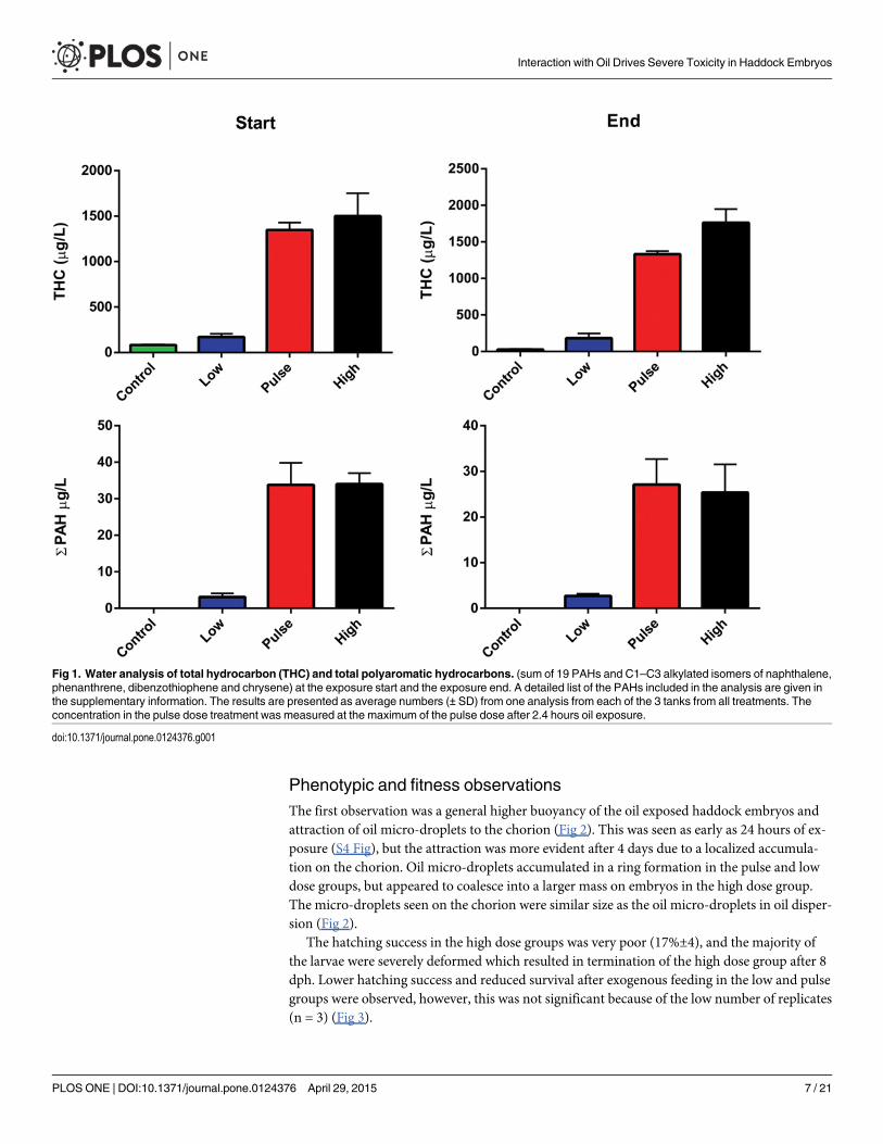
Haddock embryos and larvae were exposed to a mixture containing both oil micro-droplets(diameter 18±3 μm) andWAF. The total oil concentrations in the tanks were measured at thestart of the experiment to; 170 μg THC/L and 3 μg TPAH/L in the low dose group and 1500 μgTHC/L and 34 μg TPAH/L in the high dose. The pulse group had similar concentrations to thehigh dose when measured at the end of the 2.4 hr pulse. Average concentrations recorded atthe end of the 18 days exposure were within 75% of the initial concentration (26 μg TPAH/L inthe high and pulse groups and 2.7 μg TPAH/L in the low dose, Fig 1). The PAH profile in theexposure tanks had slightly higher levels of naphthalene and C1–C4 alkylated naphthalenes(75% of TPAH) compared with pure oil (71%, S3 Fig).
Interaction with Oil Drives Severe Toxicity in Haddock Embryos
PLOS ONE | DOI:10.1371/journal.pone.0124376 April 29, 2015 6 / 21
Phenotypic and fitness observationsThe first observation was a general higher buoyancy of the oil exposed haddock embryos andattraction of oil micro-droplets to the chorion (Fig 2). This was seen as early as 24 hours of ex-posure (S4 Fig), but the attraction was more evident after 4 days due to a localized accumula-tion on the chorion. Oil micro-droplets accumulated in a ring formation in the pulse and lowdose groups, but appeared to coalesce into a larger mass on embryos in the high dose group.The micro-droplets seen on the chorion were similar size as the oil micro-droplets in oil disper-sion (Fig 2).
The hatching success in the high dose groups was very poor (17%±4), and the majority ofthe larvae were severely deformed which resulted in termination of the high dose group after 8dph. Lower hatching success and reduced survival after exogenous feeding in the low and pulsegroups were observed, however, this was not significant because of the low number of replicates(n = 3) (Fig 3).
Fig 1. Water analysis of total hydrocarbon (THC) and total polyaromatic hydrocarbons. (sum of 19 PAHs and C1–C3 alkylated isomers of naphthalene,phenanthrene, dibenzothiophene and chrysene) at the exposure start and the exposure end. A detailed list of the PAHs included in the analysis are given inthe supplementary information. The results are presented as average numbers (± SD) from one analysis from each of the 3 tanks from all treatments. Theconcentration in the pulse dose treatment was measured at the maximum of the pulse dose after 2.4 hours oil exposure.
doi:10.1371/journal.pone.0124376.g001
Interaction with Oil Drives Severe Toxicity in Haddock Embryos
PLOS ONE | DOI:10.1371/journal.pone.0124376 April 29, 2015 7 / 21

The newly hatched larvae in the exposed groups were significantly shorter than the larvaefrom the control group (Fig 4). This reduced length compared to control increased throughoutthe period of exogenous feeding. For the high dose larvae there was no growth during the firsteight days as the larvae (plankton were given at 4 dph), did not succeed with establishment ofexogenous feeding.
Fig 2. Attraction of micro-droplets to chorion. Embryos after 4 days of exposure: (A) Control. (B) Low dose. (C) Pulse dose. (D) High dose. Arrowsindicate examples where attraction of micro-droplets appears to be more pronounced. (E) Oil droplets seen on the chorion. (F) Oil droplets in oil dispersionstock solution. The stock solution was diluted 39 and 265 times to obtain high and low dose, respectively.
doi:10.1371/journal.pone.0124376.g002
Interaction with Oil Drives Severe Toxicity in Haddock Embryos
PLOS ONE | DOI:10.1371/journal.pone.0124376 April 29, 2015 8 / 21

After 7 days of exposure newly hatched haddock larvae showed morphological defects suchas pericardial and yolk sac edema, malformation involving the jaws and other craniofacial struc-tures, and dorsally curved trunks and tails (Fig 5), and increased mortality was observed in allexposed groups. The percentage of pericardial edema observed at 8 dph were 100, 81 and 100%in low dose, pulse dose and high dose, respectively, opposed to 12% in control (S2 Table).
Fig 3. Fitness observations. (A) Average number of hatching larvae after 7 days exposure at embryo stages (start incubation of 6000 embryo in each tank)and (B) Number of surviving larvae after 10 days of exogenous feeding (14 dph, total 18 days of exposure). The numbers in brackets indicate A. averagehatching success in percent (±SD) and B. average larvae survival in percent after exogenous feeding (±SD). Asterisks indicate statistical significancedifference to the control fish, p<0.01 = **.
doi:10.1371/journal.pone.0124376.g003
Fig 4. Length curve of larvae from hatching until end of experiment. Points shown as average (±SD).Asterisks indicate statistical significance difference to the control fish, p<0.05 = *, p<0.01 = **.
doi:10.1371/journal.pone.0124376.g004
Interaction with Oil Drives Severe Toxicity in Haddock Embryos
PLOS ONE | DOI:10.1371/journal.pone.0124376 April 29, 2015 9 / 21
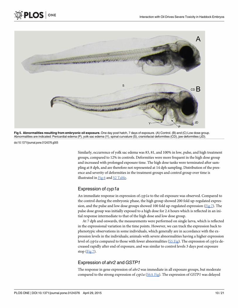
Similarly, occurrence of yolk sac edema was 83, 81, and 100% in low, pulse, and high treatmentgroups, compared to 12% in controls. Deformities were more frequent in the high dose groupand increased with prolonged exposure time. The high dose tanks were terminated after sam-pling at 8 dph, and are therefore not represented at 14 dph sampling. Distribution of the pres-ence and severity of deformities in the treatment groups and control group over time isillustrated in Fig 6 and S2 Table.
Expression of cyp1aAn immediate response in expression of cyp1a to the oil exposure was observed. Compared tothe control during the embryonic phase, the high group showed 200 fold up-regulated expres-sion, and the pulse and low dose groups showed 100 fold up-regulated expression (Fig 7). Thepulse dose group was initially exposed to a high dose for 2.4 hours which is reflected in an ini-tial response intermediate to that of the high dose and low dose group.
At 7 dph and onwards, the measurements were performed on single larva, which is reflectedin the expressional variation in the time points. However, we can track the expression back tophenotypic observations in some individuals, which generally are in accordance with the ex-pression levels in the individuals; animals with severe abnormalities having a higher expressionlevel of cyp1a compared to those with fewer abnormalities (S5 Fig). The expression of cyp1a de-creased rapidly after end of exposure, and was similar to control levels 3 days post exposurestop (Fig 7).
Expression of ahr2 andGSTP1The response in gene expression of ahr2 was immediate in all exposure groups, but moderatecompared to the strong expression of cyp1a (S6A Fig). The expression of GSTP1 was delayed
Fig 5. Abnormalities resulting from embryonic oil exposure.One day post hatch, 7 days of exposure. (A) Control. (B) and (C) Low dose group.Abnormalities are indicated: Pericardial edema (P), yolk sac edema (Y), spinal curvature (S), craniofacial deformities (CD), jaw deformities (JD).
doi:10.1371/journal.pone.0124376.g005
Interaction with Oil Drives Severe Toxicity in Haddock Embryos
PLOS ONE | DOI:10.1371/journal.pone.0124376 April 29, 2015 10 / 21

Fig 7. The relative expression of cyp1a in control, low dose, pulse dose and high dose groups at various sampling points. The expression is relativeto the 0-sample (10 dpf). Fourteen dpf was set as 50% hatching, however only embryos were analysed at this time point. Oil exposure ended at 14 dph.Points are shown as average cyp1a expression (±SD). There are statistical differences between all treatment groups and control at all time points, exceptbetween control and pulse dose groups at 15 dph (1 day post exposure stop), and control and both low dose and pulse dose at 17 dph. Asterisks indicatestatistical significance difference to the control fish, p<0.05 = *, p<0.01 = **.
doi:10.1371/journal.pone.0124376.g007
Fig 6. Distribution of deformities after hatch until end of exposure. 0: No deformity, 1: Some deformity, 2: Significant deformity and 3: Severe deformity.The figure shows the combined score for all six deformity parameters. Statistical difference between the treatment groups and control were analysed usingchi-square test and statistical difference is indicated by asterisk, p<0.01 = ** (Detailed distribution of the single deformities are given in S2 Table).
doi:10.1371/journal.pone.0124376.g006
Interaction with Oil Drives Severe Toxicity in Haddock Embryos
PLOS ONE | DOI:10.1371/journal.pone.0124376 April 29, 2015 11 / 21

and the differences between treatment groups and control were only evident starting two daysafter exposure (S6B Fig).
In situ hybridizationIn situ hybridization was performed to verify the expression and localization of cyp1a in 8 dphlarvae. Even though ISH was not used quantitatively, differences in expression levels in the var-ious treatment groups were clear (Fig 8). No cyp1a expression was observed in the controlgroup. Low and pulse dose groups showed moderate expression in the liver, kidney (Fig 8B and8C) and intestine (Fig 8B), while the high dose group showed high expression in the liver, pan-creas, gills and in a layer of nucleated cells at the midbrain-hindbrain junction and adjacent tothe hindbrain ventricle (n) (Fig 8D). The morphology of the liver was assessed after HES stain-ing in sections adjacent to those used for ISH. Lesions in the liver tissue were observed in allthe treatment groups, but not in the control (Fig 9A, 9B, 9C and 9D respectively).
Fig 8. In situ hybridization of larvae for cyp1a at 8 dph, 12 days of exposure.Only antisense hybridizations are shown.A: Control,B: Low dose,C: PulsedoseD: High dose. Organs indicated: gills (g), liver (l), intestine (i), vascular endothelial cells (n), kidney (k) and pancreas (p).
doi:10.1371/journal.pone.0124376.g008
Interaction with Oil Drives Severe Toxicity in Haddock Embryos
PLOS ONE | DOI:10.1371/journal.pone.0124376 April 29, 2015 12 / 21

DiscussionThe major finding from this study is amplified embryotoxicity observed in Atlantic haddockcompared to similar species when exposed to dispersed crude oil during early developmentalstages. In this study, haddock embryos exposed to low and high TPAH concentrations of 3 μg/L and 34 μg/L showed a suite of abnormalities typical of crude oil cardiotoxicity in other fishspecies: yolk sac and pericardial edema, craniofacial malformation, and curling of the bodyaxis. Importantly, at 8 dph presence of the deformity parameters were similar in animals ex-posed to either 3 or 34 μg/L TPAH, although the high group had generally a higher severitygrade (S2 Table and Fig 6). Pericardial edema was observed in 100% of the analysed animalsexposed to 3 μg/L TPAH, indicating a severe effect even at the low concentration. Similarly, thehigh levels of cyp1amRNA induction in the 3 μg/l exposure (~ 100-fold) relative to the 34 μg/Lexposure group (~ 200-fold) is consistent with an already high tissue PAH dose in the 3 μg/L(low) treatment group in the embryo phase. Our data indicate that haddock embryos showed amore severe toxic response to this range of TPAH concentrations than anticipated from other
Fig 9. HES staining of larvae at 8 dph, 12 days of exposure. A-D show an overview of larvae from the various treatment groups and control, with the liverboxed in and magnified. A: Control.B: Low dose.C: Pulse dose.D: High dose. The extended white areas in the livers of the exposed animalsindicate lesions.
doi:10.1371/journal.pone.0124376.g009
Interaction with Oil Drives Severe Toxicity in Haddock Embryos
PLOS ONE | DOI:10.1371/journal.pone.0124376 April 29, 2015 13 / 21

species exposed to crude oil WAFs containing dispersed micro-droplets. For example, Pacificherring embryos exposed to a high-energy mechanical dispersion of crude oil containing drop-lets showed 35% yolk sac edema at 17.3 μg/L TPAH [41]. Embryos of amberjack and yellowfintuna, both species with a buoyant pelagic egg like haddock, showed an occurrence of pericardi-al edema at 57 ± 10% at 13.8 μg/ and 75± 12% at 3.4 μg/L TPAH, respectively [16]. Althoughthe response of yellowfin tuna approaches that of haddock, tuna eggs would be expected to ac-cumulate dissolved PAHs more rapidly both because they are much smaller (~ 1 mm com-pared to 1.5 mm for haddock) with a higher surface to volume ratio, and because they incubateat a much higher temperature (27°C vs. 8°C). Based on egg size, haddock might be anticipatedto show effects in the same range as herring or amberjack. Although we did not measure tissuePAH concentrations, none of these different species would be expected to have large differ-ences in PAH toxicokinetics at the embryonic stage [42]. Thus the key difference in thesecross-species comparisons is that adherance of oil droplets to the chorion was not observed inany of these prior studies.
The most likely explanation for the high frequency and severity of defects and high levels ofcyp1a induction at such a low TPAH concentration (3μg/L) is the accumulation of oil micro-droplets on the chorion of haddock, which has been described as being generally more adher-ent than similar species like Atlantic cod [43,44]. The accumulation of micro-droplets mostlikely resulted in an amplified uptake of toxic compounds such as PAHs. This phenomenonwas seen in all treatment groups already after 24 hours of exposure, and became more evidentwith prolonged exposure time. Furthermore after five days of exposure, increased mortalitywas observed in the exposed groups; i.e. 17% and 51% hatching success in high and low/pulserespectively compared to 78%, in the controls. These results are however, not in line with previ-ous report on zebrafish (Danio rerio) [17], in which embryos were simultaneously exposed toWAFs with and without micro-droplets, indicated that dissolved PAHs alone were sufficient tocause the typical suite of defects associated with crude oil exposure and contact with dropletswas not required. Moreover, in this case also there was no observation of oil micro-droplets ac-cumulating on the chorion [17]. Additionally, toxicity of WAFs with and without dropletswere similar for Atlantic cod larvae [13,45]: EC50 values for survival probability for cod larvaewere found to be 56 μg TPAH/L for dispersed oil and 38 μg TPAH/L for WAF [13]. Adherenceof oil droplets to the haddock chorion most likely leads to a dual exposure pathway: in additionto uptake of dissolved PAHs from the WAF, attached oil droplets probably either create ahigher local dissolved PAH concentration at the eggshell, or potentially even lead to directtransfer of crude oil compounds across the eggshell. This interpretation is also consistent witha previous study on embryos of medaka (Oryzias latipes) [26] in which an enhanced toxicitywas observed after direct contact with oil and accumulation on the chorion, relative toWAF exposure.
In fish, the cytochrome P450 1a (cyp1a) gene is an established biomarker for several aryl hy-drocarbon receptor (AHR) agonists, like PAHs and selected xenobiotic compounds [46–48].CYP1A bioactivates xenobiotics by oxidation, thus preparing them for further modificationand detoxification by e.g. Glutathione S Transferases (GSTs) [49,50], particularly by GSTP1and GSTM1 [51,52]. The gene expression data indicates an induction of the metabolism of oilcomponents through the detoxification pathway by activation of cyp1a through AHR2. Accu-mulation of reactive intermediates further induces the expression of GSTP1 [50] (hence the de-layed response to oil exposure, S6 Fig), which modifies the reactive intermediates enablingthem to be safely excreted [50,53,54]. The agreement between length, the degree of deformitiesand the high expression of cyp1a in all exposed groups indicates that the effect of the exposureis higher in the late embryo phase of Atlantic haddock than expected, based on similar studiesof other species [55].
Interaction with Oil Drives Severe Toxicity in Haddock Embryos
PLOS ONE | DOI:10.1371/journal.pone.0124376 April 29, 2015 14 / 21
There was a considerable variation in cyp1a expression over time, both within and amongtreatments, and this variation has implications for understanding the role of droplets in expo-sure pathways. Over time, efficiency in hydrocarbon degradation may vary during develop-ment, i.e., embryos have been shown to have less efficient CYP1A activity from a given level ofmRNA than larval stages [56]. Variation within treatment reflected individual differences, es-pecially for the late stages from 7 dph and onward where single larvae were isolated. This waseven more pronounced in the high dose group. There was also increased expression in allgroups (including control) at the onset of independent exogenous feeding, consistent with anincreased intestinal metabolism [57] and potentially indicating an additional route of exposurein the treatment groups. The high individual variation in cyp1a expression reflect variations intissue PAH concentrations among individuals, which in turn could be due to a source of vari-ability in the exposure. If embryos and larvae were taking up dissolved PAHs only, it might beexpected that exposure would be more uniform, and variability in individual cyp1a levelswould be lower. However, variability in oil droplet size adhered to the chorion or taken upthrough feeding could explain the higher level of exposure variability.
Despite the fact that oil components can in themselves cause toxicity in the organism, inter-mediates resulting from the phase 1 detoxification pathway may be more reactive, leading toan elevated toxicity caused by a cascade of downstream reactions such as oxidative stress[50,58–60]. We observed severe lesions in the liver, an organ strongly involved in detoxificationand general metabolism, in the exposed groups (Figs 8 and 9), which could be a result of pro-longed oil exposure. Similar lesions are seen in fish and might come from excessive storage offat and glycogen. This phenomenon is commonly seen in cultured fish reared on artificial diets[61] and fish exposed to oil contaminants [62,63]. However, such lesions often appear asempty round cells, considering adipose tissue is lost during xylene treatment. In this case, thelesions seem to be extracellular, and may be the result of a disruption in the intracellular con-nection such as coagulative necrosis, fibrosis [64] or disturbance in the intercellular proteincomposition caused by oxidative stress[65]. Toxicity of oil and how it affects the organism iscomplex and much discussed [17,20,21,24,41,66–70]. As documented by other studies [71–75]and partly in the ISH in this study cyp1a seems to be expressed in tissues which act as first bar-rier organs (epidermis/gills/intestine), in tissues involved in metabolism (kidney/liver/gallbladder/pancreas), and in vascular endothelial cells including cardiac endothelium. Althoughtoxic intermediates from the CYP1A activity could play a role in the presence of liver lesions inthe exposed groups, recent data demonstrated crude oil WAFs directly disrupt ion currentsnecessary for excitation-contraction coupling in fish cardiomyocytes [25]. However, a role formetabolism of PAHs in cardiotoxicity has not been completely ruled out, in particular for me-tabolism by endocardial cells that are almost always a site of robust CYP1A induction in oil ex-posed fish embryos. It was also shown that the exposed groups had decreased growthcompared to the control and that none of the high dose groups larvae were able to start exoge-nous feeding. Deformation, unsuccessful exogenous feeding and death through starvation arealso reported in Atlantic cod larvae that were exposed for produced water (TPAH = 2 μg/L)during embryo stages [55]. Impaired growth after low dose oil exposure is found in both codand Atlantic herring larvae [11,13].
The increased effect of dispersed oil on haddock embryos in the present study may have im-portant ecological implications in oil contamination scenarios because accumulation of oildroplets on the chorion may lower the exposure time and concentration sufficient to cause tox-icity. Moreover, our data do suggest that even a short exposure to a high concentration of dis-persed oil may continue to affect the embryos even after they have been transferred into non-contaminated water by carrying along attached oil droplets as a continued source of exposure.Further studies are required to elucidate this potential for direct mass transfer of PAH from
Interaction with Oil Drives Severe Toxicity in Haddock Embryos
PLOS ONE | DOI:10.1371/journal.pone.0124376 April 29, 2015 15 / 21

adhered oil droplets to the embryo. Besides achieving detailed knowledge of the mechanisms ofsuch transfer of PAH, the extent to which this phenomenon is species specific is of importancefor ecological assessment of oil exposure. Taken together, our results illustrate that in order toobtain environmentally realistic exposure experiments for early life stages it is of high impor-tance to include dispersed oil and not only dissolved components of WAF It is also importantto clarify if the use of chemical dispersants may further contribute to the increased toxicity ofmicron-sized oil droplet. Several laboratory studies have found that dispersants increased theconcentration of toxic oil compounds within the water column both as micron-sized oil drop-lets and dissolved compounds, rather than synergistic toxicity from the combination of oil andoil dispersant [76,77]. Incorporation of dispersant surfactants into oil droplets could influencehow those droplets interact with embryos of haddock, or other species as well.
In an oil spill scenario the vertical distribution of fish eggs and larvae will be crucial for theprobability of being severely contaminated, and in the field it is likely for eggs and larvae to ex-perience intermittent exposure as a result passage through areas with variable oil concentration[7]. The pulse exposure here was designed to examine if the most important factor for toxicityis the total dose (the low and pulse treatments groups are exposed to the same amount of oilover time) or the peak concentration (the pulse group was given a similar dose as the highgroup in 2.4 h every day). We observed no differences in response between the low and thepulse treatments, suggesting that the total exposure is more important than peak concentra-tion. However, it must be pointed out that even short duration exposures of haddock embryosto dispersed oil can have severe consequences, because the accumulation of oil droplets pro-longs the exposure. Finally, the potential for enhanced PAH accumulation even with short ex-posures leading to oil droplet accumulation could render haddock embryos particularlysusceptible to the possible effect of phototoxicity in these pelagic embryos. As stated in severalstudies [78–80], UV radiation is found to markedly enhance the toxicity for some PAHs, andtherefore relevant to include in future studies.
The present study has used data from the new risk assessment model, which couples an oildrift model and a fate model (SYMBIOSES) [7], for estimating a more realistic exposure sce-nario for early life stages of haddock (embryo and larvae) after a blow-out from four hypotheti-cal platform localities around the Lofoten area. The models formed the basis to design theexposure regimes that were used in the laboratory exposure experiments. Nevertheless, it is evi-dent that even the lowest exposure doses in the current study are too high to obtain No Ob-served Effect Concentration (NOEC) and therefore further experiments especially on the earlyembryonic phase using lower exposure doses are needed.
ConclusionThis study indicates that haddock embryos are highly impacted by oil exposure when using arealistic oil exposure system that includes dispersed oil droplets in addition to the water accom-modated fraction. The haddock embryo is known to have an adherent chorion, which seems toattract micro-droplets of oil, creating a direct connection between the toxic components of thedispersed crude oil and the embryo, and thereby enhancing the exposure.
Observations and data obtained in this study therefore emphasize the overall importance ofalso considering the toxicity of the oil droplet in the risk assessment.
Supporting InformationS1 Fig. Oil exposure set up. The oil was pumped into the dispersion system using a HPLCpump (Pharmacia, LKB2150). This system generates an oil dispersant with oil droplets in thelow μm ranges. The exposure dose to the tanks was regulated by a parallel pipeline system with
Interaction with Oil Drives Severe Toxicity in Haddock Embryos
PLOS ONE | DOI:10.1371/journal.pone.0124376 April 29, 2015 16 / 21

one line with clean sea water and one line with the dispersed oil—the 2 pipelines are connectedby 3-way magnetic valve which switched between oil dispersant and clean water.(TIFF)
S2 Fig. Oil exposure regime.Oil exposure started at 10 dpf and ended at 14 dph after 18 daysof exposure. The twelve sampling points are indicated by E0–E11. Low dose (green): nominaldoses; 130 μg oil/L. High dose (black): nominal doses 1200 μg oil/L. Pulse dose (red): nominaldoses 1200 μg oil/L for 2.4 hours in a 24 hour period. Concentration of oil in the pulse tank de-creased to approx. 0 before next pulse.(TIFF)
S3 Fig. Profile of PAH (including their C1–C4 alkylated isomeres). The results are given asaverage numbers (± SD) from one analysis from each of the 3 tanks from all treatment (L, Pand H) and a pure oil sample of the weathered Heidrun oil (+250°C). Detailed abbreviationsfor PAHs are listed in supplementary information.(EPS)
S4 Fig. Exposure 24 hours, 11 dpf.Micro-droplets of dispersed oil adhere to the chorion, andare observed in all exposure groups after 24 hours of exposure. Arrows indicate examples ofmicro-droplets adhered to the chorion of the Atlantic haddock embryo.(TIFF)
S5 Fig. The relative expression of cyp1a vs. phenotypic observations. A: High dose larvaewith fewer deformities tend to have lower expression level of cyp1a (1 and 2) compared to larvaewith significant to severe deformities (3 and 4). The numbers indicate fold change in cyp1a expres-sion compared to control. B: The graph shows the cyp1a expression (black curve) and the degreeof deformity (red curve) for individuals with linked deformity-, cyp1a expression information.(TIFF)
S6 Fig. Relative expression of ahr2 and GSTP1. Pooled samples of embryo and larvae from10 dpf- 3 dph in all treatment groups and control were screened for relative expression of ahr2and GSTP1. All data is given as average (±SD). A: AhR2. B: GSTP1. Asterisks indicate statisticalsignificance difference to the control fish, p<0.05 = �, p<0.01 = ��.(EPS)
S1 Table. Primers and probes for real time qPCR.(DOCX)
S2 Table. The number of larvae with deformities (% of observed larvae). The table shows thedetailed numbers of the measurement that are combined in Fig 6 in the paper. All parametershave been graded 0–3, where 0 = no deformity, 1 = some deformity, 2 = significant deformity,3 = severe deformity or as NA (not applicable) if position of larva made deformity grading dif-ficult or impossible. � = no mouth opening yet. �� = too small to observe pericardial edema.The number shows the % of larvae that are scored from 1–3.(DOCX)
S1 Text. Analytical chemistry methods.(DOC)
AcknowledgmentsWe would like to acknowledge Stig Ove Utskot for breeding and management of the fish, thelocal fishermen at Austevoll for providing fish for the haddock broodstock, Ingrid Uglenes
Interaction with Oil Drives Severe Toxicity in Haddock Embryos
PLOS ONE | DOI:10.1371/journal.pone.0124376 April 29, 2015 17 / 21

Fiksdal for assistance in histological analysis, John Incardona for discussion of the data and re-view of the manuscript and two anonymous reviewers for thorough and helpful comments.
Author ContributionsConceived and designed the experiments: ES RBE ØK TvdM TN AT CH SJ SM. Performed theexperiments: ES ØK TvdM SM. Analyzed the data: ES SM. Contributed reagents/materials/analysis tools: TvdM TN AT CH SM. Wrote the paper: ES RBE TvdM ØK TN SJ SM.
References1. Olsen E, Aanes S, Mehl S, Holst JC, Aglen A, Gjosaeter H (2010) Cod, haddock, saithe, herring, and
capelin in the Barents Sea and adjacent waters: a review of the biological value of the area. ICES Jour-nal of Marine Science 67: 87–101.
2. Blanchard A, Hauge KH, Andersen G, Fossa JH, Grøsvik BE, Handegard NO, et al. (2013) Harmful rou-tines? Uncertainty in science and conflicting views on routine petroleum operations in Norway. MarinePolicy 43: 313–320.
3. Hauge KH, Blanchard A, Andersen G, Boland R, Grosvik BE, Howell D, et al. (2014) Inadequate risk as-sessments—A study on worst-case scenarios related to petroleum exploitation in the Lofoten area. Ma-rine Policy 44: 82–89.
4. Hjermann DO, Melsom A, Dingsor GE, Durant JM, Eikeset AM, Roed LP, et al. (2007) Fish and oil inthe Lofoten-Barents Sea system: synoptic review of the effect of oil spills on fish populations. Mar EcolProg Ser 339: 283–299.
5. Misund OA, Olsen E (2013) Lofoten-Vesterålen: for cod and cod fisheries, but not for oil? ICES Journalof Marine Science 70: 722–725.
6. Carroll J, Smith M (2011) An integrated modeling framework for decision support in ecosystemman-agement: Case study Lofoten/Barents sea. Society of Petroleum Engineers 18: 481–493.
7. Vikebo FB, Ronningen P, Lien VS, Meier S, Reed M, Adlandsvik B, et al. (2014) Spatio-termporal over-lap of oil spills and early life stages of fish. ICES Journal of Marine Science 71: 970–981.
8. Olsen GH, Klok C, Hendriks AJ, Geraudie P, De Hoop L, De Laender F, et al. (2013) Toxicity data formodeling impacts of oil components in an Arctic ecosystem. Mar Environ Res 90: 9–17. doi: 10.1016/j.marenvres.2013.05.007 PMID: 23769337
9. Carls MG, Rice SD, Hose JE (1999) Sensitivity of fish embryos to weathered crude oil: Part I. Low-levelexposure during incubation causes malformations, genetic damage, and mortality in larval Pacific her-ring (Clupea pallasi). Environ Toxicol Chem 18: 481–493.
10. Frantzen M, Falk-Petersen IB, Nahrgang J, Smith TJ, Olsen GH, Hangstad TA, et al. (2012) Toxicity ofcrude oil and pyrene to the embryos of beach spawning capelin (Mallotus villosus). Aquat Toxicol 108:42–52. doi: 10.1016/j.aquatox.2011.09.022 PMID: 22037118
11. Ingvarsdottir A, Bjorkblom C, Ravagnan E, Godal BF, Arnberg M, Joachim DL, et al. (2012) Effects ofdifferent concentrations of crude oil on first feeding larvae of Atlantic herring (Clupea harengus). J MarSyst 93: 69–76.
12. McIntosh S, King T, Wu DM, Hodson PV (2010) Toxicity of dispersed weathered crude oil to early lifestages of Atlantic herring (Clupea Harengus). Environ Toxicol Chem 29: 1160–1167. doi: 10.1002/etc.134 PMID: 20821553
13. Nordtug T, Olsen AJ, Altin D, Overrein I, Storoy W, Hansen BH, et al. (2011) Oil droplets do not affectassimilation and survival probability of first feeding larvae of North-East Arctic cod. Sci Total Environ412–413: 148–153.
14. Solberg TS, Tilseth S, Mangor-Jensen A, Serigstad B, Westrheim K (1982) Effects of low levels of Eko-fisk Crude oil on eggs and yolksac larvae of cod (Gadus morhua L.). ICES Journal of Marine Science60: 1–18.
15. Tilseth S, Solberg TS, Westrheim K (1984) Sublethal effects of the Water-Soluble Fraction of Ekofiskcrude-oil on the early larval stages of cod (Gadus-Morhua L). Mar Environ Res 11: 1–16.
16. Incardona JP, Gardner LD, Linbo TL, Brown TL, Esbaugh AJ, Mager EM, et al. (2014) Deepwater Hori-zon crude oil impacts the developing hearts of large predatory pelagic fish. Proc Natl Acad Sci U S A111: E1510–E1518. doi: 10.1073/pnas.1320950111 PMID: 24706825
17. Carls MG, Holland L, Larsen M, Collier TK, Scholz NL, Incardona JP (2008) Fish embryos are damagedby dissolved PAHs, not oil particles. Aquat Toxicol 88: 121–127. doi: 10.1016/j.aquatox.2008.03.014PMID: 18479765
Interaction with Oil Drives Severe Toxicity in Haddock Embryos
PLOS ONE | DOI:10.1371/journal.pone.0124376 April 29, 2015 18 / 21

18. Marty GD, Short JW, Dambach DM, Willits NH, Heintz RA, Rice SD, et al. (1997) Ascites, prematureemergence, increased gonadal cell apoptosis, and cytochrome P4501A induction in pink salmon larvaecontinuously exposed to oil-contaminated gravel during development. Canadian Journal of Zoology-Revue Canadienne De Zoologie 75: 989–1007.
19. Heintz RA, Short JW, Rice SD (1999) Sensitivity of fish embryos to weathered crude oil: Part II. In-creased mortality of pink salmon (Oncorhynchus gorbuscha) embryos incubating downstream fromweathered Exxon Valdez crude oil. Environ Toxicol and Chem 18: 494–503.
20. Incardona JP, Collier TK, Scholz NL (2004) Defects in cardiac function precede morphological abnor-malities in fish embryos exposed to polycyclic aromatic hydrocarbons. Toxicol Appl Pharmacol 196:191–205. PMID: 15081266
21. Incardona JP, Carls MG, Teraoka H, Sloan CA, Collier TK, Scholz NL (2005) Aryl hydrocarbon recep-tor-independent toxicity of weathered crude oil during fish development. Environ Health Perspect 113:1755–1762. PMID: 16330359
22. Incardona JP, Swarts TL, Edmunds RC, Linbo TL, Aquilina-Beck A, Sloan CA, et al. (2013) Exxon Val-dez to Deepwater Horizon: comparable toxicity of both crude oils to fish early life stages. Aquat Toxicol142–143: 303–316.
23. Jung JH, Hicken CE, Boyd D, Anulacion BF, Carls MG, ShimWJ, et al. (2013) Geologically distinctcrude oils cause a common cardiotoxicity syndrome in developing zebrafish. Chemosphere 91:1146–1155. doi: 10.1016/j.chemosphere.2013.01.019 PMID: 23481301
24. Incardona JP, Carls MG, Day HL, Sloan CA, Bolton JL, Collier TK, et al. (2009) Cardiac arrhythmia isthe primary response of embryonic Pacific herring (Clupea pallasi) exposed to crude oil during weather-ing. Environ Sci Technol 43: 201–207. PMID: 19209607
25. Brette F, Machado B, Cros C, Incardona JP, Scholz NL, Block BA (2014) Crude oil impairs cardiac exci-tation-contraction coupling in fish. Science 343: 772–776. doi: 10.1126/science.1242747 PMID:24531969
26. Gonzalez-Doncel M, Gonzalez L, Fernandez-Torija C, Navas JM, Tarazona JV (2008) Toxic effects ofan oil spill on fish early life stages may not be exclusively associated to PAHs: studies with Prestige oiland medaka (Oryzias latipes). Aquat Toxicol 87: 280–288. doi: 10.1016/j.aquatox.2008.02.013 PMID:18405983
27. Melbye AG, Brakstad OG, Hokstad JN, Gregersen IK, Hansen BH, Booth AM, et al. (2009) Chemicaland toxicological characterization of an Unresolved Complex Mixture-rich biodegraded crude oil. Envi-ron Toxicol Chem 28: 1815–1824. doi: 10.1897/08-545.1 PMID: 19413365
28. Neff JM, Ostazeski S, Gardiner W, Stejskal I (2000) Effects of weathering on the toxicity of three off-shore Australian crude oils and a diesel fuel to marine animals. Environ Toxicol Chem 19: 1809–1821.
29. Barron MG, Podrabsky T, Ogle S, Ricker RW (1999) Are aromatic hydrocarbons the primary determi-nant of petroleum toxicity to aquatic organisms? Aquat Toxicol 46: 253–268.
30. Solemdal P, Mukhina N, Knutsen T, Bjørke H, Fossum P (1997) Maturation, spawning and egg drift ofArcto-Norwegian haddock (Melanogrammus aeglefinus). Ireland: University College Galway.
31. Sundby S, Fossum P, Sandvik A, Vikebø FB, Aglen A, Buhl-Mortensen L, et al. (2013) KunnskapsInn-henting Barentshavet–Lofoten–Vesterålen (KILO). 1–186 p. PMID: 11354884
32. van der Meeren T, KarlsenØ, Liebig PJ, Mangor-Jensen A (2014) Copepod production in a saltwaterpond system: A reliable method for achievement of natural prey in start-feeding of marine fish larvae.Aquac Eng 62: 17–27.
33. van der Meeren T, Naas KE (1997) Development of rearing techniques using large enclosed ecosys-tems in the mass production of marine fish fry. Reviews in Fisheries Science 5: 367–390.
34. Naas KE, Naess T, Harboe T (1992) Enhanced 1st feeding of Halibut larvae (Hippoglossus-Hippoglos-sus L) in green water. Aquaculture 105: 143–156.
35. Stiver W, Mackay D (1984) Evaporation rate of spills of hydrocarbons and petroleummixtures. EnvironSci Technol 18: 834–840. doi: 10.1021/es00129a006 PMID: 22283213
36. Leirvik F, Wang UM, Ditlevsen MK, MoldestadØ, Faksness LG (2004) Heidrun olje- Egenskaper og for-vitring på sjøen relatert til beredskap. Trondheim, Norway: SINTEF. 1–131 p.
37. Nordtug T, Olsen AJ, Altin D, Meier S, Overrein I, Hansen BH, et al. (2011) Method for generating pa-rameterized ecotoxicity data of dispersed oil for use in environmental modelling. Mar Pollut Bull 62:2106–2113. doi: 10.1016/j.marpolbul.2011.07.015 PMID: 21835420
38. Bogerd J, Blomenrohr M, Andersson E, van der Putten HH, Tensen CP, Vischer HF, et al. (2001) Dis-crepancy between molecular structure and ligand selectivity of a testicular follicle-stimulating hormonereceptor of the African catfish (Clarias gariepinus). Biol Reprod 64: 1633–1643. PMID: 11369589
39. Weltzien FA, Norberg B, Helvik JV, Andersen O, Swanson P, Andersson E (2003) Identification and lo-calization of eight distinct hormone-producing cell types in the pituitary of male Atlantic halibut
Interaction with Oil Drives Severe Toxicity in Haddock Embryos
PLOS ONE | DOI:10.1371/journal.pone.0124376 April 29, 2015 19 / 21

(Hippoglossus hippoglossus L.). Comp Biochem Physiol A Mol Integr Physiol 134: 315–327. PMID:12547261
40. Patel S, Overgard AC, Nerland AH (2008) CD8 alpha and CD8 beta in Atlantic halibut,Hippoglossushippoglossus: Cloning, characterization and gene expression during viral and bacterial infection. FishShellfish Immunol 25: 570–580. doi: 10.1016/j.fsi.2008.08.007 PMID: 18801441
41. Barron MG, Carls MG, Short JW, Rice SD (2003) Photoenhanced toxicity of aqueous phase and chemi-cally dispersed weathered Alaska North Slope crude oil to Pacific herring eggs and larvae. Environ Tox-icol Chem 22: 650–660. PMID: 12627655
42. Petersen GI, Kristensen P (1998) Bioaccumulation of lipophilic substances in fish early life stages. En-viron Toxicol Chem 17: 1385–1395.
43. Fridgeirsson E (1978) Embryonic development of five species of gadoid fishes in Icelandic waters. RitFiskeideildar 5: 1–68.
44. Morrison C, Bird C, O'Neil D, Leggiadro C, Martin-Robichaud D, Rommens M, et al. (1999) Structure ofthe egg envelope of the haddock,Melanogrammus aeglefinus, and effects of microbial colonization dur-ing incubation. Canadian Journal of Zoology-Revue Canadienne De Zoologie 77: 890–901.
45. Olsvik PA, Lie KK, Nordtug T, Hansen BH (2012) Is chemically dispersed oil more toxic to Atlantic cod(Gadus morhua) larvae than mechanically dispersed oil? A transcriptional evaluation. BMCGenomics13: 702. doi: 10.1186/1471-2164-13-702 PMID: 23241080
46. Arukwe A, Nordtug T, Kortner TM, Mortensen AS, Brakstad OG (2008) Modulation of steroidogenesisand xenobiotic biotransformation responses in zebrafish (Danio rerio) exposed to water-soluble fractionof crude oil. Environ Res 107: 362–370. doi: 10.1016/j.envres.2008.02.009 PMID: 18396270
47. Whyte JJ, Jung RE, Schmitt CJ, Tillitt DE (2000) Ethoxyresorufin-O-deethylase (EROD) activity in fishas a biomarker of chemical exposure. Crit Rev Toxicol 30: 347–570. PMID: 10955715
48. Incardona JP, Day HL, Collier TK, Scholz NL (2006) Developmental toxicity of 4-ring polycyclic aromat-ic hydrocarbons in zebrafish is differentially dependent on AH receptor isoforms and hepatic cyto-chrome P4501Ametabolism. Toxicol Appl Pharmacol 217: 308–321. PMID: 17112560
49. Rushmore TH, Pickett CB (1993) Glutathione S-Transferases, structure, regulation, and therapeutic im-plications. J Biol Chem 268: 11475–11478. PMID: 8505281
50. Wells PG, Kim PM, Laposa RR, Nicol CJ, Parman T, Winn LM (1997) Oxidative damage in chemicalteratogenesis. Mutat Res 396: 65–78. PMID: 9434860
51. Kushman ME, Kabler SL, Ahmad S, Doehmer J, Morrow CS, Townsend AJ (2007) Protective efficacyof hGSTM1-1 against B[a]P and (+)- or (-)-B[a]P-7,8-dihydrodiol cytotoxicity, mutagenicity, and macro-molecular adducts in V79 cells coexpressing hCYP1A1. Toxicol Sci 99: 51–57. PMID: 17525473
52. LiaoWQ, Liang XF, Wang L, Lei LM, Han BP (2006) Molecular cloning and characterization of alpha-class glutathione S-transferase gene from the liver of silver carp, bighead carp, and other major Chi-nese freshwater fishes. J BiochemMol Toxicol 20: 114–126. PMID: 16788955
53. Rhee JS, Kim BM, Choi BS, Choi IY, Wu RSS, Nelson DR, et al. (2013) Whole spectrum of CytochromeP450 genes and molecular responses to Water-Accommodated Fractions exposure in the marine Me-daka. Environ Sci Technol 47: 4804–4812. doi: 10.1021/es400186r PMID: 23573833
54. Perquin M, Oster T, Maul A, Froment N, Untereiner M, Bagrel D (2001) The glutathione-related detoxifi-cation system is increased in human breast cancer in correlation with clinical and histopathological fea-tures. Journal of Cancer Research and Clinical Oncology 127: 368–374. PMID: 11414197
55. Meier S, Morton HC, Nyhammer G, Grosvik BE, Makhotin V, Geffen A, et al. (2010) Development of At-lantic cod (Gadus morhua) exposed to produced water during early life stages Effects on embryos, lar-vae, and juvenile fish. Mar Environ Res 70: 383–394. doi: 10.1016/j.marenvres.2010.08.002 PMID:20846718
56. Mattingly CJ, ToscanoWA (2001) Posttranscriptional silencing of cytochrome P4501A1 (CYP1A1) dur-ing zebrafish (Danio rerio) development. Developmental Dynamics 222: 645–654. PMID: 11748833
57. Otte JC, Schmidt AD, Hollert H, Braunbeck T (2010) Spatio-temporal development of CYP1 activity inearly life-stages of zebrafish (Danio rerio). Aquat Toxicol 100: 38–50. doi: 10.1016/j.aquatox.2010.07.006 PMID: 20674047
58. Bock KW (2012) Ah receptor- and Nrf2-gene battery members: Modulators of quinone-mediated oxida-tive and endoplasmic reticulum stress. Biochem Pharmacol 83: 833–838. doi: 10.1016/j.bcp.2011.12.006 PMID: 22192820
59. Bauder MB, Palace VP, Hodson PV (2005) Is oxidative stress the mechanism of blue sac disease inretene-exposed trout larvae? Environ Toxicol Chem 24: 694–702. PMID: 15779771
60. Fallahtafti S, Rantanen T, Brown RS, Snieckus V, Hodson PV (2012) Toxicity of hydroxylated alkyl-phenanthrenes to the early life stages of Japanese medaka (Oryzias latipes). Aquat Toxicol 106–107:56–64.
Interaction with Oil Drives Severe Toxicity in Haddock Embryos
PLOS ONE | DOI:10.1371/journal.pone.0124376 April 29, 2015 20 / 21

61. Blazer VS, Fournie JW, Wolf JC, Wolfe MJ (2006) Diagnostic criteria for proliferative hepatic lesions inbrown bullhead Ameiurus nebulosus. Diseases of Aquatic Organisms 72: 19–30. PMID: 17067070
62. Wang IC, LeeWJ (2010) Polychlorinated dibenzo-p-dioxin, polychlorinated dibenzofurans and poly-chlorinated biphenyls in farmed fish, water, sediment, and feed. Journal of Environmental Science andHealth Part a-Toxic/Hazardous Substances & Environmental Engineering 45: 201–210. doi: 10.1080/10934529.2012.667287 PMID: 22486661
63. Agamy E (2012) Histopathological Changes in the Livers of Rabbit Fish (Siganus canaliculatus) Follow-ing Exposure to Crude Oil and Dispersed Oil. Toxicologic Pathology 40: 1128–1140. doi: 10.1177/0192623312448936 PMID: 22659245
64. Mohamed FAS (2009) Histophathological Studies on Tilapia zilli and Solea vulgaris from Lake Qarun,Egypt. World Journal of Fish and Marine Sciences 1: 29–39.
65. Pietrangelo A (1998) Iron, oxidative stress and liver fibrogenesis. J Hepatol 28 Suppl 1: 8–13. PMID:9575442
66. Incardona JP, Collier TK, Scholz NL (2011) Oil spills and fish health: exposing the heart of the matter.J Expo Sci Environ Epidemiol 21: 3–4. doi: 10.1038/jes.2010.51 PMID: 21068721
67. Incardona JP, Linbo TL, Scholz NL (2011) Cardiac toxicity of 5-ring polycyclic aromatic hydrocarbons isdifferentially dependent on the aryl hydrocarbon receptor 2 isoform during zebrafish development. Toxi-col Appl Pharmacol 257: 242–249. doi: 10.1016/j.taap.2011.09.010 PMID: 21964300
68. Carls MG, Hose JE, Thomas RE, Rice SD (2000) Exposure of Pacific herring to weathered crude oil:Assessing effects on ova. Environ Toxicol Chem 19: 1649–1659.
69. Wang SY, Lum JL, Carls MG, Rice SD (1993) Relationship between growth and total nucleic-acids injuvenile Pink salmon,Oncorhynchus-Gorbuscha, fed crude-oil contaminated food. Can J Fish AquatSci 50: 996–1001.
70. Mortensen DG, Carls MG (1994) Effects of crude oil ingestion on growth and microstructure of juvenilePink salmon (Oncorhynchus-Gorbuscha) otoliths. In: Program ASGC, editor. Proceedings of the 16thNortheast Pacific Pink and Chum SalmonWorkshop. pp. 183–183.
71. Sarasquete C, Munoz-Cueto JA, Ortiz JB, Rodriguez-Gomez FJ, Dinis MT, Segner H (1999) Immuno-cytochemical distribution of cytochrome P4501A (CYP1A) in developing Gilthead seabream, Sparusaurata. Histology and Histopathology 14: 407–415. PMID: 10212801
72. Reinecke M, Segner H (1998) Immunohistochemical localization of cytochrome P4501A in developingturbot, Scophthalmus maximus. Mar Environ Res 46: 487–491.
73. Goksøyr A, Husøy AM (1998) Immunochemical approaches to studies of CYP1A localization and in-duction by xenobiotics in fish. In: Braunbeck T, Hinton DE, Streit B, editor. Fish ecotoxicology. Basel:Birkhauser. pp. 165–202.
74. Stegeman JJ, Miller MR, Hinton DE (1989) Cytochrome-P450ia1 induction and localization in endothe-lium of vertebrate (teleost) heart. Mol Pharmacol 36: 723–729. PMID: 2685543
75. Dubansky B, Whitehead A, Miller JT, Rice CD, Galvez F (2013) Multitissue molecular, genomic, anddevelopmental effects of the Deepwater Horizon oil spill on resident Gulf killifish (Fundulus grandis).Environ Sci Technol 47: 5074–5082. doi: 10.1021/es400458p PMID: 23659337
76. Adams J, Sweezey M, Hodson PV (2014) Oil and oil dispersants do not cause synergistic toxicity to fishembryos. Environ Toxicol Chem 33: 107–144. PMID: 24115182
77. WuDM,Wang ZD, Hollebone B, McIntosh S, King T, Hodson PV (2012) Comparative toxicity of fourchemically dispersed and undispersed crude oils to rainbow trout embryos. Environ Toxicol and Chem31: 754–765. doi: 10.1002/etc.1739 PMID: 22213001
78. Landrum PF, Giesy JP, Oris JT, Allred PM (1987) Photoinduced toxicity of polycyclic aromatic hydro-carbons to aquatic organisms. In: Arbor A, editor. Oil and freshwater. Oxford, UK: Pergamon.pp. 304–318.
79. Incardona JP, Vines CA, Linbo TL, Myers MS, Sloan CA, Anulacion BF, et al. (2012) Potent Phototoxic-ity of Marine Bunker Oil to Translucent Herring Embryos after ProlongedWeathering. PLOSONE 7.
80. Barron MG, Carls MG, Short JW, Rice SD, Heintz RA, Rau M, et al. (2005) Assessment of the phototox-icity of weathered Alaska North Slope crude oil to juvenile pink salmon. Chemosphere 60: 105–110.PMID: 15910909
Interaction with Oil Drives Severe Toxicity in Haddock Embryos
PLOS ONE | DOI:10.1371/journal.pone.0124376 April 29, 2015 21 / 21