Embed Size (px)
Citation preview

João Morais
Head of Cardiology Division and Research Centre
Leiria Hospital Centre
Portugal
Understanding thrombosis in venous thromboembolism
Disclosures
João Morais
On the last year JM received honoraria for consultant activities and invited speaker for pharmaceutical and device’s companies
Astra Zeneca
Bayer Healthcare
BMS / Pfizer
Boehringher Ingelheim
Boston Scientific
Daiichi Sankyo
Merck Sharp and Dhome
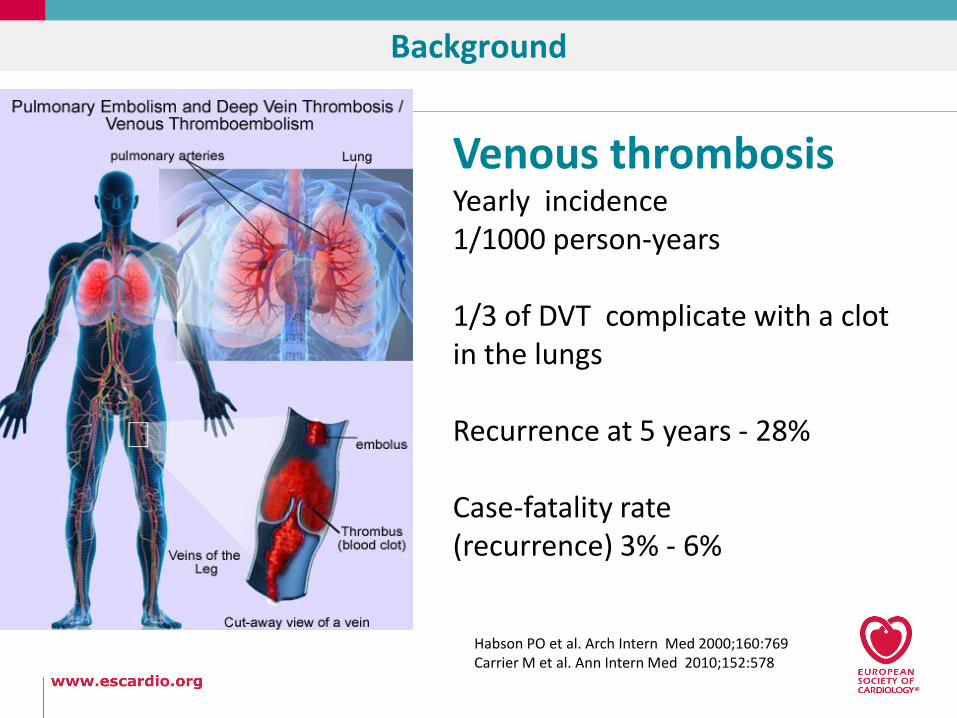
Background
Venous thrombosisYearly incidence 1/1000 person-years
1/3 of DVT complicate with a clot in the lungs
Recurrence at 5 years - 28%
Case-fatality rate (recurrence) 3% - 6%
Habson PO et al. Arch Intern Med 2000;160:769Carrier M et al. Ann Intern Med 2010;152:578

DVT by the numbers
96% arise in the lower extremities
4% arise in the upper extremities.
Chest 2008 Jan;133(1):143-8
Of symptomatic lower-extremity DVTs, 88%
involve the proximal veins; the rest only
involve the calf veins.
Almost all lower-extremity DVTs arise from
the calf veins and extend proximally.
Arch Intern Med 1993;153(24):2777-80
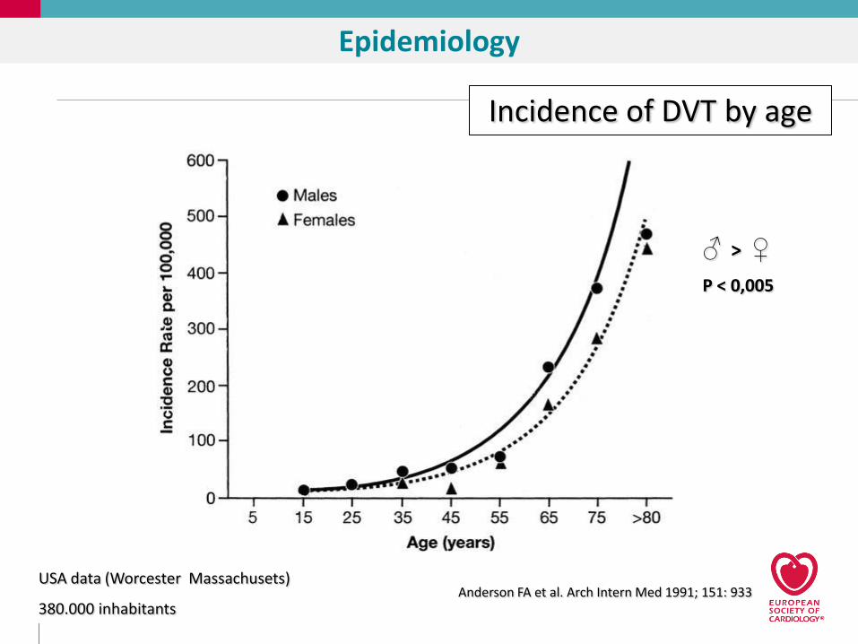
Incidência e idade Worcester
Anderson FA et al. Arch Intern Med 1991; 151: 933
♂ > ♀
P < 0,005
Incidence of DVT by age
USA data (Worcester Massachusets)
380.000 inhabitants
Epidemiology

Risk factor
History of VTE
Venous insufficiency
Chronic heart failure
Obesity
Pregnancy
Deterioration of
general condition
Immobilization
Long-distance travel
Infectious disease
Intr
insi
c fa
cto
rsTr
igge
rin
g fa
cto
rs
OR (95% CI)
15.60 (6.77-35.89)
4.45 (3.10-6.38)
2.93 (1.55-5.56)
2.39 (1.48-3.87)
11.41 (1.40-93.29)
5.75 (2.20-15.01)
5.61 (2.30-13.67)
2.35 (1.45-3.80)
1.95 (1.31-2.92)
0 10
Samama MM. Arch Intern Med. 2000;160: 3415
5 25 40
OR
Risk factors
medical outpatients presenting with DVT

Recurrence after withdrawal of AC
Eichinger S, et al. Circulation 2010;121:1630
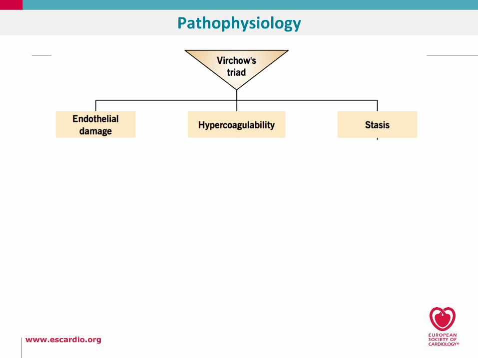
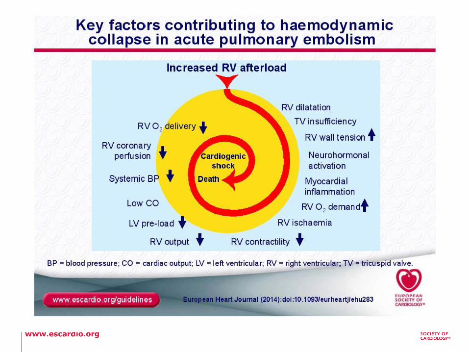
Pathophysiology
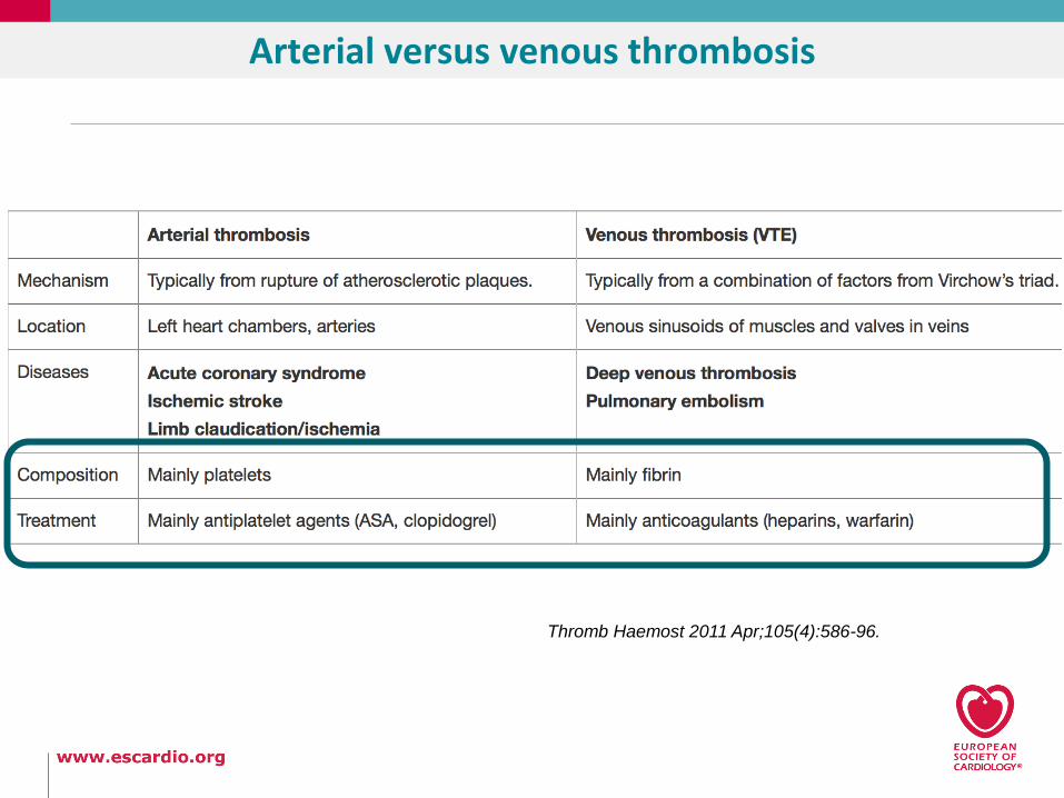
Thromb Haemost 2011 Apr;105(4):586-96.
Arterial versus venous thrombosis

Diagnosis (indirect thrombus evidence)
D-dimer is highly sensitive (more than 95%) in excluding DVT, usually below a threshold of 500 µg/L
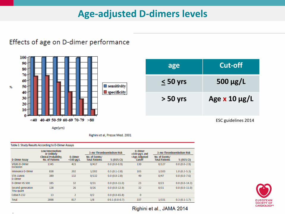
Age-adjusted D-dimers levels
age Cut-off
< 50 yrs 500 μg/L
> 50 yrs Age x 10 μg/L
ESC guidelines 2014


Diagnosis (direct thrombus evidence)
Compression Doppler Ultrasound
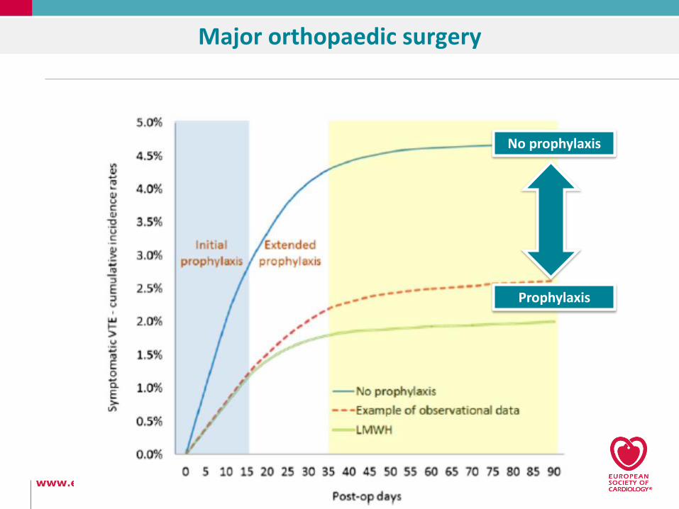
Major orthopaedic surgery
No prophylaxis
Prophylaxis

Major orthopaedic surgery
The success of thromboprophylaxis
Meta-analysis of randomized clinical trials and observational studies that
reported rates of postoperative symptomatic VTE in patients who
received recommended VTE prophylaxis after undergoing TPHA or TPKA.
44 844 cases provided by 47 studies
The pooled rates of symptomatic postoperative VTE before hospital discharge
1.09% (95% CI, 0.85% - 1.33%) for patients undergoing TPKA0.53% (95% CI, 0.35% - 0.70%) for those undergoing TPHA0.63% (95% CI, 0.47% - 0.78%) for knee arthroplasty0.26% (95% CI, 0.14% - 0.37%) for hip arthroplasty
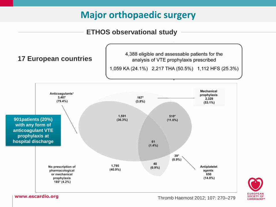
Major orthopaedic surgery
ETHOS observational study
17 European countries
Thromb Haemost 2012; 107: 270–279
901patients (20%)
with any form of
anticoagulant VTE
prophylaxis at
hospital discharge

DVT in cancer patients
Symptomatic venous thromboembolism (VTE) occurs 4-7 times morefrequently in cancer patients as compared to non-cancer patients
Thromb Haemost 2016; 116: 618–625

DVT in cancer patients
Symptomatic venous thromboembolism (VTE) occurs 4-7 times morefrequently in cancer patients as compared to non-cancer patients
Thromb Haemost 2016; 116: 618–625
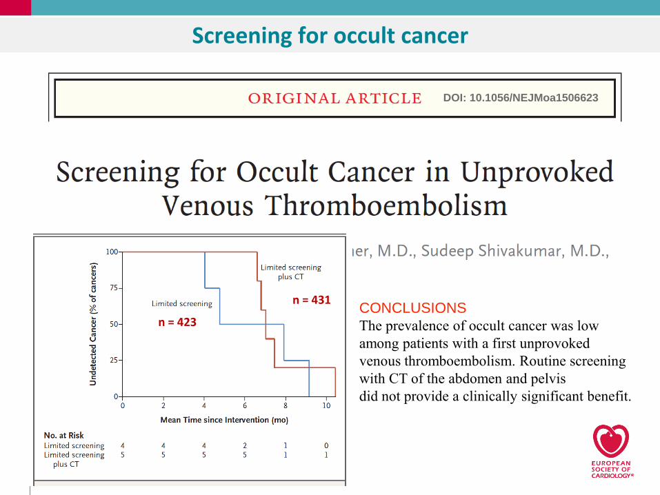
Screening for occult cancer
DOI: 10.1056/NEJMoa1506623
CONCLUSIONS
The prevalence of occult cancer was low
among patients with a first unprovoked
venous thromboembolism. Routine screening
with CT of the abdomen and pelvis
did not provide a clinically significant benefit.
n = 431
n = 423

Thrombophilia
Thrombosis and Haemostasis 109.1/2013
1905 with VTE944 with thrombophilia
78 died (4.1%)
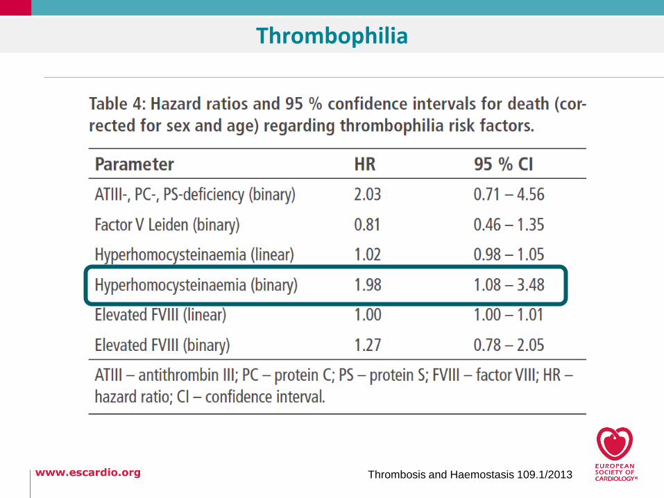
Thrombophilia
Thrombosis and Haemostasis 109.1/2013
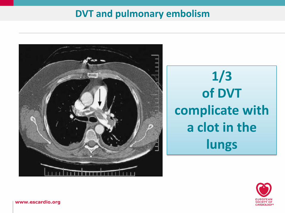
DVT and pulmonary embolism
1/3 of DVT
complicate with a clot in the
lungs


Standard of care for PE
In hospital 3 months
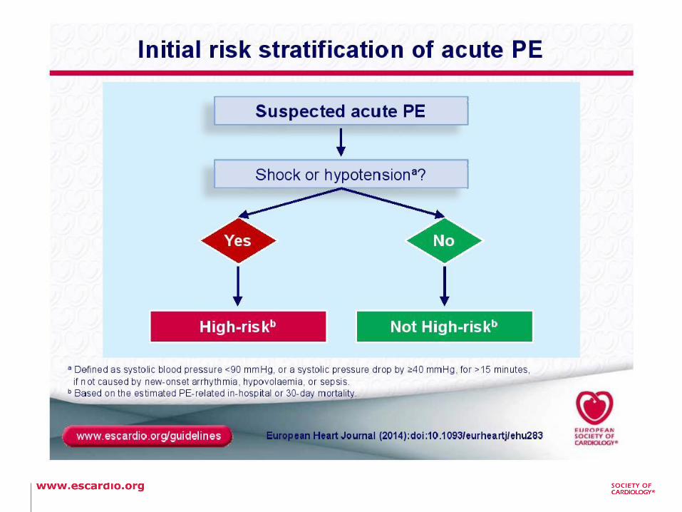
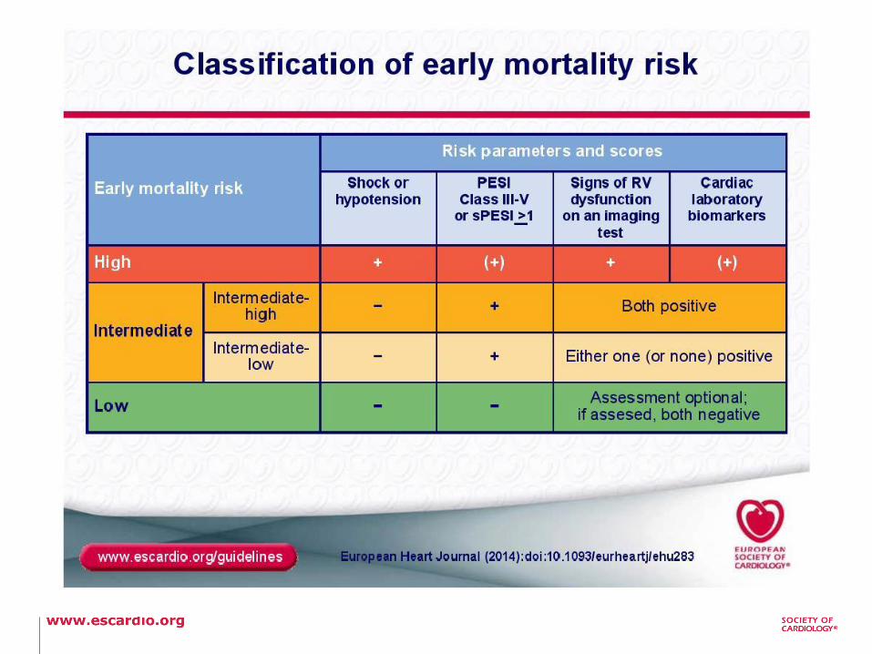
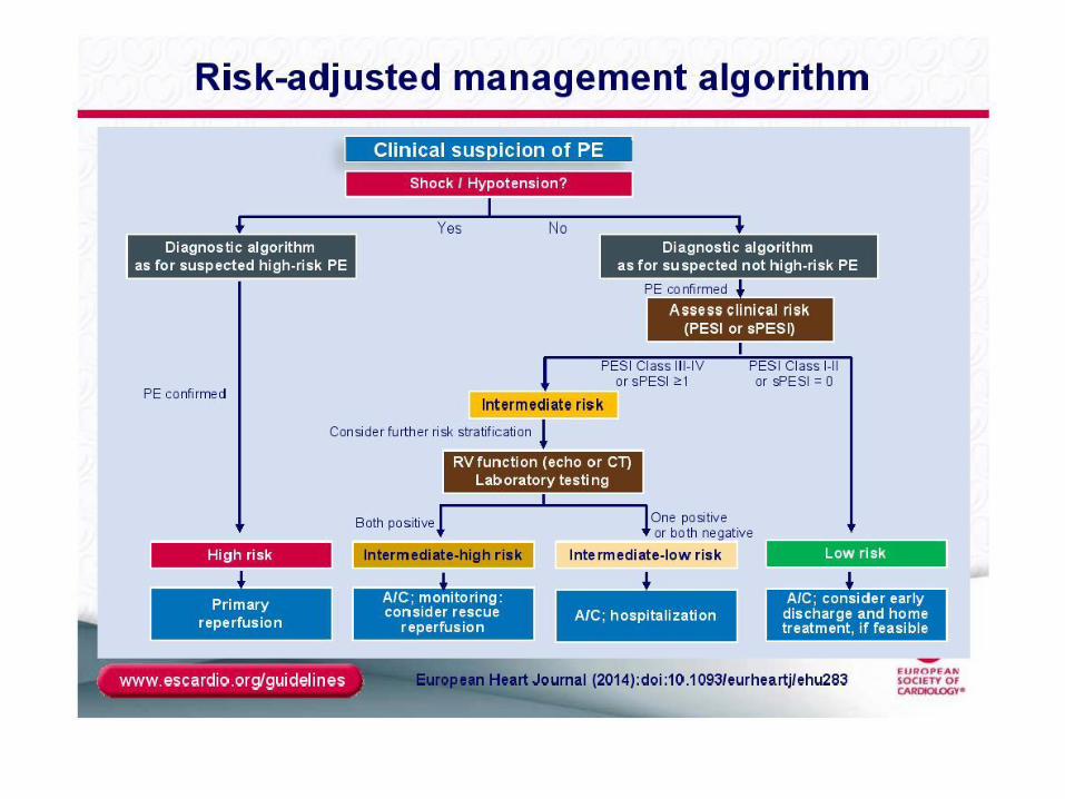
Clinical suspicion
High-risk
Non-High-risk
i.v. UFH
Confirm diagnosis
High-risk
Non High-risk
lytics
LMWH Fonda
VKAs/NOAC
LMWH Fonda
VKA/NOAC
VKA/NOAC
VKA/NOAC
LMWH Fonda
VKA/NOAC
VKA/NOAC
wait

Key messages
• Prophylaxis and early recognition are the keys to change the prognosis in patients with DVT
• The workflow is based on the risk stratification. The higher the risk more aggressive pharmacological treatment should be
• Antithrombotics mostly anticoagulants are very effective not only for prevention but also for treatment
• Pulmonary embolism should be prevented. The acute management is based on the level of risk
João Morais
Head of Cardiology Division and Research Centre
Leiria Hospital Centre
Portugal
Understanding thrombosis in venous thromboembolism