Embed Size (px)
Citation preview
JOURNAL OF BACTERIOLOGY, Nov. 2009, p. 6758–6768 Vol. 191, No. 210021-9193/09/$12.00 doi:10.1128/JB.00840-09Copyright © 2009, American Society for Microbiology. All Rights Reserved.
Uncovering the Protocatechuate 2,3-Cleavage Pathway Genes�†Daisuke Kasai,1 Toshihiro Fujinami,1 Tomokuni Abe,2 Kohei Mase,2
Yoshihiro Katayama,3 Masao Fukuda,1 and Eiji Masai1*Department of Bioengineering, Nagaoka University of Technology, Nagaoka, Niigata 940-2188,1 Toyota Industries Corporation,
Obu, Aichi 474-8601,2 and Graduate School of Bio-Applications and Systems Engineering, Tokyo University ofAgriculture and Technology, Koganei, Tokyo 184-8588,3 Japan
Received 26 June 2009/Accepted 20 August 2009
Paenibacillus sp. (formerly Bacillus macerans) strain JJ-1b is able to grow on 4-hydroxybenzoate (4HB) as asole source of carbon and energy and is known to degrade 4HB via the protocatechuate (PCA) 2,3-cleavagepathway. However, none of the genes involved in this pathway have been identified. In this study, we identifiedand characterized the JJ-1b genes for the 4HB catabolic pathway via the PCA 2,3-cleavage pathway, whichconsisted of praR and praABEGFDCHI. Based on the enzyme activities of cell extracts of Escherichia colicarrying praI, praA, praH, praB, praC, and praD, these genes were found to code for 4HB 3-hydroxylase, PCA2,3-dioxygenase, 5-carboxy-2-hydroxymuconate-6-semialdehyde decarboxylase, 2-hydroxymuconate-6-semial-dehyde dehydrogenase, 4-oxalocrotonate (OCA) tautomerase, and OCA decarboxylase, respectively, which areinvolved in the conversion of 4HB into 2-hydroxypenta-2,4-dienoate (HPD). The praE, praF, and praG geneproducts exhibited 45 to 61% amino acid sequence identity to the corresponding enzymes responsible for thecatabolism of HPD to pyruvate and acetyl coenzyme A. The deduced amino acid sequence of praR showedsimilarity with those of IclR-type transcriptional regulators. Reverse transcription-PCR analysis revealed thatpraABEGFDCHI constitute an operon, and these genes were expressed during the growth of JJ-1b on 4HB andPCA. praR-praABEGFDCHI conferred the ability to grow on 4HB to E. coli, suggesting that praEGF werefunctional for the conversion of HPD to pyruvate and acetyl coenzyme A. A promoter analysis suggested thatpraR encodes a repressor of the pra operon.
Protocatechuate (PCA) is one of the key intermediate me-tabolites in the microbial catabolic pathways for various aro-matic compounds, including phthalates, hydroxybenzoates,and lignin-derived aromatic compounds such as vanillate andferulate. It is known that the aromatic ring fission of PCA iscatalyzed by one of the three distinct dioxygenases PCA 3,4-dioxygenase (26), PCA 4,5-dioxygenase (36, 41), and PCA 2,3-dioxygenase (7). In the PCA 3,4-cleavage pathway, PCA isconverted into 2-carboxy-cis,cis-muconate by the reaction cat-alyzed by PCA 3,4-dioxygenase, and the catabolic pathway forits product (�-ketoadipate pathway) has been reported inmany bacteria (24, 26). In the case of the PCA 4,5-cleavagepathway, PCA is cleaved by PCA 4,5-dioxygenase to yield4-carboxy-2-hydroxymuconate-6-semialdehyde, and then theproduct is degraded to 2-pyrone-4,6-dicarboxylate, 4-oxalo-mesaconate, and 4-carboxy-4-hydroxy-2-oxoadipate before en-tering the Krebs cycle (36). The genes and enzymes involved inthis pathway have been recently characterized for several bac-teria, such as Sphingobium (Sphingomonas) (36), Comamonas(47), Pseudomonas (35), and Arthrobacter (13) strains. On theother hand, no genetic information on the PCA 2,3-cleavagepathway has been reported since the finding of this pathway insome bacilli (7, 8).
In 1979, Crawford et al. reported the PCA 2,3-cleavage
pathway of a 4-hydroxybenzoate (4HB) degrader, Paenibacillussp. (formerly Bacillus macerans) strain JJ-1b, which was iso-lated from a 50°C hot spring (9). In this pathway, PCA isinitially transformed to 5-carboxy-2-hydroxymuconate-6-semi-aldehyde (5CHMS) by PCA 2,3-dioxygenase (Fig. 1A).5CHMS was proposed to be subject to decarboxylation, result-ing in the formation of 2-hydroxymuconate-6-semialdehyde(HMS), which is finally degraded to pyruvate and acetyl coen-zyme A (9). Among these pathway enzymes, only the PCA2,3-dioxygenase was purified and characterized in 1993 (64);however, all the genes responsible for the PCA 2,3-cleavagepathway have not yet been identified.
In the present study, we identified and characterized the4HB catabolic gene (pra gene) cluster, including the PCA2,3-cleavage pathway genes, from Paenibacillus sp. strain JJ-1b.This is the first report on the identification and characteriza-tion of the PCA 2,3-cleavage pathway genes.
MATERIALS AND METHODS
Bacterial strains, plasmids, and culture conditions. The bacterial strains andplasmids used in this study are listed in Table 1. Paenibacillus sp. strain JJ-1b wasgrown in Luria-Bertani (LB) medium or in W minimal salt medium (44) con-taining 10 mM 4HB, 10 mM PCA, or 10 mM succinate at 37°C. Escherichia colistrains were grown in LB medium at 30°C or 37°C. For cultures of E. coli cellscarrying the ampicillin (Ap) resistance marker, the media were supplementedwith 100 mg of Ap/liter.
Cloning of the pra genes. The E2GF and E2GR primer set (see Table S1 in thesupplemental material) was used to amplify an aldehyde dehydrogenase genesequence in JJ-1b. A 563-bp PCR-amplified fragment was used for colony hy-bridization as a probe to isolate the PCA 2,3-cleavage pathway genes from JJ-1bgene libraries, which were constructed using charomid 9-36 (50) with HindIIIdigests of the JJ-1b total DNA. The flanking sequences of the 3.4-kb HindIIIfragment of pCH221 were obtained by using the 1.0-kb HindIII-KpnI fragmentand the 1.0-kb XbaI-HindIII fragment as probes as shown in Fig. 1B. Colony and
* Corresponding author. Mailing address: Department of Bioengi-neering, Nagaoka University of Technology, Nagaoka, Niigata 940-2188, Japan. Phone and fax: 81-258-47-9428. E-mail: [email protected].
† Supplemental material for this article may be found at http://jb.asm.org/.
� Published ahead of print on 28 August 2009.
6758
on Septem
ber 9, 2018 by guesthttp://jb.asm
.org/D
ownloaded from
Southern hybridizations were performed using the digoxigenin system (Roche,Mannheim, Germany).
An inverse PCR was employed to amplify a DNA fragment containing thedownstream region of pCE14 using the INVF and INVR primer set. Total DNAof JJ-1b was digested with BamHI and self-ligated, and then the ligation mixturewas used as a template for the inverse PCR. The 1.7-kb HindIII-BamHI frag-ment of the amplified product was used for colony hybridization as a probe (INVprobe shown in Fig. 1B) to isolate the 3.3-kb EcoRI fragment (pCE3) and the3.5-kb HindIII fragment (pCH1) from gene libraries of JJ-1b as shown in Fig. 1B.The INV2F and INV2R primer set was designed for inverse PCR to amplify theregion further downstream. Using BamHI-digested and self-ligated total DNA asa template, the DNA fragment was amplified. The 261-bp SmaI-ScaI fragment ofthe amplified region was cloned (pBSS) and sequenced. The nucleotide se-quences of the primer sets are shown in Table S1 in the supplemental material.
DNA manipulations and nucleotide sequencing. DNA manipulations, includ-ing total DNA isolation, 16S rRNA gene amplification, construction of deletionderivatives, and nucleotide sequencing, were performed as described in previousstudies (30, 54). Analysis of nucleotide sequences was performed using theMacVector software (MacVector, Inc., Cary, NC). Homology searches, pairwisealignment, and ClustalW multiple-sequence alignment were performed as de-scribed previously (1, 29). Analysis of the 16S rRNA gene was performed usingthe Ribosomal Database Project release 10.10 online server (6). A distancematrix and phylogenetic trees were constructed by using the neighbor-joiningmethod (51) and were visualized with the FigTree program (version 1.2; http://tree.bio.ed.ac.uk/software/figtree/).
Construction of expression plasmids of the pra genes. praA, praB, praC, praD,praH, and praI were PCR amplified and subcloned into pT7Blue. In order toachieve ligation at the NdeI site of the expression vector, all of these genes wereamplified with a forward primer designed to contain the NdeI site at its own
ATG start codon (see Table S1 in the supplemental material). After the se-quences were confirmed, each praA, praB, praC, praH, and praI fragment wasligated to an expression vector, pET21a(�), to form pETA23, pETB23, pETC23,pETH23, and pETI23, respectively. The praD fragment was ligated to pColdIVto construct pC4D23. To construct the praCH coexpression plasmid, pETCH23,the 1.8-kb EcoRI-HindIII fragment carrying praH was ligated into pETC23digested with the same restriction enzymes.
Expression of the pra genes in E. coli. The expression plasmids were intro-duced into E. coli BL21(DE3) cells. BL21(DE3) cells harboring pETA23 weregrown in 10 ml of LB medium containing 100 mg of Ap/liter at 37°C, and the cellsharboring pETB23, pETC23, pETH23, pETI23, and pETCH23 were grown inthe same medium at 30°C. Expression of praA was induced for 4 h by adding 0.1mM isopropyl-�-D-thiogalactopyranoside (IPTG) when the absorbance of theculture at 600 nm (A600) reached 0.5. Expression of praB, praC, praH, and praIand coexpression of praC and praH were induced by adding 1 mM IPTG, andthen growth was continued for 4 h. BL21(DE3) cells harboring pC4D23 weregrown in 10 ml LB medium containing Ap at 37°C until the A600 reached 0.5, andthe culture was placed at 15°C for 30 min and cultivated again at 15°C for 24 hafter the addition of 0.1 mM IPTG. Cells were harvested, resuspended in 50 mMTris-HCl buffer (pH 7.3), broken with an ultrasonic disintegrator (UD-201;Tomy Seiko Co., Tokyo, Japan), and centrifuged at 15,000 � g for 15 min. Theresulting supernatants were used as crude enzymes.
Enzyme assays. (i) PCA 2,3-dioxygenase. PCA 2,3-dioxygenase activity wasassayed by measuring the substrate-dependent oxygen consumption rate. A 2-mlassay mixture contained 50 mM GTA buffer (pH 7.3) consisting of 50 mM3,3-dimethylglutarate, 50 mM Tris, 50 mM 2-amino-2-methyl-1,3-propanediol,crude extract (2.5 �g of protein), and 100 �M PCA. The reaction mixture wasincubated at 35°C, and the oxygen consumption rate was determined with anoxygen electrode (B-505; Iijima Electronics Manufacturing Co., Ltd., Aichi,
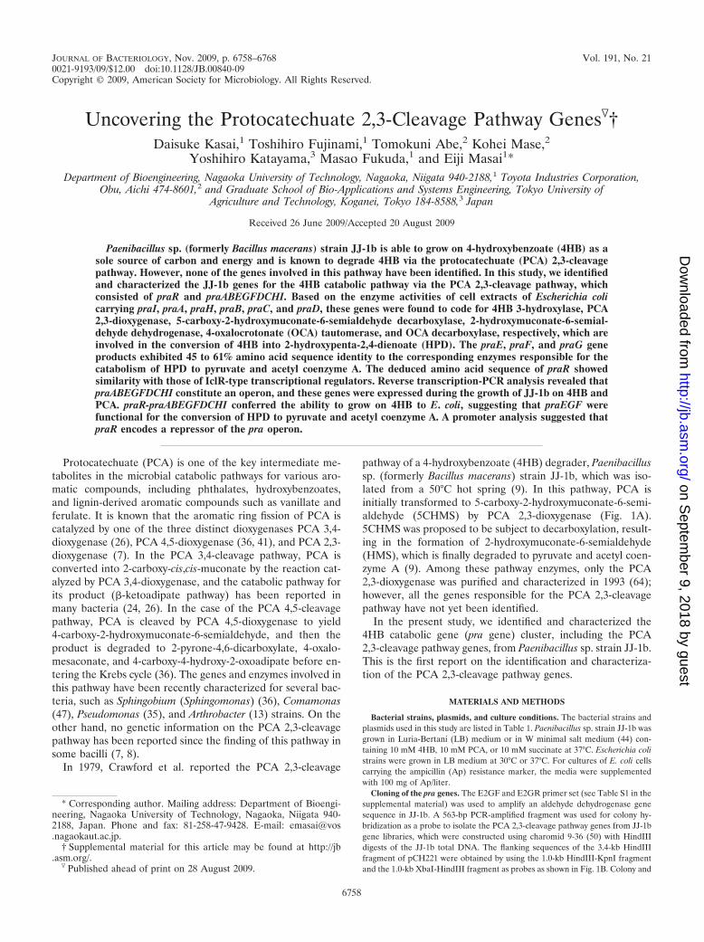
FIG. 1. Catabolic pathway of 4HB in Paenibacillus strain sp. JJ-1b (A) and organization of the pra gene cluster (B). (A) PraI, 4HB3-hydroxylase; PraA, PCA 2,3-dioxygenase; PraH, 5CHMS decarboxylase; PraB, HMS dehydrogenase; PraC, OCA tautomerase; PraD, OCAdecarboxylase; PraE, HPD hydratase; PraF, HOV aldolase; and PraG, acetaldehyde dehydrogenase (acylating). (B) Open arrows indicate the sizes,locations, and transcriptional directions of ORFs. The locations of the cloned DNA fragments are indicated above the arrows representing theORFs. Double-headed arrows indicate the positions of probes used in library screening. Boldface bars below the gene cluster diagram indicate thelocations of the amplified RT-PCR products shown in Fig. 6. Abbreviations for restriction enzymes: B, BamHI; E, EcoRI; H, HindIII; K, KpnI;Sa, SacI; Sc, ScaI; Sm, SmaI; and X, XbaI.
VOL. 191, 2009 PROTOCATECHUATE 2,3-CLEAVAGE PATHWAY GENES 6759
on Septem
ber 9, 2018 by guesthttp://jb.asm
.org/D
ownloaded from
Japan). One unit of enzyme activity was defined as the amount of enzyme thatresulted in consumption of 1 �mol of O2 per min at 35°C. Specific activity wasexpressed in units per milligram of protein. PCA 2,3-dioxygenase activity wasalso monitored using a DU-7500 spectrophotometer (Beckman Coulter, Fuller-ton, CA). The reaction mixture (final volume, 1 ml) containing 50 mM Tris-HClbuffer (pH 7.3), 50 �M PCA, and crude extract (2.5 �g of protein) was prein-cubated without the substrate for 1 min at 35°C, and then the reaction wasstarted by adding PCA.
For gas chromatography-mass spectrometry (GC-MS) analysis, the reactionmixture was acidified with 6 N hydrochloric acid to pH 2 and extracted with ethylacetate. The extract was trimethylsilylated with the TMSI-H reagent (hexameth-yldisilazane-trimethylchlorosilane-pyridine [2:1:10]; GL Science Inc., Tokyo, Ja-
pan) according to the procedure recommended by the manufacturer. The result-ing trimethylsilyl (TMS) derivatives were analyzed by GC-MS.
(ii) 5CHMS decarboxylase. 5CHMS decarboxylase activity was assayed spec-trophotometrically by monitoring the production of HMS from 5CHMS using apreassay mixture that consisted of 50 �M PCA and crude PraA (2.5 �g ofprotein) in 50 mM Tris-HCl buffer (pH 7.3) in a total volume of 900 �l. Themixture was incubated for 1 min at 35°C. After the reaction was completed, crudePraH (10 �g of protein) was added in a final volume of 1 ml, and then thedecrease in the absorbance at 350 nm and the increase in the absorbance at 375nm were monitored at 35°C.
(iii) HMS dehydrogenase. HMS dehydrogenase activity was determined spec-trophotometrically by monitoring the conversion of HMS to 2-hydroxymuconate
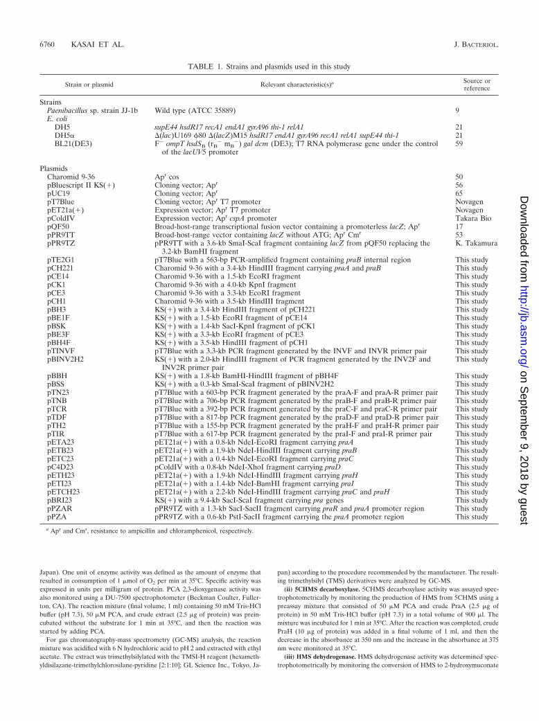
TABLE 1. Strains and plasmids used in this study
Strain or plasmid Relevant characteristic(s)a Source orreference
StrainsPaenibacillus sp. strain JJ-1b Wild type (ATCC 35889) 9E. coli
DH5 supE44 hsdR17 recA1 endA1 gyrA96 thi-1 relA1 21DH5� �(lac)U169 �80 �(lacZ)M15 hsdR17 endA1 gyrA96 recA1 relA1 supE44 thi-1 21BL21(DE3) F� ompT hsdSB (rB
� mB�) gal dcm (DE3); T7 RNA polymerase gene under the control
of the lacUV5 promoter59
PlasmidsCharomid 9-36 Apr cos 50pBluescript II KS(�) Cloning vector; Apr 56pUC19 Cloning vector; Apr 65pT7Blue Cloning vector; Apr T7 promoter NovagenpET21a(�) Expression vector; Apr T7 promoter NovagenpColdIV Expression vector; Apr cspA promoter Takara BiopQF50 Broad-host-range transcriptional fusion vector containing a promoterless lacZ; Apr 17pPR9TT Broad-host-range vector containing lacZ without ATG; Apr Cmr 53pPR9TZ pPR9TT with a 3.6-kb SmaI-ScaI fragment containing lacZ from pQF50 replacing the
3.2-kb BamHI fragmentK. Takamura
pTE2G1 pT7Blue with a 563-bp PCR-amplified fragment containing praB internal region This studypCH221 Charomid 9-36 with a 3.4-kb HindIII fragment carrying praA and praB This studypCE14 Charomid 9-36 with a 1.5-kb EcoRI fragment This studypCK1 Charomid 9-36 with a 4.0-kb KpnI fragment This studypCE3 Charomid 9-36 with a 3.3-kb EcoRI fragment This studypCH1 Charomid 9-36 with a 3.5-kb HindIII fragment This studypBH3 KS(�) with a 3.4-kb HindIII fragment of pCH221 This studypBE1F KS(�) with a 1.5-kb EcoRI fragment of pCE14 This studypBSK KS(�) with a 1.4-kb SacI-KpnI fragment of pCK1 This studypBE3F KS(�) with a 3.3-kb EcoRI fragment of pCE3 This studypBH4F KS(�) with a 3.5-kb HindIII fragment of pCH1 This studypTINVF pT7Blue with a 3.3-kb PCR fragment generated by the INVF and INVR primer pair This studypBINV2H2 KS(�) with a 2.0-kb HindIII fragment of PCR fragment generated by the INV2F and
INV2R primer pairThis study
pBBH KS(�) with a 1.8-kb BamHI-HindIII fragment of pBH4F This studypBSS KS(�) with a 0.3-kb SmaI-ScaI fragment of pBINV2H2 This studypTN23 pT7Blue with a 603-bp PCR fragment generated by the praA-F and praA-R primer pair This studypTNB pT7Blue with a 706-bp PCR fragment generated by the praB-F and praB-R primer pair This studypTCR pT7Blue with a 392-bp PCR fragment generated by the praC-F and praC-R primer pair This studypTDF pT7Blue with a 817-bp PCR fragment generated by the praD-F and praD-R primer pair This studypTH2 pT7Blue with a 155-bp PCR fragment generated by the praH-F and praH-R primer pair This studypTIR pT7Blue with a 617-bp PCR fragment generated by the praI-F and praI-R primer pair This studypETA23 pET21a(�) with a 0.8-kb NdeI-EcoRI fragment carrying praA This studypETB23 pET21a(�) with a 1.9-kb NdeI-HindIII fragment carrying praB This studypETC23 pET21a(�) with a 0.4-kb NdeI-EcoRI fragment carrying praC This studypC4D23 pColdIV with a 0.8-kb NdeI-XhoI fragment carrying praD This studypETH23 pET21a(�) with a 1.9-kb NdeI-HindIII fragment carrying praH This studypETI23 pET21a(�) with a 1.4-kb NdeI-BamHI fragment carrying praI This studypETCH23 pET21a(�) with a 2.2-kb NdeI-HindIII fragment carrying praC and praH This studypBRI23 KS(�) with a 9.4-kb SacI-ScaI fragment carrying pra genes This studypPZAR pPR9TZ with a 1.3-kb SacI-SacII fragment carrying praR and praA promoter region This studypPZA pPR9TZ with a 0.6-kb PstI-SacII fragment carrying the praA promoter region This study
a Apr and Cmr, resistance to ampicillin and chloramphenicol, respectively.
6760 KASAI ET AL. J. BACTERIOL.
on Septem
ber 9, 2018 by guesthttp://jb.asm
.org/D
ownloaded from

(HMA) using a preassay mixture that consisted of 1 mM PCA and crude PraA(4 �g of protein) in 50 mM Tris-HCl buffer (pH 7.3) in a total volume of 50 �l.The reaction mixture was incubated for 30 min at 35°C, and the increase in theabsorbance at 375 nm due to the formation of HMS from PCA was monitored.After the reaction was completed, crude PraB (25 �g of protein) and 50 �MNAD� were added to the mixture. The reaction was carried out in a 1-mlreaction mixture at 35°C. For GC-MS analysis, the reaction mixture was acidi-fied, extracted, and trimethylsilylated.
To determine the specific activity, 1 ml of reaction mixture containing 50 mMTris-HCl buffer (pH 7.3), 50 �M HMS, crude PraB (25 �g of protein), 1.2 mMpyruvate, 1.0 U lactate dehydrogenase, and 500 �M NAD(P)� was incubated at35°C, and the decrease in the absorbance at 375 nm was monitored. The specificactivity was calculated from the initial rates by using a molar extinction coeffi-cient of 35,000 M�1 cm�1 for HMS. One unit of HMS dehydrogenase activitywas defined as the amount of enzyme that converted 1 �mol of HMS per minuteunder the assay conditions used.
(iv) OCA tautomerase. 4-Oxalocrotonate (OCA) tautomerase activity wasdetermined spectrophotometrically by measuring the formation of OCA at 236nm (25). The reaction was carried out in a 1-ml reaction mixture containing 50mM Tris-HCl buffer (pH 7.3), 50 �M NAD�, 50 �l of the PraA reaction mixture,crude PraB (25 �g of protein), and crude PraC (25 �g of protein) at 35°C.
(v) OCA decarboxylase. OCA decarboxylase activity was determined by mea-suring the production of 2-hydroxypenta-2,4-dienoate (HPD) by high-perfor-mance liquid chromatography (HPLC) (Acquity Ultra Performance LC; Waters,Milford, MA). The assay was performed by using a preassay mixture that con-sisted of 50 �l of the PraA reaction mixture, 50 �M NAD�, crude PraB (25 �gof protein/ml), and crude PraC (25 �g of protein/ml) in 50 mM Tris-HCl buffer(pH 7.3) in a total volume of 990 �l. The reaction mixture was incubated for 30min at 35°C; crude PraD (25 �g of protein) and 500 �M MgSO4 were then addedto the mixture to obtain a final volume of 1 ml. The reaction mixture was furtherincubated at 35°C for 10 min, and the enzyme reaction was stopped by additionof methanol (final concentration, 25%). The stopped reaction mixture was thensubjected to HPLC (Acquity Ultra Performance LC; Waters, Milford, MA)analysis.
(vi) 4HB 3-hydroxylase. 4HB 3-hydroxylase activity was measured spectropho-tometrically by monitoring the decrease in the absorbance at 340 nm derivedfrom the consumption of NADH or NADPH at 35°C. The assay mixture (finalvolume, 1 ml) contained 50 mM Tris-HCl buffer (pH 8.0), 200 �M NAD(P)H,500 �M EDTA, 10 �M flavin adenine dinucleotide (FAD), 1 mM 4HB, andcrude PraI (200 �g of protein). The assay mixture was preincubated without thesubstrate for 1 min at 35°C, and the reaction was started by adding 4HB. Thespecific activity was calculated from the initial rates by using molar extinctioncoefficients of 6,600 and 5,070 M�1 cm�1 for NADH and NADPH, respectively(37). One unit of 4HB 3-hydroxylase activity was defined as the amount ofenzyme that consumed 1 �mol of NADH or NADPH per minute under the assayconditions used.
For GC-MS analysis, the assay was carried out in a 1-ml reaction mixturecontaining 50 mM Tris-HCl buffer (pH 8.0), 1 mM NADH, 500 �M EDTA, 10�M FAD, 100 �M 4HB, and crude PraI (200 �g of protein). After the reaction,the reaction mixture was acidified, extracted, trimethylsilylated, and analyzed byGC-MS.
RNA preparation and reverse transcription-PCR (RT-PCR) analysis. JJ-1bcells were grown in W minimal salt medium supplemented with 10 mM 4HB,PCA, or succinate at 37°C. When the A600 reached about 0.8, the cells wereharvested by centrifugation at 5,000 � g at 4°C for 10 min. Total RNA wasisolated with Isogen (Nippon Gene, Toyama, Japan) and then treated withDNase I (Takara Bio Inc.). Single-stranded cDNA was synthesized from 5.0 �gof total RNA by using PrimeScript reverse transcriptase (Takara Bio Inc.) withrandom primers in a 20-�l reaction mixture. PCR amplification was performedwith a 1.0 �l of the cDNA mixture, specific primers (see Table S1 in thesupplemental material), and Ex Taq DNA polymerase (Takara Bio Inc.). Acontrol without reverse transcriptase was used for each reaction to verify theabsence of genomic DNA contamination. PCR samples were electrophoresed ona 0.8% agarose gel and visualized with ethidium bromide.
Measurement of the promoter activity. The 1.3-kb SacI-SacII fragment carry-ing praR and the potential pra operon promoter was cloned into the promoter-probe vector pPR9TZ to obtain pPZAR. The 0.6-kb PstI-SacII fragment wasalso cloned into pPR9TZ to create pPZA carrying only the potential pra operonpromoter. Expression of the lacZ reporter gene was determined by �-galactosi-dase (�-Gal) assays performed as follows. E. coli DH5� cells harboring pPZARor pPZA were grown in 10 ml of LB medium either with or without 10 mM 4HBor PCA at 37°C. After 12 h, cells were harvested and resuspended in Z buffer,consisting of 50 mM sodium phosphate buffer (pH 7.0), 10 mM KCl, 1 mM
MgSO4, and 50 mM �-mercaptoethanol. Toluene treatment and �-Gal assaysusing o-nitrophenyl-�-D-galactopyranoside were performed as described byMiller (38). The values presented are the averages and standard deviations (errorbars) from at least three independent experiments.
Analytical methods. The protein concentration was determined by the methodof Bradford (5). The sizes of the proteins expressed in E. coli were examined bysodium dodecyl sulfate-12% polyacrylamide gel electrophoresis (SDS-PAGE).The proteins in the gels were stained with Coomassie brilliant blue R-250. Todetermine the N-terminal amino acid sequence, the crude extract was subjectedto SDS-PAGE and electroblotted onto a polyvinylidene difluoride membrane(Bio-Rad, Hercules, CA). The enzyme band was cut out and analyzed on aProcise 494 HT protein sequencing system (Applied Biosystems, Foster City,CA). GC-MS analysis was performed with a model 5971A instrument equippedwith an Ultra-2 capillary column (50 m by 0.2 mm; Agilent Technologies Co.,Palo Alto, CA). The analytical conditions were the same as those describedpreviously (29). HPLC analysis was performed with an Acquity Ultra Perfor-mance LC (Waters) equipped with a TSKgel ODS-140HTP column (2.1 by 100mm; Tosoh, Tokyo, Japan). The mobile phase was 1% (vol/vol) acetonitrile inwater containing 0.1% (vol/vol) phosphoric acid, and the flow rate was 0.5ml/min. HMA, OCA, and HPD were detected at 303, 228, and 272 nm, respec-tively.
Nucleotide sequence accession numbers. The nucleotide sequence reported inthis paper has been deposited in the DDBJ, EMBL, and GenBank nucleotidesequence databases under accession numbers AB505863 and AB505864.
RESULTS
16S rRNA gene analysis of JJ-1b. The nucleotide sequenceof the 16S rRNA gene of JJ-1b showed significant similarity tothose of Paenibacillus validus 197 (99.2% identity, 1,456-bpoverlap; accession no. EU730934), Paenibacillus sp. strain Ao3(97.0% identity, 1,515-bp overlap; EF208754), Paenibacillus sp.strain HM1 (96.9% identity, 1,515-bp overlap; AY283261), andPaenibacillus validus JCM 9077 (96.8% identity, 1,487-bp over-lap; AB073203). Accordingly, we propose to rename Bacillusmacerans JJ-1b as Paenibacillus sp. strain JJ-1b.
Cloning and nucleotide sequences of the PCA 2,3-cleavagepathway genes. In order to isolate the PCA 2,3-cleavage path-way genes from JJ-1b, we first attempted to isolate the HMSdehydrogenase gene. Based on the sequence similarity amongputative 5-carboxymethyl-2-hydroxymuconate semialdehydedehydrogenases from Bacillus cereus NVH 391-98 (ABS21296)and Geobacillus kaustophilus HTA426 (BAD77313) and a pu-tative aldehyde dehydrogenase (AAU25432) from Bacilluslicheniformis ATCC 14580, a primer set (E2GF and E2GR)was designed and used to amplify a 563-bp DNA fragmentfrom total DNA of JJ-1b. The nucleotide sequence of thisfragment showed 67% identity with that of AAU25432. Theresulting PCR fragment was then used as a probe to screen aJJ-1b genomic library, and a positive clone, pCH221, carryingthe 3.4-kb HindIII fragment was obtained. The nucleotidesequence of the 3.4-kb HindIII fragment was determined, andtwo open reading frames (ORFs), named praA and praB, werefound. The homology search revealed that the praA and praBgene products showed similarity with homoprotocatechuate2,3-dioxygenases and HMS dehydrogenases, respectively (Ta-ble 2). To acquire the flanking region of the 3.4-kb HindIIIfragment, colony hybridization and inverse PCR were per-formed. As a result, five clones, pCK1, pCE14, pCE3, pCH1,and pBSS, were obtained (Fig. 1B). Using these clones, thenucleotide sequence of the 9,354-bp region was determined. Inthis region, 10 ORFs containing praA and praB were found.Nine of these ORFs are transcribed in the same direction; theexception is praR, which appeared to encode a putative tran-
VOL. 191, 2009 PROTOCATECHUATE 2,3-CLEAVAGE PATHWAY GENES 6761
on Septem
ber 9, 2018 by guesthttp://jb.asm
.org/D
ownloaded from
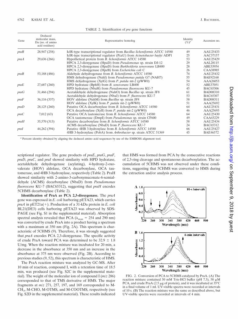
scriptional regulator. The gene products of praE, praG, praF,praD, praC, and praI showed similarity with HPD hydratase,acetaldehyde dehydrogenase (acylating), 4-hydroxy-2-oxo-valerate (HOV) aldolase, OCA decarboxylase, OCA tau-tomerase, and 4HB 3-hydroxylase, respectively (Table 2). PraHshowed similarity with 2-amino-3-carboxymuconate-6-semial-dehyde (ACMS) decarboxylase (NbaD) from Pseudomonasfluorescens KU-7 (BAC65312), suggesting that praH encodes5CHMS decarboxylase (Table 2).
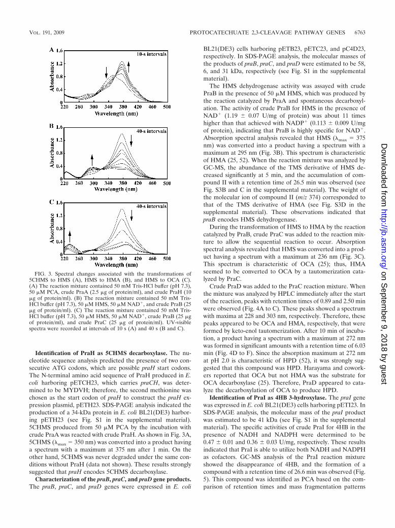
Identification of PraA as PCA 2,3-dioxygenase. The praAgene was expressed in E. coli harboring pETA23, which carriespraA in pET21a(�). Production of a 31-kDa protein in E. coliBL21(DE3) cells harboring pETA23 was observed by SDS-PAGE (see Fig. S1 in the supplemental material). Absorptionspectral analysis revealed that PCA (max 254 and 290 nm)was converted by crude PraA into a product having a spectrumwith a maximum at 350 nm (Fig. 2A). This spectrum is char-acteristic of 5CHMS (9). Therefore, it was strongly suggestedthat praA encodes PCA 2,3-dioxygenase. The specific activityof crude PraA toward PCA was determined to be 32.9 � 1.8U/mg. When the reaction mixture was incubated for 20 min, adecrease in the absorbance at 350 nm and an increase in theabsorbance at 375 nm were observed (Fig. 2B). According toprevious studies (9, 52), this spectrum is characteristic of HMS.
The PraA reaction mixture was analyzed by GC-MS. After10 min of reaction, compound I, with a retention time of 18.5min, was produced (see Fig. S2C in the supplemental mate-rial). The weight of the molecular ion of compound I (m/z 286)corresponded to that of TMS derivative of HMS. The majorfragments at m/z 271, 257, 197, and 169 corresponded to M-CH3, M-CHO, M-OTMS, and M-COOTMS, respectively (seeFig. S2D in the supplemental material). These results indicated
that HMS was formed from PCA by the consecutive reactionsof 2,3-ring cleavage and spontaneous decarboxylation. The ac-cumulation of 5CHMS was not observed under these condi-tions, suggesting that 5CHMS was converted to HMS duringthe extraction and/or analysis process.
FIG. 2. Conversion of PCA to 5CHMS catalyzed by PraA. (A) Thereaction mixture contained 50 mM Tris-HCl buffer (pH 7.3), 50 �MPCA, and crude PraA (2.5 �g of protein), and it was incubated at 35°Cin a final volume of 1 ml. UV-visible spectra were recorded at intervalsof 10 s. (B) The reaction mixture was the same as described above, butUV-visible spectra were recorded at intervals of 4 min.
TABLE 2. Identification of pra gene functions
Gene
Deducedmolecular mass,
Da (no. of aminoacid residues)
Representative homolog Identity(%)a Accession no.
praR 28,947 (258) IclR-type transcriptional regulator from Bacillus licheniformis ATCC 14580 49 AAU25433IclR-type transcriptional regulator (PcaU) from Acinetobacter baylyi ADP1 25 AAC37157
praA 29,636 (266) Hypothetical protein from B. licheniformis ATCC 14580 53 AAU25429HPCA 2,3-dioxygenase (HpaD) from Pseudomonas sp. strain DJ-12 29 AAL28115HPCA 2,3-dioxygenase (HpaD) from Burkholderia xenovorans LB400 28 ABE33954HPCA 2,3-dioxygenase (HpcB) from Escherichia coli C 26 CAA38985
praB 53,188 (486) Aldehyde dehydrogenase from B. licheniformis ATCC 14580 74 AAU25432HMS dehydrogenase (NahI) from Pseudomonas putida G7 (NAH7) 55 BAE92168HMS dehydrogenase (XylG) from P. putida mt-2 (pWW0) 54 AAA26053
praE 27,687 (260) HPD hydratase (BphH) from B. xenovorans LB400 52 ABE37051HPD hydratase (NbaH) from Pseudomonas fluorescens KU-7 45 BAC65306
praG 31,466 (294) Acetaldehyde dehydrogenase (NahO) from Bacillus sp. strain JF8 61 BAD08310Acetaldehyde dehydrogenase (NbaJ) from P. fluorescens KU-7 53 BAC65307
praF 36,116 (337) HOV aldolase (NahM) from Bacillus sp. strain JF8 58 BAD08311HOV aldolase (XylK) from P. putida mt-2 (pWW0) 51 AAA25692
praD 28,125 (260) Putative OCA decarboxylase from B. licheniformis ATCC 14580 64 AAU25431OCA decarboxylase (XylI) from P. putida mt-2 (pWW0) 46 AAA25693
praC 7,012 (63) Putative OCA tautomerase from B. licheniformis ATCC 14580 64 AAU25430OCA tautomerase (DmpI) from Pseudomonas sp. strain CF600 49 CAA43229
praH 35,570 (313) Putative decarboxylase from B. licheniformis ATCC 14580 58 AAU25434ACMS decarboxylase (NbaD) from P. fluorescens KU-7 24 BAC65312
praI 44,262 (394) Putative 4HB 3-hydroxylase from B. licheniformis ATCC 14580 66 AAU254274HB 3-hydroxylase (PobA) from Arthrobacter sp. strain ATCC 51369 45 BAE46572
a Percent identity obtained by aligning the deduced amino acid sequences by use of the EMBOSS alignment tool.
6762 KASAI ET AL. J. BACTERIOL.
on Septem
ber 9, 2018 by guesthttp://jb.asm
.org/D
ownloaded from
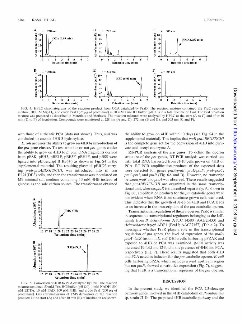
Identification of PraH as 5CHMS decarboxylase. The nu-cleotide sequence analysis predicted the presence of two con-secutive ATG codons, which are possible praH start codons.The N-terminal amino acid sequence of PraH produced in E.coli harboring pETCH23, which carries praCH, was deter-mined to be MYDVH; therefore, the second methionine waschosen as the start codon of praH to construct the praH ex-pression plasmid, pETH23. SDS-PAGE analysis indicated theproduction of a 34-kDa protein in E. coli BL21(DE3) harbor-ing pETH23 (see Fig. S1 in the supplemental material).5CHMS produced from 50 �M PCA by the incubation withcrude PraA was reacted with crude PraH. As shown in Fig. 3A,5CHMS (max 350 nm) was converted into a product havinga spectrum with a maximum at 375 nm after 1 min. On theother hand, 5CHMS was never degraded under the same con-ditions without PraH (data not shown). These results stronglysuggested that praH encodes 5CHMS decarboxylase.
Characterization of the praB, praC, and praD gene products.The praB, praC, and praD genes were expressed in E. coli
BL21(DE3) cells harboring pETB23, pETC23, and pC4D23,respectively. In SDS-PAGE analysis, the molecular masses ofthe products of praB, praC, and praD were estimated to be 58,6, and 31 kDa, respectively (see Fig. S1 in the supplementalmaterial).
The HMS dehydrogenase activity was assayed with crudePraB in the presence of 50 �M HMS, which was produced bythe reaction catalyzed by PraA and spontaneous decarboxyl-ation. The activity of crude PraB for HMS in the presence ofNAD� (1.19 � 0.07 U/mg of protein) was about 11 timeshigher than that achieved with NADP� (0.113 � 0.009 U/mgof protein), indicating that PraB is highly specific for NAD�.Absorption spectral analysis revealed that HMS (max 375nm) was converted into a product having a spectrum with amaximum at 295 nm (Fig. 3B). This spectrum is characteristicof HMA (25, 52). When the reaction mixture was analyzed byGC-MS, the abundance of the TMS derivative of HMS de-creased significantly at 5 min, and the accumulation of com-pound II with a retention time of 26.5 min was observed (seeFig. S3B and C in the supplemental material). The weight ofthe molecular ion of compound II (m/z 374) corresponded tothat of the TMS derivative of HMA (see Fig. S3D in thesupplemental material). These observations indicated thatpraB encodes HMS dehydrogenase.
During the transformation of HMS to HMA by the reactioncatalyzed by PraB, crude PraC was added to the reaction mix-ture to allow the sequential reaction to occur. Absorptionspectral analysis revealed that HMS was converted into a prod-uct having a spectrum with a maximum at 236 nm (Fig. 3C).This spectrum is characteristic of OCA (25); thus, HMAseemed to be converted to OCA by a tautomerization cata-lyzed by PraC.
Crude PraD was added to the PraC reaction mixture. Whenthe mixture was analyzed by HPLC immediately after the startof the reaction, peaks with retention times of 0.89 and 2.50 minwere observed (Fig. 4A to C). These peaks showed a spectrumwith maxima at 228 and 303 nm, respectively. Therefore, thesepeaks appeared to be OCA and HMA, respectively, that wereformed by keto-enol tautomerization. After 10 min of incuba-tion, a product having a spectrum with a maximum at 272 nmwas formed in significant amounts with a retention time of 6.03min (Fig. 4D to F). Since the absorption maximum at 272 nmat pH 2.0 is characteristic of HPD (52), it was strongly sug-gested that this compound was HPD. Harayama and cowork-ers reported that OCA but not HMA was the substrate forOCA decarboxylase (25). Therefore, PraD appeared to cata-lyze the decarboxylation of OCA to produce HPD.
Identification of PraI as 4HB 3-hydroxylase. The praI genewas expressed in E. coli BL21(DE3) cells harboring pETI23. InSDS-PAGE analysis, the molecular mass of the praI productwas estimated to be 41 kDa (see Fig. S1 in the supplementalmaterial). The specific activities of crude PraI for 4HB in thepresence of NADH and NADPH were determined to be0.47 � 0.01 and 0.36 � 0.03 U/mg, respectively. These resultsindicated that PraI is able to utilize both NADH and NADPHas cofactors. GC-MS analysis of the PraI reaction mixtureshowed the disappearance of 4HB, and the formation of acompound with a retention time of 26.6 min was observed (Fig.5). This compound was identified as PCA based on the com-parison of retention times and mass fragmentation patterns
FIG. 3. Spectral changes associated with the transformations of5CHMS to HMS (A), HMS to HMA (B), and HMS to OCA (C).(A) The reaction mixture contained 50 mM Tris-HCl buffer (pH 7.3),50 �M PCA, crude PraA (2.5 �g of protein/ml), and crude PraH (10�g of protein/ml). (B) The reaction mixture contained 50 mM Tris-HCl buffer (pH 7.3), 50 �M HMS, 50 �M NAD�, and crude PraB (25�g of protein/ml). (C) The reaction mixture contained 50 mM Tris-HCl buffer (pH 7.3), 50 �M HMS, 50 �M NAD�, crude PraB (25 �gof protein/ml), and crude PraC (25 �g of protein/ml). UV-visiblespectra were recorded at intervals of 10 s (A) and 40 s (B and C).
VOL. 191, 2009 PROTOCATECHUATE 2,3-CLEAVAGE PATHWAY GENES 6763
on Septem
ber 9, 2018 by guesthttp://jb.asm
.org/D
ownloaded from
with those of authentic PCA (data not shown). Thus, praI wasconcluded to encode 4HB 3-hydroxylase.
E. coli acquires the ability to grow on 4HB by introduction ofthe pra gene cluster. To test whether or not pra genes conferthe ability to grow on 4HB to E. coli, DNA fragments derivedfrom pBSK, pBH3, pBE1F, pBE3F, pBH4F, and pBSS wereligated into pBluescript II KS(�) as shown in Fig. S4 in thesupplemental material. The resulting plasmid, pBRI23 carry-ing praR-praABEGFDCHI, was introduced into E. coliBL21(DE3) cells, and then the transformant was inoculated onM9 minimal salt medium containing 10 mM 4HB instead ofglucose as the sole carbon source. The transformant obtained
the ability to grow on 4HB within 10 days (see Fig. S4 in thesupplemental material). This implies that praR-praABEGFDCHIis the complete gene set for the conversion of 4HB into pyru-vate and acetyl coenzyme A.
RT-PCR analysis of the pra genes. To define the operonstructure of the pra genes, RT-PCR analysis was carried outwith total RNA harvested from JJ-1b cells grown on 4HB orPCA. RT-PCR amplification products of the expected sizeswere detected for genes praA-praE, praE-praF, praF-praC,praC-praI, and praR (Fig. 6A and B). However, no transcriptbetween praR and praA was observed. These results suggestedthat praABEGFDCHI are organized in the same transcrip-tional unit, whereas praR is transcribed separately. As shown inFig. 6C, amplification products for the pra catabolic genes werenot evident when RNA from succinate-grown cells was used.This indicates that the growth of JJ-1b on 4HB and PCA leadsto an increase in the transcription of the pra catabolic operon.
Transcriptional regulation of the pra operon. PraR is similarin sequence to transcriptional regulators belonging to the IclRfamily from B. licheniformis ATCC 14580 (AAU25433) andAcinetobacter baylyi ADP1 (PcaU; AAC37157) (Table 2). Toinvestigate whether PraR plays a role in the transcriptionalregulation of pra genes, the level of expression of the praR-praA�-lacZ fusion in E. coli DH5� cells harboring pPZAR andexposed to 4HB or PCA was examined. �-Gal activity wasincreased 19-fold and 12-fold in the presence of 4HB and PCA,respectively (Fig. 7). These results suggested that both 4HBand PCA acted as inducers for the pra catabolic operon. E. colicells harboring pPZA, which includes a praA upstream regionbut not praR, showed constitutive expression (Fig. 7), suggest-ing that PraR is a transcriptional repressor of the pra operon.
DISCUSSION
In the present study, we identified the PCA 2,3-cleavagepathway genes involved in the 4HB catabolism of Paenibacillussp. strain JJ-1b. The proposed 4HB catabolic pathway and the
FIG. 4. HPLC chromatograms of the reaction product from OCA catalyzed by PraD. The reaction mixture contained the PraC reactionmixture, 500 �M MgSO4, and crude PraD (25 �g of protein/ml) in 50 mM Tris-HCl buffer (pH 7.3) in a total volume of 1 ml. The PraC reactionmixture was prepared as described in Materials and Methods. The reaction mixtures were analyzed by HPLC at the start (A to C) and after 10min (D to F) of incubation. Compounds were monitored at 228 nm (A and D), 272 nm (B and E), and 303 nm (C and F).
FIG. 5. Conversion of 4HB to PCA catalyzed by PraI. The reactionmixture contained 50 mM Tris-HCl buffer (pH 8.0), 1 mM NADH, 500�M EDTA, 10 �M FAD, 100 �M 4HB, and crude PraI (200 �g ofprotein/ml). Gas chromatograms of TMS derivatives of the reactionproducts at the start (A) and after 10 min (B) of incubation are shown.
6764 KASAI ET AL. J. BACTERIOL.
on Septem
ber 9, 2018 by guesthttp://jb.asm
.org/D
ownloaded from

corresponding genes for the enzymes of JJ-1b are indicated inFig. 1A.
4HB is transformed to PCA by the reaction catalyzed byPraI, which utilizes both NADH and NADPH as electrondonors. 4HB 3-hydroxylases can be divided into two groups onthe basis of the coenzyme specificity: (i) NADPH-specific and(ii) NAD(P)H-dependent 4HB 3-hydroxylases. A phylogeneticanalysis of 4HB 3-hydroxylases indicated that NADPH-specificand NAD(P)H-dependent enzymes are located in the differentbranches (see Fig. S5A in the supplemental material). PraIforms a cluster with NAD(P)H-dependent enzymes. The de-duced amino acid sequence alignment of PraI and NADPH-specific 4HB 3-hydroxylases indicated that Tyr38 and Arg42,which are essential for binding with NADPH (15, 16), arereplaced by glutamate and threonine residues, respectively(see Fig. S5B in the supplemental material). These residues arehighly conserved in the NAD(P)H-dependent 4HB 3-hydroxy-lases and are required for the recognition of NADH (15, 28).
PCA 2,3-dioxygenase had been previously purified and bio-chemically characterized (64), but the gene sequence has beenunavailable. The praA gene product shares amino acid se-quence similarity with extradiol dioxygenases. Extradiol dioxy-genases have been divided into three families on the basis ofamino acid sequence similarity (63). Type I extradiol dioxyge-nases belong to the vicinal oxygen superfamily, including anumber of 2,3-dihydroxybiphenyl 1,2-dioxygenases (3, 27) andcatechol 2,3-dioxygenases (40). Type II dioxygenases includeseveral homoprotocatechuate 2,3-dioxygenases (49), the � sub-unit of PCA 4,5-dioxygenase (41, 47), gallate dioxygenase (30,42), and 2-aminophenol 1,6-dioxygenase (61). Type III dioxy-
genases belong to the cupin superfamily, which includes en-zymes such as the gentisate dioxygenase, homogentisate dioxy-genase, and 1-hydroxy-2-naphthoate dioxygenase (12). Thesethree types of enzymes have similar active sites and the sameiron ligand, two histidines, and one glutamate (2-His-1-carbox-ylate structural motif), in spite of their different primary struc-tures (31). As can be seen in the phylogenetic relationships oftype II extradiol dioxygenases (see Fig. S6 in the supplementalmaterial), PraA obviously belongs to the type II extradiol di-oxygenase family and is closely related to a hypothetical pro-tein (BL03909) of B. licheniformis ATCC 14580. However, thesequence of PraA formed a deep branch with those of the �subunit of PCA 4,5-dioxygenase, suggesting that they indepen-dently evolved from a common ancestor. Crystallographicstudies revealed that the active site of the � subunit of PCA4,5-dioxygenase (LigB) contains a nonheme iron coordinatedby His12, His61, and Glu242, and His195 is thought to act as anactive-site base to facilitate deprotonation of the hydroxylgroup of the substrate (60). These amino acids are conservedamong almost all the type II extradiol dioxygenases (14). Thealignment of the deduced amino acid sequences of PraA andLigB revealed the presence of residues His11, His53, His180,and Glu227 of PraA, corresponding to His12, His61, His195,and Glu242, respectively, of LigB. These residues seemed to beinvolved in the roles described above.
When PCA was incubated with crude PraA, 5CHMS pro-duced from PCA underwent spontaneous decarboxylation toHMS. However, no production of 5CHMS from PCA wasobserved in the reaction mixture containing the crude extractof JJ-1b (9). These facts suggested that 5CHMS decarboxylaseencoded by praH plays a role in the decarboxylation of 5CHMSto HMS in JJ-1b cells. Based on the deduced amino acidsequence similarity, PraH seemed to be included in the met-allo-dependent hydrolase (amidohydrolase_2) superfamily(33). This family includes ACMS decarboxylase from bacteria,animals, and humans (19, 39, 62); uracil-5-carboxylate decar-boxylase of Neurospora crassa OR74A (57); the -resorcylatedecarboxylase GraF from Rhizobium sp. strain MTP-10005 (66,67); the 5-carboxyvanillate decarboxylase LigW (46); the bi-phenyl meta-cleavage compound hydrolase LigY (45); and the4-oxalomesaconate hydratase LigJ (22) from Sphingobium sp.strain SYK-6. A phylogenetic analysis based on the amino acidsequences of the metallo-dependent hydrolase superfamily
FIG. 6. RT-PCR analysis of the pra gene cluster in JJ-1b. TotalRNA used for cDNA synthesis was isolated from JJ-1b cells grown on4HB (A), PCA (B), and succinate (C). Agarose gel electrophoresis ofRT-PCR assays with primers targeting praR (expected size, 572 bp),praR-praA (expected size, 1,092 bp), praA-praE (expected size, 2,190bp), praE-praF (expected size, 1,940 bp), praF-praC (expected size,1,553 bp), and praC-praI (expected size, 1,779 bp) are shown. Positionsof primer pairs and primer sequences are indicated in Fig. 1B and inTable S1 in the supplemental material, respectively. Lane M, molec-ular weight markers; lanes � and �, RT-PCR with and without reversetranscriptase, respectively; lanes G, control PCR with the JJ-1bgenomic DNA. RT-PCR of the 16S rRNA gene was used as a controlto confirm equivalent quantities of template loading.
FIG. 7. Regulation of the pra operon promoter activity by PraR.(A) Schematic representation of pPZAR and pPZA. (B) Promoteractivities of the pra operon were measured in E. coli cells harboringpPZAR (shaded bars) or pPZA (open bars). These cells were grown inLB medium with or without 10 mM 4HB or PCA. Each value is theaverage � standard deviation (error bars) based on at least threeindependent experiments.
VOL. 191, 2009 PROTOCATECHUATE 2,3-CLEAVAGE PATHWAY GENES 6765
on Septem
ber 9, 2018 by guesthttp://jb.asm
.org/D
ownloaded from
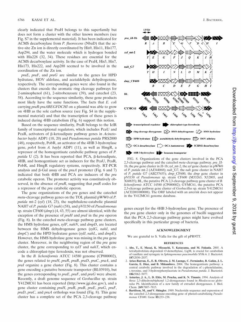
clearly indicated that PraH belongs to this superfamily butdoes not form a cluster with the other known members (seeFig. S7 in the supplemental material). It has been indicated forACMS decarboxylase from P. fluorescens (NbaD) that the ac-tive-site Zn ion is directly coordinated by His9, His11, His177,Asp294, and the water molecule which is hydrogen bondedwith His228 (32, 34). These residues are essential for theACMS decarboxylase activity. In the case of PraH, His5, His7,His173, His222, and Asp288 seemed to be involved in thecoordination of the Zn ion.
praE, praF, and praG are similar to the genes for HPDhydratase, HOV aldolase, and acetaldehyde dehydrogenase,respectively. The corresponding genes were also found in theclusters that encode the aromatic ring cleavage pathways for2-aminophenol (61), 2-nitrobenzoate (39), and catechol (23,58). According to the sequence similarity, these gene productsmost likely have the same functions. The facts that E. colicarrying praR-praABEGFDCHI on a plasmid was able to growon 4HB as the sole carbon source (see Fig. S4 in the supple-mental material) and that the transcription of these genes isinduced during 4HB catabolism (Fig. 6) support this notion.
Based on the sequence similarity, PraR belongs to the IclRfamily of transcriptional regulators, which includes PcaU andPcaR, activators of �-ketoadipate pathway genes in Acineto-bacter baylyi ADP1 (10, 20) and Pseudomonas putida PRS2000(48), respectively, PobR, an activator of the 4HB 3-hydroxylasegene, pobA from A. baylyi ADP1 (11), as well as HmgR, arepressor of the homogentisate catabolic pathway genes of P.putida U (2). It has been reported that PCA, �-ketoadipate,4HB, and homogentisate act as inducers for the PcaU, PcaR,PobR, and HmgR regulatory systems, respectively. RT-PCRanalysis and �-Gal assay of the praA promoter (Fig. 6 and 7)indicated that both 4HB and PCA are inducers of the pracatabolic operon. The promoter activity was constitutively ob-served, in the absence of praR, suggesting that praR codes fora repressor of the pra catabolic operon.
The gene organizations of the pra genes and the catecholmeta-cleavage pathway genes on the TOL plasmid pWW0 of P.putida mt-2 (xyl) (18, 23), the naphthalene-catabolic plasmidNAH7 of P. putida G7 (nah) (58), and pVI150 of Pseudomonassp. strain CF600 (dmp) (4, 43, 55) are almost identical, with theexception of the presence of praH and praI in the pra operon(Fig. 8). In the catechol meta-cleavage pathway gene clusters,the HMS hydrolase genes, xylF, nahN, and dmpD, are locatedbetween the HMS dehydrogenase genes (xylG, nahI, anddmpC) and the HPD hydratase genes (xylJ, nahL, and dmpE).However, the HMS hydrolase gene was missing in the pra genecluster. Moreover, in the neighboring region of the pra genecluster, the gene corresponding to xylT and nahT, which en-code a chloroplast-type ferredoxin, was not observed.
In the B. licheniformis ATCC 14580 genome (CP000002),the genes related to praH, praR, praB, praD, praC, praA, andpraI organize a gene cluster (Fig. 8). This cluster contains agene encoding a putative benzoate transporter (BL03910), butthe genes corresponding to praE, praF, and praG were absent.Recently, a draft genome sequence of Geobacillus sp. strainY412MC61 has been reported (http://www.jgi.doe.gov/), and agene cluster containing praH, praR, praB, praE, praG, praF,praD, praC, and praA orthologs was found (Fig. 8). This genecluster has a complete set of the PCA 2,3-cleavage pathway
genes except for the 4HB 3-hydroxylase gene. The presence ofthe pra gene cluster only in the genomes of bacilli suggestedthat the PCA 2,3-cleavage pathway genes might have evolvedspecifically in this particular group of bacteria.
ACKNOWLEDGMENT
We are grateful to S. Valla for the gift of pPR9TT.
REFERENCES
1. Abe, T., E. Masai, K. Miyauchi, Y. Katayama, and M. Fukuda. 2005. Atetrahydrofolate-dependent O-demethylase, LigM, is crucial for catabolismof vanillate and syringate in Sphingomonas paucimobilis SYK-6. J. Bacteriol.187:2030–2037.
2. Arias-Barrau, E., E. R. Olivera, J. M. Luengo, C. Fernandez, B. Galan, J. L.García, E. Díaz, and B. Minambres. 2004. The homogentisate pathway: acentral catabolic pathway involved in the degradation of L-phenylalanine,L-tyrosine, and 3-hydroxyphenylacetate in Pseudomonas putida. J. Bacteriol.186:5062–5077.
3. Asturias, J. A., L. D. Eltis, M. Prucha, and K. N. Timmis. 1994. Analysis ofthree 2,3-dihydroxybiphenyl 1,2-dioxygenases found in Rhodococcus globe-rulus P6. Identification of a new family of extradiol dioxygenases. J. Biol.Chem. 269:7807–7815.
4. Bartilson, M., and V. Shingler. 1989. Nucleotide sequence and expression ofthe catechol 2,3-dioxygenase-encoding gene of phenol-catabolizing Pseudo-monas CF600. Gene 85:233–238.
FIG. 8. Organizations of the gene clusters involved in the PCA2,3-cleavage pathway and the catechol meta-cleavage pathway. pra_JJ-1b, the pra gene cluster in JJ-1b; xyl_mt-2, the xyl gene cluster in pWW0of P. putida mt-2 (AJ344068); nah_G7, the nah gene cluster in NAH7of P. putida G7 (AB237655); dmp_CF600, the dmp gene cluster inpVI150 of Pseudomonas sp. strain CF600 (M33263, X52805, andX60835); BL, the putative PCA 2,3-cleavage pathway gene cluster of B.licheniformis ATCC 14580 (CP000002); GYMC61, the putative PCA2,3-cleavage pathway gene cluster of Geobacillus sp. strain Y412MC61(ACED01000008). The ORF labeled with an asterisk does not appearin the Y412MC61 genome database.
6766 KASAI ET AL. J. BACTERIOL.
on Septem
ber 9, 2018 by guesthttp://jb.asm
.org/D
ownloaded from
5. Bradford, M. M. 1976. A rapid and sensitive method for the quantitation ofmicrogram quantities of protein utilizing the principle of protein-dye bind-ing. Anal. Biochem. 72:248–254.
6. Cole, J. R., B. Chai, R. J. Farris, Q. Wang, S. A. Kulam, D. M. McGarrell,G. M. Garrity, and J. M. Tiedje. 2005. The Ribosomal Database Project(RDP-II): sequences and tools for high-throughput rRNA analysis. NucleicAcids Res. 33:D294–D296.
7. Crawford, R. L. 1975. Novel pathway for degradation of protocatechuic acidin Bacillus species. J. Bacteriol. 121:531–536.
8. Crawford, R. L. 1976. Pathways of 4-hydroxybenzoate degradation amongspecies of Bacillus. J. Bacteriol. 127:204–210.
9. Crawford, R. L., J. W. Bromley, and P. E. Perkins-Olson. 1979. Catabolismof protocatechuate by Bacillus macerans. Appl. Environ. Microbiol. 37:614–618.
10. Dal, S., G. Trautwein, and U. Gerischer. 2005. Transcriptional organizationof genes for protocatechuate and quinate degradation from Acinetobacter sp.strain ADP1. Appl. Environ. Microbiol. 71:1025–1034.
11. DiMarco, A. A., B. Averhoff, and L. N. Ornston. 1993. Identification of thetranscriptional activator pobR and characterization of its role in the expres-sion of pobA, the structural gene for p-hydroxybenzoate hydroxylase in Acin-etobacter calcoaceticus. J. Bacteriol. 175:4499–4506.
12. Dunwell, J. M., A. Culham, C. E. Carter, C. R. Sosa-Aguirre, and P. W.Goodenough. 2001. Evolution of functional diversity in the cupin superfam-ily. Trends Biochem. Sci. 26:740–746.
13. Eaton, R. W. 2001. Plasmid-encoded phthalate catabolic pathway in Ar-throbacter keyseri 12B. J. Bacteriol. 183:3689–3703.
14. Eltis, L. D., and J. T. Bolin. 1996. Evolutionary relationships among extradioldioxygenases. J. Bacteriol. 178:5930–5937.
15. Eppink, M. H., K. M. Overkamp, H. A. Schreuder, and W. J. Van Berkel.1999. Switch of coenzyme specificity of p-hydroxybenzoate hydroxylase. J.Mol. Biol. 292:87–96.
16. Eppink, M. H., H. A. Schreuder, and W. J. van Berkel. 1998. Lys42 and Ser42variants of p-hydroxybenzoate hydroxylase from Pseudomonas fluorescensreveal that Arg42 is essential for NADPH binding. Eur. J. Biochem. 253:194–201.
17. Farinha, M. A., and A. M. Kropinski. 1990. Construction of broad-host-range plasmid vectors for easy visible selection and analysis of promoters. J.Bacteriol. 172:3496–3499.
18. Franklin, F. C., M. Bagdasarian, M. M. Bagdasarian, and K. N. Timmis.1981. Molecular and functional analysis of the TOL plasmid pWWO fromPseudomonas putida and cloning of genes for the entire regulated aromaticring meta cleavage pathway. Proc. Natl. Acad. Sci. USA 78:7458–7462.
19. Fukuoka, S., K. Ishiguro, K. Yanagihara, A. Tanabe, Y. Egashira, H.Sanada, and K. Shibata. 2002. Identification and expression of a cDNAencoding human �-amino-�-carboxymuconate-ε-semialdehyde decarboxyl-ase (ACMSD). A key enzyme for the tryptophan-niacine pathway and“quinolinate hypothesis”. J. Biol. Chem. 277:35162–35167.
20. Gerischer, U., A. Segura, and L. N. Ornston. 1998. PcaU, a transcriptionalactivator of genes for protocatechuate utilization in Acinetobacter. J. Bacte-riol. 180:1512–1524.
21. Hanahan, D. 1983. Studies on transformation of Escherichia coli with plas-mids. J. Mol. Biol. 166:557–580.
22. Hara, H., E. Masai, Y. Katayama, and M. Fukuda. 2000. The 4-ox-alomesaconate hydratase gene, involved in the protocatechuate 4,5-cleavagepathway, is essential to vanillate and syringate degradation in Sphingomonaspaucimobilis SYK-6. J. Bacteriol. 182:6950–6957.
23. Harayama, S., P. R. Lehrbach, and K. N. Timmis. 1984. Transposon mu-tagenesis analysis of meta-cleavage pathway operon genes of the TOL plas-mid of Pseudomonas putida mt-2. J. Bacteriol. 160:251–255.
24. Harayama, S., and M. Rekik. 1989. Bacterial aromatic ring-cleavage en-zymes are classified into two different gene families. J. Biol. Chem. 264:15328–15333.
25. Harayama, S., M. Rekik, K. L. Ngai, and L. N. Ornston. 1989. Physicallyassociated enzymes produce and metabolize 2-hydroxy-2,4-dienoate, a chem-ically unstable intermediate formed in catechol metabolism via meta cleav-age in Pseudomonas putida. J. Bacteriol. 171:6251–6258.
26. Harwood, C. S., and R. E. Parales. 1996. The �-ketoadipate pathway and thebiology of self-identity. Annu. Rev. Microbiol. 50:553–590.
27. Hayase, N., K. Taira, and K. Furukawa. 1990. Pseudomonas putida KF715bphABCD operon encoding biphenyl and polychlorinated biphenyl degrada-tion: cloning, analysis, and expression in soil bacteria. J. Bacteriol. 172:1160–1164.
28. Huang, Y., K. X. Zhao, X. H. Shen, C. Y. Jiang, and S. J. Liu. 2008. Geneticand biochemical characterization of a 4-hydroxybenzoate hydroxylase fromCorynebacterium glutamicum. Appl. Microbiol. Biotechnol. 78:75–83.
29. Kasai, D., E. Masai, K. Miyauchi, Y. Katayama, and M. Fukuda. 2004.Characterization of the 3-O-methylgallate dioxygenase gene and evidence ofmultiple 3-O-methylgallate catabolic pathways in Sphingomonas paucimobilisSYK-6. J. Bacteriol. 186:4951–4959.
30. Kasai, D., E. Masai, K. Miyauchi, Y. Katayama, and M. Fukuda. 2005.Characterization of the gallate dioxygenase gene: three distinct ring cleavage
dioxygenases are involved in syringate degradation by Sphingomonas pauci-mobilis SYK-6. J. Bacteriol. 187:5067–5074.
31. Koehntop, K. D., J. P. Emerson, and L. Que, Jr. 2005. The 2-His-1-carbox-ylate facial triad: a versatile platform for dioxygen activation by mononuclearnon-heme iron(II) enzymes. J. Biol. Inorg. Chem. 10:87–93.
32. Li, T., H. Iwaki, R. Fu, Y. Hasegawa, H. Zhang, and A. Liu. 2006. �-Amino-�-carboxymuconic-ε-semialdehyde decarboxylase (ACMSD) is a new mem-ber of the amidohydrolase superfamily. Biochemistry 45:6628–6634.
33. Liu, A., and H. Zhang. 2006. Transition metal-catalyzed nonoxidative decar-boxylation reactions. Biochemistry 45:10407–10411.
34. Martynowski, D., Y. Eyobo, T. Li, K. Yang, A. Liu, and H. Zhang. 2006.Crystal structure of �-amino-�-carboxymuconate-ε-semialdehyde decarbox-ylase: insight into the active site and catalytic mechanism of a novel decar-boxylation reaction. Biochemistry 45:10412–10421.
35. Maruyama, K., T. Shibayama, A. Ichikawa, Y. Sakou, S. Yamada, and H.Sugisaki. 2004. Cloning and characterization of the genes encoding enzymesfor the protocatechuate meta-degradation pathway of Pseudomonas ochra-ceae NGJ1. Biosci. Biotechnol. Biochem. 68:1434–1441.
36. Masai, E., Y. Katayama, and M. Fukuda. 2007. Genetic and biochemicalinvestigations on bacterial catabolic pathways for lignin-derived aromaticcompounds. Biosci. Biotechnol. Biochem. 71:1–15.
37. Masai, E., K. Momose, H. Hara, S. Nishikawa, Y. Katayama, and M.Fukuda. 2000. Genetic and biochemical characterization of 4-carboxy-2-hydroxymuconate-6-semialdehyde dehydrogenase and its role in the proto-catechuate 4,5-cleavage pathway in Sphingomonas paucimobilis SYK-6. J.Bacteriol. 182:6651–6658.
38. Miller, J. H. 1972. Experiments in molecular genetics. Cold Spring HarborLaboratory, Cold Spring Harbor, NY.
39. Muraki, T., M. Taki, Y. Hasegawa, H. Iwaki, and P. C. Lau. 2003. Prokary-otic homologs of the eukaryotic 3-hydroxyanthranilate 3,4-dioxygenase and2-amino-3-carboxymuconate-6-semialdehyde decarboxylase in the 2-nitro-benzoate degradation pathway of Pseudomonas fluorescens strain KU-7.Appl. Environ. Microbiol. 69:1564–1572.
40. Nakai, C., H. Kagamiyama, M. Nozaki, T. Nakazawa, S. Inouye, Y. Ebina,and A. Nakazawa. 1983. Complete nucleotide sequence of the metapyrocat-echase gene on the TOL plasmid of Pseudomonas putida mt-2. J. Biol. Chem.258:2923–2928.
41. Noda, Y., S. Nishikawa, K. Shiozuka, H. Kadokura, H. Nakajima, K. Yoda,Y. Katayama, N. Morohoshi, T. Haraguchi, and M. Yamasaki. 1990. Mo-lecular cloning of the protocatechuate 4,5-dioxygenase genes of Pseudomo-nas paucimobilis. J. Bacteriol. 172:2704–2709.
42. Nogales, J., A. Canales, J. Jimenez-Barbero, J. L. García, and E. Díaz. 2005.Molecular characterization of the gallate dioxygenase from Pseudomonasputida KT2440. The prototype of a new subgroup of extradiol dioxygenases.J. Biol. Chem. 280:35382–35390.
43. Nordlund, I., and V. Shingler. 1990. Nucleotide sequences of the meta-cleavage pathway enzymes 2-hydroxymuconic semialdehyde dehydrogenaseand 2-hydroxymuconic semialdehyde hydrolase from Pseudomonas CF600.Biochim. Biophys. Acta 1049:227–230.
44. Peng, X., T. Egashira, K. Hanashiro, E. Masai, S. Nishikawa, Y. Katayama,K. Kimbara, and M. Fukuda. 1998. Cloning of a Sphingomonas paucimobilisSYK-6 gene encoding a novel oxygenase that cleaves lignin-related biphenyland characterization of the enzyme. Appl. Environ. Microbiol. 64:2520–2527.
45. Peng, X., E. Masai, Y. Katayama, and M. Fukuda. 1999. Characterization ofthe meta-cleavage compound hydrolase gene involved in degradation of thelignin-related biphenyl structure by Sphingomonas paucimobilis SYK-6.Appl. Environ. Microbiol. 65:2789–2793.
46. Peng, X., E. Masai, H. Kitayama, K. Harada, Y. Katayama, and M. Fukuda.2002. Characterization of the 5-carboxyvanillate decarboxylase gene and itsrole in lignin-related biphenyl catabolism in Sphingomonas paucimobilisSYK-6. Appl. Environ. Microbiol. 68:4407–4415.
47. Providenti, M. A., J. Mampel, S. MacSween, A. M. Cook, and R. C.Wyndham. 2001. Comamonas testosteroni BR6020 possesses a single geneticlocus for extradiol cleavage of protocatechuate. Microbiology 147:2157–2167.
48. Romero-Steiner, S., R. E. Parales, C. S. Harwood, and J. E. Houghton. 1994.Characterization of the pcaR regulatory gene from Pseudomonas putida,which is required for the complete degradation of p-hydroxybenzoate. J.Bacteriol. 176:5771–5779.
49. Roper, D. I., and R. A. Cooper. 1990. Subcloning and nucleotide sequence ofthe 3,4-dihydroxyphenylacetate (homoprotocatechuate) 2,3-dioxygenasegene from Escherichia coli C. FEBS Lett. 275:53–57.
50. Saito, I., and G. R. Stark. 1986. Charomids: cosmid vectors for efficientcloning and mapping of large or small restriction fragments. Proc. Natl.Acad. Sci. USA 83:8664–8668.
51. Saitou, N., and M. Nei. 1987. The neighbor-joining method: a new methodfor reconstructing phylogenetic trees. Mol. Biol. Evol. 4:406–425.
52. Sala-Trepat, J. M., and W. C. Evans. 1971. The meta cleavage of catechol byAzotobacter species. 4-Oxalocrotonate pathway. Eur. J. Biochem. 20:400–413.
53. Santos, P. M., I. Di Bartolo, J. M. Blatny, E. Zennaro, and S. Valla. 2001.
VOL. 191, 2009 PROTOCATECHUATE 2,3-CLEAVAGE PATHWAY GENES 6767
on Septem
ber 9, 2018 by guesthttp://jb.asm
.org/D
ownloaded from
New broad-host-range promoter probe vectors based on the plasmid RK2replicon. FEMS Microbiol. Lett. 195:91–96.
54. Sasoh, M., E. Masai, S. Ishibashi, H. Hara, N. Kamimura, K. Miyauchi, andM. Fukuda. 2006. Characterization of the terephthalate degradation genes ofComamonas sp. strain E6. Appl. Environ. Microbiol. 72:1825–1832.
55. Shingler, V., J. Powlowski, and U. Marklund. 1992. Nucleotide sequence andfunctional analysis of the complete phenol/3,4-dimethylphenol catabolicpathway of Pseudomonas sp. strain CF600. J. Bacteriol. 174:711–724.
56. Short, J. M., J. M. Fernandez, J. A. Sorge, and W. D. Huse. 1988. ZAP: abacteriophage expression vector with in vivo excision properties. NucleicAcids Res. 16:7583–7600.
57. Smiley, J. A., M. Kundracik, D. A. Landfried, V. R. Barnes, Sr., and A. A.Axhemi. 2005. Genes of the thymidine salvage pathway: thymine-7-hydrox-ylase from a Rhodotorula glutinis cDNA library and iso-orotate decarboxylasefrom Neurospora crassa. Biochim. Biophys. Acta 1723:256–264.
58. Sota, M., H. Yano, A. Ono, R. Miyazaki, H. Ishii, H. Genka, E. M. Top, andM. Tsuda. 2006. Genomic and functional analysis of the IncP-9 naphthalene-catabolic plasmid NAH7 and its transposon Tn4655 suggests catabolic genespread by a tyrosine recombinase. J. Bacteriol. 188:4057–4067.
59. Studier, F. W., and B. A. Moffatt. 1986. Use of bacteriophage T7 RNApolymerase to direct selective high-level expression of cloned genes. J. Mol.Biol. 189:113–130.
60. Sugimoto, K., T. Senda, H. Aoshima, E. Masai, M. Fukuda, and Y. Mitsui. 1999.Crystal structure of an aromatic ring opening dioxygenase LigAB, a protocatechuate4,5-dioxygenase, under aerobic conditions. Structure Fold. Des. 7:953–965.
61. Takenaka, S., S. Murakami, R. Shinke, K. Hatakeyama, H. Yukawa, and K.Aoki. 1997. Novel genes encoding 2-aminophenol 1,6-dioxygenase fromPseudomonas species AP-3 growing on 2-aminophenol and catalytic proper-ties of the purified enzyme. J. Biol. Chem. 272:14727–14732.
62. Tanabe, A., Y. Egashira, S. Fukuoka, K. Shibata, and H. Sanada. 2002.Purification and molecular cloning of rat 2-amino-3-carboxymuconate-6-semialdehyde decarboxylase. Biochem. J. 361:567–575.
63. Vaillancourt, F. H., J. T. Bolin, and L. D. Eltis. 2006. The ins and outs ofring-cleaving dioxygenases. Crit. Rev. Biochem. Mol. Biol. 41:241–267.
64. Wolgel, S. A., J. E. Dege, P. E. Perkins-Olson, C. H. Jaurez-Garcia, R. L.Crawford, E. Munck, and J. D. Lipscomb. 1993. Purification and character-ization of protocatechuate 2,3-dioxygenase from Bacillus macerans: a newextradiol catecholic dioxygenase. J. Bacteriol. 175:4414–4426.
65. Yanisch-Perron, C., J. Vieira, and J. Messing. 1985. Improved M13 phagecloning vectors and host strains: nucleotide sequences of the M13mp18 andpUC19 vectors. Gene 33:103–119.
66. Yoshida, M., N. Fukuhara, and T. Oikawa. 2004. Thermophilic, reversible -resorcylate decarboxylase from Rhizobium sp. strain MTP-10005: purifica-tion, molecular characterization, and expression. J. Bacteriol. 186:6855–6863.
67. Yoshida, M., T. Oikawa, H. Obata, K. Abe, H. Mihara, and N. Esaki. 2007.Biochemical and genetic analysis of the -resorcylate (2,6-dihydroxybenzo-ate) catabolic pathway in Rhizobium sp. strain MTP-10005: identification andfunctional analysis of its gene cluster. J. Bacteriol. 189:1573–1581.
6768 KASAI ET AL. J. BACTERIOL.
on Septem
ber 9, 2018 by guesthttp://jb.asm
.org/D
ownloaded from