Embed Size (px)
Citation preview
Uncertainty-Encoded Augmented Reality forRobot-Assisted Partial Nephrectomy:
A Phantom Study
Alborz Amir-Khalili1, Masoud S. Nosrati2, Jean-Marc Peyrat3,Ghassan Hamarneh2, and Rafeef Abugharbieh1
1 University of British Columbia, Vancouver, Canada2 Simon Fraser University, Burnaby, Canada
3 Qatar Robotic Surgery Centre, Qatar Science and Technology Park, Doha, Qatar
Abstract. In most robot-assisted surgical interventions, multimodal fu-sion of pre- and intra-operative data is highly valuable, affording thesurgeon a more comprehensive understanding of the surgical scene ob-served through the stereo endoscopic camera. More specifically, in thecase of partial nephrectomy, fusing pre-operative segmentations of kid-ney and tumor with the stereo endoscopic view can guide tumor localiza-tion and the identification of resection margins. However, the surgeonsare often unable to reliably assess the levels of trust they can bestowon what is overlaid on the screen. In this paper, we present the proof-of-concept of an uncertainty-encoded augmented reality framework andnovel visualizations of the uncertainties derived from the pre-operativeCT segmentation onto the surgeon’s stereo endoscopic view. To verify itsclinical potential, the proposed method is applied to an ex vivo lamb kid-ney. The results are contrasted to different visualization solutions basedon crisp segmentation demonstrating that our method provides valuableadditional information that can help the surgeon during the resectionplanning.
1 Introduction
The emergence of robot-assisted interventions using medical robots (e.g. da VinciSurgical System, Intuitive Surgical, Inc., Sunnyvale, CA, USA), has been shownto increase the accuracy and reduce the operative trauma associated with com-plex interventions. In partial nephrectomies, for instance, a crucial step is tumoridentification during which the surgeon localizes the kidney tumor mass andidentifies the resection margins. This step is important to properly plan andspeed up the succeeding stage of tumor mass excision during which blood flowcan only be safely obstructed for a limited time. More importantly, the accuracyof this step is necessary not only to preserve kidney function by sparing as muchhealthy tissue as possible, but also to avoid tumor recurrence by resecting allcancerous tissue.
The tumor identification step is usually performed with the help of multi-modal source of information at the surgeon’s disposal: pre-operative scans (typi-cally 3D CT and/or MR) and intra-operative data (2.5D stereo endoscopic data
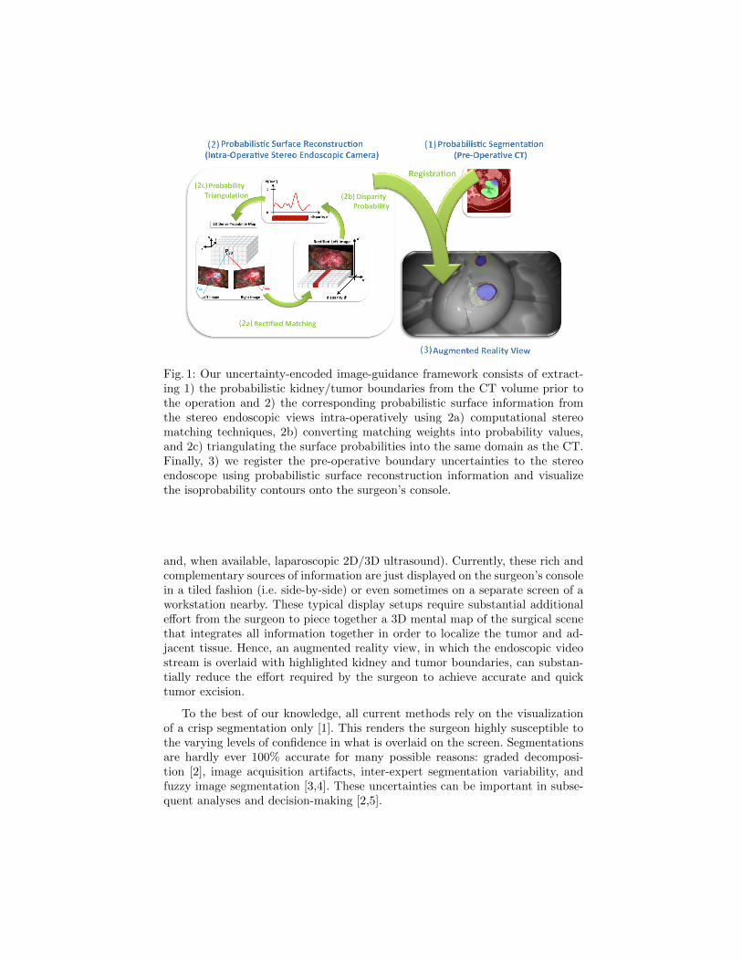
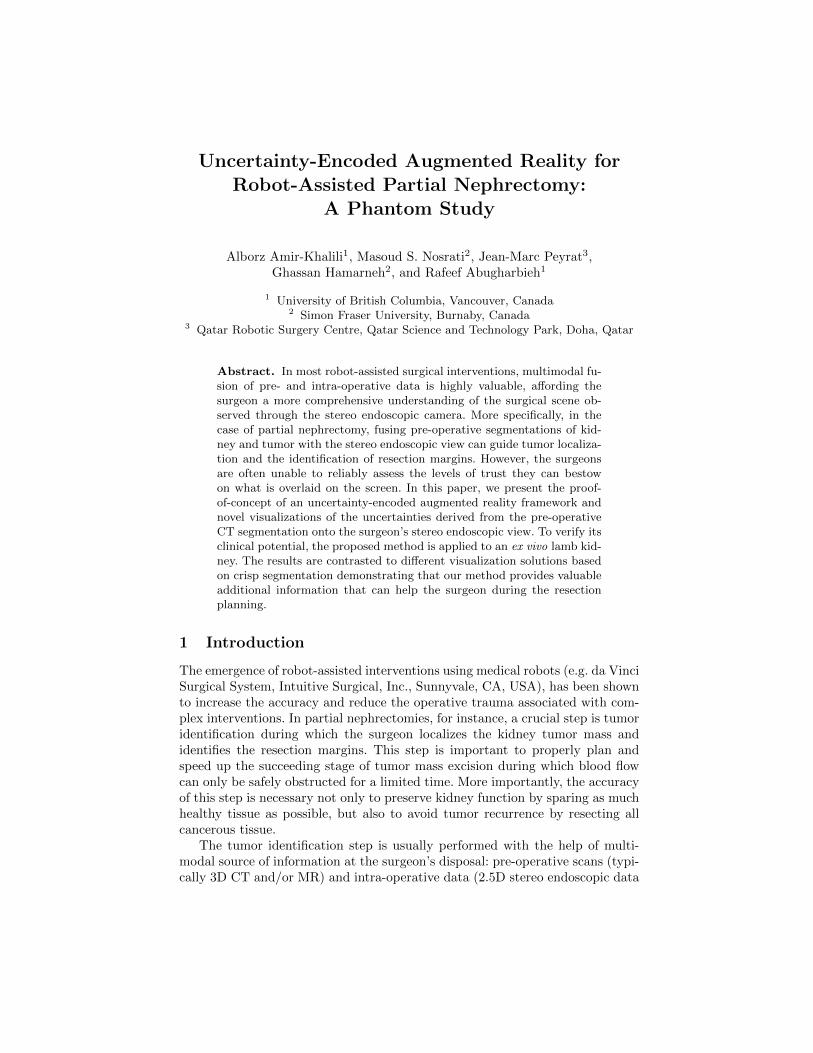
Fig. 1: Our uncertainty-encoded image-guidance framework consists of extract-ing 1) the probabilistic kidney/tumor boundaries from the CT volume prior tothe operation and 2) the corresponding probabilistic surface information fromthe stereo endoscopic views intra-operatively using 2a) computational stereomatching techniques, 2b) converting matching weights into probability values,and 2c) triangulating the surface probabilities into the same domain as the CT.Finally, 3) we register the pre-operative boundary uncertainties to the stereoendoscope using probabilistic surface reconstruction information and visualizethe isoprobability contours onto the surgeon’s console.
and, when available, laparoscopic 2D/3D ultrasound). Currently, these rich andcomplementary sources of information are just displayed on the surgeon’s consolein a tiled fashion (i.e. side-by-side) or even sometimes on a separate screen of aworkstation nearby. These typical display setups require substantial additionaleffort from the surgeon to piece together a 3D mental map of the surgical scenethat integrates all information together in order to localize the tumor and ad-jacent tissue. Hence, an augmented reality view, in which the endoscopic videostream is overlaid with highlighted kidney and tumor boundaries, can substan-tially reduce the effort required by the surgeon to achieve accurate and quicktumor excision.
To the best of our knowledge, all current methods rely on the visualizationof a crisp segmentation only [1]. This renders the surgeon highly susceptible tothe varying levels of confidence in what is overlaid on the screen. Segmentationsare hardly ever 100% accurate for many possible reasons: graded decomposi-tion [2], image acquisition artifacts, inter-expert segmentation variability, andfuzzy image segmentation [3,4]. These uncertainties can be important in subse-quent analyses and decision-making [2,5].
(a) (b) (c)
Fig. 2: Probabilistic pre-operative CT segmentation. (a) Original CT. (b) Mem-bership probabilities of kidney (green), tumor (blue), and background (red).(c) Background boundary location probability (0 in black and 1 in white).
In this paper, we propose to provide a visualization of uncertainties at thekidney and tumor boundaries as a visual cue to assist the surgeon in finding theoptimal resection strategy. This is similar in concept to what is currently beingexplored in radiotherapy for brain tumors when extrapolating glioma invasionwith variable margins [6]. Our visual cues are derived from shape boundaryuncertainties in the probabilistic segmentation of the pre-operative CT. Thisinformation is then registered to the endoscopic view as explained in Fig. 1. Weapply our method to an ex vivo lamb kidney to create an uncertainty-encodedaugmented reality view. We compare our results to standard guidance methodsthat use crisp segmentations and clearly demonstrate the benefits of our methodand its utility for resection planning.
2 Methods
We first describe the probabilistic segmentation of the pre-operative CT thatprovides uncertainties about the boundary localization of kidney and tumor.Secondly, we perform a probabilistic 3D surface reconstruction from stereo en-doscopy to which the probabilistic segmentation is directly registered.
2.1 Probabilistic Segmentation of Pre-Operative CT Scans
The probabilistic segmentation of the pre-operative CT is based on the randomwalker segmentation algorithm [4,7] that generates membership probabilities ofthree manually seeded regions: background (BG: red), kidney (KD: green), andtumor (TM: blue) (Fig. 2b).
We denote the resulting multi-label probabilistic CT segmentation by:
PCTseg : Ω ⊂ R3 → p ∈ S2 ,
where p = [pBG, pKD, pTM ] belongs to the simplex of order 2, and Ω is thespatial domain of the CT. From this multi-label probabilistic segmentation, we
can extract the membership probability map of background PCTBG , kidney PCT
KD
and tumor PCTTM regions.
We also compute the likelihood PCTsurface of the surface union of kidney and
tumor in the pre-operative CT (Fig. 2c) by combining the membership proba-bilities of being inside the kidney PCT
KD and inside the tumor PCTTM as follows:
PCTsurface = 1− |(P
CTKD + PCT
TM )− 0.5|0.5
. (1)
2.2 Probabilistic Stereo-Endoscopic Surface Reconstruction
We propose an extension of traditional computational stereo techniques of sur-face reconstruction from a single crisp surface [8] to a probabilistic representationof surfaces in 3-space.
Dense Matching of Left and Right Stereo Images Using polar rectifica-tion [9] with the camera calibration parameters, the 2D dense matching of leftand right stereo images is simplified to a 1D matching along parallel epipolarlines in the left and right rectified images. We use the normalized cross correlation(NCC) ratio on greyscale images as a matching similarity metric. This metrichas the advantage of being less prone to changes in illumination. In contrastwith current state-of-the-art methods, e.g. [10,11,12], instead of computing oneset of robust and optimal matches, we retain all possible matches with their as-sociated disparity (displacement d ∈ Z between matching points along the samehorizontal line of the recitified images) and similarity measure (c ∈ [−1, 1]).
Construction of a 3D Probabilistic Voxel Map In order to facilitate thepre-op to intra-op registration detailed in Section 2.3, we first create a 3D prob-abilistic voxel map in which each voxel stores the probability of being at thesurface of the stereo endoscopic scene. To achieve this, we compute the disparityprobability values by converting the NCC profile c = [c1, c2, · · · , cNd
] computedpreviously at every pixel (u, v) ∈ Ω2D ⊂ R2 in one of the rectified images fordifferent disparities d ∈ D = d1, d2, · · · , dNd
, where Nd is the total number ofdisparities. Basically, the NCC profiles are stacked into a 3D correlation map:
NCCstereo3D : (u, v, di) ∈ Ω3D → ci ∈ [−1, 1] (2)
and converted into a 3D probabilitistic voxel map using the Gibbs measure asfollows:
P stereo3D (u, v, di) =
exp (−β (maxd (NCCstereo3D (u, v, d))−NCCstereo
3D (u, v, di)))
W (β),
(3)where W (β) =
∑d exp (−β (maxd (NCCstereo
3D (u, v, d))−NCCstereo3D (u, v, di)))
is the partition function, and β is a free parameter.
Finally, the 3D position of each matched pair of points in the stereo viewsis triangulated with the camera projection matrices to transform P stereo
3D into aprobabilistic voxel map P stereo
surface in real world 3D space:
P stereosurface : (x, y, z) ∈ Ω3D → [p, 1− p] ∈ S1 , (4)
where p ∈ [0, 1] is the likelihood of a surface at voxel (x, y, z) in real world 3Dspace.
2.3 Registration of Stereo Camera and CT
We initialize the registration of the CT to the stereo camera in a semi-automaticmanner using manually matched landmarks between the original CT, left andright camera views. In this first step, we use a similarity transformation to modelthe combination of (1) a rigid transformation to cope with different referenceframes between stereo camera and CT acquisitions and (2) a global scaling tocope with ambiguities resulting from possible camera calibration errors. The re-sulting transformation is then refined with an automatic similarity registration ofPCTsurface to P stereo
surface obtained respectively from (1) and (4). Finally, a non-linearregistration step of these two volumes with a B-Spline transformation model isperformed to cope with deformations occurring between the pre-operative CTacquisition and the surgical scene. We used elastix [13] with the sum of squareddifferences (SSD) similarity metric for the two last automatic registration steps.
3 Results
3.1 Materials
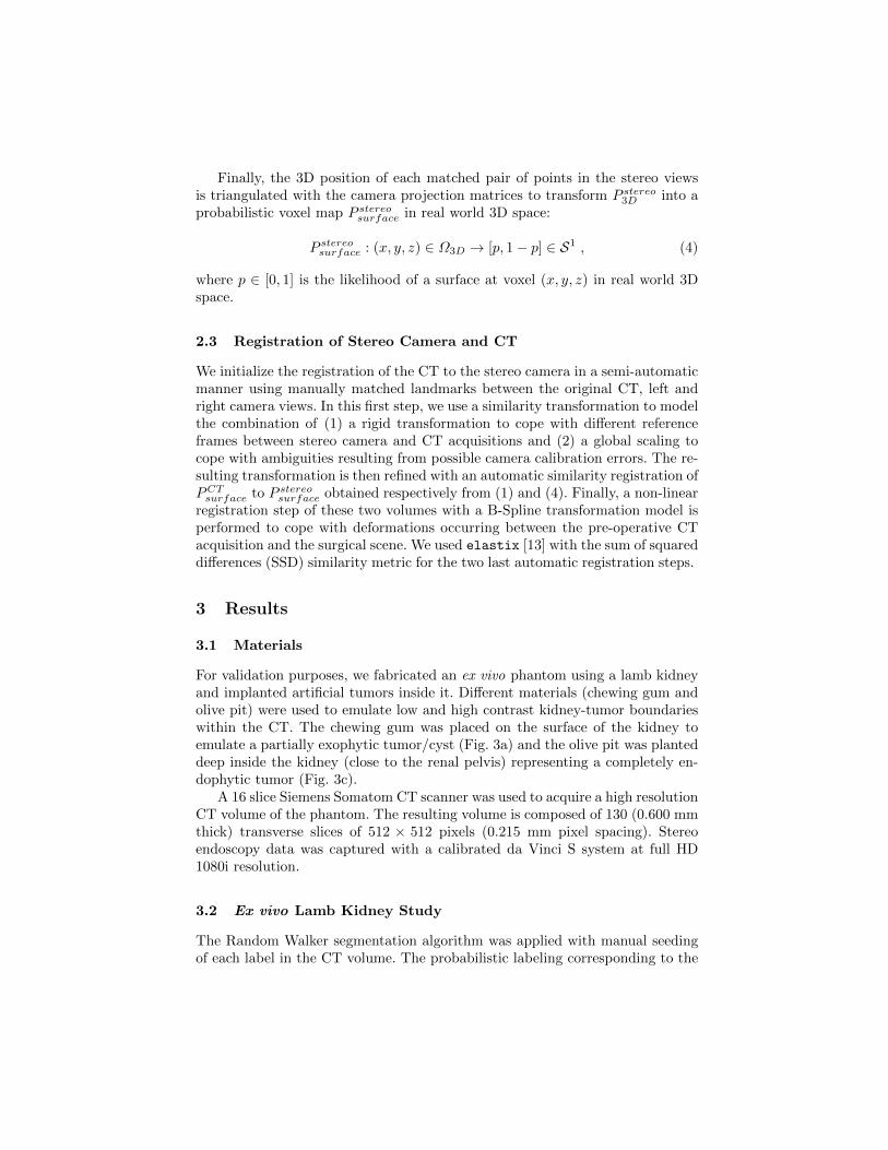
For validation purposes, we fabricated an ex vivo phantom using a lamb kidneyand implanted artificial tumors inside it. Different materials (chewing gum andolive pit) were used to emulate low and high contrast kidney-tumor boundarieswithin the CT. The chewing gum was placed on the surface of the kidney toemulate a partially exophytic tumor/cyst (Fig. 3a) and the olive pit was planteddeep inside the kidney (close to the renal pelvis) representing a completely en-dophytic tumor (Fig. 3c).
A 16 slice Siemens Somatom CT scanner was used to acquire a high resolutionCT volume of the phantom. The resulting volume is composed of 130 (0.600 mmthick) transverse slices of 512 × 512 pixels (0.215 mm pixel spacing). Stereoendoscopy data was captured with a calibrated da Vinci S system at full HD1080i resolution.
3.2 Ex vivo Lamb Kidney Study
The Random Walker segmentation algorithm was applied with manual seedingof each label in the CT volume. The probabilistic labeling corresponding to the
(a) (b) (c) (d)
Fig. 3: Transverse slices of CT volume depicting our ex vivo lamb kidney phantomwith (a) an exophytic and (c) an endophytic artificial tumor. (b) and (d) areprobabilistic Random Walker segmentations of (a) and (c), respectively. Tumorlabels are colored blue, kidney is colored green, and the background is red.
(a) (b)
(c) (d) (e)
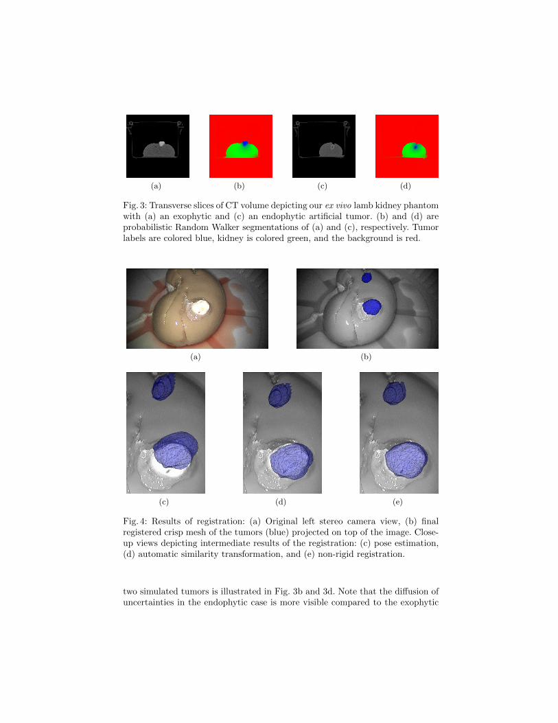
Fig. 4: Results of registration: (a) Original left stereo camera view, (b) finalregistered crisp mesh of the tumors (blue) projected on top of the image. Close-up views depicting intermediate results of the registration: (c) pose estimation,(d) automatic similarity transformation, and (e) non-rigid registration.
two simulated tumors is illustrated in Fig. 3b and 3d. Note that the diffusion ofuncertainties in the endophytic case is more visible compared to the exophytic
tumor; this is a direct result of weaker contrast (CT intensity values: differencein pit/gum composition) at the kidney-tumor boundary. We were careful to keepthe distances between the manually placed seeds and the visible boundaries con-stant to decrease the influence of seed placement on the resulting segmentations.
As illustrated in Fig. 4a, our phantom is quite smooth and lacks uniquefeatures on its surface. This results in a largely uncertain reconstruction from ourstereo matching algorithm, which in turn causes the registration to be sensitive tothe initial pose estimation. Successful registration was achieved after estimatingthe pose (Fig. 4c) using only four manually selected corresponding landmarks.The outcome of the registration was verified visually (Fig. 4) by projecting thekidney and tumor surfaces on both left and right endoscopy views. A small errorin alignment (< 1 mm) is observed in the resulting registration, this is due to theerror in reconstruction which is attributed to lack of texture on the phantom.
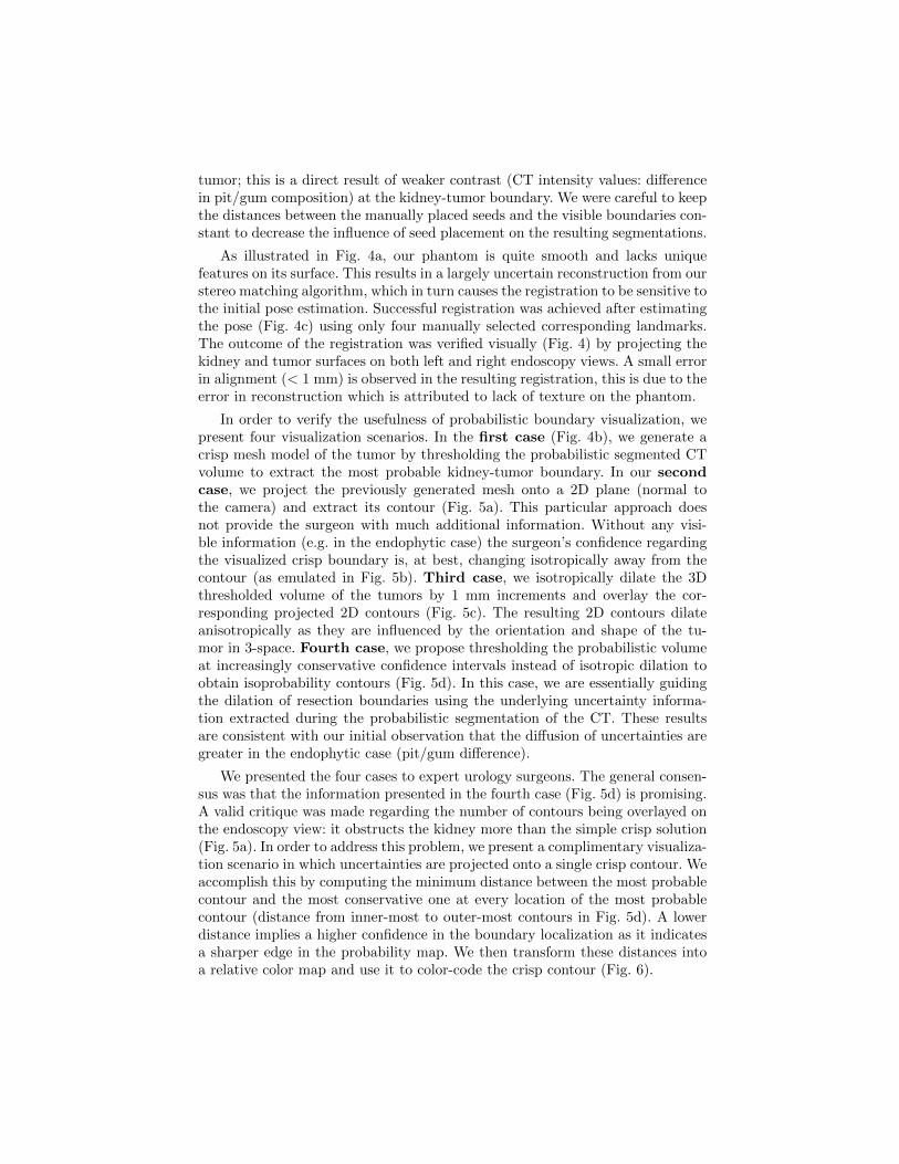
In order to verify the usefulness of probabilistic boundary visualization, wepresent four visualization scenarios. In the first case (Fig. 4b), we generate acrisp mesh model of the tumor by thresholding the probabilistic segmented CTvolume to extract the most probable kidney-tumor boundary. In our secondcase, we project the previously generated mesh onto a 2D plane (normal tothe camera) and extract its contour (Fig. 5a). This particular approach doesnot provide the surgeon with much additional information. Without any visi-ble information (e.g. in the endophytic case) the surgeon’s confidence regardingthe visualized crisp boundary is, at best, changing isotropically away from thecontour (as emulated in Fig. 5b). Third case, we isotropically dilate the 3Dthresholded volume of the tumors by 1 mm increments and overlay the cor-responding projected 2D contours (Fig. 5c). The resulting 2D contours dilateanisotropically as they are influenced by the orientation and shape of the tu-mor in 3-space. Fourth case, we propose thresholding the probabilistic volumeat increasingly conservative confidence intervals instead of isotropic dilation toobtain isoprobability contours (Fig. 5d). In this case, we are essentially guidingthe dilation of resection boundaries using the underlying uncertainty informa-tion extracted during the probabilistic segmentation of the CT. These resultsare consistent with our initial observation that the diffusion of uncertainties aregreater in the endophytic case (pit/gum difference).
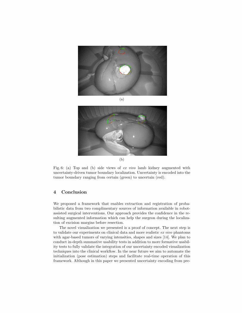
We presented the four cases to expert urology surgeons. The general consen-sus was that the information presented in the fourth case (Fig. 5d) is promising.A valid critique was made regarding the number of contours being overlayed onthe endoscopy view: it obstructs the kidney more than the simple crisp solution(Fig. 5a). In order to address this problem, we present a complimentary visualiza-tion scenario in which uncertainties are projected onto a single crisp contour. Weaccomplish this by computing the minimum distance between the most probablecontour and the most conservative one at every location of the most probablecontour (distance from inner-most to outer-most contours in Fig. 5d). A lowerdistance implies a higher confidence in the boundary localization as it indicatesa sharper edge in the probability map. We then transform these distances intoa relative color map and use it to color-code the crisp contour (Fig. 6).
(a) (b)
(c) (d)
Fig. 5: Augmented reality view of Ex vivo lamb kidney with endophytic andexophytic artificial tumors showing different visualization scenarios: (a) crispcontour of projected mesh, (b) isotropic 2D diffusion of the crisp contour, (c) 2Dprojections of the crisp mesh dilated in 3D by 1 mm increments, (d) 2D pro-jections of 3D isoprobabilities from 0.5 to 0.15. Contours range from the mostprobable boundary (red) to the most conservative boundary (green).
This final visualization scenario does not only provide the most probabletumor boundary localization, but also provide information about its local con-fidence. This visualization can guide the surgeon to quickly identify the best(most confident) place to start the resection. During the resection, the surgeoncan always opt for the fourth case to see exactly how the uncertainty is diffusedspatially.
(a)
(b)
Fig. 6: (a) Top and (b) side views of ex vivo lamb kidney augmented withuncertainty-driven tumor boundary localization. Uncertainty is encoded into thetumor boundary ranging from certain (green) to uncertain (red).
4 Conclusion
We proposed a framework that enables extraction and registration of proba-bilistic data from two complimentary sources of information available in robot-assisted surgical interventions. Our approach provides the confidence in the re-sulting augmented information which can help the surgeon during the localiza-tion of excision margins before resection.
The novel visualization we presented is a proof of concept. The next step isto validate our experiments on clinical data and more realistic ex vivo phantomswith agar-based tumors of varying intensities, shapes and sizes [14]. We plan toconduct in-depth summative usability tests in addition to more formative usabil-ity tests to fully validate the integration of our uncertainty encoded visualizationtechniques into the clinical workflow. In the near future we aim to automate theinitialization (pose estimation) steps and facilitate real-time operation of thisframework. Although in this paper we presented uncertainty encoding from pre-
operative CT, we will be taking advantage of other intra-operative sources ofuncertainty to improve the confidence at the localized boundary while new datais acquired during the resection.
Acknowledgement The authors would like to thank Dr. Abdulla Al Ansariand Dr. Osama Al-Alao for their expert opinion and assistance with the dataacquisition. This publication was made possible by NPRP Grant from the QatarNational Research Fund (a member of the Qatar Foundation). The statementsmade herein are solely the responsibility of the authors.
References
1. P. Pratt, E. Mayer, J. Vale, D. Cohen, E. Edwards, A. Darzi, and G.-Z. Yang,“An effective visualisation and registration system for image-guided robotic partialnephrectomy,” Journal of Robotic Surgery, pp. 1–9, 2012.
2. J. Udupa and G. Grevera, “Go digital, go fuzzy,” Pattern Recognition Letters,vol. 23, no. 6, pp. 743–754, 2002.
3. Y. Zhang, M. Brady, and S. Smith, “Segmentation of brain MR images through ahidden markov random field model and the expectation-maximization algorithm,”IEEE Transactions on Medical Imaging, vol. 20, no. 1, pp. 45–57, 2001.
4. L. Grady, “Random walks for image segmentation,” IEEE Transactions on PatternAnalysis and Machine Intelligence, vol. 28, no. 11, pp. 1768–1783, 2006.
5. S. Warfield, K. Zou, and W. Wells, “Simultaneous truth and performance levelestimation (STAPLE): an algorithm for the validation of image segmentation,”IEEE Transactions on Medical Imaging, vol. 23, no. 7, pp. 903–921, 2004.
6. E. Konukoglu, O. Clatz, P. Bondiau, H. Delingette, and N. Ayache, “Extrapolat-ing glioma invasion margin in brain magnetic resonance images: suggesting newirradiation margins,” Medical Image Analysis, vol. 14, no. 2, pp. 111–125, 2010.
7. A. K. Sinop and L. Grady, “A seeded image segmentation framework unifyinggraph cuts and random walker which yields a new algorithm,” in ICCV, pp. 1–8,2007.
8. R. Hartley and A. Zisserman, Multiple view geometry in computer vision. Cam-bridge Univ Press, 2nd ed., 2004.
9. M. Pollefeys, R. Koch, and L. Van Gool, “A simple and efficient rectificationmethod for general motion,” in ICCV, pp. 496–501, 1999.
10. D. Stoyanov, M. V. Scarzanella, P. Pratt, and G.-Z. Yang, “Real-time stereo recon-struction in robotically assisted minimally invasive surgery,” in MICCAI, pp. 275–282, 2010.
11. S. Bernhardt, J. Abi-Nahed, and R. Abugharbieh, “Robust dense endoscopic stereoreconstruction for minimally invasive surgery,” MICCAI workshop on MedicalComputer Vision (MCV), pp. 198–207, 2012.
12. S. Rohl, S. Bodenstedt, S. Suwelack, H. Kenngott, B. Muller-Stich, R. Dillmann,and S. Speidel, “Real-time surface reconstruction from stereo endoscopic imagesfor intraoperative registration,” in Proc. SPIE, vol. 7964, p. 796414, 2011.
13. S. Klein, M. Staring, K. Murphy, M. Viergever, and J. Pluim, “Elastix: a toolboxfor intensity based medical image registration,” IEEE Transactions on MedicalImaging, vol. 29, pp. 196–205, 2010.
14. J. S. Huber, Q. Peng, and W. W. Moses, “Multi-modality phantom development,”IEEE Transactions on Nuclear Science, vol. 56, no. 5, pp. 2722–2727, 2009.