Embed Size (px)
Citation preview

Uhrf1 controls the self-renewal versus differentiationof hematopoietic stem cells by epigeneticallyregulating the cell-division modesJingyao Zhaoa,1, Xufeng Chena,1, Guangrong Songb, Jiali Zhanga, Haifeng Liua, and Xiaolong Liua,b,2
aState Key Laboratory of Cell Biology, Chinese Academy of Sciences Center for Excellence in Molecular Cell Science, Institute of Biochemistry and CellBiology, Shanghai Institutes for Biological Sciences, Chinese Academy of Sciences, Shanghai 200031, China; and bSchool of Life Science and Technology,ShanghaiTech University, Shanghai 200031, China
Edited by Ioannis Aifantis, New York University School of Medicine, New York, NY, and accepted by Editorial Board Member Tak W. Mak November 18, 2016(received for review August 4, 2016)
Hematopoietic stem cells (HSCs) are able to both self-renew anddifferentiate. However, how individual HSC makes the decisionbetween self-renewal and differentiation remains largely unknown.Here we report that ablation of the key epigenetic regulator Uhrf1 inthe hematopoietic system depletes the HSC pool, leading to hemato-poietic failure and lethality. Uhrf1-deficient HSCs display normal sur-vival and proliferation, yet undergo erythroid-biased differentiation atthe expense of self-renewal capacity. Notably, Uhrf1 is required forthe establishment of DNA methylation patterns of erythroid-specificgenes during HSC division. The expression of these genes is enhancedin the absence of Uhrf1, which disrupts the HSC-division modes bypromoting the symmetric differentiation and suppressing the sym-metric self-renewal. Moreover, overexpression of one of the up-reg-ulated genes, Gata1, in HSCs is sufficient to phenocopy Uhrf1-deficientHSCs, which show impaired HSC symmetric self-renewal and increaseddifferentiation commitment. Taken together, our findings suggestthat Uhrf1 controls the self-renewal versus differentiation of HSCthrough epigenetically regulating the cell-division modes, thus provid-ing unique insights into the relationship among Uhrf1-mediated DNAmethylation, cell-division mode, and HSC fate decision.
Uhrf1 | HSCs | epigenetic regulation | cell-division mode | cell fate decision
Hematopoietic stem cells (HSCs) harbor the capacities of bothself-renewal and differentiation to sustain life-long hemato-
poiesis (1). Although differentiation is responsible for producingall functional blood cells, self-renewal is critical in maintaining thesize of the HSC pool (2). It has been reported that extrinsic cellsignals, such as stem cell factor (SCF)/c-Kit signaling, Notch sig-naling, and Wnt signaling, contribute to the maintenance of HSCself-renewal (3–5). Moreover, the transcription factors Id2 (in-hibitor of DNA binding 2) and Hoxa9 (homeobox A9) are re-quired for HSC self-renewal and expansion (6, 7), and Hmga2(high mobility group AT-hook 2) overexpression endows HSCswith higher self-renewal potential (8). Additionally, many tran-scription factors are involved in HSC differentiation. For example,the enforced expression of the erythroid master gene, Gata1(GATA binding protein 1), in HSCs results in the exclusive gen-eration of megakaryocyte and erythrocyte lineages (9). Consis-tently, Gfi1b (growth factor independent 1B), a downstream targetof Gata1, controls erythroid and megakaryocytic differentiation byregulating TGF-β signaling (10). Recently, increasing research hasfocused on the functions of epigenetic regulation in HSCs. Theabsence of Dnmt1 (DNA methyltransferase 1) in HSCs impairstheir self-renewal capacity (11, 12), whereas shortages of Dnmt3aand Dnmt3b block the differentiation process (13).The self-renewal and the differentiation of HSCs were considered
as two independent fate choices (14). Intriguingly, upon each di-vision, HSCs undergo only one of the three mutually exclusivecell-division modes [symmetric self-renewal (SS), symmetric dif-ferentiation (SD), and asymmetric self-renewal (AS)] (15, 16),thus indicating that the regulation of HSC self-renewal cannot be
separated from that of differentiation (17, 18). However, the keyfactors that regulate the HSC-division modes and the detailedmechanisms underlying how individual HSC accomplishes the de-cision of self-renewal versus differentiation remain largely unknown.The epigenetic regulator Uhrf1 (ubiquitin-like, containing PHD
and RING finger domains, 1) contains multiple functional domainsthat enable it to participate in various molecular processes (19–21).Among these processes, Uhrf1 is believed to be critical for maintainingDNA methylation (19). During DNA replication, Uhrf1 recognizesand binds to the hemimethylated CG residues generated at replicationfoci via the Set and Ring Associated domain, after which it recruitsDNA methyltransferases and sustains the methylation of the newlysynthesized DNA strand (22, 23). Previous research has reported thatUhrf1 facilitates the proliferation and maturation of colonic regulatoryT cells (24), and our recent findings have suggested that Uhrf1 isrequired for invariant natural killer T cell development by regulatingthe Akt-mammalian target of rapamycin signaling pathway (25).To investigate the functions of Uhrf1 in the hematopoietic
system, we conditionally deleted Uhrf1 from hematopoietic cells.Uhrf1 deficiency leads to the exhaustion of the HSC pool and to asevere reduction in hematopoiesis. Uhrf1-deficient HSCs undergoerythroid-biased differentiation at the expense of self-renewal.
Significance
Hematopoietic stem cells (HSCs) harbor the capacities of both self-renewal and differentiation to sustain life-long production of allblood cells. However, how individual HSCs accomplish the de-cision of self-renewal versus differentiation remains largely un-known. Here, we find that Uhrf1, a key epigenetic regulator ofDNA methylation, specifically controls this critical process. In theabsence of Uhrf1, HSCs undergo erythroid-biased differentiationat the expense of self-renewal capacity, leading to hematopoieticfailure and lethality. Mechanistically, Uhrf1 regulates the HSC-division mode by DNA methylation-mediated repression of theexpression of certain erythroid-specific genes, and thus modulatesthe cell fate decision of HSCs. This study provides unique insightsinto the relationship among Uhrf1-mediated DNA methylation,cell-division mode, and HSC fate decision.
Author contributions: J. Zhao, X.C., and X.L. designed research; J. Zhao, X.C., G.S., J. Zhang,and H.L. performed research; J. Zhao, X.C., G.S., J. Zhang, and X.L. analyzed data;and J.Zhao, X.C., and X.L. wrote the paper.
The authors declare no conflict of interest.
This article is a PNAS Direct Submission. I.A. is a Guest Editor invited by the Editorial Board.
Data deposition: The data reported in this paper have been deposited in the Gene Ex-pression Omnibus (GEO) database, www.ncbi.nlm.nih.gov/geo (accession no. GSE85450).
See Commentary on page 192.1J. Zhao and X.C. contributed equally to this work.2To whom correspondence should be addressed. Email: [email protected].
This article contains supporting information online at www.pnas.org/lookup/suppl/doi:10.1073/pnas.1612967114/-/DCSupplemental.
E142–E151 | PNAS | Published online December 12, 2016 www.pnas.org/cgi/doi/10.1073/pnas.1612967114

Notably, Uhrf1 plays essential roles in the establishment of DNAmethylation patterns of differentiation-promoting genes duringHSC division and in the regulation of HSC-division modes, andhence is critical for the decision of self-renewal versus differenti-ation of individual HSC. Altogether, our findings identified Uhrf1as an essential regulator that controls the cell fate decision ofindividual HSC through epigenetically regulating the HSC-division modes.
ResultsUhrf1 Is Required to Maintain the Fetal Liver-HSC Pool. To study thefunction of Uhrf1 in hematopoiesis, we bred conditional Uhrf1L/L
mice with the Vav1-cre strain. Uhrf1 was efficiently ablated fromthe hematopoietic system from 12.5 d postcoitum (dpc) mice (Fig.S1 A–D). Weaned Vav1-cre+Uhrf1L/L mice (designated Uhrf1−/−
mice) were not observed and litters did not have Mendelian ge-notype ratios (Fig. 1A). However, perinatal Uhrf1−/− mice withnormal morphology but pale bodies (Fig. 1B) were identified.Uhrf1-deficient mice showed a significant decrease in fetal liver(FL) cellularity (Fig. 1C), compared with their WT control lit-termates (Uhrf1L/L or Uhrf1L/+). Further analysis revealed adramatic decrease in the absolute cell numbers of multilineagehematopoietic cells in the Uhrf1-deficient fetal liver (Fig. 1D).These phenotypes inspired us to evaluate the roles of Uhrf1 in
hematopoietic stem and progenitor cells. Consistent with the re-duction in multilineage hematopoietic cells, Uhrf1-deficient fetallivers contained decreased megakaryocyte/erythroid progenitors(MEPs), common myeloid progenitors (CMPs), granulocyte-macrophage progenitors (GMPs), and common lymphoid progen-itors (CLPs) (Fig. 1E). Moreover, Uhrf1-deficient mice displayedreduced hematopoietic stem/progenitor cells (HSPCs) [lineage−
cKit+ Sca1+ (LSK)] after 12.5 dpc (Fig. 1 F andG). Consistent withthis result, FL-HSCs (CD150+ CD48− Mac1low LSKs) (26) alsodeclined at 16.5 dpc (Fig. 1H and Fig. S2A). The aorta–gonad–mesonephros (AGM) region is a source of definitive HSCs beforethey colonize the fetal liver at 12.5 dpc (27). Notably, both theproportion and cell number of HSCs in the Uhrf1-deficientAGM were comparable to those in the control AGM at 11.5 dpc
(Fig. S3 A and B). Together, these results indicate that Uhrf1 is re-quired for the maintenance of the FL-HSC pool in the fetal liver.
Uhrf1 Is Essential for FL-HSC Self-Renewal. Given that the ablation ofUhrf1 in FL-HSCs results in HSC depletion, the survival and pro-liferation capacity of Uhrf1-deficient FL-HSCs was evaluated.Freshly isolated Uhrf1-deficient FL-HSCs showed similar levels ofapoptosis compared with control FL-HSCs (Fig. 2 A and B), thusindicating that Uhrf1 deficiency did not impair the survival capacityof FL-HSCs. BrdU incorporation experiments were then performedto assess the proliferation capacity of FL-HSCs. Uhrf1-deficientFL-HSCs incorporated similar amounts of BrdU compared withcontrol HSCs, a result suggesting that FL-HSCs can normallyenter the cell cycle in the absence of Uhrf1 (Fig. 2 C and D).Collectively, these data show that Uhrf1-deficient FL-HSCs retaina normal survival and proliferation capacity.We then wondered whether the defects in the establishment of the
FL-HSC pool and in the consequential hematopoiesis resulted fromthe compromised self-renewal capacity of FL-HSCs in the absenceof Uhrf1. To this end, we performed colony-forming unit (CFU)assays. The colonies formed by Uhrf1-deficient FL-HSPCs weremuch smaller than control colonies (Fig. 2E). Additionally, Uhrf1-deficient HSPCs formed more erythroid colonies (BFU-E) butfewer multilineage colonies (CFU-GEMM) and myeloid colonies(CFU-GM/G/M) compared with control HSPCs (Fig. 2F). Com-petitive bone marrow transplantations were then carried out to fur-ther analyze the self-renewal capacity of Uhrf1-deficient FL-HSCs.Uhrf1-deficient FL-HSPCs, compared with control FL-HSPCs,did not achieve multilineage reconstitution in irradiated mice(Fig. 2 G and H and Fig. S3 C and D). Moreover, Uhrf1-deficientFL-HSPCs could not reconstitute the HSC pool after trans-plantation (Fig. 2 I and J). Together, these data suggest thatUhrf1 is essential for FL-HSC self-renewal.
The HSC-Division Mode Is Impaired After Uhrf1 Ablation. Because theproliferation and survival of FL-HSCs remained unchanged in theabsence of Uhrf1, cell fate analysis was performed to addresswhether HSC differentiation was affected. Notably, Uhrf1-de-
B
Cel
l num
ber (
X10
6 )
F
**
Sca1
cKit
WT Uhrf1 /
A
45.3 2.31 57.4 1.05
0
50
150
100
Mic
e nu
mbe
r
Uhrf1fl/+
Vav1-Cre-Uhrf1fl/fl
Vav1-Cre-Uhrf1fl/+
Vav1-Cre+Uhrf1fl/fl
Vav1-Cre+WT Uhrf1 /
0
20
40
60
80
100C
WT Uhrf1 /0
20
40
60
80
100
0
50
100
150
0
1
2
3
4
5
0.0
0.5
1.0
1.5
2.0
Cel
l num
ber (
X10
6 )
Cel
l num
ber (
X10
4 )
Cel
l num
ber (
X10
4 )
Cel
l num
ber (
X10
4 )
D
WT Uhrf1 / WT Uhrf1 / WT Uhrf1 / WT Uhrf1 /
Cel
l num
ber (
X10
3 )
G
Ter119 Gr1/Mac1 B cell T cellExpected Actual
*** *
*
0
5
10
15
11.5 12.5 13.5 14.5
WTUhrf1 /
**
***
N.S.N.S.
0
1
2
3
4
0
5
10
15
20
25
0
5
10
15
20
25
0
50
100
150
E
Cel
l num
ber (
X10
4 )
Cel
l num
ber (
X10
4 )
Cel
l num
ber (
X10
4 )
Cel
l num
ber (
X10
4 )
WT Uhrf1 /WT Uhrf1 / WT Uhrf1 /WT Uhrf1 /
MEP GMPCMP CLP
***** ***
*
H FL-HSC
WT Uhrf1 /0
5
10
15
Cel
l num
ber (
X10
2 ) ***
Fig. 1. Loss of Uhrf1 leads to embryonic lethality because of FL-HSC depletion. (A) Expected (according to Mendel’s law) and actual weaned mouse numbersof different genotypes. (B) Representative images of control littermates and Uhrf1−/− mice at birth. (Scale bar, 1 cm.) (C) Cellularity of fetal livers from controllittermates and Uhrf1−/− mice at 16.5 dpc (n = 3). (D and E) Absolute cell numbers of hematopoietic cells (D) and HPCs (MEPs, CMPs, GMPs, CLPs) (E) fromcontrol and Uhrf1-deficient fetal livers at 14.5 dpc (n = 3). (F) Flow cytometric analysis of HSPCs (lineage− cKit+ Sca1+, LSK) and HPCs (lineage− cKit+ Sca1−, LK)from control and Uhrf1-deficient fetal livers at 14.5 dpc. Lineage− viable cells are shown. (G) Absolute cell numbers of HSPCs (LSKs) from control and Uhrf1-deficient fetal livers at different gestational ages (n = 3–4 for each genotype for each gestational age). (H) Absolute cell numbers of FL-HSCs (CD150+ CD48−
Mac1low LSKs) from control and Uhrf1-deficient fetal livers at 16.5 dpc (n = 3). The data are means ± standard deviation, for all panels: *P < 0.05, **P < 0.01,***P < 0.001 by Student’s t test; N.S.: no significance.
Zhao et al. PNAS | Published online December 12, 2016 | E143
CELL
BIOLO
GY
PNASPL
US
SEECO
MMEN
TARY

ficient FL-HSPCs could not sustain the HSPC pool, whereas morehematopoietic progenitor cells [HPCs (lineage− cKit+ Sca1−,LK), referred to as HPC(LK)s] were generated (Fig. 3 A–C).These results suggested that FL-HSPCs are more prone toundergoing differentiation when Uhrf1 is deleted.Given that self-renewal and differentiation are coordinately reg-
ulated in HSCs through the exclusive choice among the three stem-cell division modes (SS, SD, and AS) during HSC division (15, 16),we then sought to determine whether the attenuated self-renewaland the increased differentiation is resulted from abnormal HSC-division modes. To address this possibility, we stained the cell-fatedeterminant Numb [numb homolog (Drosophila)] in individualUhrf1-deficient and WT FL-HSPC and measured the ratios of SSdivisions (low expression of Numb in both daughter cells), SD (highNumb expression in both daughter cells), and AS divisions (higherexpression of Numb protein in one of two daughter cells) (Fig. 3D)(16, 17). Notably, significantly fewer SS divisions (25.82% ± 9.16%vs. 41.18% ± 3.93% in WT FL-HSPCs) and more SD divisions(46.15% ± 10.46% vs. 30.96% ± 8.12% in WT FL-HSPCs) wereobserved in Uhrf1-deficient FL-HSPCs (Fig. 3E). Consistently,Uhrf1-deficient FL-HSCs (CD150+ CD48− LSKs) also underwentfewer SS divisions (31.77% ± 3.59% vs. 55.37% ± 4.33% in WTFL-HSCs) and more SD divisions (50.93% ± 2.04% vs. 26.36% ±5.64% in WT FL-HSCs) (Fig. 3F). Moreover, Numb mRNAs werecomparable between Uhrf1-deficient and control FL-HSCs, in-dicating that the up-regulation of Numb in the daughter cells wasnot as a direct consequence of Uhrf1 ablation (Fig. S3E). Thesefindings suggested that Uhrf1 is involved in the regulation of HSC-division modes and controls the decision of self-renewal and dif-ferentiation of FL-HSCs.To a lesser extent, some Uhrf1-deficient FL-HSCs still under-
went SS and AS divisions (Fig. 3 E and F), thus raising the questionof how the FL-HSC pool was depleted under such a division pat-tern. Therefore, we stained FL-HSPCs with the cell division in-dicator 5-(and 6)-Carboxyfluorescein diacetate succinimidyl ester(CFSE), and then assessed the maintenance of the HSC pool afterdivision. The proportion of FL-HSPCs declined as cell divisions
continued (28). Interestingly, Uhrf1-deficient FL-HSPCs declinedfaster than their WT counterparts, thus eventually leading to thedepletion of the HSC pool after multiple rounds of division (Fig. 3G and H).Given that MEPs declined to a much lesser extent than CMPs,
GMPs, and CLPs in Uhrf1-deficient fetal livers (Fig. 1E), andUhrf1-deficient HSPCs formed excessive erythroid colonies in theCFU assays (Fig. 2F), we then sought to explore whether the in-creased differentiation potential of Uhrf1-deficient FL-HSCs wasaccompanied by biased lineage commitment. Notably, amongthose assessed progenitors, only MEP in Uhrf1-deficient fetallivers displayed an increased proportion compared with the WTcontrol (Fig. 3 I and J and Fig. S3 F andG), suggesting that Uhrf1-deficient FL-HSCs underwent erythroid-biased differentiation.
FL-HSCs Up-Regulate Erythroid-Specific Gene Expression in the Absenceof Uhrf1. To investigate the molecular mechanism of Uhrf1 incontrolling the self-renewal versus differentiation of FL-HSCs,high-throughput sequencing was performed to analyze the tran-scriptomes of WT and Uhrf1-deficient FL-HSPCs. Consistent withthe erythroid-biased differentiation of Uhrf1-deficient FL-HSCs(Fig. 3 I and J), genes involved in erythrocyte differentiation weresignificantly enriched in Uhrf1-deficient FL-HSPCs according tothe Gene Ontology (GO) enrichment analysis. Although thiscluster was ranked seventh among the top 24, with a score greaterthan 5, it was ranked first in the cell fate commitment category(Fig. 4A and Table S1). Gene-set enrichment analysis using genesets from lineage-restricted progenitors defined by Sanjuan-Plaet al. (29) revealed that genes associated with myeloid and lym-phoid (CLP) programming were enriched in WT HSPCs, whereasgenes associated with erythroid progenitors (were enriched inUhrf1-deficient HSPCs. Moreover, the HSC self-renewal associ-ated genes defined by Krivtsov et al. (30) were enriched in WTHSPCs but not Uhrf1-deficient HSPCs (Fig. S4A). We then gen-erated gene signatures specific for stemness of HSPCs (stemsignature) or myeloerythroid progenitors (MEP signature)by subtracting the genes expressed in WT HPC(LK)s from those
A
HG
B
39
58.2
97.7
0.013
25.5
68.6
97.8
0.17
Donor
Com
petit
or
Annexin V
DA
PI
DAPI
Brd
uDonor
Com
petit
or
0
10
15
20
0
20
40
60
80
0
20
40
60
80
0
20
40
60
80
100
WT Uhrf1 / WT Uhrf1 /
WT Uhrf1 / WT Uhrf1 /
WT Uhrf1 /
WT Uhrf1 / WT Uhrf1 /
WT Uhrf1 / WT Uhrf1 /
Ann
exin
V+ D
AP
I-ce
ll (%
)
Col
onie
s pe
r10
0 FL
-HS
PC
s
Don
or c
him
eris
m (%
)
Brd
u+ce
ll (%
)
Don
or c
him
eris
m (%
)
C
JI
D E
F
N.S. N.S.
*** ***
GEMMGM/G/MBFU-E
0
10
20
30
40
WT Uhrf1 /
*******
0.86 0.86
6.0192.3
0.89 0.15
7.4191.6
53.456.9
5
Fig. 2. Impaired FL-HSC self-renewal in the absence of Uhrf1. (A and B) Representative dot plots of the Annexin V staining profile (A) and the percentage ofcells that undergo apoptosis (Annexin V+ DAPI−) (B) of control and Uhrf1-deficient FL-HSCs (CD150+ CD48− LSKs) at 13.5 dpc (n = 3). (C and D) BrdU in-corporation profiles (C) and the percentages of cells that entered the S phase (D) were assessed 2 h after BrdU injection (n = 6). (E) Representative images ofcolonies generated from control and Uhrf1-deficient FL-HSPCs (LSKs) at 13.5 dpc. Images were captured on day 10. (Scale bars, 500 μm.) (F) Numbers ofmultilineage colonies (CFU-GEMM), erythroid colonies (BFU-E), and myeloid colonies (CFU-GM/G/M) generated by 100 FL-HSPCs (LSKs) sorted from control andUhrf1-deficient fetal livers at 13.5 dpc (n = 3). (G and H) Representative dot plots (G) and percentages (H) of donor chimerism of peripheral blood mono-nuclear cells (PBMCs) (8 wk) in recipients transplanted with 1,000 sorted FL-HSPCs (LSKs) from control or Uhrf1-deficient fetal livers at 13.5 dpc together with500,000 competitor bone marrow cells (WT, n = 9; Uhrf1−/−, n = 8). (I and J) Representative dot plots (I) and percentages (J) of donor chimerism of HSPCs (LSKs;8 wk) in recipients transplanted as described above (WT, n = 9; Uhrf1−/−, n = 8). The data are means ± standard deviation, for all panels: *P < 0.05, **P < 0.01.***P < 0.001 by Student’s t test; N.S.: no significance.
E144 | www.pnas.org/cgi/doi/10.1073/pnas.1612967114 Zhao et al.

expressed in WT FL-HSPCs or vice versa (12). Of the stem sig-nature genes, 71.28% showed lower expression and 28.72% showedhigher expression in Uhrf1-deficient FL-HSPCs compared withWT FL-HSPCs (Fig. 4B). Interestingly, among the MEP signaturegenes, genes enriched in erythroid differentiation (27.54%) wereup-regulated in the absence of Uhrf1, whereas the remaining genesenriched in myeloid-specific genes (72.46%) were suppressed,consistent with previous research (9) (Fig. S4B). Particularly, incomparison with the WT control, Uhrf1-deficient FL-HSPCsup-regulated certain erythrocyte differentiation-related genes
[e.g., Gata1, Gata2 (GATA binding protein 2), Gfi1b, Car1 (car-bonic anhydrase 1), Zfpm1 (zinc finger protein, multitype 1), andItga2b (integrin alpha 2b)] (Fig. 4 C and E), most of which werephysiologically up-regulated in HPC(LK)s (Fig. S4C), whereassome genes [Id2, Satb1 (special AT-rich sequence binding protein1), Hmga2] that play critical roles in HSC maintenance were down-regulated (Fig. 4 C and D). These results suggested that Uhrf1controls the self-renewal versus differentiation of FL-HSCs bysuppressing the expression of the erythroid-specific genes andmaintaining the expression of HSC stemness genes.
B
95.4
91.1
Sca1
cKit
22.5
3651.3
46.7
CFSE
Sca
1
0
1
2
3
4
0
5
10
15
0
5
10
15
20
Cel
l num
ber (
X10
3 )
Cel
l num
ber (
X10
3 )
HPC
(LK)
/FL-
HSP
CR
atio
WT Uhrf1 / WT Uhrf1 / WT Uhrf1 /
C
A Before culture After culture
WT
Uhrf1
/
DW
TUhrf1
/
G18 hourS
12
2.69
48 hours30 hours
0
5
10
15
0
5
10
15
0
50
100
150
Pop
ulat
ion
(%)
Pop
ulat
ion
(%)
Pop
ulat
ion
(%)
J
WT Uhrf1 / WT Uhrf1 / WT Uhrf1 /
FL-HSPC HPC(LK)
7.85
10.678.2
0.66
0.5897.3
CD34
CD
16/3
2
I
WT
Uhrf1 /
Myeloid progenitors
MEPGMP
CMP
MEPGMPCMP
** ****
*** *** ***
Numb DAPI Merge
SS
AS
SD
Numb DAPI Merge
WT Uhrf1 / E
020406080
100120
Per
cent
age
WT Uhrf1 /
44.8 40.5
81.4 9.64
0
5
10
15
0
20
40
60
0
10
20
30
WT Uhrf1KO WT Uhrf1KO WT Uhrf1KO
48 hours30 hours18 hours* *
**
Pop
ulat
ion
(%)
Pop
ulat
ion
(%)
Pop
ulat
ion
(%)
40
*N.S.
*
AS SDSS
F H
020406080
100120
Per
cent
age
WT Uhrf1 /
***N.S.
***
AS SDSS
Fig. 3. Uhrf1 ablation promotes the symmetric differentiation and suppresses the SS of HSCs. (A–C) Analysis of the self-renewal capacity of FL-HSPCs inculture. Two-thousand sorted FL-HSPCs (LSKs) from control or Uhrf1-deficient fetal livers at 13.5 dpc were cultured in IMDM supplemented with 10% (vol/vol)FBS, 50 μM 2-mercaptoethanol, 50 ng/mL SCF, 10 ng/mL mIL-3, and 10 ng/mL mIL-6 for 40 h before flow cytometric analysis. (A) Representative dot plots of theFL-HSPCs (LSKs) and HPC(LK) proportions before (Left) and after (Right) culture are shown. (B) Absolute numbers of FL-HSPCs (LSKs) and HPC(LK)s yielded bycontrol or Uhrf1-deficient FL-HSPCs (LSKs) in culture. (C) The ratios of HPC(LK) related to FL-HSPCs (LSKs) are shown (n = 4). (D) Staining of Numb (green) insorted WT (Left) and Uhrf1-deficient (Right) FL-HSPCs (LSKs) after one division to identify SS (Top), SD (Middle), and AS (Bottom) division. DNA was coun-terstained with DAPI (blue). (Scale bar, 5 μm.) (E) Cell-division mode of control and Uhrf1-deficient FL-HSPCs (LSKs) (n = 220 (WT) or 214 (Uhrf1-deficient) celldoublets from five independent experiments). (F) Cell-division mode of control and Uhrf1-deficient FL-HSCs (CD150+ CD48− LSKs) (n = 143 (WT) or 128 (Uhrf1-deficient) cell doublets from four independent experiments). (G and H) In vitro assessment of the maintenance of the HSPC (LSK) pool. Fetal liver cells fromcontrol or Uhrf1-deficient embryos at 12.5 dpc were stained with CFSE and cultured overnight in the presence of 10 ng/mL TPO and 100 ng/mL nocodazolebefore sorting. Sorted CFSE+ FL-HSPCs (LSKs) were cultured for the indicated time periods before analysis of the maintenance of the HSPC (LSK) pool.Representative dot plots (G) and percentages of Lin− cKit+ population (H) of sorted CFSE+ FL-HSPCs (LSKs) cultured for the indicated time points are shown(n = 3–5 for each time point). (I and J) Representative dot plots (I) and percentages (J) of myeloid progenitors in control and Uhrf1-deficient fetal livers at14.5 dpc (n = 3). The data are means ± standard deviation, for all panels: *P < 0.05, **P < 0.01, ***P < 0.001 by Student’s t test; N.S.: no significance.
Zhao et al. PNAS | Published online December 12, 2016 | E145
CELL
BIOLO
GY
PNASPL
US
SEECO
MMEN
TARY

Uhrf1 Coordinates the HSC-Division Mode with the DNA MethylationPatterns of Erythroid-Specific Genes.Uhrf1 has been reported to actby recruiting DNA methyltransferases to the newly synthesizedDNA strand, thereby facilitating the maintenance of DNA meth-ylation during cell division (19, 22). Indeed, Uhrf1-deficient HSPCsshowed reduced global DNA methylation level compared withcontrol HSPCs. (Fig. S5A). Notably, the methylation level of genesunrelated to hematopoiesis (e.g., Ctla4) remained unchanged afterUhrf1 ablation (Fig. S5 B–D). To further address whether Uhrf1plays a role in the establishment of the DNA methylation patternsof erythroid-specific genes, we performed bisulfite sequencing toassess the DNA methylation levels of several up-regulated ery-throid-specific genes in Uhrf1-deficient FL-HSPCs. The CpG sitesaround the transcription start sites of Gata1, Gfi1b, and Car1,which are critical for regulating the expression of these genes (11,31, 32), showed decreased DNA methylation levels compared withthose in WT controls (Fig. 5 A and B). This decreased methylation
might lead to the increased expression of these genes in Uhrf1-deficient FL-HSPCs (Fig. 4 C and E). Moreover, we found that theCpG sites around the transcription start sites of several down-regulated stemness genes (Satb1, Id2) stayed almost as unmethy-lated in Uhrf1-deficient FL-HSPCs as those in WT controls (Fig.S5 E and F), suggesting that the down-regulation of these stemnessgenes is unlikely to be relevant to the DNAmethylation regulationsand may be the consequence of enhanced expression of multipleerythroid-specific genes.We then wondered whether the DNA methylation levels of the
erythroid-specific genes were progressively decreased through gen-erations, given that the FL-HSC pool was exhausted after multiplerounds of cell division in the absence of Uhrf1 (Fig. 3 G and H). Toaddress this possibility, the DNA methylation levels of the mastererythroid-specific gene Gata1, as an example, were analyzed inFL-HSPCs from different cell generations. Indeed, the DNA meth-ylation level of Gata1 in Uhrf1-deficient FL-HSPCs progressively
A
C
Satb1 Hmga2 Id2
Gata1 Gata2
Klf1 Icam4
Hbb-b1
Gfi1b Car1
Epor
Zfpm1 Itga2b
Gata1Gata2Gfi1bCar1Zfpm1Itga2bIsg15Hbb-b1Hbb-b2EporKlf1Icam4Satb1Hmga2Hoxa9Id2Kit
Exp.1 Exp.2 Exp.3 Exp.1 Exp.2 Exp.3
WT Uhrf1
Ste
m s
igna
ture
WT Uhrf1 WT
FL-HSPC HPC(LK)
Color Key
1-1Row Z-Score
B
1-1 0
Color Key
Row Z-Score
innate immune responsenegative regulation of viral genome replication
chemotaxisdefense response to virus
immune responseresponse to interferon-beta
positive regulation of erythrocyte differentiationinactivation of MAPK activity
platelet formationresponse to virus
leukocyte chemotaxistranscriptional activation by promoter-enhancer looping
eosinophil fate commitment
neutrophil chemotaxisMAP kinase tyrosine/serine/threonine phosphatase activity
protein bindingdense fibrillar component
very-low-density lipoprotein particle clearanceinterferon-beta production
modification-dependent protein catabolic processnegative regulation of B cell differentiation
regulation of definitive erythrocyte differentiation
neutrophil aggregationregulation of interferon-gamma production
0 2 4 6 8 10 12-log10 (P. value)
0.00
0.01
0.02
0.03
0.04
0.00
0.02
0.04
0.06
0.000
0.005
0.010
0.015
0.00
0.01
0.02
0.03
0.000
0.001
0.002
0.003
0.004
0.005
0.000
0.005
0.010
0.015
0.020
0.000
0.005
0.010
0.015
0.00
0.02
0.04
0.06
0.08
0.10
0.000
0.002
0.004
0.006
0.008
0.010
0.000
0.002
0.004
0.006
0.000
0.002
0.004
0.006
0.008
0.010
0.000
0.005
0.010
0.015
0.020
0.000
0.005
0.010
0.015
Rel
ativ
e ex
pres
sion
leve
l
D
Rel
ativ
e ex
pres
sion
leve
lR
elat
ive
expr
essi
on le
vel
Rel
ativ
e ex
pres
sion
leve
l
WT Uhrf1 WT Uhrf1 WT Uhrf1
WT Uhrf1 WT Uhrf1 WT Uhrf1 WT Uhrf1
WT Uhrf1 WT Uhrf1 WT Uhrf1 WT Uhrf1
WT Uhrf1 WT Uhrf1
E
** *
***
**
* * N.S. N.S.
N.S. N.S.
*
Fig. 4. Uhrf1-deficient FL-HSCs up-regulate a cluster of genes involved in erythroid development. (A) Pathway enrichment analysis of differentially expressedgenes (P < 0.05, fold-change > 2.0) in FL-HSPCs (LSKs) from control and Uhrf1-deficient fetal livers. (B) Stem signature gene sets were generated by subtractingthe genes expressed in WT HPC(LK)s from those expressed in WT FL-HSPCs (LSKs), as calculated from P < 0.05 and log fold-change > 2.0. Absolute expressionvalues were transformed to z-scores before visualization. (C) Expression of representative genes with known functions in stem cells (Satb1, Hmga2, Hoxa9,Id2, Kit) and erythroid cells (Gata1, Gata2, Gfi1b, Car1, Zfpm1, Itga2b, Isg15, Hbb-b1, Hbb-b2, Epor, Klf1, Icam4). (D and E) Validation of the transcriptionallevels of representative stemness genes (D) and erythroid genes (E) in FL-HSPCs (LSKs) from control and Uhrf1-deficient fetal livers (n = 3–5). The data aremeans ± standard deviation, for all panels: *P < 0.05, **P < 0.01 by Student’s t test; N.S.: no significance.
E146 | www.pnas.org/cgi/doi/10.1073/pnas.1612967114 Zhao et al.

decreased through each generation, whereas it remained roughlyunchanged in normal FL-HSPCs (Fig. 5 C and D).Considering that Uhrf1 ablation impairs the balanced cell-
division mode (Fig. 3 D–F) and abolishes the maintenance of DNAmethylation imprints of erythroid-specific genes in FL-HSPCs, wesought to explore the relationship between Uhrf1-mediated DNAmethylation and HSC-division mode. To this end, Gata1 methyl-ation level was analyzed in FACS-sorted Numbhigh daughter cellsand Numblow daughter cells after single-round division. Interestingly,the DNAmethylation level ofGata1 in Numblow daughter cells wassignificantly higher than those in Numbhigh daughter cells in bothWT and Uhrf1-deficient FL-HSPCs (Fig. 5 E and F), suggestingthat Uhrf1 coordinates the HSC-division mode with the DNAmethylation patterns of erythroid-specific genes. Taken together,the aforementioned data suggested that Uhrf1 establishes theDNA methylation patterns of erythroid-specific genes, which bal-ances the HSC-division modes, therefore ensuring the precise cellfate decision of FL-HSCs.
Uhrf1 Controls the Self-Renewal Versus Differentiation of Adult HSCs.To further explore whether Uhrf1 serves similar functions in con-trolling the self-renewal versus differentiation and the cell-divisionmodes of adult HSCs, we bred conditional Uhrf1L/L mice with theinducibleMx1-cre strain to ablate Uhrf1 in adult HSCs after poly(I:C)administration. The survival capacity of Mx1-cre+Uhrf1L/L mice
(designated Uhrf1KO mice) was significantly reduced comparedwith that ofMx1-cre−Uhrf1L/L mice (designatedWTmice) (Fig. 6A),a result consistent with the significantly lower hematopoietic line-ages and HSPCs in Uhrf1KO mice (Fig. S6 A–C). Notably, flowcytometric analysis performed on day 13 after low-dosage poly(I:C)injection, when Uhrf1 had been efficiently ablated from HSPCs(Fig. S1 E and F), showed that both HPC(LK)s and HSCs(CD150+ CD34− CD48− LSKs) in Uhrf1KO mice were dramaticallyreduced compared with those in WT mice (Fig. 6 B and C andFig. S2 B and C).CFU assays and competitive bone marrow transplantations (Fig.
S6D) were then performed to evaluate the self-renewal capacityof Uhrf1-deficient adult HSCs. Compared with control HSPCs,Uhrf1-deficient HSPCs formed more BFU-E colonies but fewerCFU-GEMM and CFU-GM/G/M colonies (Fig. 6D). Consistently,Uhrf1-deficient adult HSCs failed to achieve multilineage re-constitution in irradiated mice compared with WT adult HSCs(Fig. 6 E and F and Fig. S6 E and F). Moreover, the HSC pool wasnot maintained after poly(I:C) administration (Fig. 6 G and H).Similarly to Uhrf1-dificient FL-HSCs, adult HSCs (CD150+
CD48− LSKs) from Uhrf1KO mice displayed impaired self-renewalcapacity and were more prone to undergoing erythroid-biaseddifferentiation compared with that in WT controls (Fig. 6 I–L).Moreover, adult HSCs from Uhrf1KO mice underwent significantlyfewer SS divisions (23.61% ± 11.51% vs. 45.29% ± 7.21% in WT
0
20
40
60
80
100
0
50
100
150
WT Uhrf1
Gata1
Gfi1b
Met
hyla
tion
leve
l (%
)M
ethy
latio
n le
vel (
%)
WT Uhrf1
WT Uhrf1
Unmethylated Methylated
A
EG0 G1 G2
WT
Uhrf1
Unmethylated Methylated
Gata1
Gfi1b
*
*
0
20
40
60
80
100
G0 G1 G2
Met
hyla
tion
leve
l(%
)
WTUhrf1
* ****
Car1 Car1
WT Uhrf1Met
hyla
tion
leve
l (%
)
0
50
100
150
**
WT Uhrf1
Unmethylated Methylated
Num
blow
Num
bhig
h
C
020406080
100120
Met
hyla
tion
leve
l (%
)
***
WT Uhrf1
Numbhigh
Numblow
B
F
D/
/
/
//
/
/
/
Fig. 5. The DNA methylation patterns of erythroid-specific genes are impaired during FL-HSC division in the absence of Uhrf1. (A and B) Methylation analysisof selected erythroid-specific genes (Gata1, Gfi1b, Car1) was performed by bisulfite conversion of genomic DNA extracted from FACS sorted control andUhrf1-deficient FL-HSPCs (LSKs) at 13.5 dpc (n = 3). (A) The relative positions of the selected CpG sites in each gene (Left) and clonal bisulfite sequencing results(Right) are shown. (B) The methylation levels of the selected genes in A. (C and D) Clonal bisulfite sequencing results (C) and methylation levels (D) of theGata1 promoter in sorted 12.5-dpc control and Uhrf1-deficient FL-HSPCs (LSKs) after the indicated times of divisions. G0: generation 0; G1: generation 1; G2:generation 2 (n = 3). (E and F) Clonal bisulfite sequencing results (E) and methylation levels (F) of the Gata1 promoter in Numblow (stem-like) and Numbhigh
(committed) progenies from sorted control and Uhrf1-deficient FL-HSPCs (LSKs) after single division, as in Fig. 3 D,**P < 0.01, ***P < 0.001 by Student’s t test.
Zhao et al. PNAS | Published online December 12, 2016 | E147
CELL
BIOLO
GY
PNASPL
US
SEECO
MMEN
TARY

HSCs) and more SD divisions (62.56% ± 5.36% vs. 34.81% ± 5.78%in WT HSCs) (Fig. 6M). These data suggested that Uhrf1 servesconserved functions to control the self-renewal versus differenti-ation of adult HSCs through regulating the HSC-division modes.
Uhrf1 Controls the Cell Fate Decision of Adult HSCs ThroughEpigenetically Regulating the HSC-Division Modes. To further in-vestigate whether adult HSCs use similar molecular mechanismsto control the decision of self-renewal versus differentiation, theexpression of erythroid-specific genes was assessed in adult HSCs.Erythroid-specific genes were consistently up-regulated in Uhrf1-deficient adult HSCs compared with WT controls, whereas thestemness gene Id2 was down-regulated (Fig. 7A). Moreover, bi-sulfite sequencing revealed that the DNA methylation of Gata1was significantly decreased in Uhrf1-deficient adult HSCs com-pared with WT controls (Fig. 7 B and C). These data indicatedthat adult HSCs use the conserved epigenetic regulation con-ducted by Uhrf1 to ensure precise cell fate decision.Finally, retrovirus-mediated overexpression of Gata1 in adult
HSCs was performed to determine whether the enhanced ex-pression of erythroid-specific genes observed in Uhrf1-deficientHSCs accounts for the imbalanced cell-division mode and thesubsequent increased differentiation commitment. Consistent with
previous research (9), Gata1-overexpressing HSPCs formed moreBFU-E colonies but fewer CFU-GEMM and CFU-GM/G/Mcolonies compared with vector transduced HSPCs in CFU as-says (Fig. 7D), and Gata1-overexpressing HSCs did not achievemultilineage reconstitution in irradiated mice (Fig. 7 E and Fand Fig. S6G). Notably, Gata1-overexpressing HSCs underwentfewer SS divisions (20.49% ± 5.37% vs. 56.35% ± 9.01% in vectortransduced HSCs) and more SD divisions (61.70% ± 13.32% vs.28.69% ± 8.75% in vector transduced HSCs) (Fig. 7G), whicheventually led to increased differentiation of HSCs (Fig. 7H); thus,Gata1-overexpressing in HSCs is sufficient to phenocopy theUhrf1-deficient HSCs. Altogether, we concluded that Uhrf1controls the decision of self-renewal versus differentiation ofHSCs through epigenetically regulating the HSC-division modes.
DiscussionThe mechanism by which HSCs decide between self-renewal anddifferentiation is a long-standing question in the field. Here, weshow that Uhrf1 is a bona fide epigenetic regulator that controls theself-renewal versus differentiation of HSCs. Uhrf1 epigeneticallyrepresses the expression of differentiation-promoting genes duringdivision, which in turn balances the HSC-division modes, and thuscontrols the decision of self-renewal versus differentiation of HSCs.
A
71
21.9
30.4
62.7
Donor
Com
petit
or
0
20
40
60
80
0
20
40
60
80
100
WT Uhrf1KO
WT Uhrf1KO
WT Uhrf1KO
Days
WTUhrf1KO
Sur
viva
l (%
)D
27.5
66.1
90.5
1.34
Donor
Com
petit
or
WT Uhrf1KO
FE
Don
or c
him
eris
m (%
)
Don
or c
him
eris
m (%
)
G
H
PBMC
HSPC
*****
***
0 10 20 30 40 500
50
100
125
75
25
B C
K
I
J
0
40
60
20
WT Uhrf1KO
Cel
l num
ber (
X10
4 )
HPC(LK)***
0
10
15
5
Cel
l num
ber (
X10
2 )
WT Uhrf1KO
*HSC
M
020406080
100120
Per
cent
age
WT Uhrf1KO
***N.S.
*
0
2
4
6
8
10
0
1
2
3
4
5
0
5
10
15
20
Cel
l num
ber (
X10
2 )
Cel
l num
ber (
X10
2 )
HPC
(LK)
/HSC
Rat
io
WT Uhrf1KO WT Uhrf1KO
WT Uhrf1KO
***
***
HPC(LK)HSC
AS SDSS
0
10
20
30
0
5
10
15
20
0
10
20
30
WT Uhrf1KO WT Uhrf1KO WT Uhrf1KO
GMPMEPCMP
*** *
0
20
40
60
0
10
20
30
40
0
20
40
60
Pop
ulat
ion
(%)
Pop
ulat
ion
(%)
Pop
ulat
ion
(%)
WT Uhrf1KO WT Uhrf1KO WT Uhrf1KO
GMPMEPCMP*** * ***
80
100 ***C
ell n
umbe
r ( X
104 )
Cel
l num
ber (
X10
4 )
Cel
l num
ber (
X10
4 )
0
10
20
30
40
50
WT Uhrf1KO
*****
**
GEMMGM/G/MBFU-EC
olon
ies
Per
100
HS
PC
s
L
Fig. 6. Uhrf1 regulates the self-renewal versus differentiation of adult HSCs. (A) Kaplan–Meier survival curve of Mx1-cre−Uhrf1L/L (WT, n = 5) and Mx1-cre+Uhrf1L/L (Uhrf1KO, n = 4) mice injected with poly(I:C). (B and C) Absolute cell numbers of HPC(LK)s (B) and HSCs (CD150+ CD34− CD48− LSKs) (C ) fromWTand Uhrf1KO bone marrow 13 d after poly(I:C) injection. (D) Numbers of multilineage colonies (CFU-GEMM), erythroid colonies (BFU-E), and myeloid colonies(CFU-GM/G/M) generated by 100 HSPCs (LSKs) sorted from control and Uhrf1-deficient bone marrows 10 d after poly(I:C) injection. (n = 4). (E and F) Repre-sentative dot plots (E) and donor chimerism (F) of PBMCs in recipients transplanted as described in Fig. S6D (n = 5). (G and H) Representative dot plots (G) anddonor chimerism (H) of HSPCs (LSKs) in recipients transplanted as described in Fig. S6D (n = 5). (I and J) Analysis of the self-renewal capacity of adult HSCs in culture.Sorted HSCs (CD150+ CD48− LSKs) fromWT or Uhrf1KO bonemarrow samples were cultured for 42 h before flow cytometric analysis. (I) Absolute numbers of HSCs andHPC(LK)s yielded by WT or Uhrf1KO HSCs in culture. (J) The ratios of HPC(LK)s related to HSCs are shown (n = 4). (K) Percentages of CMPs, MEPs, and GMPs in WT andUhrf1KO bonemarrow samples 10 d after poly(I:C) injection (WT, n = 10; Uhrf1KO, n = 9). (L) Absolute cell numbers of CMPs, MEPs, and GMPs inWT and Uhrf1KO bonemarrow 10 d after poly(I:C) injection (n = 5). (M) Cell-divisionmode of WT and Uhrf1KO HSCs (CD150+ CD48− LSKs) (n = 259 (WT) or 134 (Uhrf1KO) cell doublets fromfour independent experiments). The data are means ± standard deviation, for all panels: *P < 0.05, **P < 0.01, ***P < 0.001 by Student’s t test; N.S.: no significance.
E148 | www.pnas.org/cgi/doi/10.1073/pnas.1612967114 Zhao et al.

Uhrf1 has been reported to regulate the survival and pro-liferation of cancer cells and invariant natural killer T cells (25,33, 34); however, Uhrf1-deficient HSCs display normal survivaland proliferation compared with WT HSCs. Intriguingly, Uhrf1-deficient HSCs undergo significantly more SD divisions at theexpense of SS divisions, which is correlated with the enhancedexpression of erythroid-specific genes. These findings emphasizethat Uhrf1 specifically regulates the decision of self-renewalversus differentiation rather than other cell fate decisionof HSCs.The cell fate decision is regulated by both cell-intrinsic and
cell-extrinsic mechanisms in various cell types (35–37), amongwhich epigenetic regulation is the only heritable mechanismthat controls stem cell fate (38, 39). As one of the epigenetic
modifications, DNA methylation has been reported to maintainHSC self-renewal capacity (13, 40, 41). The present study sup-ports this theory by highlighting that Uhrf1-mediated DNAmethylation controls the self-renewal versus differentiation ofindividual HSC through epigenetically regulating the HSC-divisionmodes. Our study highlights the possibility that the methylationlevel of certain key differentiation-promoting genes (such asGata1)contributes to determining the HSC-division mode (choices amongSS, SD, and AS). Briefly, HSCs that contain a high-methylationlevel of these genes (express low level of these genes) prefer un-dergoing SS division, whereas HSCs that harbor a low-methylationlevel are forced to undergo SD division, with the HSCs containing amedium-methylation level undergoing AS division. Further effortsshould be made to address this hypothesis by using genetic models
0.00
0.02
0.04
0.06
0.08
WT Uhrf1KO
0
20
40
60
80
100
CB
H
A
0.000
0.005
0.010
0.015
0.000
0.005
0.010
0.015
0.020
0.025
0.00
0.05
0.10
0.15
0.00
0.02
0.04
0.06
0.000
0.005
0.010
0.015
0.000
0.002
0.004
0.008
0.010
G
***
Rel
ativ
e ex
pres
sion
leve
l
WT Uhrf1KO WT Uhrf1KO WT Uhrf1KO WT Uhrf1KO WT Uhrf1KO WT Uhrf1KO WT Uhrf1KO
Gata1 Gata2 Gfi1b Id2EporZfpm1Itga2b
****
**
** *
Met
hyla
tion
leve
l (%
)
WT Uhrf1KO
**
020406080
100120
Per
cent
age
Vec Gata1
0.006
*
0
10
20
40
Vec Gata1
HPC
(LK)
/HSC
Rat
io **
30
0
5
10
15
0
2
4
6
0
5
10
Vec Gata1
HSCLSK
PBMC
***
* **
GFP
(%)
15
8
10
GFP
(%)
GFP
(%)
*N.S.
**
AS SDSS
0
10
20
30
50
***
**
GEMMGM/G/MBFU-E
D
Col
onie
s pe
r 100
Gat
a1ov
erex
pres
sing
HS
PC
s
E
F
Vec Gata1
Vec Gata1 Vec Gata1
Fig. 7. Uhrf1 controls the cell fate decision of adult HSCs through epigenetically regulating HSC-division modes. (A) The transcriptional levels of representativeerythroid-specific genes and stemness gene Id2 in HSCs (CD150+ CD48− LSKs) from WT and Uhrf1KO bone marrow samples (n = 4). (B and C) Clonal bisulfitesequencing results (B) and methylation levels (C) of the Gata1 promoter in HSCs (CD150+ CD48− LSKs) fromWT and Uhrf1KO bonemarrow samples (n = 3). (D) CFUassays of Gata1-overexpressing HSCs. Sorted HSCs (CD150+ CD48− LSKs) fromWT bonemarrows were transducedwith vector orGata1 cDNA and cultured for 36 h.One-hundred FACS-sorted transduced HSPCs (GFP+ LSKs) were then seeded to the MethoCult media. Numbers of multilineage colonies (CFU-GEMM), erythroidcolonies (BFU-E), and myeloid colonies (CFU-GM/G/M) were counted at day 8 (n = 3). (E) Percentages of donor chimerism of PBMCs (4 wk) in recipients trans-planted with 200 sorted transduced HSCs (GFP+ CD150+ CD48− LSKs) together with 500,000 competitor bone marrow cells (vector, n = 7; Gata1, n = 6).(F) Percentages of donor chimerism of HSPCs (LSKs; 4 wk) and HSCs (CD150+ CD48− LSKs; 4 wk) in recipients transplanted as described above (vector, n = 7; Gata1,n = 6). (G) Cell-division mode of HSCs (CD150+ CD48− LSKs) transduced with a Gata1-overexpressing construct or vector (n = 44 (vector) or 61 (Gata1-overexpressingconstruct) cell doublets from three independent experiments). (H) Analysis of the self-renewal capacity of Gata1-overexpressing HSCs in culture. Sorted HSCs(CD150+ CD48− LSKs) from WT bone marrows were transduced with vector or Gata1 cDNA and cultured for 48 h. FACS-sorted transduced HSCs (GFP+ CD150+
CD48− LSKs) were then cultured for another 42 h before flow cytometric analysis. The ratio of HPC(LK)s related to HSCs is shown (n = 3). The data are means ±standard deviation, for all panels: *P < 0.05, **P < 0.01, ***P < 0.001 by Student’s t test; N.S.: no significance.
Zhao et al. PNAS | Published online December 12, 2016 | E149
CELL
BIOLO
GY
PNASPL
US
SEECO
MMEN
TARY

that are able to quantitatively fine-tune the DNA methylation levelof certain key differentiation-promoting genes.FL-HSCs and adult HSCs differ in terms of cell-cycle dynamics,
surface-marker expression, differentiation potential, and gene-expression profile (42–45). FL-HSCs and adult HSCs might usedifferent mechanisms to control self-renewal and differentiation.Thus far, it has been reported that multiple factors serve differentfunctions in fetal and adult HSCs. Pten, Bmi1, and Cebpa are re-quired for adult HSC maintenance (46–48), whereas Sox17, Ezh2,and Hmga2 regulate the self-renewal potential in fetal HSCs but notadult HSCs (8, 49, 50). In the current study, we not only identifiedUhrf1 as a key factor that controls the self-renewal versus differen-tiation of FL-HSCs, but also extended the functions of Uhrf1 to adultHSCs, which use similar mechanisms as FL-HSCs. Therefore, Uhrf1-mediated DNA methylation and the coordinated cell-division modeshave conserved functions in hematopoiesis throughout the lifespan.Maintaining the balance between self-renewal and differentiation
is a critical feature of HSCs, whereas aberrant balance can be ahallmark of oncogenesis (51). Leukemic stem cells (LSCs), whichpossess extensive self-renewal properties and have the capacity toundergo limited differentiation into leukemic blasts, are critical forthe initiation and progression of all types of leukemia. Therefore,investigating the molecular wiring of LSCs is important because itmay be possible to cure leukemia by targeting LSCs (52, 53). Pre-vious research has identified Uhrf1 as an oncogene in various typesof cancers, and the hypomethylation of the global genome driven byUhrf1 overexpression contributes to cancer initiation and progression(33, 54, 55). In this study, our findings uncovered the vital functionsof Uhrf1 in regulating the self-renewal versus differentiation of HSCsthrough epigenetically regulating their division patterns. The con-served functions of Uhrf1 in both fetal liver and adult HSCs raise thepossibility that Uhrf1 may play a role in maintaining LSC self-renewal.Further investigations should be conducted to elucidate the un-derlying mechanisms and to determine whether Uhrf1 can serve asa potential target for defining new approaches to leukemia therapy.
Materials and MethodsMice. TheUhrf1 floxedmicewere obtained as describedpreviously (25). TheMx1-cre mice (strain: B6.Cg-Tg(Mx1-cre)1Cgn/J), the Vav1-cre transgenic mice (strain:B6.Cg-Tg(Vav1-cre)A2Kio/J), and CD45.1 mice (strain: B6.SJL-PtprcaPepcb/BoyJ)were obtained from The Jackson Laboratory. All mice were bred and housed inspecific pathogen-free conditions. The experimental embryos were generatedby crossing Uhrf1L/L mice with Vav1-cre+Uhrf1L/+ mice. Mx1-cre–induced genedeletion was done by intraperitoneal injection of poly(I:C) (300 μg per mice) fourtimes at 2-d intervals. For low-dosage poly(I:C) administration, mice were in-traperitoneally injected with poly(I:C) (100 μg per mice) three times at 2-d in-tervals. All mice were genotyped using PCR analysis before experimentation(primer pairs for genotyping are listed in Table S2). All animal experiments wereapproved by the Institutional Animal Care and Use Committee of the ShanghaiInstitutes for Biological Sciences, Chinese Academy of Sciences.
Cell Culture and Methylcellulose Colony Formation Assay. Unless indicated, sortedFL-HSCs, FL-HSPCs, and adult HSCs were seeded into a 96-well U-bottom plate inIMDM supplemented with 10% (vol/vol) fetal bovine serum (FBS), 50 μM2-mercaptoethanol, 100 IU/mL penicillin, and 100 μg/mL streptomycin (Gibco),SCF (50 ng/mL), murine interleukin-3 (mIL-3; 10 ng/mL) and murine interleukin-6 (mIL-6; 10 ng/mL). At indicated time points, cells were harvested and sub-jected to flow cytometric analysis. All cytokines were from PeproTech. Formethylcellulose assays, 100 sorted FL-HSPCs (LSKs) were plated in duplicate inIscove’s modified medium-based methylcellulose medium (Methocult M3434,StemCell Technologies). Erythroid (BFU-E), myeloid (CFU-GM), and multilineage(CFU-GEMM) colonies were counted on day 8 or day 10.
BrdU Incorporation, Apoptosis Analysis, and CFSE Labeling. In vivo BrdU in-corporation was performed as described previously (56). BrdU (100 mg/kg bodyweight; Sigma) was injected intraperitoneally into 13.5-dpc pregnant mice 2 hbefore killing the mice, then fetal livers were isolated from embryos. BrdUstaining was performed using the APC BrdU Flow Kit (BD Pharmingen). AnnexinV (Biolegend) and DAPI (Cell Signaling Technology) were used in apoptosisassays. In some experiments, fetal liver cells were stained with CFSE (5 μM; Sigma)and cultured overnight in IMDM supplemented with 10% (vol/vol) FBS, 50 μM
2-mercaptoethanol, antibiotics, TPO (10 ng/mL), and nocodazole (100 ng/mL;Sigma). CFSE+ FL-HSPCs (LSKs) were sorted and cultured for the indicated timepoints before analysis or FACS sorting.
Transplantation Assays. For FL-HSPC transplantation, 1,000 sorted FL-HSPCs(LSKs) (CD45.2+) were transplanted into lethally radiated (10.8 Gy) recipientmice (CD45.1+) along with 500,000 competitive bone marrow cells derivedfrom nonirradiated recipient mice (CD45.1+) by tail vein injection. Recipientmice received donor cells derived from one individual embryo of a givengenotype. Peripheral blood of recipient mice was collected at 8 wk aftertransplantation. For comparative transplantation of adult HSCs, total bonemarrow cells from CD45.1+ competitor and either CD45.2+ Mx1-cre−Uhrf1L/L
or CD45.2+ Mx1-cre+Uhrf1L/L mice was mixed at a ratio of 1:1 and a total of2,000,000 cells were injected intravenously into lethally radiated recipientmice. Uhrf1 deletion was achieved by poly(I:C) administration 4-wk post-transplantation and donor chimerism was assessed 4 wk after Uhrf1 de-letion. For comparative transplantation of Gata1-overexpressing HSCs, 200sorted transduced HSCs (GFP+ CD150+ CD48− LSKs) together with 500,000competitor bone marrow cells were injected intravenously into lethallyradiated recipient mice.
Cell Isolation, FACS Analysis, and Cell Sorting. Fetal livers were isolated fromexperimental embryos at the indicated gestational age. Single-cell suspen-sions were prepared by triturating with IMDM (Gibco) supplemented with10% (vol/vol) FBS (HyClone), then filtering through nylon screen (40 μM; BDBiosciences). Bone marrow cells were obtained by flushing tibias and femursfrom experimental mice, followed by red blood cell lysis before filtration.Surface staining was carried out as described previously (57). The followingantibodies were used to define lineage+ cells: anti-CD3e (145-2C11), anti-CD4(GK1.5), anti-CD8 (53-6.7), anti-Gr1 (RB6-8C5), anti-TER119 (TER119), and anti-B220 (RA3-6B2). The following additional antibodies were used to defineFL-HSCs (CD150+ CD48− Mac1low LSKs), FL-HSPCs (LSKs), HPCs(LKs), and AGM-HSCs: anti-cKit (2B8), anti-Sca1 (D7), anti-CD48 (HM48-1), anti-CD150 (TC15-12F12.2), anti-Mac1 (M1/70), and anti-CD34 (RAM34). For identifying adult HSCs,anti-Mac1 (M1/70) was added to the lineage mixture. For identifying CMPs,GMPs, and MEPs, anti-CD16/32 (93) were used. To measure donor-derived chi-merism, peripheral blood from recipients was obtained by the tail vein-bleedingmethod and prepared as previously described (58), the following antibodies wereused to assess multilineage reconstitution: anti-CD3e (145-2C11), anti-B220 (RA3-6B2), anti-Gr1 (RB6-8C5), anti-Mac1 (M1/70), anti-CD45.1 (A20), and anti-CD45.2(104). All antibodies were from BD Pharmingen, Biolegend, and eBioscience. Cellfluorescence was acquired on a four-laser BD LSRFortessa II or a two-laser BDFACSCalibur and was analyzed with FlowJo software. Cell sorting was carried bya BD FACSAria II after surface staining. Sorted cell purity was over 90%.
Retroviral Production and Transduction. Mouse Gata1 cDNAs were cloned intothe pMCs-IRES-GFP retroviral vector. Virus was packaged in PlatE cell line and theviral supernatants were collected at 4 d after transfection. For retroviral trans-duction, fleshly isolated whole fetal liver cells or 5-FU–treated whole bone mar-row cells were suspended with retroviral supernatant in the presence of 8 μg/mLpolybrene (Sigma) and then centrifuged at 1,500 × g for 2 h at 32 °C. Retroviralsupernatants were then replaced by flesh culture medium after transduction.
Quantitative Real-Time PCR Analysis. Total RNAwas extracted from FACS-sortedcells using the Quick-RNA MicroPrep Kit (Zymo Research), then reverse-transcribedwith theHiScript IIQRTSuperMix for quantitativePCR (qPCR) (+gDNAwiper) Kit (Vazyme). Real-Time PCR was performed using SYBE Green RealtimePCR master Mix (TOYOBO) on a Rotor-Gene Q machine. β-Actin was used asinternal control. The primer pairs for the genes examined are listed in Table S2.
Immunofluorescence Staining for Numb Distribution. Immunofluorescencestaining for Numb distribution was performed as previously described (17).Briefly, sorted HSCs and HSPCs were stimulated in IMDM supplemented with20% (vol/vol) BIT 9500 serum substitute (STEMCELL, 09500), 50 μM 2-mercap-toethanol, 50 ng/mL SCF, and 50 ng/mL TPO for 16 h then treated with 10 nMnocodazole for 24 h to arrest cells in late telophase. Cells were then harvested,fixed with 4% (vol/vol) paraformaldehyde, and settled on coverslips coatedwith poly-L-lysine (Sigma) at 37 °C for 1 h. Cells were permeabilized with 0.5%Triton X-100 and blocked with 1% BSA in Tris-Buffered Saline and Tween 20(TBST). Coverslips were stained overnight at 4 °C with antibody against Numb(1:100; ABcam, ab14140) diluted in blocking buffer. Primary antibody stainingwas developed with secondary antibody conjugated to FITC together withDAPI (1 μg/mL). Slides were analyzed on a Leica confocal microscope. ImageJsoftware was used to determine fluorescence intensity of pixels followingstaining for Numb.
E150 | www.pnas.org/cgi/doi/10.1073/pnas.1612967114 Zhao et al.
Western Blot Analysis. Cell lysates from WT (Uhrf1fl/+ or Uhrf1fl/fl) and Uhrf1-deficient lineage− cKit+ cells were separated by SDS/PAGE. Proteins were detectedby antibodies against Uhrf1 (mouse polyclonal, generated in our own laboratory)or β-actin (Sigma, A1978). First antibodies were developed by HRP-conjugatedgoat anti-mouse IgG1 (Santa Cruz, sc-2005).
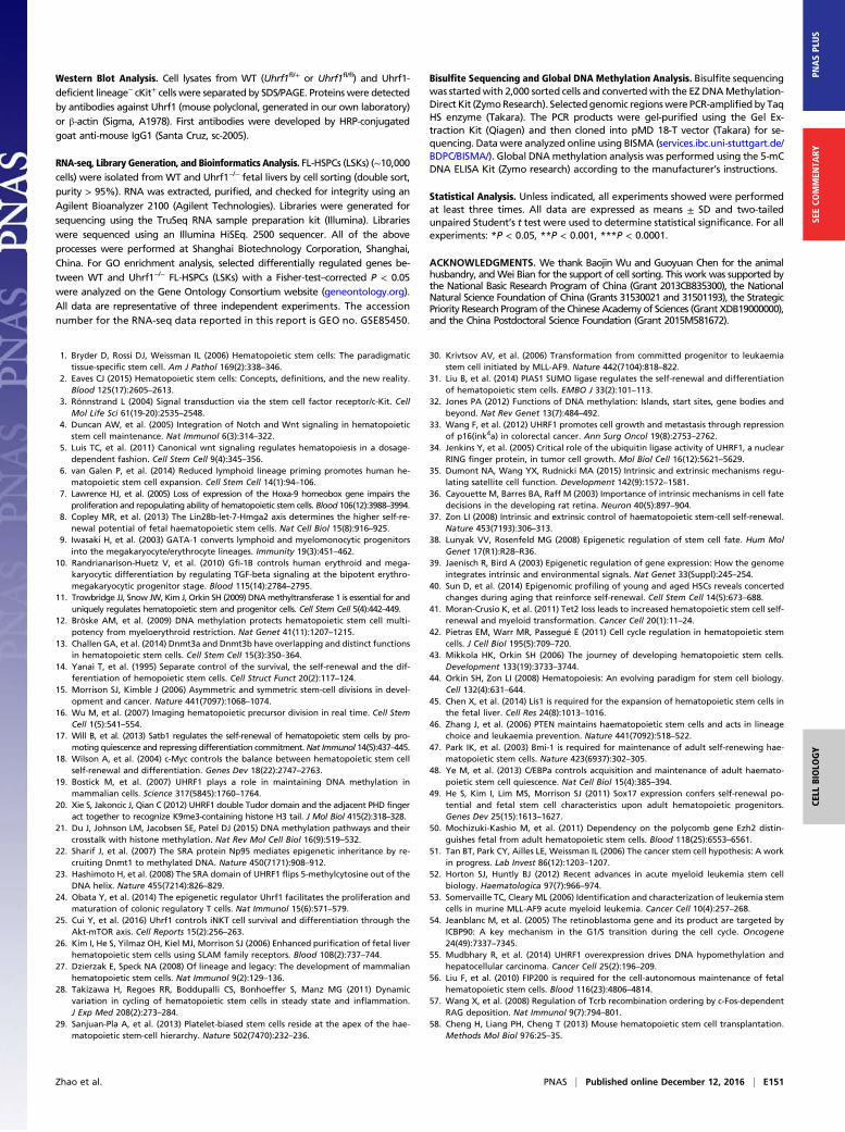
RNA-seq, Library Generation, and Bioinformatics Analysis. FL-HSPCs (LSKs) (∼10,000cells) were isolated fromWT and Uhrf1−/− fetal livers by cell sorting (double sort,purity > 95%). RNA was extracted, purified, and checked for integrity using anAgilent Bioanalyzer 2100 (Agilent Technologies). Libraries were generated forsequencing using the TruSeq RNA sample preparation kit (Illumina). Librarieswere sequenced using an Illumina HiSEq. 2500 sequencer. All of the aboveprocesses were performed at Shanghai Biotechnology Corporation, Shanghai,China. For GO enrichment analysis, selected differentially regulated genes be-tween WT and Uhrf1−/− FL-HSPCs (LSKs) with a Fisher-test–corrected P < 0.05were analyzed on the Gene Ontology Consortium website (geneontology.org).All data are representative of three independent experiments. The accessionnumber for the RNA-seq data reported in this report is GEO no. GSE85450.
Bisulfite Sequencing and Global DNA Methylation Analysis. Bisulfite sequencingwas startedwith 2,000 sorted cells and convertedwith the EZ DNAMethylation-Direct Kit (ZymoResearch). Selectedgenomic regionswere PCR-amplified by TaqHS enzyme (Takara). The PCR products were gel-purified using the Gel Ex-traction Kit (Qiagen) and then cloned into pMD 18-T vector (Takara) for se-quencing. Data were analyzed online using BISMA (services.ibc.uni-stuttgart.de/BDPC/BISMA/). Global DNAmethylation analysis was performed using the 5-mCDNA ELISA Kit (Zymo research) according to the manufacturer’s instructions.
Statistical Analysis. Unless indicated, all experiments showed were performedat least three times. All data are expressed as means ± SD and two-tailedunpaired Student’s t test were used to determine statistical significance. For allexperiments: *P < 0.05, **P < 0.001, ***P < 0.0001.
ACKNOWLEDGMENTS. We thank Baojin Wu and Guoyuan Chen for the animalhusbandry, andWei Bian for the support of cell sorting. This work was supported bythe National Basic Research Program of China (Grant 2013CB835300), the NationalNatural Science Foundation of China (Grants 31530021 and 31501193), the StrategicPriority Research Programof the ChineseAcademy of Sciences (Grant XDB19000000),and the China Postdoctoral Science Foundation (Grant 2015M581672).
1. Bryder D, Rossi DJ, Weissman IL (2006) Hematopoietic stem cells: The paradigmatictissue-specific stem cell. Am J Pathol 169(2):338–346.
2. Eaves CJ (2015) Hematopoietic stem cells: Concepts, definitions, and the new reality.Blood 125(17):2605–2613.
3. Rönnstrand L (2004) Signal transduction via the stem cell factor receptor/c-Kit. CellMol Life Sci 61(19-20):2535–2548.
4. Duncan AW, et al. (2005) Integration of Notch and Wnt signaling in hematopoieticstem cell maintenance. Nat Immunol 6(3):314–322.
5. Luis TC, et al. (2011) Canonical wnt signaling regulates hematopoiesis in a dosage-dependent fashion. Cell Stem Cell 9(4):345–356.
6. van Galen P, et al. (2014) Reduced lymphoid lineage priming promotes human he-matopoietic stem cell expansion. Cell Stem Cell 14(1):94–106.
7. Lawrence HJ, et al. (2005) Loss of expression of the Hoxa-9 homeobox gene impairs theproliferation and repopulating ability of hematopoietic stem cells. Blood 106(12):3988–3994.
8. Copley MR, et al. (2013) The Lin28b-let-7-Hmga2 axis determines the higher self-re-newal potential of fetal haematopoietic stem cells. Nat Cell Biol 15(8):916–925.
9. Iwasaki H, et al. (2003) GATA-1 converts lymphoid and myelomonocytic progenitorsinto the megakaryocyte/erythrocyte lineages. Immunity 19(3):451–462.
10. Randrianarison-Huetz V, et al. (2010) Gfi-1B controls human erythroid and mega-karyocytic differentiation by regulating TGF-beta signaling at the bipotent erythro-megakaryocytic progenitor stage. Blood 115(14):2784–2795.
11. Trowbridge JJ, Snow JW, Kim J, Orkin SH (2009) DNAmethyltransferase 1 is essential for anduniquely regulates hematopoietic stem and progenitor cells. Cell Stem Cell 5(4):442–449.
12. Bröske AM, et al. (2009) DNA methylation protects hematopoietic stem cell multi-potency from myeloerythroid restriction. Nat Genet 41(11):1207–1215.
13. Challen GA, et al. (2014) Dnmt3a and Dnmt3b have overlapping and distinct functionsin hematopoietic stem cells. Cell Stem Cell 15(3):350–364.
14. Yanai T, et al. (1995) Separate control of the survival, the self-renewal and the dif-ferentiation of hemopoietic stem cells. Cell Struct Funct 20(2):117–124.
15. Morrison SJ, Kimble J (2006) Asymmetric and symmetric stem-cell divisions in devel-opment and cancer. Nature 441(7097):1068–1074.
16. Wu M, et al. (2007) Imaging hematopoietic precursor division in real time. Cell StemCell 1(5):541–554.
17. Will B, et al. (2013) Satb1 regulates the self-renewal of hematopoietic stem cells by pro-moting quiescence and repressing differentiation commitment.Nat Immunol 14(5):437–445.
18. Wilson A, et al. (2004) c-Myc controls the balance between hematopoietic stem cellself-renewal and differentiation. Genes Dev 18(22):2747–2763.
19. Bostick M, et al. (2007) UHRF1 plays a role in maintaining DNA methylation inmammalian cells. Science 317(5845):1760–1764.
20. Xie S, Jakoncic J, Qian C (2012) UHRF1 double Tudor domain and the adjacent PHD fingeract together to recognize K9me3-containing histone H3 tail. J Mol Biol 415(2):318–328.
21. Du J, Johnson LM, Jacobsen SE, Patel DJ (2015) DNA methylation pathways and theircrosstalk with histone methylation. Nat Rev Mol Cell Biol 16(9):519–532.
22. Sharif J, et al. (2007) The SRA protein Np95 mediates epigenetic inheritance by re-cruiting Dnmt1 to methylated DNA. Nature 450(7171):908–912.
23. Hashimoto H, et al. (2008) The SRA domain of UHRF1 flips 5-methylcytosine out of theDNA helix. Nature 455(7214):826–829.
24. Obata Y, et al. (2014) The epigenetic regulator Uhrf1 facilitates the proliferation andmaturation of colonic regulatory T cells. Nat Immunol 15(6):571–579.
25. Cui Y, et al. (2016) Uhrf1 controls iNKT cell survival and differentiation through theAkt-mTOR axis. Cell Reports 15(2):256–263.
26. Kim I, He S, Yilmaz OH, Kiel MJ, Morrison SJ (2006) Enhanced purification of fetal liverhematopoietic stem cells using SLAM family receptors. Blood 108(2):737–744.
27. Dzierzak E, Speck NA (2008) Of lineage and legacy: The development of mammalianhematopoietic stem cells. Nat Immunol 9(2):129–136.
28. Takizawa H, Regoes RR, Boddupalli CS, Bonhoeffer S, Manz MG (2011) Dynamicvariation in cycling of hematopoietic stem cells in steady state and inflammation.J Exp Med 208(2):273–284.
29. Sanjuan-Pla A, et al. (2013) Platelet-biased stem cells reside at the apex of the hae-matopoietic stem-cell hierarchy. Nature 502(7470):232–236.
30. Krivtsov AV, et al. (2006) Transformation from committed progenitor to leukaemiastem cell initiated by MLL-AF9. Nature 442(7104):818–822.
31. Liu B, et al. (2014) PIAS1 SUMO ligase regulates the self-renewal and differentiationof hematopoietic stem cells. EMBO J 33(2):101–113.
32. Jones PA (2012) Functions of DNA methylation: Islands, start sites, gene bodies andbeyond. Nat Rev Genet 13(7):484–492.
33. Wang F, et al. (2012) UHRF1 promotes cell growth and metastasis through repressionof p16(ink4a) in colorectal cancer. Ann Surg Oncol 19(8):2753–2762.
34. Jenkins Y, et al. (2005) Critical role of the ubiquitin ligase activity of UHRF1, a nuclearRING finger protein, in tumor cell growth. Mol Biol Cell 16(12):5621–5629.
35. Dumont NA, Wang YX, Rudnicki MA (2015) Intrinsic and extrinsic mechanisms regu-lating satellite cell function. Development 142(9):1572–1581.
36. Cayouette M, Barres BA, Raff M (2003) Importance of intrinsic mechanisms in cell fatedecisions in the developing rat retina. Neuron 40(5):897–904.
37. Zon LI (2008) Intrinsic and extrinsic control of haematopoietic stem-cell self-renewal.Nature 453(7193):306–313.
38. Lunyak VV, Rosenfeld MG (2008) Epigenetic regulation of stem cell fate. Hum MolGenet 17(R1):R28–R36.
39. Jaenisch R, Bird A (2003) Epigenetic regulation of gene expression: How the genomeintegrates intrinsic and environmental signals. Nat Genet 33(Suppl):245–254.
40. Sun D, et al. (2014) Epigenomic profiling of young and aged HSCs reveals concertedchanges during aging that reinforce self-renewal. Cell Stem Cell 14(5):673–688.
41. Moran-Crusio K, et al. (2011) Tet2 loss leads to increased hematopoietic stem cell self-renewal and myeloid transformation. Cancer Cell 20(1):11–24.
42. Pietras EM, Warr MR, Passegué E (2011) Cell cycle regulation in hematopoietic stemcells. J Cell Biol 195(5):709–720.
43. Mikkola HK, Orkin SH (2006) The journey of developing hematopoietic stem cells.Development 133(19):3733–3744.
44. Orkin SH, Zon LI (2008) Hematopoiesis: An evolving paradigm for stem cell biology.Cell 132(4):631–644.
45. Chen X, et al. (2014) Lis1 is required for the expansion of hematopoietic stem cells inthe fetal liver. Cell Res 24(8):1013–1016.
46. Zhang J, et al. (2006) PTEN maintains haematopoietic stem cells and acts in lineagechoice and leukaemia prevention. Nature 441(7092):518–522.
47. Park IK, et al. (2003) Bmi-1 is required for maintenance of adult self-renewing hae-matopoietic stem cells. Nature 423(6937):302–305.
48. Ye M, et al. (2013) C/EBPa controls acquisition and maintenance of adult haemato-poietic stem cell quiescence. Nat Cell Biol 15(4):385–394.
49. He S, Kim I, Lim MS, Morrison SJ (2011) Sox17 expression confers self-renewal po-tential and fetal stem cell characteristics upon adult hematopoietic progenitors.Genes Dev 25(15):1613–1627.
50. Mochizuki-Kashio M, et al. (2011) Dependency on the polycomb gene Ezh2 distin-guishes fetal from adult hematopoietic stem cells. Blood 118(25):6553–6561.
51. Tan BT, Park CY, Ailles LE, Weissman IL (2006) The cancer stem cell hypothesis: A workin progress. Lab Invest 86(12):1203–1207.
52. Horton SJ, Huntly BJ (2012) Recent advances in acute myeloid leukemia stem cellbiology. Haematologica 97(7):966–974.
53. Somervaille TC, Cleary ML (2006) Identification and characterization of leukemia stemcells in murine MLL-AF9 acute myeloid leukemia. Cancer Cell 10(4):257–268.
54. Jeanblanc M, et al. (2005) The retinoblastoma gene and its product are targeted byICBP90: A key mechanism in the G1/S transition during the cell cycle. Oncogene24(49):7337–7345.
55. Mudbhary R, et al. (2014) UHRF1 overexpression drives DNA hypomethylation andhepatocellular carcinoma. Cancer Cell 25(2):196–209.
56. Liu F, et al. (2010) FIP200 is required for the cell-autonomous maintenance of fetalhematopoietic stem cells. Blood 116(23):4806–4814.
57. Wang X, et al. (2008) Regulation of Tcrb recombination ordering by c-Fos-dependentRAG deposition. Nat Immunol 9(7):794–801.
58. Cheng H, Liang PH, Cheng T (2013) Mouse hematopoietic stem cell transplantation.Methods Mol Biol 976:25–35.
Zhao et al. PNAS | Published online December 12, 2016 | E151
CELL
BIOLO
GY
PNASPL
US
SEECO
MMEN
TARY