Embed Size (px)
Citation preview
Tuning Topology of Dirac and Weyl Semimetals by Pressure
A.K. SoodIndian Institute of Science, Bengaluru
Abstract
Dirac and Weyl semimetals are a new class of topological materials with non-trivial topology in
their bulk electronic structure. The bulk valence and conduction bands cross at discrete points in the
Brillouin zone, called Dirac points, and disperse linearly along all the directions in three
dimensional (3D) momentum space. The Dirac point splits into a pair of opposite chirality Weyl
points if either crystal inversion or time-reversal symmetry is broken as in Weyl semimetals.
My talk will present our recent studies of Dirac semimetal Cd3As2 [1] and Weyl semimetals NbP,
TaP, NbAs and TaAs [2,3] covering Raman spectroscopy, synchrotron x-ray diffraction and
electrical resistivity under pressure using diamond anvil cell and their quantitatively understanding
using first principle density functional theory.
I acknowledge all my collaborators as listed in references below.
[1] Satyendra Nath Gupta, D.V.S. Muthu, C. Shekhar, R. Sankar, C. Felser and A.K. Sood,
Pressure-induced electronic and structural phase transitions in Dirac semimetal Cd3As2: Raman
study; Euro Physics Letter 120, 57003 (2017)
[2] Satyendra Nath Gupta, Anjali Singh, Koushik Pal, D.V.S. Muthu, C. Shekhar, Yanpeng Qi, Pavel
G. Naumov, Sergey A. Medvedev, C. Felser, U.V. Waghmare and A.K. Sood, Pressure-induced
Lifshitz transition in NbP: Raman, x-ray diffraction, electrical transport and density functional
theory; Phys. Rev B 97, 064102 (2018)
[3] Satyendra Nath Gupta, Anjali Singh, Koushik Pal, D.V.S. Muthu, C. Shekhar, Moaz A.
Elghazali, Pavel G. Naumov, Sergey A. Medvedev, C. Felser, U.V. Waghmare and A.K. Sood,
Pressure-induced Lifshitz transition in NbAs: Experiments and Theory; Under Review (2018)
Indo-Italian Synchrotron Collaboration
Andrea LausiHead, Xpress Beamline, Elettra Sincrotrone Trieste
Abstract
Andrea Lausi1, Boby Joseph1, Andrea Goldoni2, Maurizio Polentarutti2, Dipankar Das Sarma3 1Xpress
Beamline, Elettra Sincrotrone Trieste, Trieste, Italy, 2Beamlines Group, Elettra Sincrotrone Trieste, TRIESTE, Italy, 3Solid State and Structural Chemistry Unit, Indian Institute of Science, 560012, Bengaluru, India
Indian research groups have been collaborating with researchers at the Italian Synchrotron radiation source
Elettra since past two decades. The collaboration between Elettra and Indian research institutions is a part of
a wider collaboration between the countries sponsored by the Department of Science and Technology,
Government of India and the Italian Ministry for Foreign Affairs. The high standard of the collaboration in
the field of synchrotron radiation was recognized in the joint statement by the prime ministers of both the
countries, on the occasion of Italian prime minister's visit to India in February 2007. Statistics till 2015
indicate that the collaboration has led few hundred visits of Indian groups to Elettra (with an average of three
scientists per visit) in the last 20 years and to the publication of more than 500 articles in peer-reviewed
scientific journals. Till 2016, Elettra received around 959 experimental proposals from Indian research
groups, corresponding to about 8% of the total: in third place, just after Italy and France (4873 and 1034
proposals respectively). This makes Elettra the most requested European national synchrotron radiation
laboratory by Indian users.
This collaboration has been the earliest possibility for Indian scientists to access synchrotron facilities and
played the most crucial role in building up the synchrotron community within India by making it available to
even those who did not have any prior access or knowledge of synchrotron-based techniques. As a part of
this project, the community was nurtured to become competitive in preparing their proposals that were
evaluated internationally to obtain synchrotron beam-time in various areas of research ranging from solid
state and surface physics to nanotechnology, materials science and life sciences. Great success of these
activities prompted both sides to take a further significant step forward involving the development of two
dedicated beamlines: a macromolecular and a high pressure X-ray diffraction facilities, respectively XRD2
and Xpress, under the partnership between Elettra and Indian Institute of Sciences, Bangalore. Both these
beamlines share a superconducting wiggler as a high flux source. From the beginning of 2016 Xpress
beamline started accepting proposals from external users. This milestone is going play a new chapter in the
scientific collaborations involving Elettra and Indian research community.
Structural Transitions at High Pressures
Goutam Dev MukherjeeIndian Institute of Science Education and Research Kolkata, Mohanpur
AbstractMy journey into the field of study of structural transitions at high pressures started in the XRD1
beamline with the photon flux from bending magnet and now there is a dedicated high pressure
beamline EXPRESS with high brilliance. In this talk I shall first discuss pressure induced structural
behaviour in frame work structured negative thermal expansion material NbOPO4, which shows an
anomaly at 0.6 GPa. Pressure behaviour of unit cell volume changes in linear fashion, and is
followed by a structural amorphization above 20 GPa. This work was published in ELETTRA
Highlights in 2002-2003. Next I shall present our recent work in high pressure XRD studies on
layered WS2 obtained by liquid exfoliation. High pressure X-ray diffraction measurements in
conjunction with Raman studies show the emergence of a triclinic modification along with the
hexagonal phase of WS2 above about 5.8 GPa. However analysis of our ambient and high pressure
studies on monolayer WS2 along with those of the nanocrystalline sample indicate to the presence
of the triclinic phase in patches embedded in the parent hexagonal phase raising a question mark
over the structural purity of the exfoliated monolayered materials beyond a certain stress conditions.
This work has been carried out in EXPRESS beamline.
Structural biology @Elettra: an integrated approach to DNA replication and repair
Silvia OnestiHead, Structural Biology Lab, Elettra Sincrotrone Trieste
AbstractThe Structural Biology Laboratory at Elettra applies molecular and structural biology tools to study
the basic genetic processes within the cell and to characterise some of the proteins involved. We use
protein crystallography to determine the atomic structure of these proteins, as well as biochemical
and biophysical approaches to understand how they work. Crystallographic studies are
complemented by the concomitant use of electron microscopy to visualise the architecture of large
complexes, NMR spectroscopy to study small, disordered regions and small-angle X-ray scattering
(SAXS) to obtain additional structural information. Data from these techniques can be combined
with crystallographic atomic structures to get a three-dimensional picture of the complex
architecture.
Our research focus on DNA replication, recombination and DNA repair. These are crucial events in
the cell cycle, underpinning cellular processes with important consequences such as cell
proliferation and genome stability. Failure to control these processes causes chromosome instability,
which can lead to the development of cellular abnormalities, genetic diseases and the onset of
cancer.
I will present examples derived from our own work aimed at unravelling the structural and
functional aspects of key proteins in these processed, such as the MCM helicase, Cdc45, GINS,
PCNA, and RecQ4. All of these are present in proliferating cells and are highly expressed in
malignant human cancer cells and pre-cancerous cells undergoing malignant transformation.
Therefore, they are ideal diagnostic markers for cancer and possibly targets for anti-cancer drug
development.
http://www.elettra.trieste.it/labs/structural-biology
Structural studies for target characterisation against sepsis causing gram negative bacteria.Ramaswamy S
Institute for Stem Cell Biology and Regenerative Medicine, Bengaluru
AbstractSialic acids (5-N-acetylneuraminic acids, Neu5Ac) are a family of related nine-carbon sugar acids
that are used by bacteria for molecular mimicry, as nutrition, and cell signaling. The motivation for
our program is to understand how bacteria scavenge, transport and incorporate sialic acid into the
lipooligosaccharide (LOS) or lipopolysaccharides (LPS) (Vimr et al, 2004). While some pathogens
have evolved de novo biosynthesis pathways for Neu5Ac many bacteria rely on the acquisition of
Neu5Ac from the environment and hence require high-affinity transport systems. Bacteria
incorporate this scavenged Neu5Ac into the LOS/LPS as the terminal non-reducing sugar. This
sugar is recognized by the complement system in the serum as “self”. Several opportunistic bacteria
use this type of molecular mimicry to evade the immune system, resulting in serious pathological
consequences. It has been shown in H. influenzae that inhibition of the sialic acid transport system
leads to non-virulence in continuous flow biofilm chambers (Allen et al, 2005; Severi et al, 2010).
We propose that the pathway of Sialic acid uptake, catabolism and incorporation would be a very
good drug targets and we should be able to develop anti-microbial agents that act under a variety of
conditions. In order to test this, we have determined structures of proteins involved in scavenging,
transport, catabolism and silica acid incorporation into cell surface glycolipids. I will discuss our
structure-function studies on these proteins from several gram negative bacteria and current efforts
on drug discovery.
The beamlines XAFS and XRF at Elettra:recent highlights and future opportunities
Giuliana AquilantiHead, EXAFS Beamline, Elettra Sincrotrone Trieste
Abstract
The XAFS beamline at Elettra is dedicated to XAS and can operate in a large energy range from 2.4
to 25 keV. The simple optical scheme makes the beamline particularly suitable for transmission
geometry measurements on homogeneous samples for which a large and spatially uniform beam
guarantees high quality data. A fair variety of sample environments and the possibility to practically
adapt any sample environment in the experimental hutch makes the XAFS beamline a quite
appealing instrument as evidenced by the high oversubscription ratio.
The XRF beamline has been conceived to be a multipurpose beamline for x-ray spectrometry and
its endstation hosts an ultra-high vacuum chamber owned by IAEA. This instrument should offer
the possibility to use different x-ray spectrometry techniques such as: x-ray fluorescence, total
reflection x-ray fluorescence, grazing incidence x-ray fluorescence, x-ray reflectometry and x-ray
absorption spectroscopy with submillimetric spatial resolution.
Research highlights for the two beamlines will be shown along with their complementarity. A recent
reorganization of the beamlines’ personnel will allow the user community access to the two
beamlines in a coordinated manner.
Understanding Magneto-structural interactions in Ni-Mn based Heusler alloysK. R. Priolkar
Goa University, Taleigao Plateau, Goa
AbstractNi-Mn based Heusler alloys undergoing austenitic to martensitic transition have been of interest due
to technologically important phenomena like large magnetic field induced strain, large
magnetoresistance, large normal and inverse magnetocaloric effect, etc. These properties are
extremely sensitive to composition of the Heusler alloy as well as the degree of chemical order.
Extended X-ray absorption fine structure (EXAFS) spectroscopy and X-ray magnetic circular
dichroism (XMCD) in conjunction with diffraction techniques have been employed to gain an
understanding of atomistic picture of chemical order and effect of composition on martensitic
transition in such Heusler alloys. Systematic investigations on Ni2Mn1+xIn1-x (0 ≤ x ≤ 0.6) reveal
presence of local structural disorder even in the austenitic phase. Despite geometric constraints, Ni-
Mn and Ni-In distances are not equal and the difference between them progressively increases with
increasing Mn content. Presence of chemical disorder results in formation Mn 3d – Ni 3d
hybridized states which are responsible for antiferromagnetic interactions in the martensitic state.
Using Synchrotron to understand properties of doped Nanomaterials: EXAFS and XRD.
Ranjani ViswanathaJawaharlal Nehru Centre for Advanced Scientific Research, Bangalore
AbstractDoping in semiconductor quantum dots as well as nanocrystal heterostructures have been shown to
have distinct advantages for applications. However, diffusion of dopants or impurities in host
nanocrystals out of the host or within the host pose new challenges in determining the nature of
interfaces in heterostructures as well as the interaction environment of the dopant with the host
nanocrystal. While long range phenomena like X-ray diffraction, particularly as a function of
temperature can provide answers to some questions, these long range techniques are not suitable for
analysis such short range phenomena. In this talk, I discuss the use of the use of XRD and EXAFS
to understand properties correlated to doped nanomaterials.
Small angle scattering to investigate the nm-scale: from biology to hard condensed matter
H. AmenitschAustria & Austrian SAXS beamline, Elettra Sincrotrone Trieste
AbstractSmall and Wide Angle Scattering has developed as a standard method to investigate nanostructures
in the range of 0.1 to >100 nm in situ and under in-operando conditions. The enormous benefit of
SAXS compared to many other structural methods is the limited requirements for sample
preparation and environment. This quality enables to study liquids, solids, aerosols and thin films
close the conditions occurring in chemical reactions, temperature treatments and even under more
complicated constrains of in operando investigations such as in supercapacitors or micro chambers
for nanomaterials growth. Therefore, the method allows getting molecular movies on the
nanometer-scale.
This presentation will discuss highlights of the current research program of the Austrian SAXS
beamline covering fields as diverse as structural biology, drug delivery systems or energy materials.
Further, an outlook will be given to the new experimental development as well as emerging
perspectives such as ultra-fast pump probe experiments.
Influence of Confinement on Heme-Protein Function and it’s Consequences on SensingAninda J. Bhattacharyya
Indian Institute of Science, Bengaluru
Abstract
The electroactive centre or site of a biomolecule controls the effective molecular properties at
various length scales. For example, Fe-containing heme proteins, which control storage, transport,
and release of oxygen in most higher forms of life, in biological buffer display a characteristic
quasi-reversible electrochemical behaviour of the FeIIIII couple located in the porphyrin ring.
Encapsulation of these proteins in a variety of hosts (dhost >> dbiomolecule) renders a reversible
electrochemical FeIIIII response. Thus, the reversible redox phenomenon under confinement is
functionally different from that of the protein molecules in the solution. Our objective here is to
understand the fundamental mechanisms behind these changes in the electrochemical response in
the context of protein structure and dynamics under confinement. To develop a comprehensive
mechanistic model, the structure and dynamics of encapsulated proteins are studied using
electrochemical and various techniques including synchrotron and neutron measurements. Control
experiments involving interactions of small molecules, which chemically mimic the protein, with
individual amino acids are also performed. The purpose here is to deconvolute the complex
electrochemical function of heme proteins into simpler interactions. Outcomes of these studies
provided useful inputs on the chemical design of practical electrochemical biosensors.
In-situ SAXS and Diffraction Studies of Nanostructure-Self-Assembly ProcessesMilan K. Sanyal
Saha Institute of Nuclear Physics, Kolkata
AbstractFormation of ordered structures with nanoparticles is an evolving field of research as control over
this self assembly process is essential to develop cost-effective nanostructures in robust engineering
form and to investigate predictions of low dimensional physics. Physical/chemical properties of
nanoparticles, ranging from chemically-formed novel materials to physically deposited quantum
dots grown in extremely controlled environment, can be tuned by altering shape, size and
compositions. However understanding structural features of individual nano-sized materials and
assembly of these particles is a nontrivial exercise. Availability of intense photon sources in
synchrotrons is giving us the opportunity to investigate in-situ individual nanoparticles and the self-
assembly processes with small angle x-ray scattering (SAXS) and x-ray diffraction techniques. We
shall illustrate this topic with few of our recent results.
References:
1. In-Situ GISAXS Study of Supramolecular Nanofibers having Ultrafast Humidity Sensitivity,
A. Bhattacharyya, M. K. Sanyal, U. Mogera, S. J.George, M. K. Mukhopadhyay, S. Maiti
and G.U. Kulkarni, Scientific Reports 7, 246 (2017).
2. Evidence of contact epitaxy in the self-assembly of HgSe nanocrystals formed at a liquid–
liquid interface, S. Maiti, M. K. Sanyal, M. K. Jana, B. Runge, B. M. Murphy, K. Biswas
and C. N. R. Rao,
J. Phys.: Condens. Matter 29, 095101 (2017).
Synchrotron Radiation and Cultural Heritage:The Experience of Elettra
Franco ZaniniHead, SYRMEP-Imaging Beamline, Elettra Sincrotrone Trieste
Abstract
The use of synchrotron radiation for the analysis of samples of historical and artistic importance has
been increasing over the past years, and experiments related to the study of our cultural heritage
(CH) have been routinely performed at many beamlines of Elettra, the Italian synchrotron radiation
facility. The laboratory now offers a platform dedicated to CH researchers in order to support both
the proposal application phase and the different steps of the experiment, from sample preparation to
data analysis.
TwinMic, the European Soft X-ray Transmission and Emission Microscope, integrates the
advantages of complementary scanning and full-field imaging modes into a single instrument.
TwinMic operates in the 400-2200 eV energy range. This microscope station has been designed as
highly modular in its optical configuration and specimen environment, and an international
community of scientists and technicians continuously improves the instruments performance and
versatility to suit the experimenter's requirements.
X-Ray Fluorescence is a highly versatile beamline working in an energy range between 2 and 14
keV. The beamline is optically designed to present beam parameters needed for high level
measurements in spectroscopy as well as in microscopy. The beamline hosts an ultra-high vacuum
chamber, in partnership with the IAEA, which will allows the synergistic application of various X-
Ray Fluorescence techniques together with X-ray Absorption Near Edge Structure (XANES) and X-
Ray Reflectometry.
The flexible design of the MCX beamline allows a wide range of non-single crystal diffraction
experiments relevant for the cultural heritage, from phase identification to atomic structural studies.
Other powder diffraction techniques that can be applied to cultural heritage materials comprise
texture and orientation determination of the crystalline phases and the determination of the
microstructure of the sample in terms of crystallite size, defects characterization and accumulated
strain.
SYRMEP, the X-ray microimaging and microtomography station, with an energy range of 8.3 - 35
keV, is a highly flexible beamline allowing the analysis of samples in both absorption and phase
contrast mode. The versatility of this instrument has attracted researchers from different
communities, from restoration and conservation to art history, archaeology and paleoanthropology.
The XAFS beamline, working in the energy range between 2.4 and 27 keV, provides microscopic
structural information through the analysis of a sample X-ray absorption spectrum, with unique
applications in the field of cultural heritage. It is a powerful local structural probe, which does not
require long-range order, and allows the determination of the chemical environment of a single
element in terms of number and type of neighbours, interatomic distances and structural disorder.
UV resonant Raman spectroscopy performed at IUVS beamline has been demonstrated to be a non-
destructive powerful tool for obtaining a detailed compositional characterization of pigments
present in artworks of historical-artistic interest. Thanks to the exploiting of the resonant conditions
occurring for specific functional groups at different excitation wavelengths in the UV-range and the
strong reduction of the interfering fluorescence background that often seriously limits the use of
conventional Raman spectroscopy in cultural heritage studies, UV resonant Raman scattering
constitutes an alternative spectroscopic approach for achieving a non-destructive identification of
the main pigmenting agents.
Infrared Microscopy techniques at SISSI beamline offer the possibility to correlate the sample
morphological features with its vibrational local pattern at diffraction limited spatial resolution.
Chemical information on both organic and inorganic constituents of art pieces can be obtained at
SISSI, exploiting several sampling methodologies, suitable for probing thin sections and material
surface properties as well.
The Scanning photoelectron microscope (SPEM) hosted at the ESCA microscopy beamline allows
to combine chemically surface sensitive measurements with high spatial resolution. A beam spot
down to 120 nm and energy sensitivity within 180 meV using a third generation X-ray source
providing more than 109 photons/s in the probe has opened the opportunity for CH to perform
surface micro-characterization. The experimental apparatus allows to carry out a manifold of
experiments, aiming at quantitative and qualitative chemical characterisation of morphologically
complex materials.
Evolution of Structures at the Air-Water Interface
Alokmay DattaSaha Institute of Nuclear Physics, kolkata
Abstract
The air-water interface is itself a dynamic structure with a hierarchy of timescales. This is related to
the coexistence of interchanging hydrogen-bonded structures of water which give rise to a slightly
negatively charged surface with an unusually high free energy. Introduction of charged or polar
bodies to this interface sets up new dynamics that lead to correlated structures ranging from sub-
nanometer to several hundred micrometers and even beyond.
These structures can be transferred onto suitable substrates and probed. Understanding the role of
molecular bonding on these structures possibly holds the key to control and functionalise them.
Ultraviolet and Soft X-ray spectroscopy is an invaluable tool to probe the bonding and correlating
this information with structural and morphological data. In this talk I shall present the results on
structures evolving under ion-molecule and nanoparticle-molecule forces.
Biophysics and Nanomedicine at the Elettra NanoInnovation LabLoredana Casalis
NanoInnovation Lab, Elettra Sincrotrone Trieste
Abstract
The NanoInnovation Lab of Elettra works on applications of nanotechnologies to biophysics and
translational medicine. The focus is mainly on world spread diseases, as cancer and
neurodegenerative diseases. We implement innovative sensing strategies for the detection of disease
molecular biomarkers at the early stage of disease progression, as well as integrated platforms to
decipher the mechanisms of diseases development. Our platforms are based on Atomic Force
Microscopy (AFM), to study biorecognition events as well as cell and tissue biomechanics, and are
complemented by Fluorescence Microscopy, Electrochemical Impedance Spectroscopy and other
Synchrotron Radiation-based techniques.
Spectroscopy of self-assembled networks and nano systems
Dinesh TopwalInstitute of Physics, Bhubaneswar
Abstract
Abstract: The confinement effects of electrons in ultra-thin films and nanowires grown on metallic
and semiconducting substrates will be discussed. Finite electron reflectivity at the interface is
sufficient to sustain the formation of quantum well states and weak quantum well resonance states
even in closely matched metals. The expected parabolic dispersion of sp derived quantum well
states for free standing layers undergoes deviations from parabolic behaviour and undergoes
modifications due to the underlying substrate bands, suggesting the effects of strong hybridization
between the quantum well states and the substrate bands. I will also discuss electronic structure of
ordered Ag-Bi alloy and their surface-state (SS) bands, spin split by the Rashba interaction,
selectively coupled to the quantum-well state (QWS) bands, originally spin degenerate, in the metal
film.

XRD2 - the new indo-italian beamline for macromolecular crystallographyMaurizio Polentarutti
XRD1 Beamline, Elettra Sincrotrone TriesteAbstract
The new beamline XRD2 at the Elettra synchrotron, dedicated to macromolecular cryustallography,is ending the commissioning phase. The XRD2 project is developed in partnership with the Indianscientific community and administered through Elettra and Indian Institute of Science (IISc),Bengaluru.
A new beamline has been constructed at section 11.2 at the Elettra synchrotron and is nowcompleting the commissioning phase. The high photon flux provided by the SuperConducting multipole Wiggler (SCW), installed at the 11.2 section, permits the operation of threebeamlines, a central tunable and two fixed energy beamlines.
The XRD2 beamline will be complementary to the existing general-purpose XRD1. XRD2beamline will be dedicated to high throughput macromolecular crystallography experiments: largetunable energy range (8.0 - 35.0 keV) for SAD/MAD experiments, automated sample mounting in acryogenic environment and high speed large area detector are some of the important features ofthis beamline.
Beamline features (energy resolution, flux, spot dimensions) as well as sample changer capabilitieswill be reported together with data quality indicators from data collected on standard-test (lysozyme) and real-life samples.
Crystal structure determination of nucleotide-dependent restriction-modification enzymes
SaikrishnanIndian Institute of Science Education and Research, Pune
Abstract
Nucleoside triphosphate-dependent restriction-modification enzymes are the most prominent
bacterial defence against foreign DNA invasion. The enzymes have a nucleotide-dependent
endonuclease activity that cleaves foreign DNA that enters the host cell. Molecular details of the
activities of these enzymes have remained unravelled due to lack of structural information in atomic
detail. Efforts to obtain the structural data has been hindered by the large size and complex nature of
these enzymes. My laboratory has over the years been carrying out structural studies on these
enzymes primarily using X-ray crystallography. These studies have led to the first snapshots of the
enzymes in action. I will discuss the ways and means employed by us towards obtaining these
snapshots, and also the mechanistic details that have emerged from these studies.
Applications of synchrotron radiation for integral membrane protein structures.Aravind Penmatsa
National Centre for Biological Sciences, Bengaluru
AbstractSynchrotron radiation from insertion devices is highly successful at getting high resolution data
from crystals of membrane proteins which tend to have high levels of solvent content and/or are
very small in size. Using the case study of the dopamine transporter, I will put forth the immense
potential of obtaining high resolution structural information for membrane proteins, that are not
very easy to crystallize. Integral membrane proteins are also crystallized in lipidic cubic phase that
allows formation of type-I crystals. Cubic phase crystals tend to be micron sized and are prone to
radiation damage. Attempts to crystallize the dopamine transporter in complex with an antibody
yielded cubic phase crystals from which a dataset could be stitched together using data from seven
crystals. My talk will focus on the extensive modification done to the protein, crystallization trials
and the use of synchrotron radiation that helped reveal the structure of the dopamine transporter.
Molecular Orbital Tomography: adsorption geometry and electronic structure
V. FeyerHead, NanoESCA Beamline, Elettra Sincrotrone Trieste
Abstract
Angle-resolved photoelectron spectroscopy (ARPES) is a well-established method to study the band
structure of and recently it has been shown that it can also provide an alternative route to obtain
information regarding the charge distribution of individual molecular orbitals [1, 2]. Under specific
assumptions about the final state of the photoemitted electron, molecular orbital structures can be
investigated using data from the angle-resolved photoemission. This approach, based on the
comparison between ARPES measurements and theoretical calculations, forms the basis of
molecular orbital tomography (MOT).
The photoemission current (I) within the dipole approximation is proportional to:
where and are the polar and azimuthal emission angles, respectively. I is given by the sum over
all transitions from the occupied initial states i to the final states f characterized by the direction
( ) and the kinetic energy of the photoemitted electron. The final state, in the most simple
approach, can be approximated by a plane wave (PW) only characterized by the direction and wave
number of the emitted electron [1] :
For organic molecules, the photocurrent of a well-defined initial state can be selectively measured,
when the energetic separation of individual molecular orbitals is bigger than the intermolecular
band dispersion. This allows the measurement of |i(k)| and, via a subsequent inverse Fourier
transform, the reconstruction of molecular orbital densities in real space, providing a new tool for
the investigation of organic molecular films on metallic substrates.
As example, in the talk I will show the investigation of the adsorption behavior of nickel
tetraphenyl porphyrin (NiTPP) molecules on the Cu(100) surface by applying a comprehensive
multi-technique approach, photoemission electron microscopy (PEEM), MOT and scanning
tunneling microscopy (STM), complemented density functional theory (DFT) calculations [3]. The
adsorbed NiTPP arrange in two different geometrical configurations with different orientation
respect to [100] crystal direction. By combining STM with DFT calculations, we demonstrate that
the contrast, in STM images, arises mainly from the phenyl peripheral groups, which are tilted
upwards. This adsorption configuration prevents the macrocycle, where frontier orbitals are
localized, to be resolved by the STM tip. For this reason, STM cannot be used for mapping the
charge distribution of the highest occupied and lowest unoccupied molecular orbitals (HOMO and
LUMO, respectively). Therefore, as a complementary technique, we exploited the capability of our
PEEM to directly image a wide reciprocal space in one single shoot. The ARPES data were then
compared to DFT calculations, within the MOT framework. The molecular orbitals exhibit peculiar
features which can be used to unambiguously identify them in the valence band spectra. This is
particularly useful to determine the molecule-substrate interaction and the charge transfer
phenomena between adsorbed molecules and metal surfaces. The comparison between experiment
and theory shows that the former gas-phase LUMO+3 becomes occupied upon adsorption on the
metal surface. This unexpected result suggests that a multi-technique approach is mandatory in
order to obtain a consistent picture of the adsorption behavior and electronic properties of the
molecular system.
[1] Puschnig, P. et al. Science 326, 702–706 (2009).
[2] Ziroff, J., et al. Physical Review Letters 104, 233004.
[3] Zamborlini, G. et al. Nature Communicationsvolume 8, Article number: 335 (2017)
Infrared Spectroscopy using Synchrotron RadiationHimali Bhatt
Bhabha Atomic Research Centre, Trombay, Mumbai
AbstractInfrared spectroscopy, in particular, IR microspectroscopy, has matured from a simple analytical
technique to a rigorous micro-structural technique, owing primarily to the advent of Fourier
Transform interferometry and synchrotron radiation as the illuminating source, among other factors.
It is the brightness advantage of the synchrotron radiation, the directed emission from relativistic
electrons in the storage ring, which leads to the focussing capabilities of the long wavelength IR
light to a spot size comparable with the diffraction limit. This results in a specific advantage of
enhanced signal to noise ratio from microscopic specimens, thus benefitting the fields of high
pressure physics, IR imaging of tiny biological samples like tissues, hair cross sections etc., optical
properties of single crystals and surface science. In this talk, the design and capabilities of
synchrotron IR beamlines, BL-6 at the Indian synchrotron radiation source, Indus-1 and the Italian
SISSI beamline at Elettra synchrotron radiation source will be discussed and compared. Typical
case studies on synchrotron high pressure infrared studies of proton dynamics in strong hydrogen
bonds of molecular solids and pressure effects on the superconducting materials will be presented.
In addition, the use of in-situ low temperature technique for deducing vibrational structure and
optical properties will also presented, highlighting the crucial micro-structural information extracted
using infrared spectroscopy of materials under extreme thermodynamic environments. The
information, thus obtained, can be used with the complementary techniques using x-ray and neutron
sources, for extracting comprehensive structural details of the samples.
Superconductivity in Ferro-pnictides
Kalobaran MaitiTata Institute of Fundamental Research, Homi Bhabha Road, Colaba, Mumbai
Abstract
Superconductivity in Fe-based compounds is believed to appear due to spin fluctuations. Recent
studies, however, revealed mysterious superconductivity with high transition temperature under
pressure and/or chemical substitution, where the compound do not show magnetic order. There are
controversies on the relation between hydrostaticity of the pressure and superconductivity. We
studied the Fermiology of some of the 122 class of compounds employing angle resolved
photoemission spectroscopy (ARPES). The Fermi surfaces at different temperatures reveal
importance of strain induced effects in the electronic structure. We show that the evolution of FS
topology in CaFe2As2 is not directly driven by the structural transition. In addition, we discover the
existence in ambient conditions of energy bands related to the collapsed tetragonal phase. These
bands are distinctly resolved in the high-photon energy spectra exhibiting strong Fe 3d character.
They gradually move to higher binding energies due to thermal compression with cooling, leading
to the emergence of 3D topology in the Fermi surface. The results of EuFe2As2 exhibit signature of
coherent polaron at low temperatures. Our time resolved ARPES results show orbital selective
behavior in EuFe2As2.These results reveal the existence of hidden parameters, which are argued to
lead to quantum fluctuations responsible for the exotic electronic properties in Ferro pnictide
superconductors.
References:
[1] Khadiza Ali, Swapnil Patil, Ganesh Adhikary, Sangeeta Thakur, S. K. Mahatha, A. Thamizhavel, G.
De Ninno, Paolo Moras, Polina M. Sheverdyaeva, C. Carbone, L. Petaccia and Kalobaran Maiti,
Phys. Rev. B 97, 054505 (2018). [2] Khadiza Ali and Kalobaran Maiti, Scientific Reports 7, 6298 (2017).[3] G. Adhikary, B. Ressel, M. Stupar, P. R. Ribič, J. Urbančič, G. De Ninno, D. Krizmancic, A.
Thamizhavel, and Kalobaran Maiti, arXiv:1711.05464v2.[4] G. Adhikary et al., J. Appl. Phys. 115, 123901 (2014).[5] G. Adhikary et al., J. Phys.: Condens. Matter 25, 225701 (2013).[6] G. Adhikary et al., J. Appl. Phys. 114, 163906 (2013).
A low-photon-energy ARPES system for band mapping and many-body effects in solid materials
Luca PetacciaBaDElph Beamline, Elettra Sincrotrone Trieste
AbstractThe BaDElPh beamline is an undulator-based normal incidence monochromator
(NIM) instrument which provides photons in the energy range 4.6-40 eV with
high flux, high energy resolution, and horizontal-vertical linear polarization [1].
The beamline serves an end station to perform primarily high-resolution angle-
resolved photoemission spectroscopy (ARPES) experiments from solids in the
low photon energy regime. Photon energies lower than 15 eV provide enhanced
bulk sensitivity, allow for the highest momentum and energy resolution, and
are useful for tuning matrix elements which vary rapidly at low energy. The
availability of such low photon energies for high-resolution ARPES studies
makes BaDElPh unique at Elettra and it is one of the few beamlines available
worldwide. In this talk, the status and performance of the system will be
presented with also a review of some recent scientific results.
[1] www.elettra.eu/elettra-beamlines/badelph.html
Atomic, molecular and cluster physics with novel XUV light sources
Dr. M. Coreno ISM-CNR and Elettra Sincrotrone Trieste
Abstract
XUV facilities at third generation synchrotron laboratory, like the Gas Phase Photoemission
beamline of the Elettra storage ring (Trieste, Italy) have enabled thorough photoionization studies of
isolated systems for more than 20 years [1]. More recently, the interest of the atomic and molecular
physics community has been attracted by the opportunity of exploring the temporal dynamics of
isolated systems by means of novel state-of-the-art light sources. Thus, our group has taken active
part in the development of two novel XUV facilities: the Low Density Matter (LDM) beamline [2]
at the FERMI free electron laser [3], and CITIUS [4], a state-of-the-art fs-VUV source, based on
laser High Harmonic Generation on rare gases. Recent experiments on photoionization
spectroscopy of gas phase molecular targets of increasing complexity will be presented, as well as
research opportunities opened in the field of atomic, molecular and cluster physics by these novel
ultrafast light sources.
Keywords: Photoionization Spectroscopy; Photoelectron Spectroscopy; Synchrotron Radiation
References
[1] K.C. Prince, et al J. Synch. Rad. 5 (1998) 565;
see also at http://www.elettra.eu/elettra-beamlines/gasphase.html
[2] V. Lyamayev, et al. J. Phys. B 46 (2013) 164007. See also
http://www.elettra.eu/lightsources/fermi/fermi-beamlines/ldm/ldmhome-page.html
[3] “Free Electron laser Radiation for Multidisciplinary Investigations” . See e.g.
E. Allaria, et al. New J. Phys. 14 (2012) 113009.
[4] C. Grazioli, et al. , Rev. Sci. Instr. 85 (2014) 23104. See also
https://www.elettra.eu/lightsources/labs-and-services/citius/citius.html
Unravelling the photodynamics of mesoscopic aggregates hosted on quantum fluid clusters:from synchrotrons towards free-electron lasers
Sivarama KrishnanIndian Institute of Technology, Madras
AbstractAdvanced modern light sources, especially the third generation, to which the Elettra synchrotron
belongs, offer unique opportunities to probe quantum systems. When combined with powerful
coincidence spectroscopy techniques we can find answers to generic questions as well as specific
details of the photodynamics of two- and, indeed, multi-component systems. In particular, we have
probed in a series of experiments over the last five years aggregate mesoscopic systems formed in
or on quantum fluid clusters, superfluid He nanodroplets [1]. Beginning with the first
photoelectron-photoion coincidence (PEPICO) spectroscopy on these systems enabled by the
workstations at Elettra, we will present key results and finding [2, 3].
Current and future directions in investigating the photodynamics of these systems is focussing on
multi-electron processes in these complex systems where classic Fano profiles [4] have been
observed and studied, inter-atomic Coulomb decay and electron transfer mediated decay [5] has
been evidenced and even multiple ionization following Penning and excitation transfer has been
found to occur [6].
We will present both published results as well as findings from ongoing work. These pave way and
motivate further experiments involving free electron lasers where the dynamics and timescales of
these processes can be probe directly and unambiguously.
References:[1] J. P. Toennies et al., Annual review of physical chemistry 49.1, 1-41 ((1998)[2] D Buchta et al., The Journal of chemical physics 139 (8), 084301 (2013)[3] D Buchta et al., The Journal of Physical Chemistry A 117 (21), 4394-4403 (2013)[4] A C LaForge et al., Physical Review A 93 (5), 050502 (2016)[5] A C LaForge et al., Physical review letters 116 (20), 203001 (2016) [6] S. R. Krishnan et al., (manuscript under review).
High Pressure studies to unravel exotic states of matter
Chandrabhas NarayanaJawaharlal Nehru Centre for Advanced Scientific Research, Bengaluru
Abstract
The talk will be providing an introduction to the area of high pressure in condense matter with
examples which brought out exotic understanding of science of materials. Specifically, how
pressure is a very excellent tool for perturb a system and studying exotic phase of the materials. As
an example, Ice like phase of NH4F will be shown to suggest how tetrahedrallly coordinated system
with different bonding can mimic the rich Ice phase diagram. At the end, we will be providing the
latest result of high pressure results on TiTe2 studied at ELETTRA which shows topological phases
under pressure.
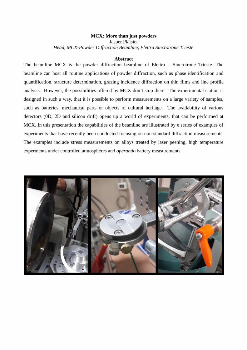
MCX: More than just powdersJasper Plaisier
Head, MCX-Powder Diffraction Beamline, Elettra Sincrotrone Trieste
AbstractThe beamline MCX is the powder diffraction beamline of Elettra – Sincrotrone Trieste. The
beamline can host all routine applications of powder diffraction, such as phase identification and
quantification, structure determination, grazing incidence diffraction on thin films and line profile
analysis. However, the possibilities offered by MCX don’t stop there. The experimental station is
designed in such a way, that it is possible to perform measurements on a large variety of samples,
such as batteries, mechanical parts or objects of cultural heritage. The availability of various
detectors (0D, 2D and silicon drift) opens up a world of experiments, that can be performed at
MCX. In this presentation the capabilities of the beamline are illustrated by e series of examples of
experiments that have recently been conducted focusing on non-standard diffraction measurements.
The examples include stress measurements on alloys treated by laser peening, high temperature
experments under controlled atmospheres and operando battery measurements.
Investigation of polymorphism under pressure using synchrotron x-ray diffractionH. K. Poswal
High Pressure and Synchrotron Radiation Physics Division, Bhabha Atomic Research Centre, Mumbai
AbstractPhysical state of a material is uniquely defined by the thermodynamic conditions (i.e., pressure and
temperature) it is subjected to. At particular state of material, the physical properties are governed
by the inter-atomic or intermolecular bondings, which in turn depend primarily on the electronic
structure. These electronic states, arising from the overlap of the electronic wave functions, can be
significantly altered by the pressure and temperature, as these affect the inter-atomic distances.
These distances can be substantially altered during structural transition. The advancement in the
development of diamond anvil cell (DAC) and synchrotron radiation sources made it possible to
investigate the materials under extreme high pressure conditions. The structural response of
materials to high pressures can be investigated using bright synchrotron x-ray sources. In this
presentation, pressure induced polymorphism in the materials will be discussed through a few
representative studies carried out at ECXRD beamline Indus-2 and Elettra synchrotron radiation
sources.


![PACS numbers: 03.65.Pm, 05.60.Gg, 05.20.Dd, 52.65.Ff, 03 ...3. Modeling environmental e ects in Dirac materi-als such as topological insulators [8, 70, 71], Weyl semimetals [72, 73],](https://img.dokumen.tips/doc/110x75/5e5ebb0c0102ca59575a3ffc/pacs-numbers-0365pm-0560gg-0520dd-5265ff-03-3-modeling-environmental.jpg)