Embed Size (px)
Citation preview
Tumor imaging
Assoc. prof. V. Marković, MD, PhD
Assoc. prof. A. Punda, MD, PhD
A. Barić, MD, nucl. med. spec.
Radiotracers
1. Ga-67
2. Tc-99m-diphosphonate
3. J-131
4. J-131-MIBG
5. In-111-pentetreotide (Octreotide, Octreoscan)-
somatostatin receptor imaging
6. F-18-FDG
7. Labeled monoclonal antibodies imunoscintography
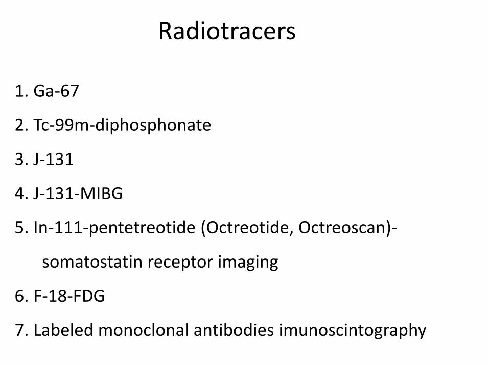
Tumor diagnostic
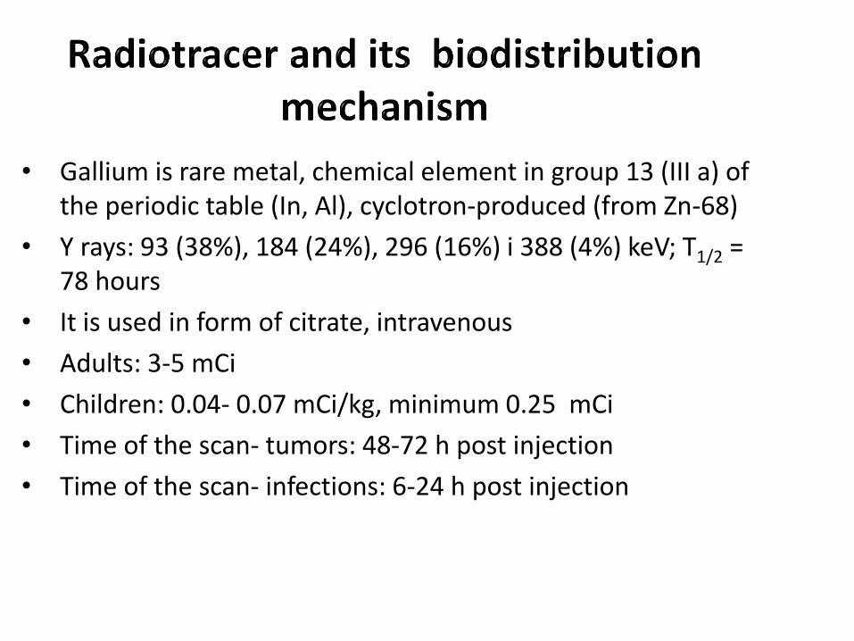
• Gallium is rare metal, chemical element in group 13 (III a) of the periodic table (In, Al), cyclotron-produced (from Zn-68)
• Y rays: 93 (38%), 184 (24%), 296 (16%) i 388 (4%) keV; T1/2 = 78 hours
• It is used in form of citrate, intravenous
• Adults: 3-5 mCi
• Children: 0.04- 0.07 mCi/kg, minimum 0.25 mCi
• Time of the scan- tumors: 48-72 h post injection
• Time of the scan- infections: 6-24 h post injection
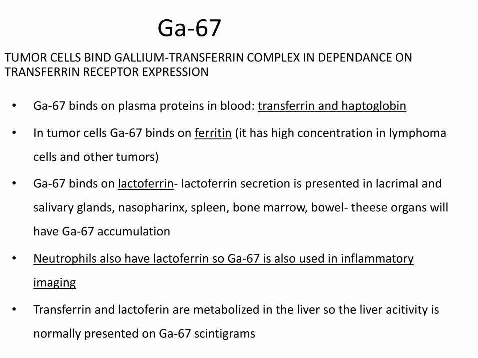
TUMOR CELLS BIND GALLIUM-TRANSFERRIN COMPLEX IN DEPENDANCE ON TRANSFERRIN RECEPTOR EXPRESSION
• Ga-67 binds on plasma proteins in blood: transferrin and haptoglobin
• In tumor cells Ga-67 binds on ferritin (it has high concentration in lymphoma
cells and other tumors)
• Ga-67 binds on lactoferrin- lactoferrin secretion is presented in lacrimal and
salivary glands, nasopharinx, spleen, bone marrow, bowel- theese organs will
have Ga-67 accumulation
• Neutrophils also have lactoferrin so Ga-67 is also used in inflammatory
imaging
• Transferrin and lactoferin are metabolized in the liver so the liver acitivity is
normally presented on Ga-67 scintigrams
Ga-67
Ga-67
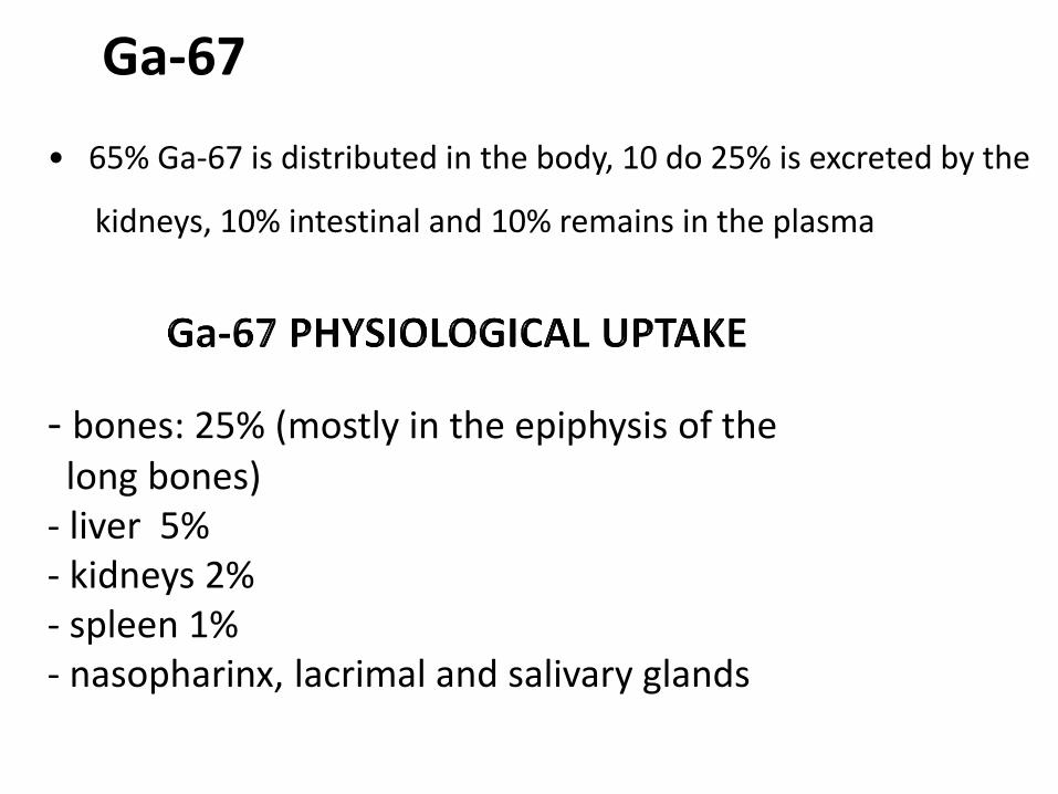
• 65% Ga-67 is distributed in the body, 10 do 25% is excreted by the
kidneys, 10% intestinal and 10% remains in the plasma
- bones: 25% (mostly in the epiphysis of the long bones) - liver 5% - kidneys 2% - spleen 1% - nasopharinx, lacrimal and salivary glands
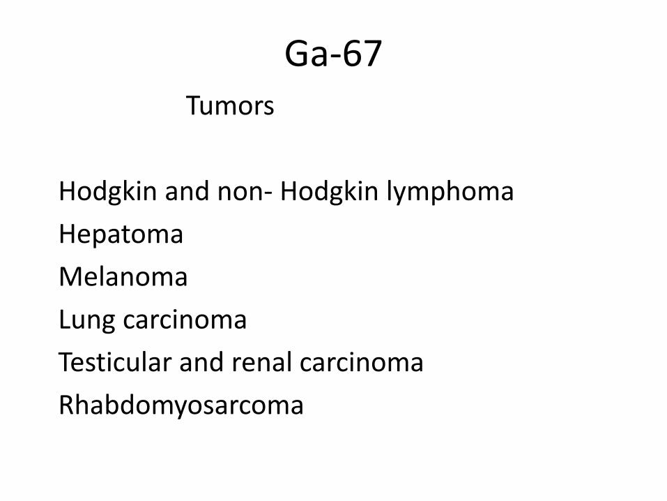
Ga-67 Tumors
Hodgkin and non- Hodgkin lymphoma
Hepatoma
Melanoma
Lung carcinoma
Testicular and renal carcinoma
Rhabdomyosarcoma
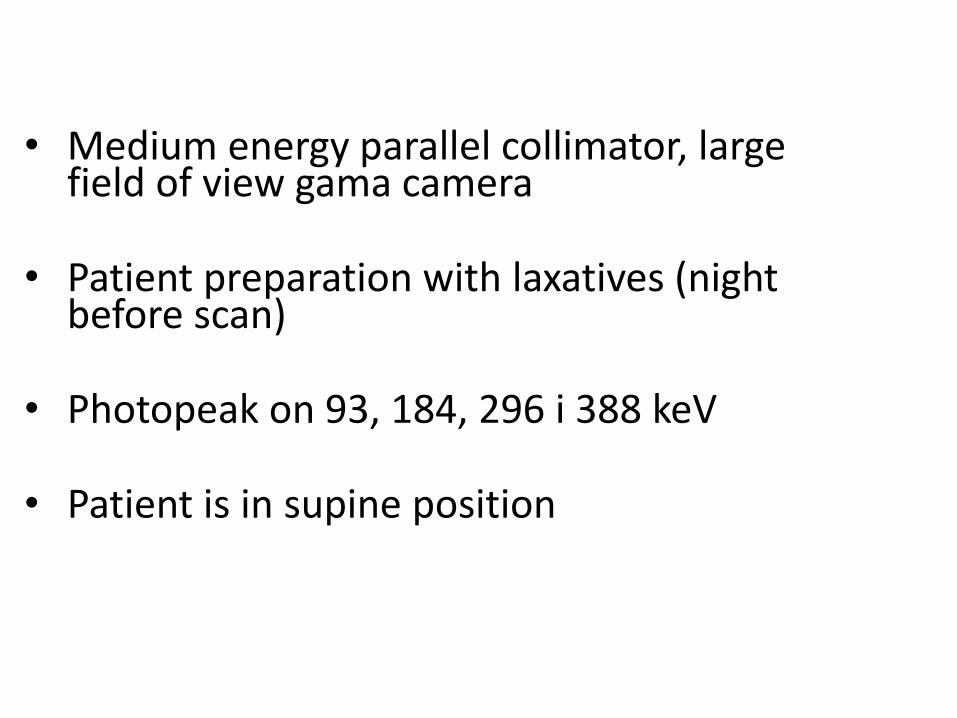
• Medium energy parallel collimator, large field of view gama camera
• Patient preparation with laxatives (night before scan)
• Photopeak on 93, 184, 296 i 388 keV
• Patient is in supine position
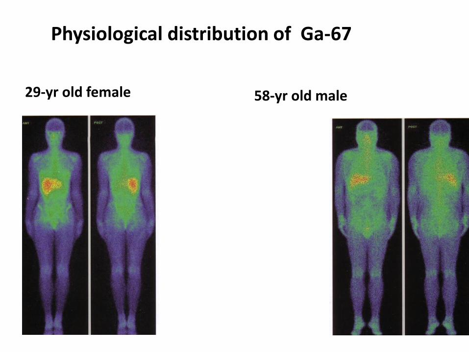
Physiological distribution of Ga-67
29-yr old female 58-yr old male
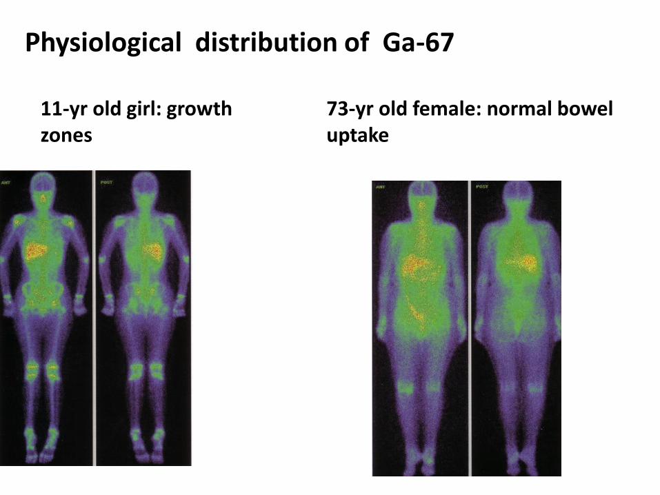
Physiological distribution of Ga-67
11-yr old girl: growth zones
73-yr old female: normal bowel uptake


Postoperative location • Ga-67 citrate accumulation
Fracture healing haematoma, wound healing Pregnancy, hormonal th, menarche Elderly, smokers Iron supplement, hemodyalisis, chemotherapy
• Ga-67 accumulation
• Accumulation in breasts
• Symetrically accumulation in the lungs
• Bone accumulation
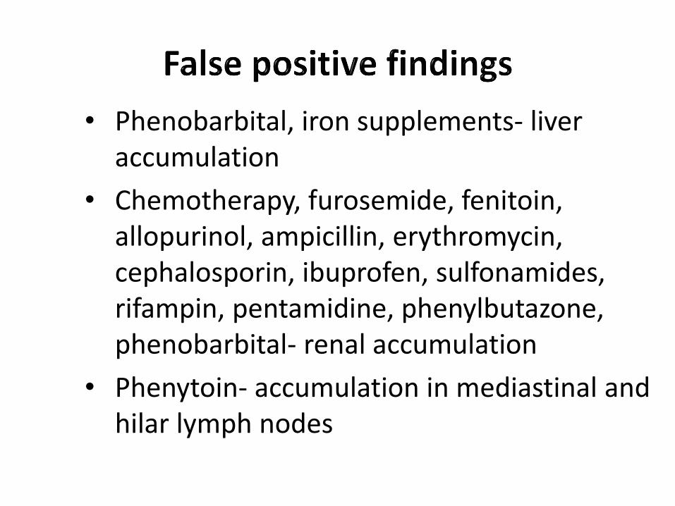
• Phenobarbital, iron supplements- liver accumulation
• Chemotherapy, furosemide, fenitoin, allopurinol, ampicillin, erythromycin, cephalosporin, ibuprofen, sulfonamides, rifampin, pentamidine, phenylbutazone, phenobarbital- renal accumulation
• Phenytoin- accumulation in mediastinal and hilar lymph nodes
• Lymphoma, NHL an HL (nowedays it is widely replaced by F-18-FDG PET), usually in follow-up after therapy (decreased or disappearance of Ga-67 pathological uptake)
• Ga-67 scan must not be performed earlier then 4-6 weeks after chemotherapy
• HEPATOMA- increased focal uptake of Ca-67 on the place of scintigraphic cold areas on Tc-99m liver coloid scan
Indications
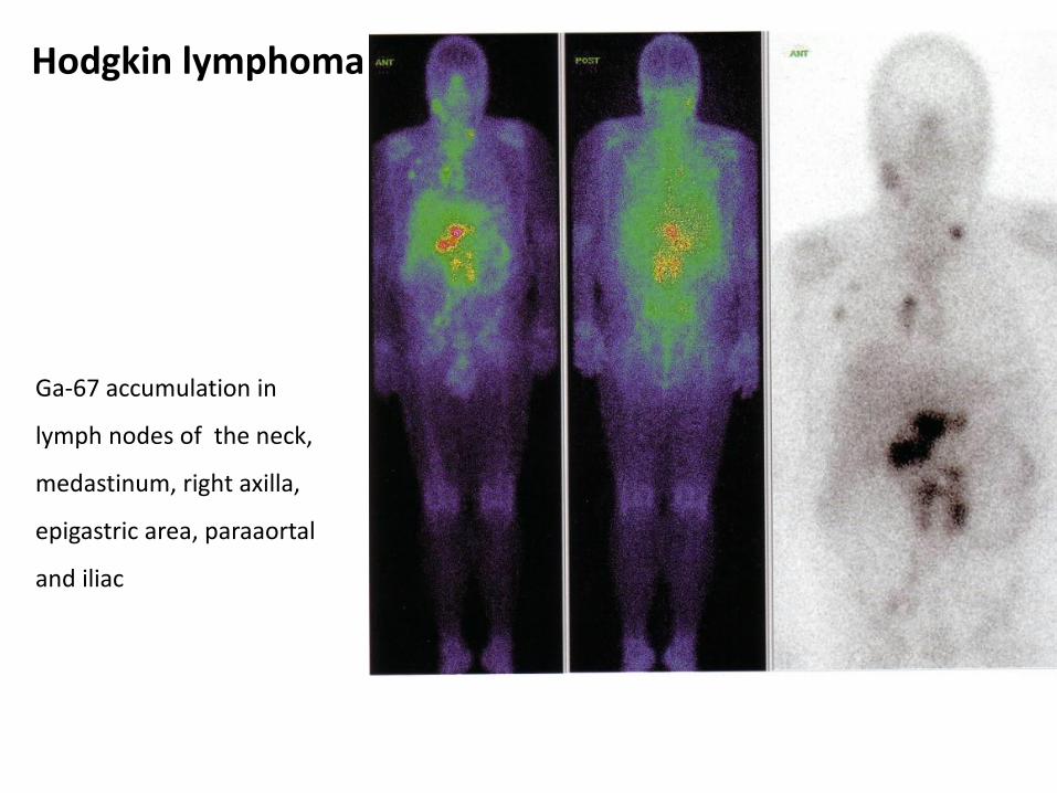
Hodgkin lymphoma
Ga-67 accumulation in
lymph nodes of the neck,
medastinum, right axilla,
epigastric area, paraaortal
and iliac
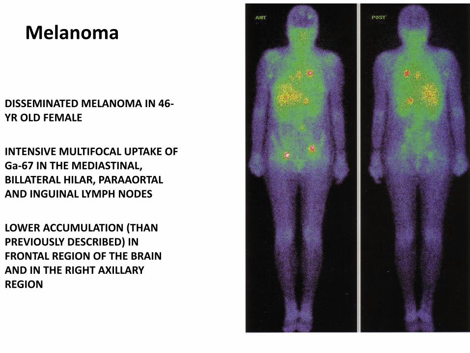
DISSEMINATED MELANOMA IN 46-YR OLD FEMALE
INTENSIVE MULTIFOCAL UPTAKE OF Ga-67 IN THE MEDIASTINAL, BILLATERAL HILAR, PARAAORTAL AND INGUINAL LYMPH NODES
LOWER ACCUMULATION (THAN PREVIOUSLY DESCRIBED) IN FRONTAL REGION OF THE BRAIN AND IN THE RIGHT AXILLARY REGION
Melanoma
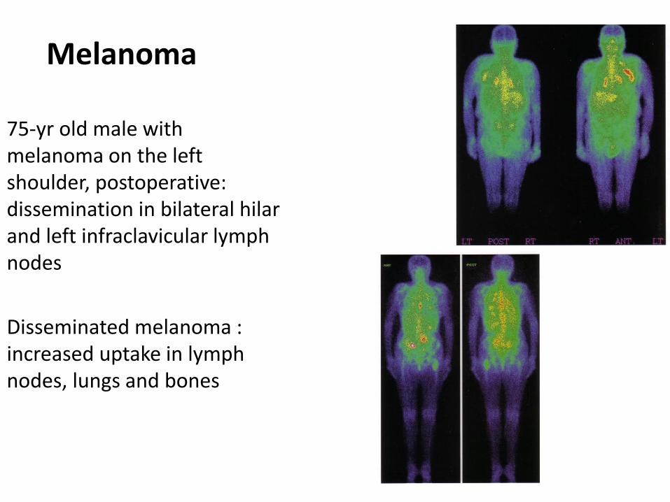
75-yr old male with melanoma on the left shoulder, postoperative: dissemination in bilateral hilar and left infraclavicular lymph nodes
Disseminated melanoma : increased uptake in lymph nodes, lungs and bones
Melanoma
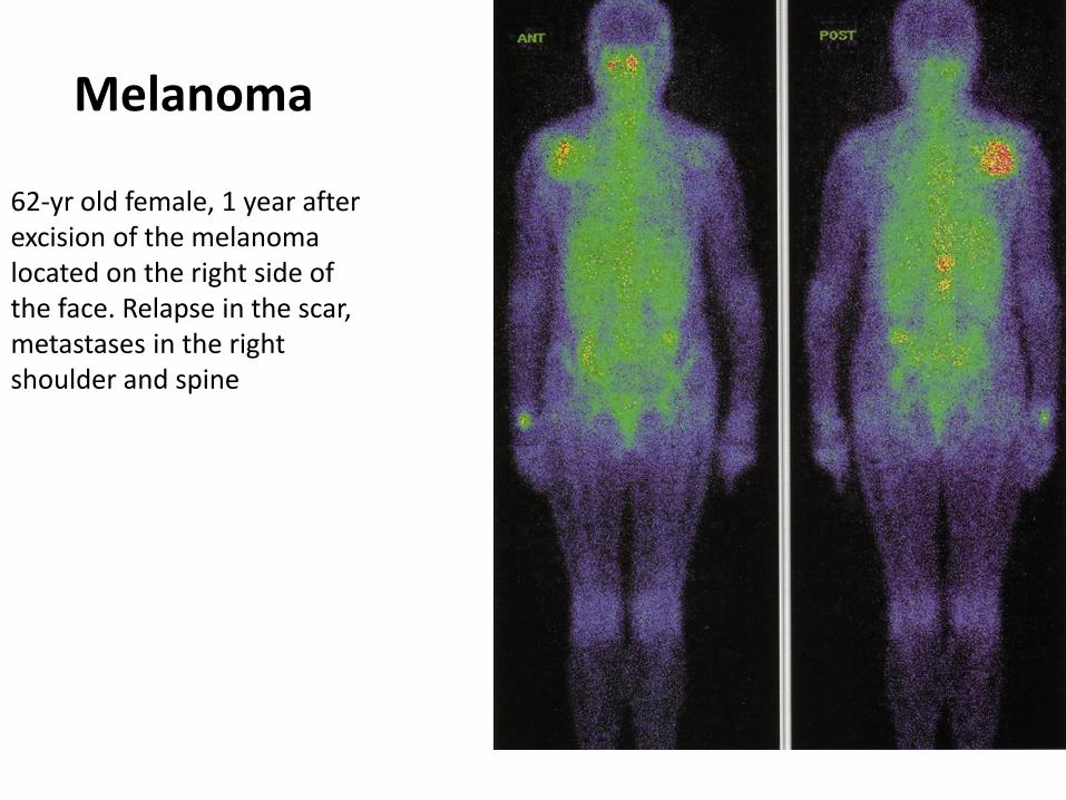
62-yr old female, 1 year after excision of the melanoma located on the right side of the face. Relapse in the scar, metastases in the right shoulder and spine
Melanoma
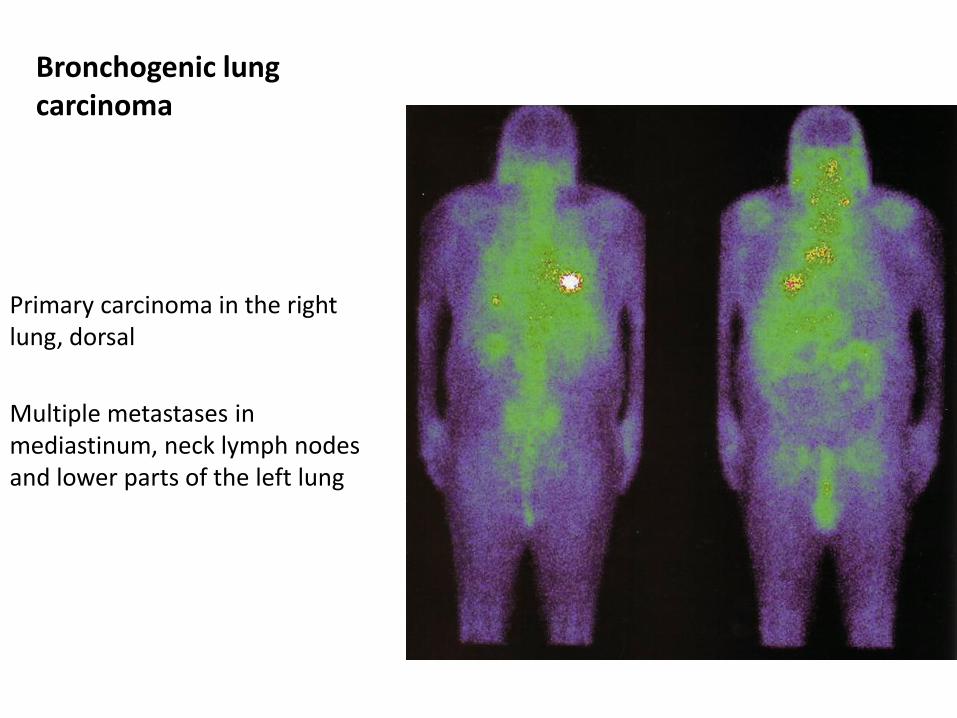
Primary carcinoma in the right lung, dorsal
Multiple metastases in mediastinum, neck lymph nodes and lower parts of the left lung
Bronchogenic lung carcinoma

68-yr old female: tumor in the left kidney with central necrosis
Adenocarcinoma of the left kidney
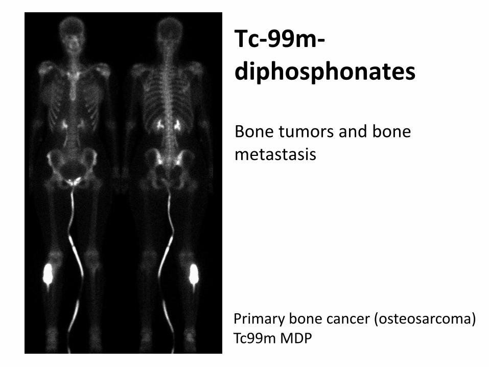
Primary bone cancer (osteosarcoma) Tc99m MDP
Tc-99m-diphosphonates Bone tumors and bone metastasis
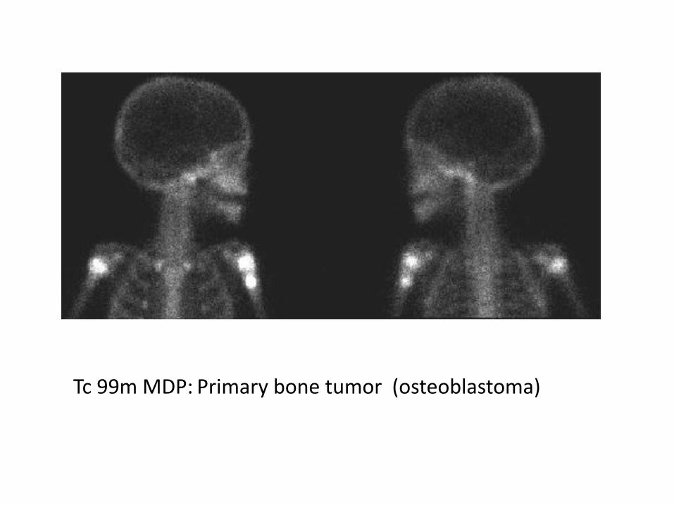
Tc 99m MDP: Primary bone tumor (osteoblastoma)
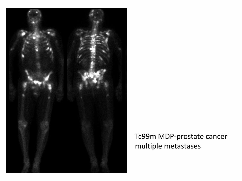
Tc99m MDP-prostate cancer multiple metastases
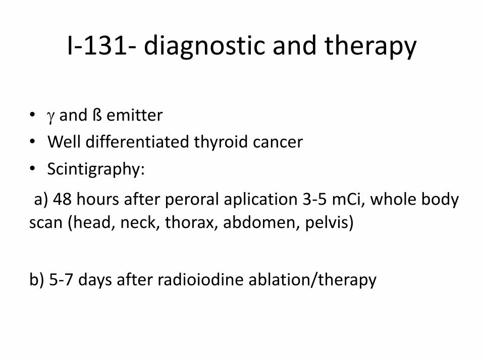
I-131- diagnostic and therapy
• and ß emitter
• Well differentiated thyroid cancer
• Scintigraphy:
a) 48 hours after peroral aplication 3-5 mCi, whole body scan (head, neck, thorax, abdomen, pelvis)
b) 5-7 days after radioiodine ablation/therapy
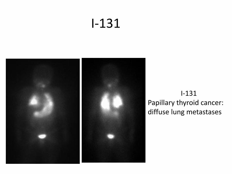
I-131
I-131 Papillary thyroid cancer: diffuse lung metastases

I-131-MIBG
• Metaiodobenzylguanidine (MIBG): norepinephrine analog
• Selective accumulation in tumors of neuroectodermal origin:
- Neuroblastoma
- Malignant pheochromocitoma
- Medullary thyroid cancer
- Carcinod metastases
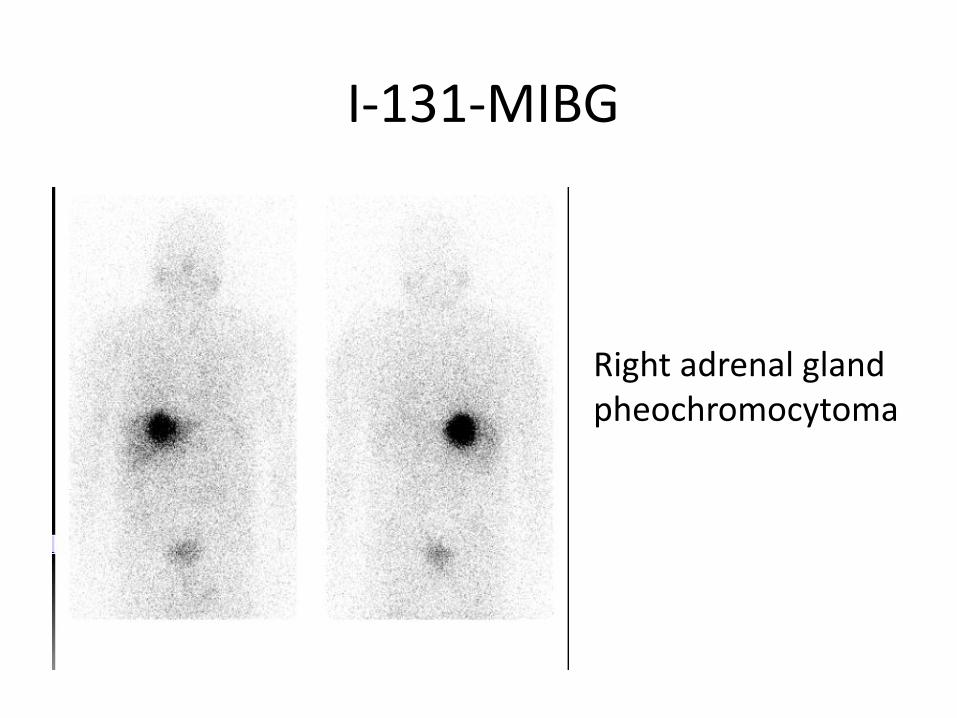
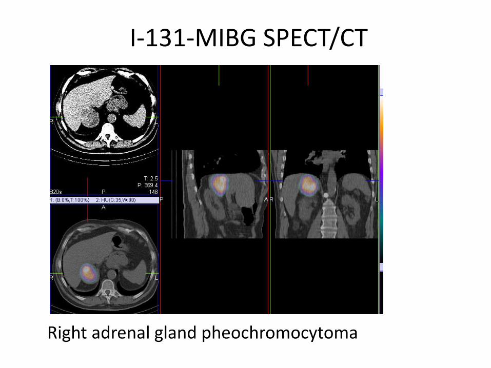
Right adrenal gland pheochromocytoma
I-131-MIBG

Right adrenal gland pheochromocytoma
I-131-MIBG SPECT/CT

Right adrenal gland pheochromocytoma
I-131-MIBG SPECT/CT
Right adrenal gland pheochromocytoma
I-131-MIBG SPECT/CT
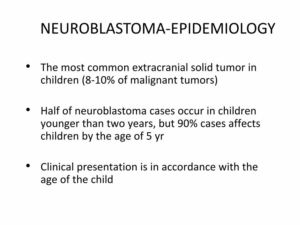
NEUROBLASTOMA-EPIDEMIOLOGY
• The most common extracranial solid tumor in children (8-10% of malignant tumors)
• Half of neuroblastoma cases occur in children younger than two years, but 90% cases affects children by the age of 5 yr
• Clinical presentation is in accordance with the age of the child
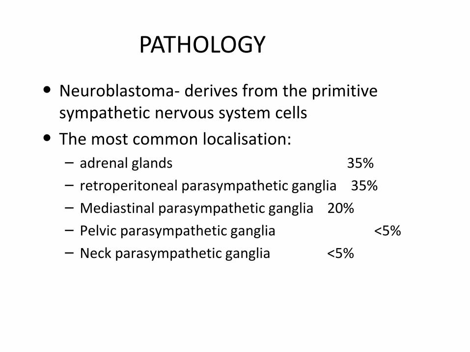
PATHOLOGY
• Neuroblastoma- derives from the primitive sympathetic nervous system cells
• The most common localisation:
– adrenal glands 35%
– retroperitoneal parasympathetic ganglia 35%
– Mediastinal parasympathetic ganglia 20%
– Pelvic parasympathetic ganglia <5%
– Neck parasympathetic ganglia <5%
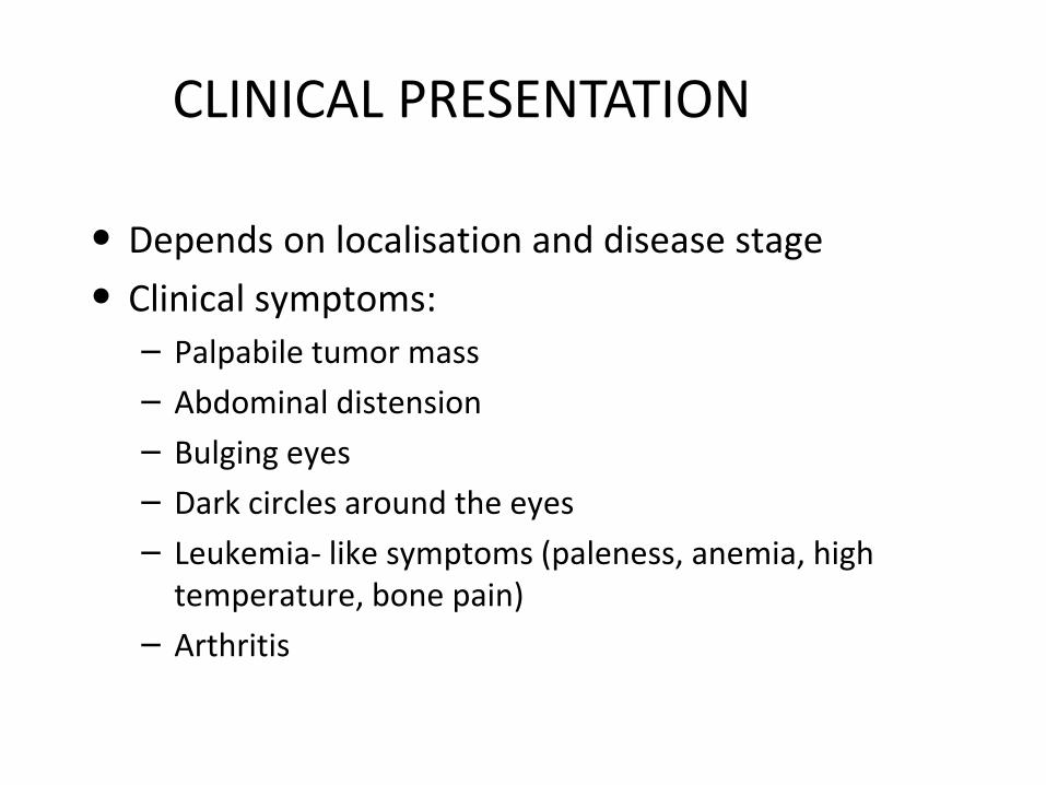
CLINICAL PRESENTATION
• Depends on localisation and disease stage
• Clinical symptoms:
– Palpabile tumor mass
– Abdominal distension
– Bulging eyes
– Dark circles around the eyes
– Leukemia- like symptoms (paleness, anemia, high temperature, bone pain)
– Arthritis
DIAGNOSTIC
• Anamnesis, clinical examination
• Laboratory paremeters (↑ LDH, NSE (neuron specific
enolase), ferritin → bad prognostic sign)
• Genetic testing (partial deletion of chromosome 1. i 11., amplification of the MYCN oncogene)
DIAGNOSTIC
Diagnostic imaging
– CT (initial staging, localised or diseminated disease)
– MRI (better estimation of soft tissue, especially in evaluation of expansion into spinal cord and epidural space)
– US
DIAGNOSTIC
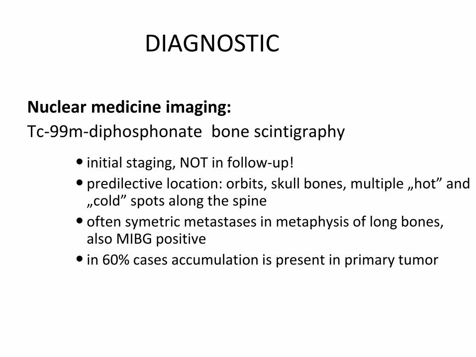
Nuclear medicine imaging:
Tc-99m-diphosphonate bone scintigraphy
• initial staging, NOT in follow-up!
• predilective location: orbits, skull bones, multiple „hot” and „cold” spots along the spine
• often symetric metastases in metaphysis of long bones, also MIBG positive
• in 60% cases accumulation is present in primary tumor
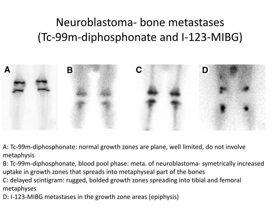
Neuroblastoma- bone metastases (Tc-99m-diphosphonate and I-123-MIBG)
A: Tc-99m-diphosphonate: normal growth zones are plane, well limited, do not involve metaphysis B: Tc-99m-diphosphonate, blood pool phase: meta. of neuroblastoma- symetrically increased uptake in growth zones that spreads into metaphyseal part of the bones C: delayed scintigram: rugged, bolded growth zones spreading into tibial and femoral metaphyses D: I-123-MIBG metastases in the growth zone areas (epiphysis)
DIAGNOSTIC
Nuclear medicine imaging:
– Somatostatin receptor scintigraphy (octreotide)
• positive octreotide indicates a better prognosis
– Labeled antibodies scintigraphy • relapse, bone metastases
– PET-FDG • Accumulation in dependence on tumor proliferation and
diferentiation
• Initially
• I-131 MIBG scintigraphy is more specific
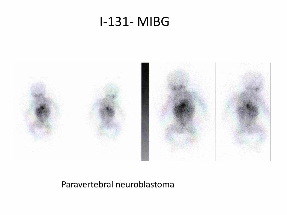
I-131- MIBG
Paravertebral neuroblastoma
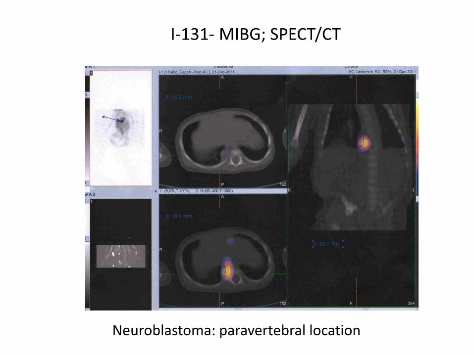
I-131- MIBG; SPECT/CT
Neuroblastoma: paravertebral location
Preoperative Postoperative
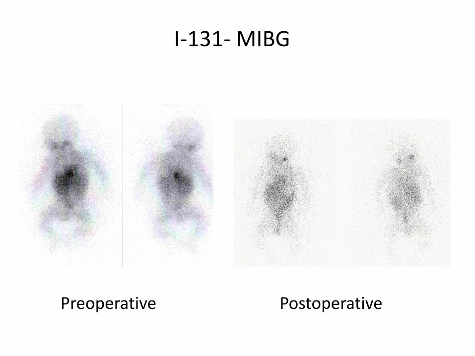
I-131- MIBG
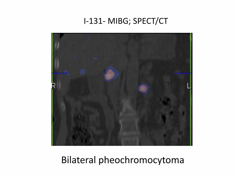
Bilateral pheochromocytoma
I-131- MIBG; SPECT/CT
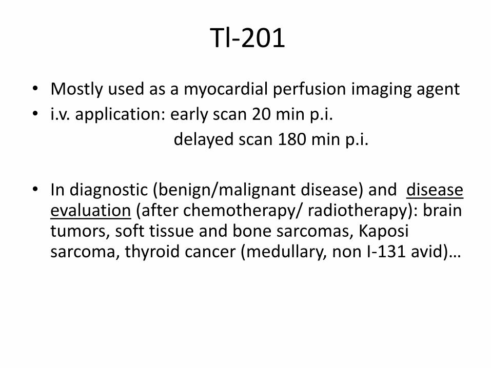
Tl-201
• Mostly used as a myocardial perfusion imaging agent
• i.v. application: early scan 20 min p.i.
delayed scan 180 min p.i.
• In diagnostic (benign/malignant disease) and disease evaluation (after chemotherapy/ radiotherapy): brain tumors, soft tissue and bone sarcomas, Kaposi sarcoma, thyroid cancer (medullary, non I-131 avid)…
Receptor scintigraphy
• Receptor imaging using specific agonists or radiolabeled agonists
• Oncology related receptors:
- transferrin: malignant tumors, sarcoidosis, tbc, inflammatory changes
- somatostatine: neuroendocrine and neuroendocrine related tumors
SOMATOSTATIN RECEPTOR SINTIGRAPHY
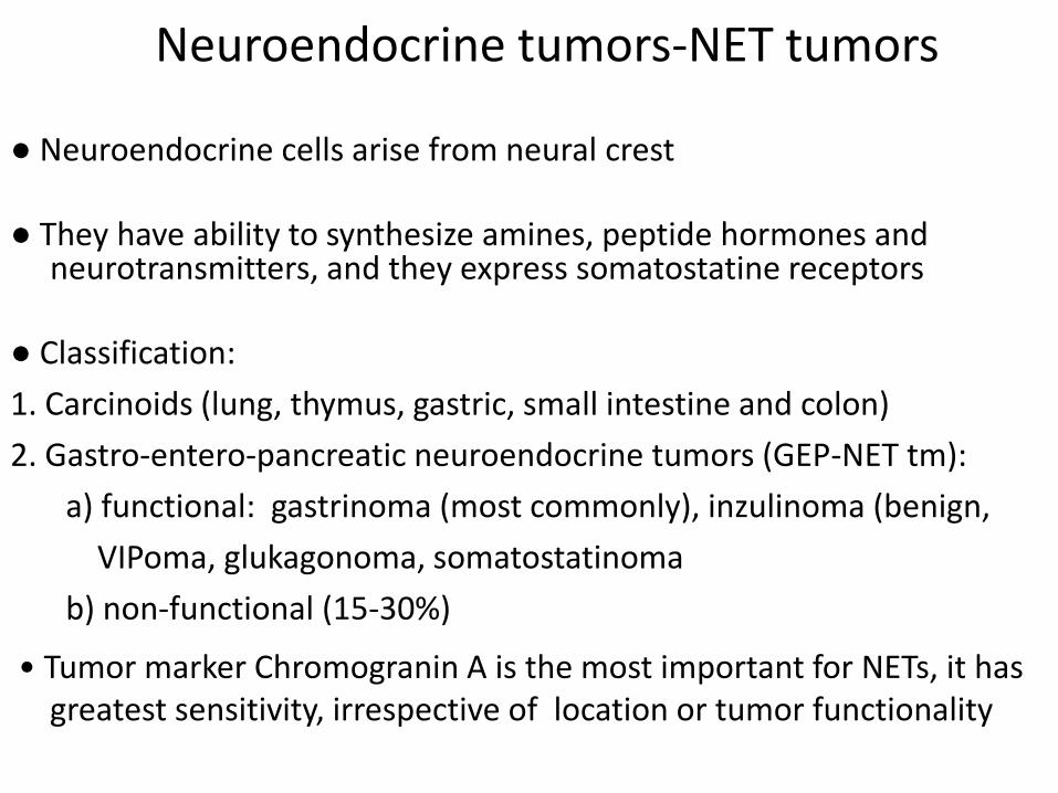
Neuroendocrine tumors-NET tumors
● Neuroendocrine cells arise from neural crest ● They have ability to synthesize amines, peptide hormones and
neurotransmitters, and they express somatostatine receptors ● Classification:
1. Carcinoids (lung, thymus, gastric, small intestine and colon)
2. Gastro-entero-pancreatic neuroendocrine tumors (GEP-NET tm):
a) functional: gastrinoma (most commonly), inzulinoma (benign,
VIPoma, glukagonoma, somatostatinoma
b) non-functional (15-30%)
• Tumor marker Chromogranin A is the most important for NETs, it has greatest sensitivity, irrespective of location or tumor functionality
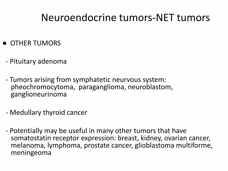
Neuroendocrine tumors-NET tumors
● OTHER TUMORS - Pituitary adenoma
- Tumors arising from symphatetic neurvous system:
pheochromocytoma, paraganglioma, neuroblastom, ganglioneurinoma
- Medullary thyroid cancer
- Potentially may be useful in many other tumors that have somatostatin receptor expression: breast, kidney, ovarian cancer, melanoma, lymphoma, prostate cancer, glioblastoma multiforme, meningeoma

Somatostatin
- hormone, 14 amino acids, T1/2 = 1-3 min
- normaly expressed in hypothalamus, cerebral cortex, brainstem, GI
system, pancreas
- function: neurotransmitter or growth hormone-inhibiting hormone
(GHIH) but it also inhibits insuline, glucagon and other neuropeptide
secretion
- somatostatin reseptors (SSR) are expressed on many cells and
tumors of neuroedocrine origin
- 5 SSR subtypes
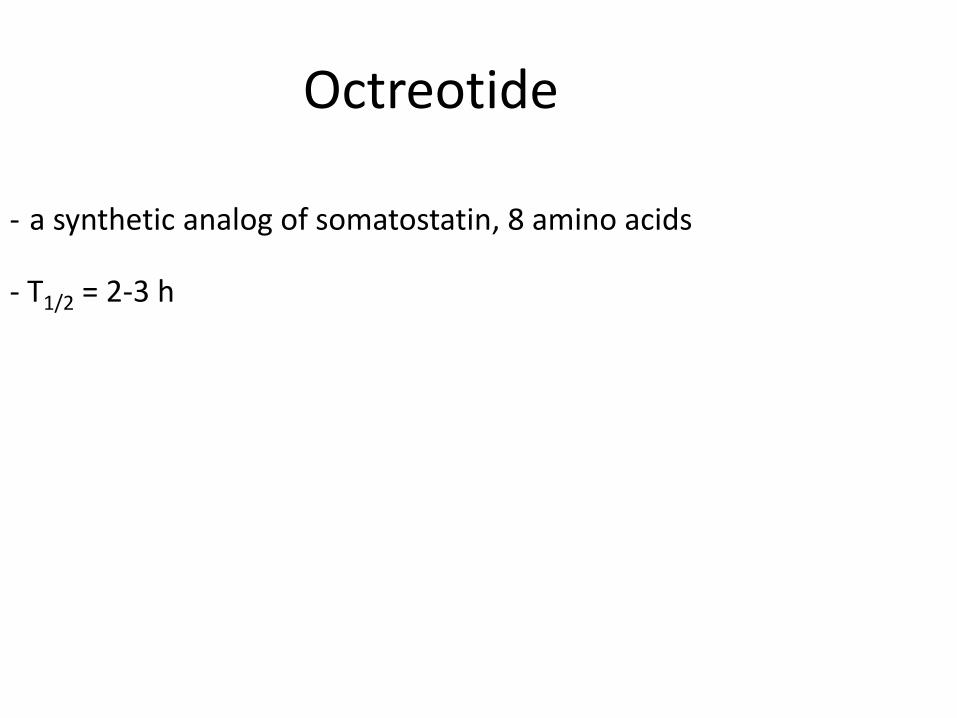
Octreotide
- a synthetic analog of somatostatin, 8 amino acids
- T1/2 = 2-3 h
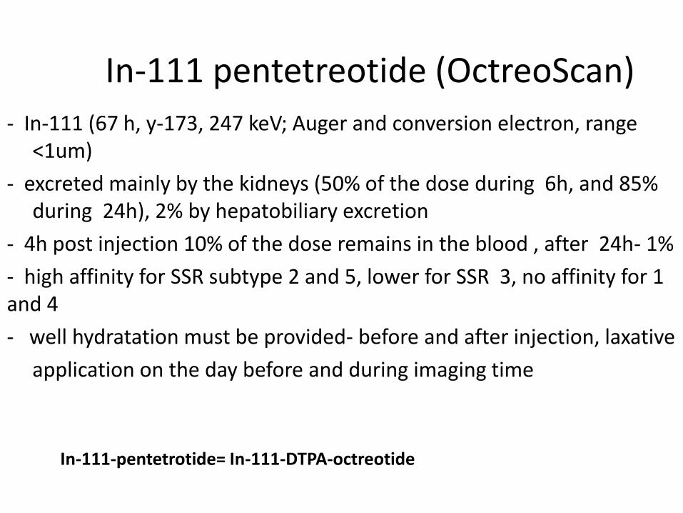
In-111 pentetreotide (OctreoScan)
- In-111 (67 h, y-173, 247 keV; Auger and conversion electron, range <1um)
- excreted mainly by the kidneys (50% of the dose during 6h, and 85% during 24h), 2% by hepatobiliary excretion
- 4h post injection 10% of the dose remains in the blood , after 24h- 1%
- high affinity for SSR subtype 2 and 5, lower for SSR 3, no affinity for 1 and 4
- well hydratation must be provided- before and after injection, laxative
application on the day before and during imaging time
In-111-pentetrotide= In-111-DTPA-octreotide

Patient preparation
- it is preferable to discontinue Sandostatin therapy the
day before injection, and in case of an depo preparation
OctreoScan may be provided just before the next
treatment
-- well hydratation must be provided, laxative preparation before injection and during imaging (caution in patients with diarrheal syndroma)
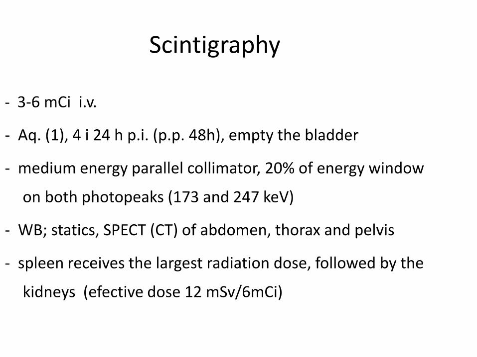
Scintigraphy
- 3-6 mCi i.v.
- Aq. (1), 4 i 24 h p.i. (p.p. 48h), empty the bladder
- medium energy parallel collimator, 20% of energy window
on both photopeaks (173 and 247 keV)
- WB; statics, SPECT (CT) of abdomen, thorax and pelvis
- spleen receives the largest radiation dose, followed by the
kidneys (efective dose 12 mSv/6mCi)
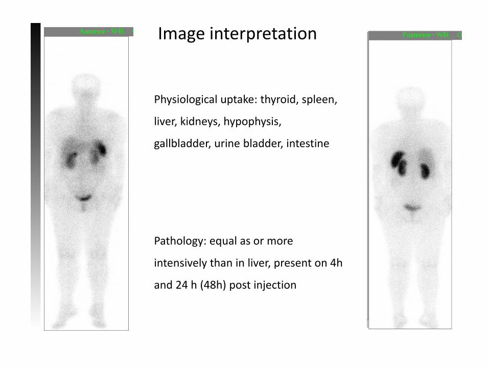
Image interpretation
Physiological uptake: thyroid, spleen,
liver, kidneys, hypophysis,
gallbladder, urine bladder, intestine
Pathology: equal as or more
intensively than in liver, present on 4h
and 24 h (48h) post injection

Indications
- localisation of primary tumor
- evaluation of disease stage
- post therapy follow up
- evaluation of relapse
- assessment of radionuclide therapy
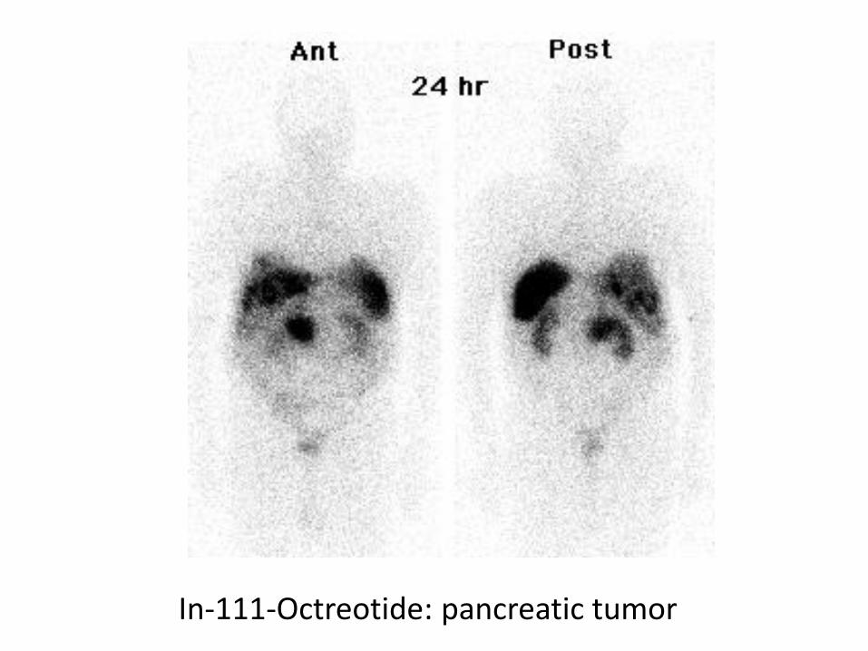
In-111-Octreotide: pancreatic tumor
Pancreatic NET, palliative surgical treatment was provided. Liver metastases.
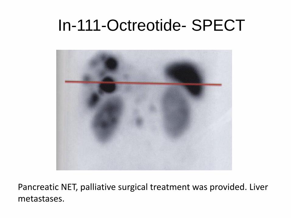
In-111-Octreotide- SPECT
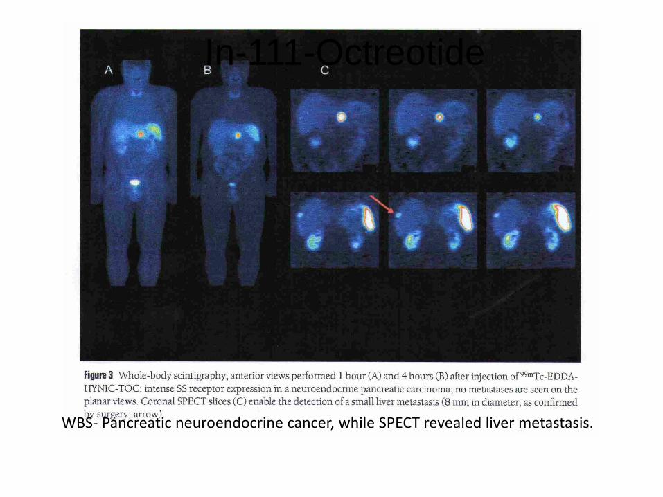
WBS- Pancreatic neuroendocrine cancer, while SPECT revealed liver metastasis.
In-111-Octreotide
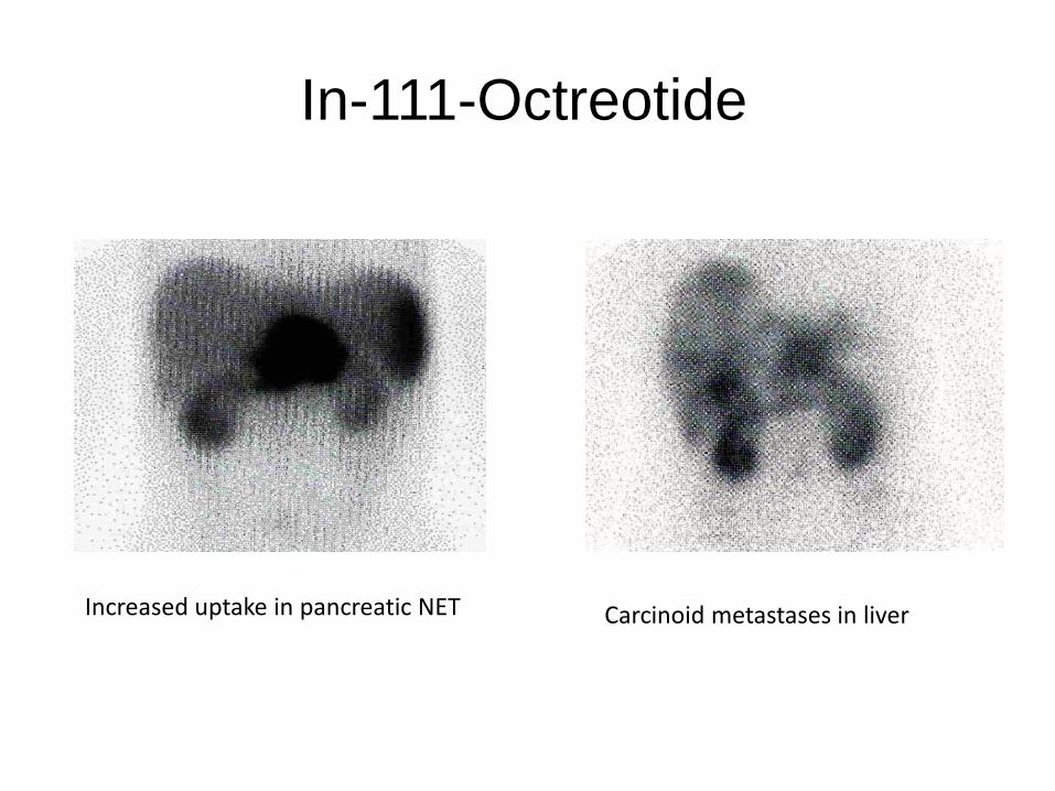
Increased uptake in pancreatic NET Carcinoid metastases in liver
In-111-Octreotide
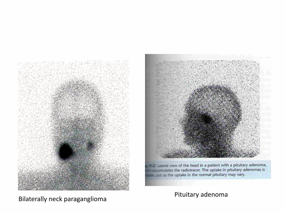
Bilaterally neck paraganglioma Pituitary adenoma

Octreotide sensitivity in NETs
carcinoid 80%
insulinoma 31%
gastrinoma 95%
SCLC 100%
PHEO 100%
MTC 54%
pituitary adenoma 80%
The Requisites, 2006.
Tc -99m Tektrotyde
- Tc-99m labeled somatostatin receptor analogue subtypes
2,3, and 5
- i.v. 15-20 mCi, empty bladder previously
- Aquisition 2 i 4 h p.i.: WB, SPECT of abdomen, thorax and
pelvis, patient may eat and drink after first scan
- Whole diagnostic procedure is done in one day, equivalent
dose is lower (4, 2 mSv/20 mCi), as well as price
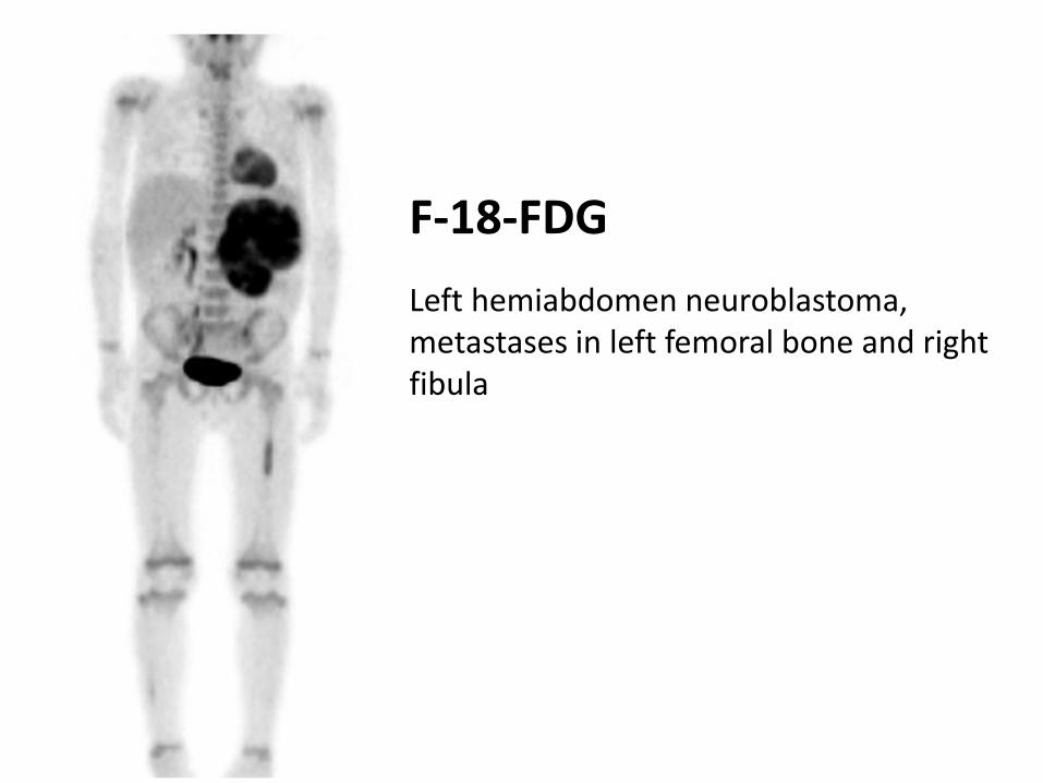
F-18-FDG
Left hemiabdomen neuroblastoma, metastases in left femoral bone and right fibula
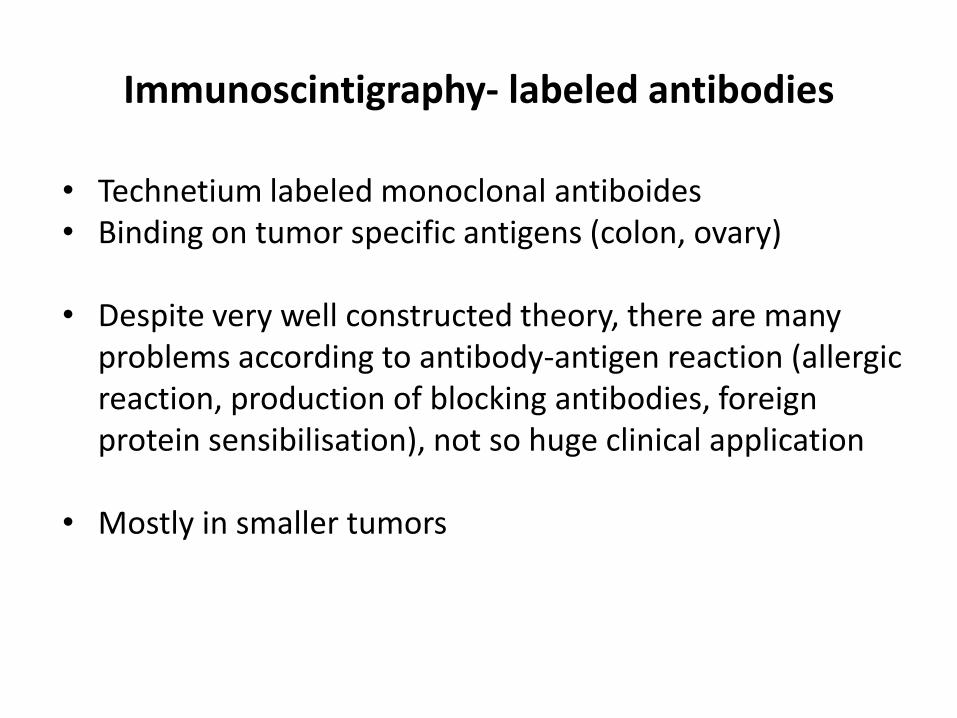
Immunoscintigraphy- labeled antibodies
• Technetium labeled monoclonal antiboides • Binding on tumor specific antigens (colon, ovary)
• Despite very well constructed theory, there are many
problems according to antibody-antigen reaction (allergic reaction, production of blocking antibodies, foreign protein sensibilisation), not so huge clinical application
• Mostly in smaller tumors
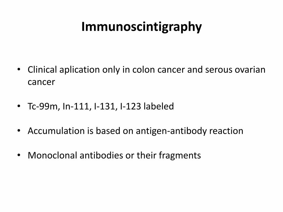
Immunoscintigraphy
• Clinical aplication only in colon cancer and serous ovarian
cancer • Tc-99m, In-111, I-131, I-123 labeled
• Accumulation is based on antigen-antibody reaction
• Monoclonal antibodies or their fragments

Tumor markers
• Tumor cell necrosis and cytolysis lead to release of tumor markers in the blood and other body fluids
• Monoclonal antibodies (previously labeled with tracers) are used for determining the tumor markers concentration
• In accordance of tracer: immunoradiometric, enzymatic, fluorometric and luminimetric methods
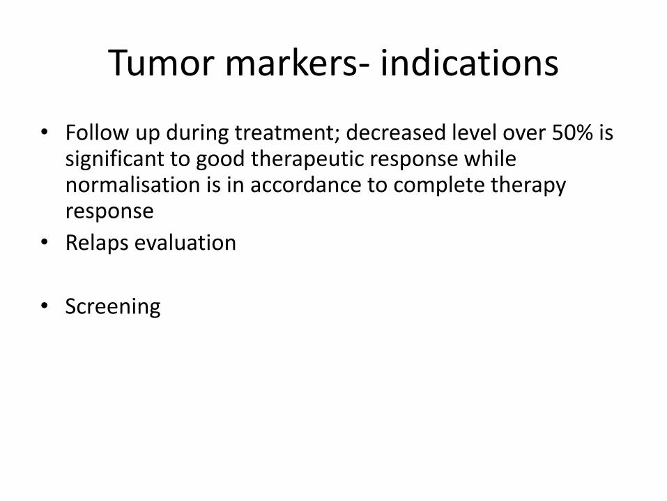
Tumor markers- indications
• Follow up during treatment; decreased level over 50% is significant to good therapeutic response while normalisation is in accordance to complete therapy response
• Relaps evaluation
• Screening
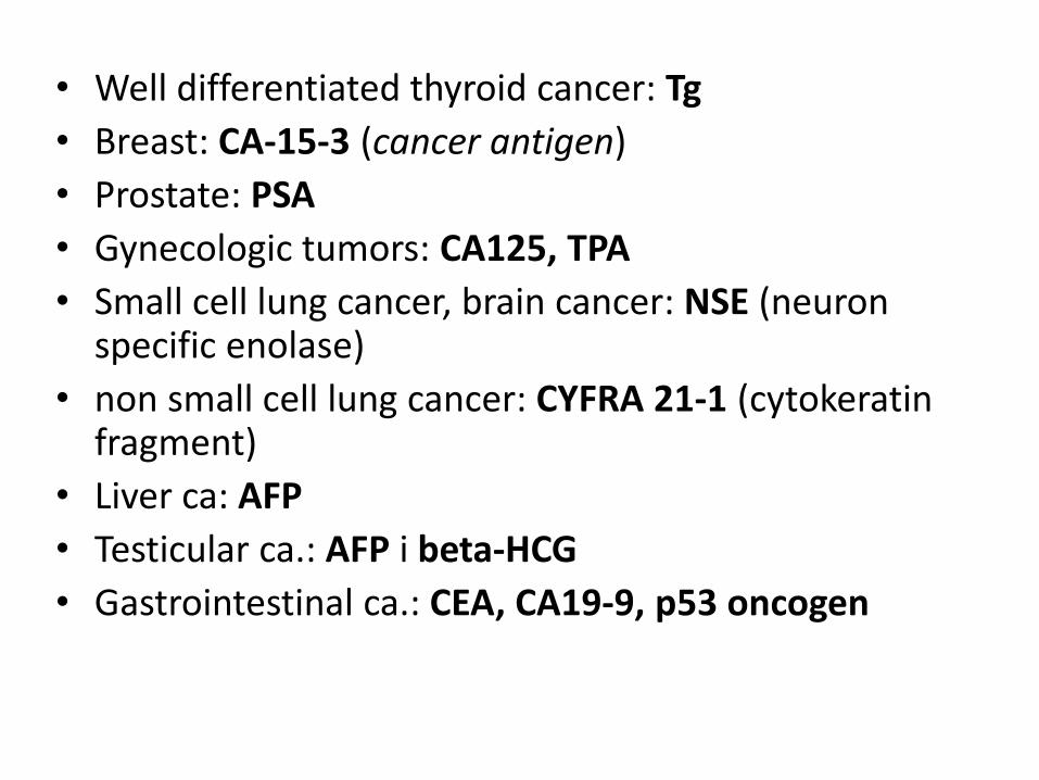
• Well differentiated thyroid cancer: Tg
• Breast: CA-15-3 (cancer antigen)
• Prostate: PSA
• Gynecologic tumors: CA125, TPA
• Small cell lung cancer, brain cancer: NSE (neuron specific enolase)
• non small cell lung cancer: CYFRA 21-1 (cytokeratin fragment)
• Liver ca: AFP
• Testicular ca.: AFP i beta-HCG
• Gastrointestinal ca.: CEA, CA19-9, p53 oncogen
THE END
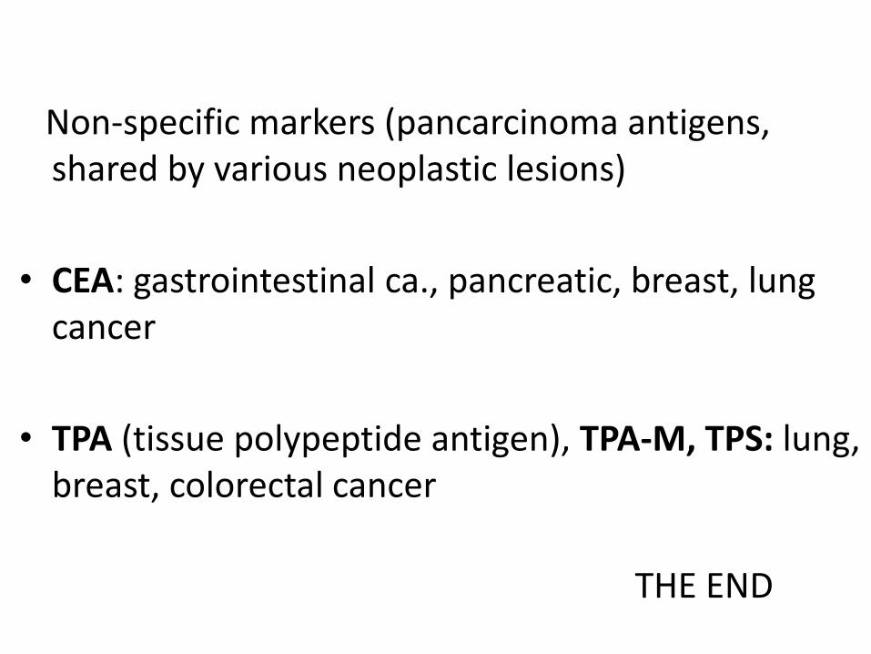
Non-specific markers (pancarcinoma antigens, shared by various neoplastic lesions)
• CEA: gastrointestinal ca., pancreatic, breast, lung cancer
• TPA (tissue polypeptide antigen), TPA-M, TPS: lung, breast, colorectal cancer