Embed Size (px)
Citation preview
TUBERCULOUS PERICARDIAL EFFUSION
BY
S. SUZMAN
From the Cardiographic Department, Guy's Hospital
Received June 22, 1942
Tuberculous pericardial effusion is not common; and so the case described is of interest on accountof the patient's age, the frequency with which the pericardial effusion required tapping, the relativeabsence of symptoms during most of the illness, and the stages through which the case passed fromgross effusion to relative dryness at autopsy with only about 2 oz. of fluid in a greatly thickenedpericardium.
DESCRIPTION OF CASE
A sailor, aged 23, was admitted into a hospital on August 16, 1940, for epigastric pain and frequentvomiting, which had begun three days previously. There had been a similar attack three monthspreviously. There was continued pyrexia up to 102. The diagnosis was apparently difficult until hecomplained of some sub-sternal tightness a few days later, when attention was directed to his heart.This was found to be enlarged, 1 inch outside the nipple line, but it was only after an X-ray that thediagnosis of pericardial effusion was established. Before this, on account of dullness at the left baseof the lung with diminished breath sounds, a pleural effusion was suspected, but the X-ray did notconfirm this. The blood pressure was typically low, 100/70.
Repeated tappings were performed as under:August 23, 300 c.c. ; August 27, 400 c.c.; August 30, 500 c.c.; the fluid each time being sterile and
clear and straw-coloured; September 6, 300 c.c.; and September 13, 550 c.c., the fluid being deeplyblood-stained on these two occasions. (For later tappings see below.)
On Sept. 16 he was admitted to the Southern Hospital, where he came under my observation.The heart was greatly enlarged, both to left and right. The heart sounds were faint and muffled.Continued pyrexia was still present, varying between 99 and 103. Symptoms were absent, except forsub-sternal tightness which he seemed to experience whenever the effusion increased; this was re-lieved after tapping. Tachycardia was a constant feature throughout, the pulse rate being about100 to 120. The blood sedimentation rate was 78 mm. after one hour. Blood culture was sterile.A Mantoux test (1 in 10,000) was negative. And in spite of a negative guinea-pig test on two occa-sions, the diagnosis of tuberculous pericardial disease was made.
September 24, aspiration of 520 c.c. of old blood-stained fluid. There was no change in his bloodpressure, 105/80, before and after, or in the cardiogram which showed the characteristic T waveinversions.
November 6, aspiration of 600 c.c. of fluid, less blood-stained. He had remained fairly comfortableand without sternal pain. On this occasion an air replacement was done, and an X-ray afterwardsshowed the parietal layer of the pericardium, which measured half an inch thick. The cardiac shadowwas not diminished after the tapping. X-ray on December 10, showed the air replacement to beabsorbed, and an increase in the size of the effusion. A course of sulphapyridine (17 g.) was givenabout this stage, and brought down the temperature for a time. Towards the end of December hedeveloped a dry cough and the temperature rose again to 102. He began losing weight and he hadnight sweats.
December 28, aspiration of 600 c.c. of amber-coloured fluid; air replacement of 100 c.c. Finecrepitations were now heard, and an additiconal. diagnosis of miliary tuberculosis of the lungs wasmade. lHe gradually became worse about the middle of January 1941, and was mildly delusional attimes. On January 19, 1941, he became drowsy; coma set in and he died the next day.
Post-mortem examination. Pleura: light fibrous adhesions, obliterating both pleurae.Heart: Great dilatation of the pericardial sac, which contained only 2 oz. of amber
fluid, but was much thickened by granulomatous tissue. A layer of friable yellow fibrin, uniting thethickened layers of the pericardium, up to 1 cm. thick; no loculations. Heart itself only slightlyenlarged; valves normal.
19
on February 16, 2020 by guest. P
rotected by copyright.http://heart.bm
j.com/
Br H
eart J: first published as 10.1136/hrt.5.1.19 on 1 January 1943. Dow
nloaded from
S. SUZMAN
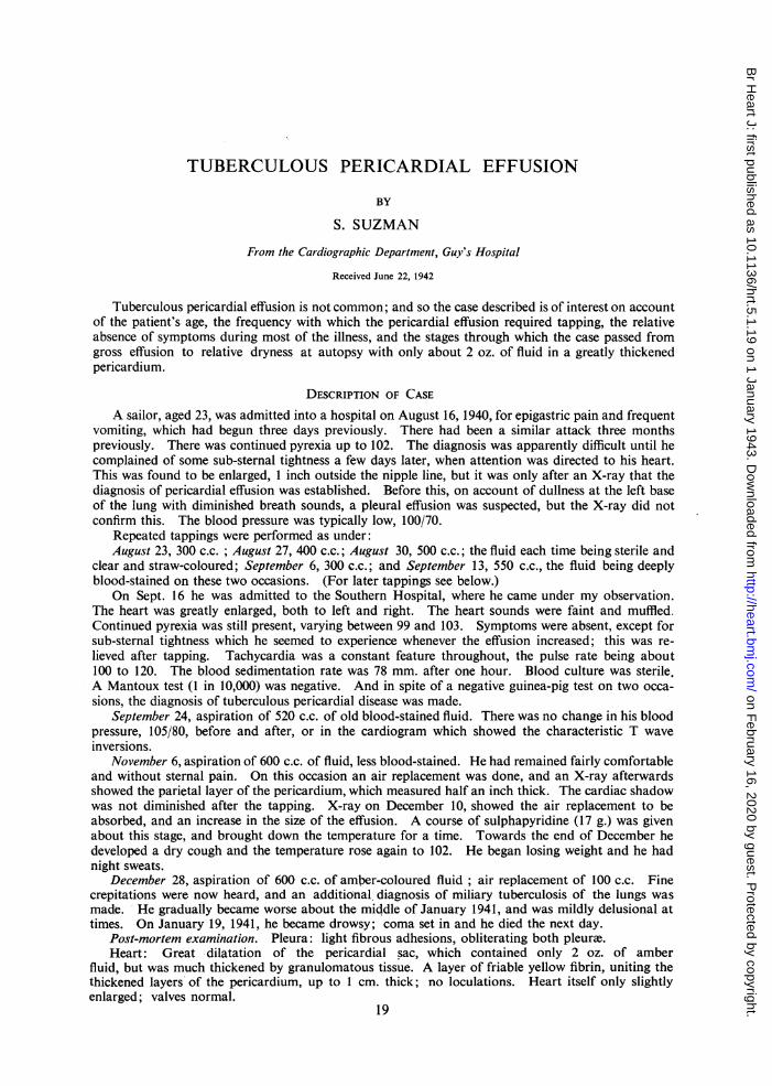
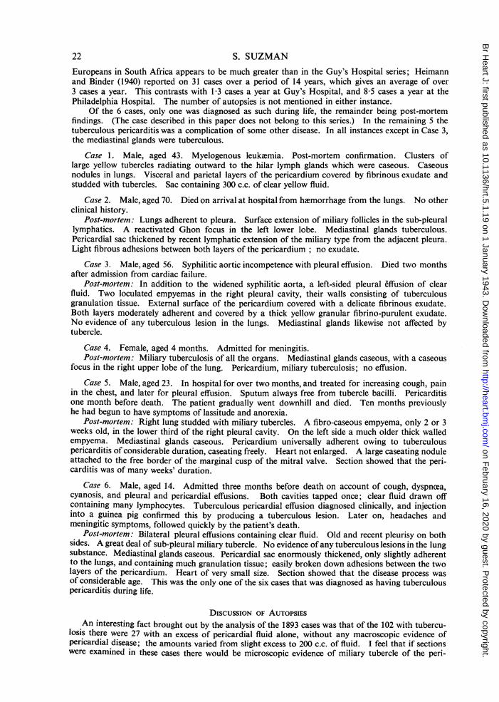
FIG. 1 -Radiogram of the heart showing much dilatation of the pericardial sac.
Lungs: Congested and closely studded with granulomatous miliary tubercles; no cavities.Caseous glands in both hila and at the bifurcation of the bronchi, two inches by one quarter inch insize on each side.
Liver: Cloudy swelling and a few miliary tubercles.Spleen: Three times normal size; some tubercles.Kidneys: Cloudy swelling with streaks of pus in the pyramids; studded with miliary tubercles.Bladder: Infected with pus.Prostate: Caseous tuberculosis present.Brain: General cedema. Sero-meningitis over the base of the brain with a few scattered tubercles
in the fissures. A tuberculoma, 05 cm. in diameter, in the left occipital cortex.Histological examination confirmed tuberculosis.
DISCUSSION OF CASEThere is no doubt that in this case the primary focus was a tuberculous gland in the mediastinum,
secondarily affecting the pericardium. And later as the disease progressed, further disseminationoccurred, causing generalised miliary tuberculosis. Another point of interest was the absence oflung symptoms until the final phase of his illness. HWmorrhagic pericardial effusions are said to becharacteristic of tuberculosis. I think this is an open question, and the probability of trauma whenneedling must be taken into full account. In this case the first three tappings were not blood-stained.The next two were deeply blood-stained, and the succeeding ones clearly pointed to the presence ofold blood in the sac. The last specimen of pericardial fluid was only amber-coloured.
Another point of interest is that the cardiac shadow did not diminish after paracentesis, owing tothe very thick and rigid pericardium.
20
on February 16, 2020 by guest. P
rotected by copyright.http://heart.bm
j.com/
Br H
eart J: first published as 10.1136/hrt.5.1.19 on 1 January 1943. Dow
nloaded from
TUBERCULOUS PERICARDIAL EFFUSION
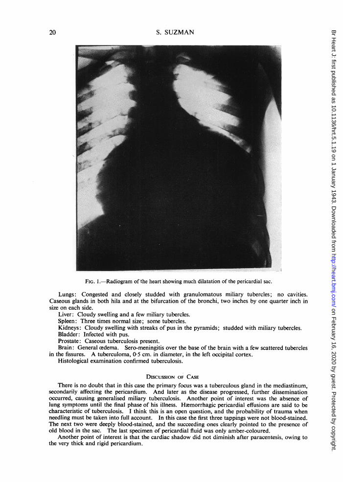
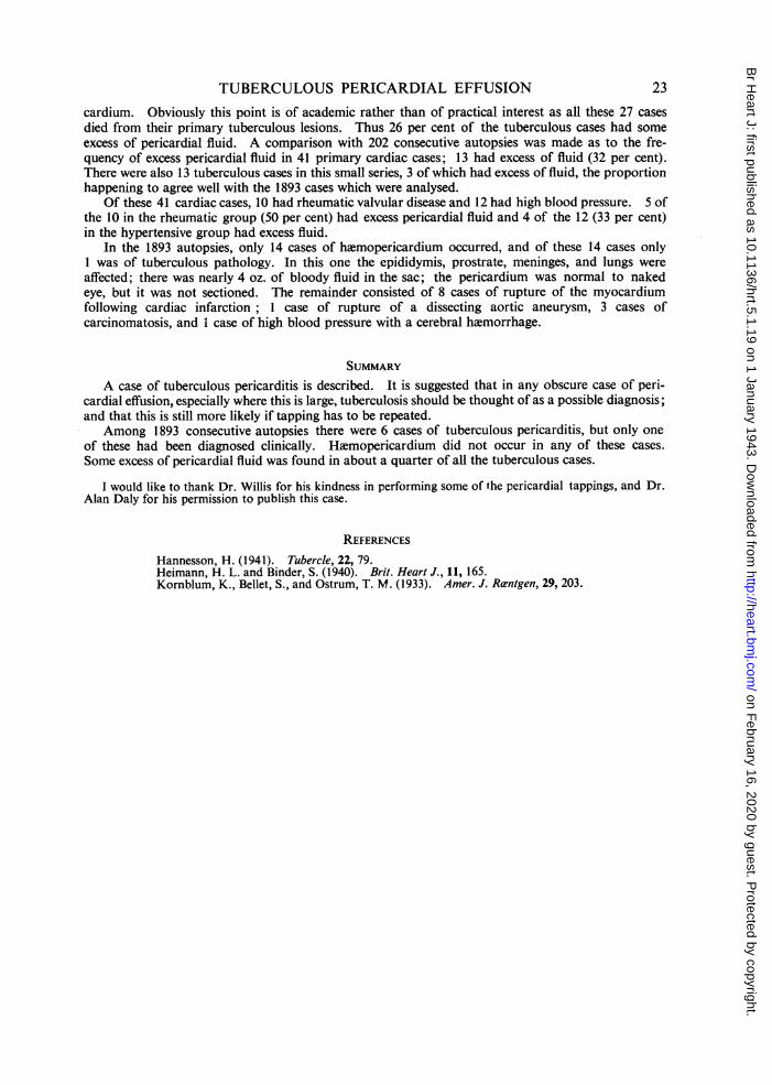
FIG. 2.-Photograph of the heart showing much thickening of the pericardium.
The negative results of laboratory findings, such as the absence of tubercle bacilli in the fluids andthe failure of inoculation of guinea pigs to produce any result, should also be stressed.
POST-MORTEM STATISTICS AT Guy's HOSPITALTo assess the incidence of tuberculous pericarditis I have analysed 1893 autopsies at Guy's Hospital
from January 1935 to June 1939. There were 102 tuberculous cases with lung involvement. Therewere 6 cases of tuberculous pericarditis, confirmed by histological section; 2 others with pericarditiswere most probably tuberculous, judging from the post-mortem description, but cannot be includedas there was no mention of tuberculosis macroscopically or microscopically. There was also onecase of myocardial tubercle alone, without pericardial involvement. This makes an incidence of0-3 per cent of all the cases that came to autopsy during this period, and 6 per cent of all cases thathad tuberculous involvement in any organ. If the two probable cases were included it would raisethe incidence among general cases only to slightly over 0 4 per cent, but it would raise the incidenceamong tuberculous cases to as high as 8 per cent.
As a comparison for incidence, Kornblum, Bellet, and Ostrum (1933) give figures of 1 per centamong general cases and 4 per cent among tuberculous cases (Philadelphia Hospital): they reported17 cases during a period of two years. The incidence of tuberculous pericarditis among non-
21
on February 16, 2020 by guest. P
rotected by copyright.http://heart.bm
j.com/
Br H
eart J: first published as 10.1136/hrt.5.1.19 on 1 January 1943. Dow
nloaded from
Europeans in South Africa appears to be much greater than in the Guy's Hospital series; Heimannand Binder (1940) reported on 31 cases over a period of 14 years, which gives an average of over3 cases a year. This contrasts with 1-3 cases a year at Guy's Hospital, and 8-5 cases a year at thePhiladelphia Hospital. The number of autopsies is not mentioned in either instance.
Of the 6 cases, only one was diagnosed as such during life, the remainder being post-mortemfindings. (The case described in this paper does not belong to this series.) In the remaining 5 thetuberculous pericarditis was a complication of some other disease. In all instances except in Case 3,the mediastinal glands were tuberculous.
Case 1. Male, aged 43. Myelogenous leukemia. Post-mortem confirmation. Clusters oflarge yellow tubercles radiating outward to the hilar lymph glands which were caseous. Caseousnodules in lungs. Visceral and parietal layers of the pericardium covered by fibrinous exudate andstudded with tubercles. Sac containing 300 c.c. of clear yellow fluid.
Case 2. Male, aged 70. Died on arrival at hospital from hemorrhage from the lungs. No otherclinical history.
Post-mortem: Lungs adherent to pleura. Surface extension of miliary follicles in the sub-pleurallymphatics. A reactivated Ghon focus in the left lower lobe. Mediastinal glands tuberculous.Pericardial sac thickened by recent lymphatic extension of the miliary type from the adjacent pleura.Light fibrous adhesions between both layers of the pericardium; no exudate.
Case 3. Male, aged 56. Syphilitic aortic incompetence with pleural effusion. Died two monthsafter admission from cardiac failure.
Post-mortem: In addition to the widened syphilitic aorta, a left-sided pleural effusion of clearfluid. Two loculated empyemas in the right pleural cavity, their walls consisting of tuberculousgranulation tissue. External surface of the pericardium covered with a delicate fibrinous exudate.Both layers moderately adherent and covered by a thick yellow granular fibrino-purulent exudate.No evidence of any tuberculous lesion in the lungs. Mediastinal glands likewise not affected bytubercle.
Case 4. Female, aged 4 months. Admitted for meningitis.Post-mortem: Miliary tuberculosis of all the organs. Mediastinal glands caseous, with a caseous
focus in the right upper lobe of the lung. Pericardium, miliary tuberculosis; no effusion.
Case 5. Male, aged 23. In hospital for over two months, and treated for increasing cough, painin the chest, and later for pleural effusion. Sputum always free from tubercle bacilli. Pericarditisone month before death. The patient gradually went downhill and died. Ten months previouslyhe had begun to have symptoms of lassitude and anorexia.
Post-mortem: Right lung studded with miliary tubercles. A fibro-caseous empyema, only 2 or 3weeks old, in the lower third of the right pleural cavity. On the left side a much older thick walledempyema. Mediastinal glands caseous. Pericardium universally adherent owing to tuberculouspericarditis of considerable duration, caseating freely. Heart not enlarged. A large caseating noduleattached to the free border of the marginal cusp of the mitral valve. Section showed that the peri-carditis was of many weeks' duration.
Case 6. Male, aged 14. Admitted three months before death on account of cough, dyspnea,cyanosis, and pleural and pericardial effusions. Both cavities tapped once; clear fluid drawn offcontaining many lymphocytes. Tuberculous pericardial effusion diagnosed clinically, and injectioninto a guinea pig confirmed this by producing a tuberculous lesion. Later on, headaches andmeningitic symptoms, followed quickly by the patient's death.
Post-mortem: Bilateral pleural effusions containing clear fluid. Old and recent pleurisy on bothsides. A great deal of sub-pleural miliary tubercle. No evidence of any tuberculous lesions in the lungsubstance. Mediastinal glands caseous. Pericardial sac enormously thickened, only slightly adherentto the lungs, and containing much granulation tissue; easily broken down adhesions between the twolayers of the pericardium. Heart of very small size. Section showed that the disease process wasof considerable age. This was the only one of the six cases that was diagnosed as having tuberculouspericarditis during life.
DIscUSSION OF AUTOPSIESAn interesting fact brought out by the analysis of the 1893 cases was that of the 102 with tubercu-
losis there were 27 with an excess of pericardial fluid alone, without any macroscopic evidence ofpericardial disease; the amounts varied from slight excess to 200 c.c. of fluid. I feel that if sectionswere examined in these cases there would be microscopic evidence of miliary tubercle of the peri-
22 S. SUZMAN
on February 16, 2020 by guest. P
rotected by copyright.http://heart.bm
j.com/
Br H
eart J: first published as 10.1136/hrt.5.1.19 on 1 January 1943. Dow
nloaded from
TUBERCULOUS PERICARDIAL EFFUSION
cardium. Obviously this point is of academic rather than of practical interest as all these 27 casesdied from their primary tuberculous lesions. Thus 26 per cent of the tuberculous cases had someexcess of pericardial fluid. A comparison with 202 consecutive autopsies was made as to the fre-quency of excess pericardial fluid in 41 primary cardiac cases; 13 had excess of fluid (32 per cent).There were also 13 tuberculous cases in this small series, 3 of which had excess of fluid, the proportionhappening to agree well with the 1893 cases which were analysed.
Of these 41 cardiac cases, 10 had rheumatic valvular disease and 12 had high blood pressure. 5 ofthe 10 in the rheumatic group (50 per cent) had excess pericardial fluid and 4 of the 12 (33 per cent)in the hypertensive group had excess fluid.
In the 1893 autopsies, only 14 cases of hemopericardium occurred, and of these 14 cases only1 was of tuberculous pathology. In this one the epididymis, prostrate, meninges, and lungs wereaffected; there was nearly 4 oz. of bloody fluid in the sac; the pericardium was normal to nakedeye, but it was not sectioned. The remainder consisted of 8 cases of rupture of the myocardiumfollowing cardiac infarction ; 1 case of rupture of a dissecting aortic aneurysm, 3 cases ofcarcinomatosis, and i case of high blood pressure with a cerebral hiemorrhage.
SUMMARYA case of tuberculous pericarditis is described. It is suggested that in any obscure case of peri-
cardial effusion, especially where this is large, tuberculosis should be thought of as a possible diagnosis;and that this is still more likely if tapping has to be repeated.
Among 1893 consecutive autopsies there were 6 cases of tuberculous pericarditis, but only oneof these had been diagnosed clinically. HWmopericardium did not occur in any of these cases.Some excess of pericardial fluid was found in about a quarter of all the tuberculous cases.
I would like to thank Dr. Willis for his kindness in performing some of the pericardial tappings, and Dr.Alan Daly for his permission to publish this case.
REFERENCES
Hannesson, H. (1941). Tubercle, 22, 79.Heimann, H. L. and Binder, S. (1940). Brit. Heart J., 11, 165.Kornblum, K., Bellet, S., and Ostrum, T. M. (1933). Amer. J. Raentgen, 29, 203.
23
on February 16, 2020 by guest. P
rotected by copyright.http://heart.bm
j.com/
Br H
eart J: first published as 10.1136/hrt.5.1.19 on 1 January 1943. Dow
nloaded from