Embed Size (px)
Citation preview
Treatment of Articular Cartilage Defects of the Knee With Autologous Chondrocyte Implantation Scott D. Gillogly, M D ' Michael Voigbt, DPT, ATC2 Tab Blackburn, PT, ATC3
amage to the articular cartilage in the knee has long been thought to be a precipitating event leading to accel- .,
erated onset of osteoarthritis. Al- though the true natural history of full thickness chondral defects in the knee has yet to be defined and the clinical course may be unpredictable, it is nonetheless recognized that ar- ticular cartilage lesions are often as- sociated with continued patient syrnp toms and followed by degenerative changes typical of osteoarthritis (16, 34,35). The inability of the articular cartilage to heal traumatic defects with a similar quality tissue has been appreciated by physicians since the time of Hippocrates. The deficiency of human articular cartilage repair was succinctly noted by Hunter in the 18th century when he stated, "Ulcer- ated cartilage is a troublesome thing; once destroyed, it is not repairedn (30). In the 20th century, surgeons began utilizing various techniques to attempt to stimulate the damaged joint surfaces into a repair mecha- nism. The advent of the arthroscope not only led to a greater appreciation of the extent of cartilage lesions asso- ciated with various mechanisms of knee injury but also provided the ave- nue for further attempts at treatment trying to alter the otherwise often dismal natural history of full thick- ness chondral defects of the knee. It
The treatment of focal full thickness articular defects in the knee has continued to present a challenge, with no traditional treatment method providing consistent acceptable long-term clinical results. Patients with significant chondral defects frequently have persistent joint line pain, swelling, and catching in the knee. In contrast to marrow stimulation treatment techniques, such as abrasion arthroplasty, drilling, or microfracture which populate the defect with pluripotential stem cells, the use of cultured autologous chondrocytes fills the defect with cells of a committed pathway to develop hyaline-like cartilage. This hyaline-like cartilage more closely recreates the wear characteristics and durability of normal hyaline cartilage than the fibrous or fibrocartilage repair tissue fonned by pluripotential stem cells. The purpose of this paper is to review the efficacy of available treatment options as well as the basic science rationale, indications, technique, postoperative rehabilitation, and clinical results of using cultured autologous chondrocytes in the treatment of focal full thickness chondral defects of the knee.
Key Words: articular cartilage surgery, knee joint disease, autologous chondrocyte, cell transplantation ' Director of S p m Medicine, Atlanta Knee and Shoulder Clinic, Georgia Baptist Orthopaedic Residency Program, 3200 Downwood Circle, Suite 530, Atlanta, GA 30327
Associate Professor, Program of Physical Therapy, Belmont University, Nashville, TN Director of Sports Rehabilitation, Julane Institute of Sports Medicine, New Orleans, LA
is these full thickness defects down to the subchondral bone which invari- ably progress in size and often lead to early osteoarthritis. Despite numer- ous different techniques aimed at repair, no method has been able to create the characteristics of normal hyaline cartilage which can withstand the demands placed on the chondral surfaces within the knee (9,16,18,33, 34,50). Because of the deficiencies of various traditional repair techniques, research has focused on new ways to create a more durable repair tissue that more closely approximates hya- line cartilage able to alter the other- wise downward spiral leading to early onset osteoarthritis.
Since the early work of Pirdie and Insall, who combined open joint debridement with stimulation of the subchondral bone to treat osteoar- thritis of the knee, numerous tech- niques were developed using arthro- scopic methods (31,54). As with many other procedures, the use of arthroscopic techniques reduced the morbidity of treatment methods for cartilage repair and allowed for early detection of chondral defects of the knee. The work of Noyes et al (44), Casteleyn et al (12), and Dehaven (1 7) independently documented the incidence of articular cartilage le- sions associated with acute hemar- throses of the knee. They found an
JOSPT Volume 28 Number 4 October 1998
C L I N I C A L C O M M E N T A R Y
incidence of between 6 and 20% of chondral damage in these patients. Chondral lesions were particularly associated with the type of injury mechanisms producing anterior cru- ciate ligament tears. The natural his- tory of these chondral defects was found to lead to a higher incidence of persistent symptoms, such as pain, catching, and swelling, particularly in patients continuing with impact load- ing activities (35,44). More recently, Curl et al have noted chondral le- sions present in 63% of 31,516 knee arthroscopies performed by 136 sur- geons between June 1991 and Octo- ber 1995 (15). The average age of the patients with chondral lesions was 43 years. The prevalence of chondral injury in this series noted 41% of grade 111 (partial thickness involve- ment greater than 50% of the depth of cartilage) and 19.2% of grade IV (full thickness involvement down to exposed bone) lesions. The most common sites were the medial femo- ral condyle and patella. Because of the significant incidence and clinical problems of cartilage lesions, there has been much interest focused on new treatments for articular cartilage defects.
Current Treatment Methods
The most basic of current arthro- scopic techniques to treat articular cartilage damage in the knee is la- vage and debridement. The initial clinical benefits of "washing out" the loose cartilage fragments, catabolic enzymes, and loose bodies and re- moving flaps of damaged cartilage have been demonstrated since the wide-spread use of the arthroscope became available in the early 1980s (16,34). Jackson et al showed that up to 88% experienced some initial im- provement following this procedure although, by 3 years, only 68% still maintained improvement (32). Baumgartner et al showed 52% of 44 patients with osteoarthritis were ini- tially improved after arthroscopic de- bridement; however, only 40% still
had benefit at 33 months (4). Other studies by Bert and Maschka (5), Timoney et al (60), and Rand (55) have demonstrated good results in up to 66% of patients in up to a 5-year follow-up. Hubbard has shown that arthroscopic debridement was supe- rior to lavage alone when assessing Lysholm scores over 5 years following the procedures (29,37). Gibson et al (24) showed little clinical benefit from arthroscopic lavage and de- bridement in patients with osteoar- thritis, especially in those with ex- posed bonean-bone contact. Longer- term follow-up for debridement and/or lavage of predominantly par- tial thickness articular cartilage le- sions greater than 1 cm in young adults has been reported by Messner and Maletius (39). They noted 21 of 28 patients demonstrated continued functional improvement at a 14year follow-up on Lysholm score, but 16 patients demonstrated joint space narrowing or other signs of early ar- thritis (39).
In an attempt to improve on the results of debridement and lavage and, particularly, the longevity of treatment, repair mechanisms were directed toward marrow stimulation to provide blood elements to form a fibrin clot in the defect leading to differentiation into a fibrocartilage repair. Different methods to accom- plish this stimulation include arthro- scopic abrasion, drilling, and micro- fracture (5,9,16,33,34,56). Each of these techniques includes debride- ment of damaged articular cartilage remnants and any fibrous tissue within the defect back to the demar- cation of surrounding more healthy cartilage to allow the clot to bind to the edges of the normal remaining cartilage. Abrasion arthroplasty is per- formed by abrading the surface of the exposed subchondral bone with an arthroscopic burr to create a bleeding bony surface. Arthroscopic drilling relies on multiple drill holes through the exposed subchondral bone to provide access to the marrow blood elements and formation of a
clot in the defect. The microfracture technique achieves access to the vas- cularity of the bone by creating small holes with the use of sharp awl- shaped picks which penetrate the subchondral bone and propagate small microfractures around the holes, thereby increasing access to the marrow blood supply (56).
Each of these techniques relies on some degree of violation of the subchondral bone to provide access to the vascular channels of the bone to bring blood elements to the sur- face of the defect. The essential cells provided by the marrow blood are mesenchymal stem cells which are pluripotential in nature and can dif- ferentiate into cartilage, fibrous tis- sue, or bone according to the bio- logic and mechanical environment in the knee. Results of various studies reporting biopsy of the reparative tissue indicate predominantly fibrous tissue and/or fibrocartilage (18,33,34, 50). Unfortunately, these types of repair tissue do not exhibit the wear characteristics of hyaline cartilage. The repair tissue becomes unable to withstand the load demands of the knee and again breaks down, leading to recurrence of symptoms and pro- gression of the size of the defect. The clinical results of these various ar- throscopic techniques to produce marrow stimulation in the base of full thickness chondral defects as a method of stimulating the growth of fibrous or fibrocartilage repair tissue have been reflective of the poor wear characteristics and inadequate dura- bility. Factors associated with poorer results include defects larger than 1 cm and increased impact loading ac- tivity (16). Numerous reports docu- ment that results with these methods steadily deteriorate over a relatively short period postoperatively (4,5,18, 22,24,34,50,55).
Johnson, in the most extensive report on the results of abrasion ar- throplasty, noted poor results in an older population of almost 400 pa- tients with an average age of 60 years (33). Postoperatively, 66% of patients
Volume 28 Number 4 October 1998 JOSPT
C L I N I C A L C O M M E N T A R Y
continued to have pain, and only 12% were without symptoms. Virtu- ally all the patients had restriction in activity levels. In separate studies, Friedman et al, Bert and Maschka, and Rand all demonstrate only 50- 60% satisfactory results at a 3 to 5-year follow-up using arthroscopic abrasion arthroplasty, with better re- sults occurring in younger patients (5,22,55). Rodrigo et a1 reported on the benefit of continuous passive mo- tion after using the microfracture technique, noting that 45% of pa- tients in the noncontinuous passive motion group showed no improve- ment, whereas, in the group using continuous passive motion for 8 weeks postoperatively, only 15% failed to show improvement (56). The results of second-look arthros- copy showed a significant improve- ment in the quality of the repair tis- sue in the continuous passive motion group vs. the noncontinuous passive motion group when grading by a p pearance of the tissue. Because of the inconsistent results and overall high rate of recurrent symptoms and in- ability to progress in functional activi- ties, other methods of articular carti- lage repair have continued to be sought.
Biologic Repair With Autologous Tissue
In an attempt to improve the re- sults of patients with full thickness chondral defects, Homminga et a1 (27) used autologous perichondrial grafts obtained from rib cartilage to repair grades I11 and lV chondral defects in the knees of 25 patients. The perichondral grafts were cut to fit the shape of the defect and held in place with fibrin glue. Arthro- scopic assessment at almost 1 year postoperatively showed the defects had completely filled with cartilage- like tissue in 90% of the defects. The patients showed marked functional improvement as measured by Hospi- tal for Special Surgery knee scores,
and 18 of 25 patients were essentially symptom-free at 1 year. However, with over 80 patients treated and fol- lowed for up to 8 years, 20% of the grafts had developed endochondral ossification with bone forming at the repair site, and 60% of the patients had failed this treatment (28). A sim- ilar experience with late endochon- dral ossification and delamination of the perichondral grafts has been noted by Minas, leading to failure of 6-10 grafts at 4 years (41).
O'Driscoll et al studied the ef- fects on rabbits using periosteum to resurface articular cartilage defects (47). then later expanded the use of this technique in humans (46). O'Driscoll has reported on the use of autologous periosteal grafts for the treatment of chondral defects of the knee in 23 patients using debride- ment of the bone defect to a bleed- ing surface and suturing the perios- teum to the base of the defect with the cambium layer toward the joint (46). Of 15 patients followed clini- cally, there were nine satisfactory re- sults and six unsatisfactory results due to graft failure (46). The procedure was not recommended for general use until further research was avail- able. Angermann and Riegels-Nielsen reported 80% of 14 young patients with femoral condyle osteochondritis dissecans were pain-free while walk- ing and 64% were pain-free while running 1 year after undergoing peri- osteal transplantation over the osteo- chondritis dessicans defects (1). How- ever, by an &year follow-up, 75% of patients had a poor or fair outcome, and over 50% of patients had pro- gressed to osteoarthritis (2). Hoikka et a1 used autologous periosteal grafts and subchondral drilling to treat 13 patients with full thickness chondral defects of the patella (26). At 4 years, eight patients were rated as good, four as fair, and one as a failure. Nine of these patients were later fol- lowed at 9 years, and 64% had devel- oped worse pain (59). Biopsies in six of eight patients failed to demon- strate hyaline-like repair tissue (59).
Another option which has re- cently stimulated considerable inter- est is osteochondral autografts (6,38). In this procedure, cylindrical osteo- chondral plugs are harvested from areas of normal cartilage on non- weight-bearing surfaces about the intercondylar notch or the anterior superior lateral femoral condyle and then transferred to matching holes made in the base of the chondral defect. Matsusue et a1 (38) first de- scribed treatment of a 15mm defect of the medial femoral condyle using three 5mm autologous osteochon- dral plugs. Arthroscopy at 2 years postoperatively showed good healing of the transferred plugs. Yamashita et al (63) demonstrated that osteochon- dral transfers retained the viability of the hyaline tissue and had fibrocarti- lage filling the borders between the graft and articular cartilage. Bobic treated 12 patients with anterior cru- ciate ligament deficiency and chon- dral defects ranging in size from 10 to 22 mm with three to five autolo- gous osteochondral plugs (6). Ten of 12 patients were reported as excel- lent at 1-year follow-up. This tech- nique appears promising for smaller defects less than 2 cm2 in size, al- though no longer term follow-up is presently available. Osteochondral allografts have also been used to treat massive osteochondral defect$ or os- teochondritis dissecans of the femo- ral condyles with fresh or frozen tis- sue. Results of up to 80% success rates have been reported by Garrett (23), Convery et a1 (13). and Zukor and Gross (64) for defects which fre- quently have accompanying large ar- eas of bone loss.
Autologous Chondrocyte Implantation
Based on encouraging results in animal studies in a rabbit model per- formed both at the Hospital for Joint Disease in New York and later at the University of Gothenburg in Sweden, Brittberg et al and Grande et al re-
JOSPT * Volume 28 * Number 4. October 1998
C L I N I C A L C O M M E N T A R Y --.-
fined a technique to culture autolo- gous chondrocytes from a small sam- ple of human patients' own normal articular cartilage (7,8,25). In 1987, human clinical trials were begun to treat full thickness chondral defects in the knee with a periosteal graft and autologous chondrocytes (7).
The recommended indications for this treatment have evolved to include patients with focal full thick- ness chondral defects of the femoral condyles, trochlea, and osteochondri- tis dissecans. Relative indications are for patellar, tibial, or multiple defects and are evaluated by surgeons based on the available treatment options for each clinical situation. The pre- requisites for this technique require appropriate biomechanical align- ment, ligamentous stability, and range of motion. Patients with abnor- mal biomechanical alignment such as a varus knee may require corrective high tibial osteotomy to alleviate the abnormal force concentration within the involved knee compartment (14, 42,52). Patients with patellar tracking abnormalities would require realign- ment of the extensor mechanism prior to or in conjunction with autol- ogous chondrocyte implantation for a patellar or trochlea defect. Anterior- cruciate-ligamentdeficient patients likewise require ligament reconstruc- tion in conjunction with autologous chondrocyte implantation for femoral condyle defects. It should be cau- tioned, however, that patients with longstanding ligamentous deficiency or biomechanical abnormalities are more likely to have greater degrees of coexisting degenerative changes and, therefore, may not be suitable candidates for this reason. The same is true with patients who have long- standing changes following a distant total meniscectomy. For patients with inflammatory arthritis and moderate to severe degenerative joint disease. autologous chondrocyte implantation is not recommended. This procedure is not intended as a treatment for osteoarthritis; rather, the goal of the treatment is to prevent a symptomatic
cartilage defect from possibly p r e gressing to advanced degenerative arthritis while providing longer term relief of patient symptoms and allow- ing them to return to physical activi- ties (42,52,53).
Surgical Procedure
At the time of arthroscopic as- sessment of the joint, either as an evaluation of the degree of suspected articular damage or when an articu- lar defect is found in conjunction with some other intra-articular pa- thology such as an anterior cruciate ligament or meniscal tear, a chondral biopsy is obtained for autologous chondrocyte tissue culture. The bi- opsy is obtained from the outer edge of the superior medial or lateral fem- oral condyle or the inner edge of the lateral femoral condyle at the inter- condylar notch. Two to three fill- thickness samples of healthy articular cartilage are necessary for culture. weighing 200-300 mg or about the volume of a pencil eraser. The biopsy specimen is then placed in the biopsy vial and sent to a commercial facility (Genzyme Tissue Repair, Cambridge, MA), where the culture process oc- curs, leading to a 10- to 12-fold in- crease in the number of viable autol- ogous chondrocytes. This process usually takes about 3 weeks, although the process can be safely temporarily suspended for months if necessary until the patient is ready for implan- tation. At the time of implantation. the cells are delivered to the surgeon in a vial containing about S . 4 cc of medium with 12 million autologous chondrocytes.
At the implantation procedure, an arthrotomy is necessary to gain exposure to the site of the chondral defect. This can usually be accom- plished with a limited exposure de- pending on the location of the de- fect. Any other planned procedures are also accomplished in conjunction with the implantation. Anterior cruci- ate ligament reconstruction, patel- lofemoral realignment, and even me-
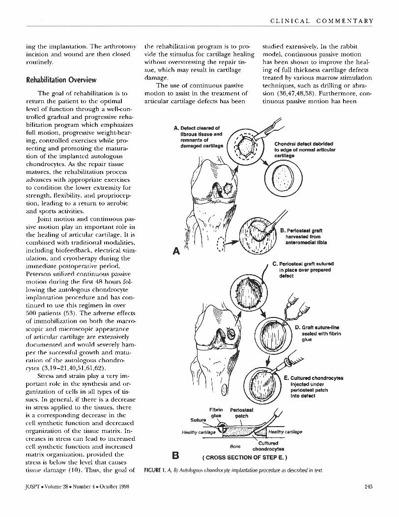
niscal transplant can be accom- plished at the time of autologous chondrocyte implantation. The chon- dral defect is first debrided circum- ferentially back to a healthy rim of surrounding normal cartilage (Figure 1A and B). Any fibrous tissue or re- maining damaged cartilage is re- moved from the base of the defect with a curette, with careful attention to avoid violating the subchondral bone in order to keep the bone from bleeding. Any punctate bleeding that might occur is controlled with com- pression sponges impregnated with epinephrine or thrombin. Once the defect has been debrided and conve- niently shaped, it is carefully mea- sured for sizing of the periosteal patch.
The periosteum is obtained through a small separate incision over the anteromedial tibia just distal to the insertion of the pes tendons. The periosteum from the proximal tibia and distal femur have been shown to be chondrogenic and p r e vide a paracrine effect to chondro- cyte growth, as well as providing a water-tight seal to contain the cells as they attach to the subchondral bone and populate the defect. The perios- teal patch is harvested slightly over- sized and then placed over the defect with the cambium layer down to the bone and secured in place to the sur- rounding normal cartilage with multi- ple interrupted 6-0 absorbable su- tures. The suture line around the periosteum is then further sealed with fibrin glue made from the pa- tient's own cryoprecipitate mixed with thrombin and calcium chloride. A small opening at the superior por- tion of the defect is left open to al- low for injection of the cells.
The patient's autologous chon- drocytes are then aspirated from the vial into a tuberculin syringe and in- jected with an 1 8 p u g e plastic angio- cath into the defect, ensuring com- plete fill of the defect with the cells. The small remaining opening is then closed with one or two final sutures and sealed with fibrin glue, complet-
Volume 28 Number 4 October 1998 JOSPT
ing the implantation. The arthrotomy incision and wound are then closed routinely.
Rehabilitation Overview
The goal of rehabilitation is to return the patient to the optimal level of function through a wellcon- trolled gradual and progressive reha- bilitation program which emphasizes full motion, progressive weight-bear- ing, controlled exercises while pro- tecting and promoting the matura- tion of the implanted autologous chondrocytes. As the repair tissue matures, the rehabilitation process advances with appropriate exercises to condition the lower extremity for strength, flexibility, and propriocep tion, leading to a return to aerobic and sports activities.
Joint motion and continuous pas- sive motion play an important role in the healing of articular cartilage. It is combined with traditional modalities, including biofeedback, electrical stim- ulation, and cryotherapy during the immediate postoperative period. Peterson utilized continuous passive motion during the first 48 hours fol- lowing the autologous chondrocyte implantation procedure and has con- tinued to use this regimen in over 500 patients (53). The adverse effects of immobilization on both the macro- scopic and microscopic appearance of articular cartilage are extensively documented and would severely ham- per the successful growth and matu- ration of the autologous chondro- cytes (3,19-21,40,51,61,62).
Stress and strain play a very im- portant role in the synthesis and or- ganization of cells in all types of tis- sues. In general, if there is a decrease in stress applied to the tissues, there is a corresponding decrease in the cell synthetic function and decreased organization of the tissue matrix. In- creases in stress can lead to increased cell synthetic function and increased matrix organization, provided the stress is below the level that causes tissue damage (10). Thus, the goal of
the rehabilitation program is to pro- vide the stimulus for cartilage healing without overstressing the repair tis- sue, which may result in cartilage damage.
The use of continuous passive motion to assist in the treatment of articular cartilage defects has been
studied extensively. In the rabbit model, continuous passive motion has been shown to improve the heal- ing of full thickness cartilage defects treated by various marrow stimulation techniques, such as drilling or abra- sion (36,47,48,58). Furthermore, con- tinuous passive motion has been
A. Defect cleared of fibrous tissue and remnants of damaged cadlage Chondml defect debrlded
to edge of normal articular % K o n d , defect debrlded
to edge of normal artlcular
P . . . ,' . ..
hswested from anteromedial tibia
C. Perlostd gmn sutured / in lace over ~rODat'ed
sealed with flbrln
. Cultured chondrocytea
periosteal patch
Bone Cultured
chondrocytea
B ( CROSS SECTION OF STEP E. )
FIGURE 1. A, B) Autologous chondrocyte implantation procedure as described in text.
JOSPT Volume 28 Number 4 October 1998

Phase I: Protective Phase (Weeks 0-6) Goals:
Protect healing tissue from load and shear forces Restoration of full passive knee extension Gradual improvement of knee flexion Regaining quadriceps control
Brace: Locked at Oquring weight-bearing activities Sleep in locked brace for 2-4 weeks
Weight bearing: Nonweight bearing for 2 weeks, progress to toe touch weight bearing (approximately 20-30 Ibs) for
4 weeks Toe touch weight bearing (approximately 114 body weight) at week 5
Range of motion (ROM): Immediate motion exercise Continuous passive motion after 4-12 hours (0-40°), 4-12 hours per day for 2-3 weeks Progress continuous passive motion ROM as tolerated 5-10" per day Patellar mobilization Passive knee flexion ROM 2-3 x daily Knee flexion ROM goal is 9 0 % ~ 2 weeks Knee flexion ROM goal is 105" by 4 weeks and 120%~ 6 weeks Stretch hamstrings, calf, and quadriceps
Stretching program: Ankle pump using rubber tubing Multiangle isometrics (co-contractions quadriceps and hamstrings) Active knee extension 90-40" (no resistance) Straight leg raises (four directions) Stationary bicycle when ROM allows Biofeedback and electrical muscle stimulation as needed Isometric leg press at week 4 (multiangle)
Functional activities: Gradual return to daily activities If symptoms occur, patient should reduce activities to reduce pain and inflammation Extended standing should be avoided
Phase II: Transition Phase (Weeks 6-12) Goals:
Gradually increase ROM Gradual improvement in quadriceps strength and endurance Gradual increase in functional activities
Criteria to Prorrress to Phase II 1. Full passive knee extension 2. Knee flexion to 11 5-1 20" 3. Minimal pain and swelling
Brace: Discontinue brace at 4-6 weeks
Weight bearing: Progress weight bearing as tolerated Half of body weight with crutches at 6 weeks Progress to full weight bearing 8-9 weeks Discontinue crutches at 8-9 weeks
Range of motion: Gradual increase in ROM Maintain full passive knee extension Progress knee flexion to 120-1 25" Continue patellar and soft tissue mobilization as needed Continue stretching program
Strengthening exercises: Initiate minisquats 0-45" Closed kinetic chain exercises (leg press) Toe-calf raises Open kinetic chain knee extension (passive resistance exercises) 1 Ib per week Stationary bicycle (gradually increase time) Continued
shown to increase cell proliferation and the total amount of type I1 colla- gen when used in rabbits treated with periosteal grafts placed over cartilage defects (43,47,49). Conversely, immo- bilization after the periosteal grafting has been shown to inhibit chondro- genesis (57). Continuous passive mo- tion also plays a role in ensuring the distribution of synovial fluid neces- sary for articular cartilage nutrition to the remainder of the joint (36,57). Another as yet not fully understood mechanism thought to be derived from continuous passive motion is that of modulation of the chondro- cyte precursors, prechondrocytes, to develop and initiate matrix produc- tion necessary for type I1 cartilage (7.36). There is no definitive answer as to the optimal time that continu- ous passive motion needs to be used. Continuous passive motion used 8 hours/day appears to be as effective as 24 hours/day and is certainly bet- ter tolerated by patients. Peterson (52,53) and Brittberg et a1 (7) adve cated using continuous passive mo- tion for the first 48 hours after sur- gery and have reported over 90% good and excellent results for iso- lated femoral condyle lesions. Per- haps the initial effect on the pre- chondrocytes shortly after implan- tation is the most important mecha- nism for autologous chondrocyte im- plantation. Further use of continuous passive motion may benefit the pa- tient in achieving motion, diminish- ing adhesions, and reducing pain.
Rehabilitation Program
The postoperative protocol is ac- companied by the use of immediate cryotherapy, elevation, compression, and continuous passive motion. The knee is immobilized in a standard postoperative dial-lock brace locked at 0" for functional activities. With nonweight-bearing activities, the brace may be unlocked for comfort,
TABLE 1. Rehabilitation program following autologous chondrocyte implantation procedure. and motion is encouraged. Active ankle pumping should also be en-
246 Volume 28 Number 4 October 1998 JOSPT

. .. .. C L I N I C A L C O M M E N T A R Y .- -- - - - - . ..
TABLE 1. Continued from previous page.
couraged in the first week after sur- gery to help reduce lower extremity swelling. Until biking or swimming can be safely performed for cardio- vascular training, an upper body er- gometer should be utilized.
JOSPT Volume 28 Number 4 October 1998
The rehabilitation program is outlined in Table 1. The rate of pro- gression may vary based on the pa- tient's defect size, location, and con- comitant procedures. Additionally, the patient's response to the surgery
will also determine the rate of p r e gression. The rehabilitation specialist must consider all of these factors when designing the postoperative program.
Through a well-controlled reha- bilitation program, patients under- going autologous chondrocyte im- plantation will optimally achieve the goals of the procedure to alleviate symptoms and return to a higher functional level of participation. The above outlined program allows for overcoming the effects of sur- gery, controlling swelling and in- flammation, advancing motion with gradual return of weight bearing, and progressive strengthening and agility training corresponding to the known maturation process of implanted autologous chondrocytes in an environment that encourages healing of articular cartilage with- out overloading the repair tissue. It is stressed that this program is a guide that needs to be modified for the specific needs of each patient's situation.
Clinical Results With Autologous Chondrocyte Implantation
Brittberg et al (7) reported on the first 23 patients with full-thickness chondral defects, 16 with femoral condyle and seven with patellar de- fects. The results in 14 of 16 femoral condyle patients (87%) were graded as excellent and good at the 39- month follow-up. Second-look arthro- scopy and biopsy demonstrated for- mation of new cartilage that was similar to surrounding normal carti- lage and had an abundance of type I1 collagen and metachromatic staining of the matrix. Results were less satis- factory in treatment of defects of the patella. Although five of seven pa- tients were improved, only two were good or excellent. The group with patellar lesions did not undergo re- alignment procedures (7).
Encouraged by these promising initial results, the treatment was ex-
C L I N I C A L C O M M E N T A R Y
Site of Defect Number of Follow-up (Years): Excellent and Patients Range = 2-10 years Good Results
Femoral condyle, isolated 57 4.0 90% Osteochondritis dissecans 32 3.9 84% Femoral condyle with any ACL 2 7 4.3 74%
reconstruction (Only primary ACL reconstruction) (85%) -
ACL = Anterior cruciate ligament.
TABLE 2. Autologous chondrocyte implantation: Overall clinical assessment of consecutive patients (53).
panded to include over 500 patients by 1997. Peterson reported on 219 consecutive patients with an average 4year follow-up (range = 2-10 years) treated with autologous chondrocyte implantation for isolated femoral condyle defects, femoral condyle os- teochondritis dessicans, and for fem- oral condyle defect. with anterior cruciate ligament reconstruction, which indicated consistently good to excellent results (53) (Table 2). The majority of these patient. had under-
gone prior attempts at repair of their chondral defects using either arthro- scopic debridement and/or marrow stimulation techniques with failed results. The clinical results were eval- uated by multiple methods of clinical testing to include the Modified Cin- cinnati, Tegner, Lysholm, VAS (visual analog scale), and the Brittberg scales, all of which indicated a statisti- cally significant improvement (37.45, 52,53). Of 19 biopsy specimens, 14 demonstrated hyaline-like tissue with
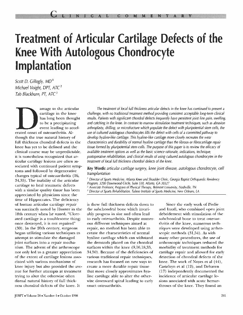
8.8 8.4
Very Good 8 6.7
2
1 r V
Pre op 1 yr 2 Yr N=25 N=25 N=8
Clinician Patient -pre-op vs 1 yr, 2 yr, p < .001 -pre-op vs 1 yr, 2 yr, p c.001 -1 yr vs 2 yr, p < .O1
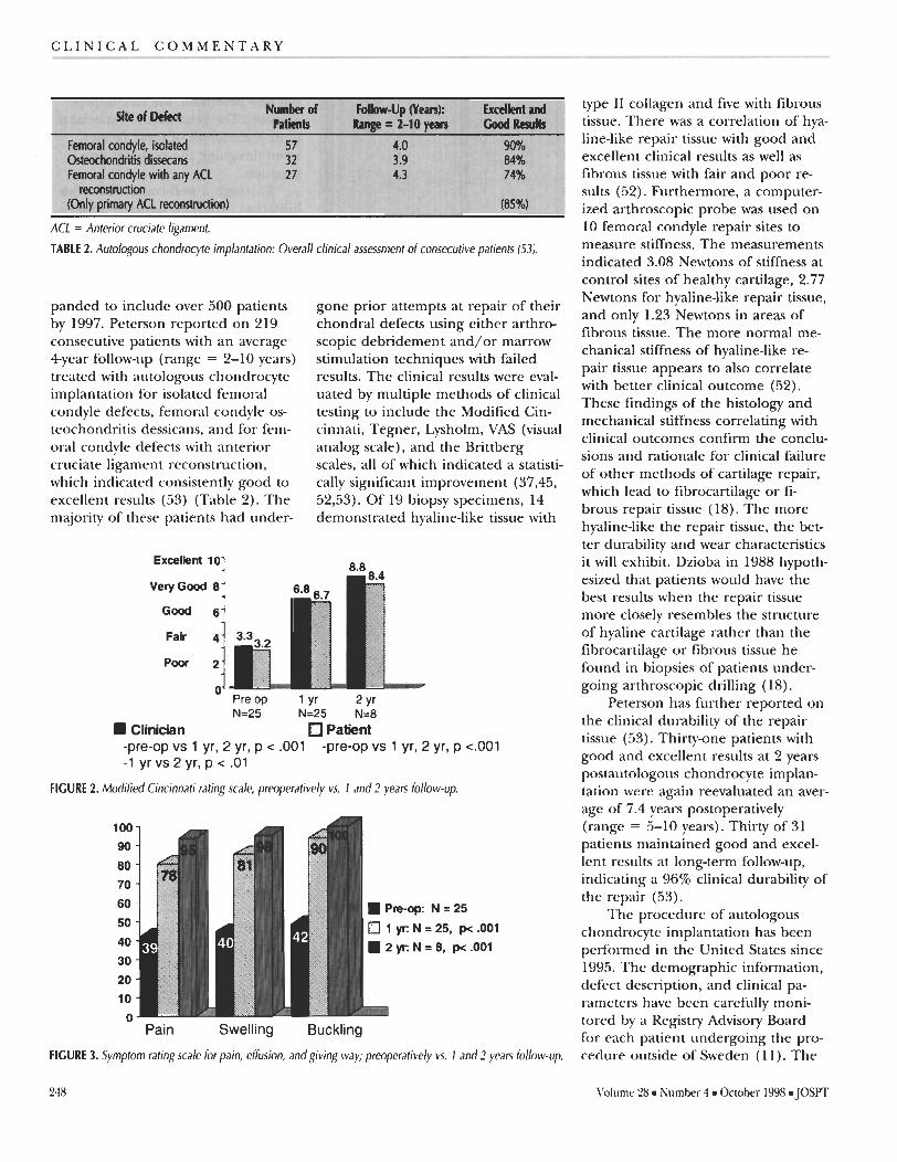
FIGURE 2. Modified Cincinnati rating scale, preoperatively vs. 1 and 2 years follow-up.
- Pain Swelling Buckling
FIGURE 3. Symptom rating scale for pain, effusion, andgiving way; preoperatively vs. 1 and 2 years follow-up.
type I1 collagen and five with fibrous tissue. There was a correlation of hya- line-like repair tissue with good and excellent clinical results as well as fibrous tissue with fair and poor re- sults (52). Furthermore, a computer- ized arthroscopic probe was used on 10 femoral condyle repair sites to measure stiffness. The measurements indicated 3.08 Newtons of stiffness at control sites of healthy cartilage, 2.77 Newtons for hyaline-like repair tissue, and only 1.23 Newtons in areas of fibrous tissue. The more normal me- chanical stiffness of hyaline-like re- pair tissue appears to also correlate with better clinical outcome (52). These findings of the histology and mechanical stiffness correlating with clinical outcomes confirm the conclu- sions and rationale for clinical failure of other methods of cartilage repair, which lead to fibrocartilage or fi- brous repair tissue (18). The more hyaline-like the repair tissue, the bet- ter durability and wear characteristics it will exhibit. Dzioba in 1988 hypoth- esized that patients would have the best results when the repair tissue more closely resembles the structure of hyaline cartilage rather than the fibrocartilage or fibrous tissue he found in biopsies of patients under- going arthroscopic drilling (1 8).
Peterson has further reported on the clinical durability of the repair tissue (53). Thirty-one patients with good and excellent results at 2 years postautologous chondrocyte implan- tation were again reevaluated an aver- age of 7.4 years postoperatively (range = 5-10 years). Thirty of 31 patients maintained good and excel- lent results at longterm follow-up, indicating a 96% clinical durability of the repair (53).
The procedure of autologous chondrocyte implantation has been performed in the United States since 1995. The demographic information, defect description, and clinical pa- rameters have been carefully moni- tored by a Registry Advisory Board for each patient undergoing the pro- cedure outside of Sweden ( 1 1 ). The
Volume 28 Number 4 October 1998 JOSPT
- - . +-. , - . C L I N I C A L C O M M E N T A R Y
-N=25
1 Yr: N = 25, p< -001 2 Y r N=8, pe.001
- Pre -op 1 yr 2 yr
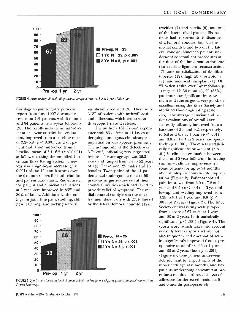
FIGURE 4. Knee Society clinical rating system, preoperatively vs. 1 and 2 years follow-up.
Cartilage Repair Registry periodic report from June 1997 documents results on 191 patients with 6 months and 84 patients with 1-year follow-up (9). The results indicate an improve- ment at 1 year on clinician evalua- tion, improved from a baseline mean of 3.2-6.9 (p < 0.001), and on pa- tient evaluation, improved from a baseline mean of 3.1-6.5 (P < 0.001) at follow-up, using the modified Cin- cinnati Knee Rating System. There was also a significant increase (p < 0.001 ) of the 12-month scores over the &month scores for both clinician and patient evaluations. Respectively, the patient and clinician evaluations at 1 year were improved in 85% and 80% of knees. Additionally, the rat- ings for joint line pain, swelling, stiff- ness, catching, and locking were all
significantly reduced (9). There were 3.3% of patients with arthrofibrosis and adhesions, which required ar- throscopic lysis and release.
The author's (SDG) own experi- ence with 53 defects in 41 knees un- dergoing autologous chondrocyte implantation also appears promising. The average size of the defects was 5.74 cm2, indicating very large-sized lesions. The average age was 36.2 years and ranged from 14 to 52 years of age. There were 25 males and 16 females. Twenty-nine of the 41 pa- tients had undergone a total of 50 previous surgeries directed at their chondral injuries which had failed to provide relief of symptoms. The me- dial femoral condyle was the most frequent defect site with 27, followed by the lateral femoral condyle (12),
90
80
70
60
50 0 1 Yt: N=25,p<.001
40 m2Yt: N=8,p<.001
30
20
10 n
FIGURE 5. Sports score based on level of fitness activity and frequency ofparticipation, preoperatively vs. 1 and 2 years follow-up.
trochlea (7) and patella (6), and one of the lateral tibial plateau. Six pa- tients had osteochondritis dissecans of a femoral condyle, four on the medial condyle and two on the lat- eral condyle. Nineteen patients un- derwent concomitant procedures at the time of the implantation for ante- rior cruciate ligament reconstruction (7), anteromedialization of the tibial tubercle (12), high tibial osteotomy (I ) , and meniscal transplant (1). Of 25 patients with over 1-year follow-up (range = 12-36 months), 22 (88%) patients show significant improve- ment and rate as good, very good, or excellent using the Knee Society and Modified Cincinnati rating scales (45). The average clinician and pa- tient evaluations of overall knee scores significantly improved from a baseline of 3.3 and 3.2, respectively, to 6.8 and 6.7 at 1 year (p < .001) and 8.8 and 8.4 at 2 years postopera- tively (p < .001). There was a statisti- cally significant improvement (p < .01) in clinician evaluation between the 1- and 2-year follow-up, indicating continued clinical improvement in some patients for up to 24 months after autologous chondrocyte implan- tation (Figure 2). Patient-reported pain improved from 3.9 to 7.8 at 1 year and 9.5 (p < .001) at 2-year fol- low-up, and swelling improved from 4.25 to 8.1 at 1 year and 9.8 (p < .001) at 2 years (Figure 3). The Knee Society clinical rating scale jumped from a score of 67 to 89 at 1 year and 98 at 2 years, both statistically significant (p < .001) (Figure 4). The sports score, which takes into account not only level of sports activity but also frequency and duration of activ- ity, significantly improved from a pre- operative score of 38-66 at 1 year and 88 at 2 years (both p < .001) (Figure 5). One patient underwent debridement for hypertrophy of the repair cartilage at 6 months, and two patients undergoing concomitant pr* cedures required arthroscopic Iysis of adhesions for decreased motion at 5 and 6 months postoperatively.
JOSPT Volume 28 Number 4 October 1998 249

C L I N I C A L C O M M E N T A R Y
Minas and Nehrer have recently reported on their early experience with autologous chondrocyte implan- tation in 50 patients (42). They have noted a gradual time-related improve- ment in patient-reported symptoms. By 12 months, there is a 90% im- provement, and, by 18 months, there is near complete resolution of the preoperative pain. This was felt to be reflective of the repair tissue matura- tion over time and corresponds to the findings at second-look arthros- copy from an indentable softer tissue at 3-6 months to a firm nonindent- able tissue at 18 months. These find- ings parallel the Swedish experience as well as the author's (7,52,53).
These early good to excellent clinical results noted in the Cartilage Repair Registry and by Minas and Nehrer and the author mirror the early results noted in the Swedish group of patients that have contin- ued to demonstrate good and excel- lent results up to and over 10 years, with an average of 4 years follow-up (1 1,42,52,53).
CONCLUSIONS
Full thickness chondral defects of the knee are a relatively frequent finding in patients less than 50 years of age and do not spontaneously heal with similar quality hyaline tissue. These defects are often associated with continued patient symptoms and can lead to progressive onset of early osteoarthritis, although the exact in- cidence is unknown. The treatment of these defects by the traditional methods of marrow stimulation, which provide access to the bone- blood supply by penetrating the sub- chondral bone to provide mesenchy- ma1 stem cells within the fibrin clot, have demonstrated mixed early re- sults and only fair to poor long-term results. The goal of achieving hyaline- like repair tissue, which would pro- vide better wear characteristics and durability, is seldom achieved with these treatment techniques. Autolo- gous chondrocyte implantation, on
the other hand, by providing chon- drocytes committed to forming type I1 cartilage, appears to provide hya- line-like repair tissue, with corre- sponding improvement in the histo- logic, biomechanical, and durability characteristics as reflected by better clinical outcomes in up to 90% of patients with femoral condyle defects with an average of 4year follow-up and up to 10 years postoperatively (53). The renewed interest by the medical field and the recent interest by the public underscore the scope of the clinical problems of cartilage defects in the knee in an active popu- lation and will undoubtedly lead to further refinements and developments, ultimately providing even better results for this difficult clinical problem. JOSm
REFERENCES Angermann P, Riegels-Nielsen P: Os- teochondritis dissecans of the femoral condyle treated with periosteal trans- plantation: A preliminary clinical study of 14 cases. Orthop Int 2:425-428, 1994 Angermann P: Periosteal transplanta- tion for the treatment of osteochondritis dissecans of the femoral condyles: Long-term results. Cartilage Repair Symposium, Bermuda, August, 1997 Baker WC, Thomas TG, Kirkaldy-Willis WH: Changes in the cartilage of the posterior intervertebral joints after an- terior fusion. J Bone Joint Surg 518: 736-746, 1969 Baumgartner MR, Cannon WD, Vittori JM, Schmidt ES, Maurer RC: Arthro- scopic debridement of the arthritic knee. Clin Orthop 253: 197-202, 1990 Bert JM, Maschka K: The arthroscopic treatment of unicompartmental gonar- throsis: A five-year follow-up study of abrasion arthroplasty plus arthroscopic debridement and arthroscopic debride- ment alone. Arthroscopy 5:25-32, 1989 Bobic V: Arthroscopic osteochondral autograft transplantation in anterior cruciate ligament reconstruction: A preliminary clinical study. Knee Surg Sports Traumatol Arthroscopy 3:262- 264, 1996 Brittberg M, Lindahl A, Nilsson A, Ohlsson C, Isaksson 0, Peterson L: Treatment of deep cartilage defects in the knee with autologous chondrocyte transplantation. N EngI J Med 33 1 :889- 895, 1994
8. Brittberg M, Nilsson A, Lindahl A, Ohlsson C, Peterson L: Rabbit articular cartilage defects treated with autolo- gous cultured chondrocytes. Clin Or- thop 326:270-283, 1996
9. Buckwalter ]A, Lohmander S: Opera- tive treatment of osteoarthritis. J Bone Joint Surg 76A: 1405- 14 18, 1994
10. Buckwalter ]A, Woo SL-Y: Effects of re- petitive loading and motion on the musculoskeletal tissues. In: Delee JC, Drez D (eds), Orthopaedic Sports Med- icine: Principles and Practice, pp 60- 72. Philadelphia: W.B. Saunders Com- pany, 1994
11. Cartilage Repair Registry (Volume 31, Cambridge, MA: Genzyme Tissue Re- pair, 1997
12. Casteleyn PP, Handelberg F, Opdecam P: Traumatic haemarthrosis of the knee. J Bone Joint Surg 70B:404-406, 1988
13. Convery R, Akeson WH, Keown GH: Fresh osteochondral allograhing of the femoral condyle. Clin Orthop 273: 139-145, 1991
14. Coventry MB, llstrup DM, Wallrichs SL: Proximal tibia1 osteotomy. I Bone Joint Surg 75A: 196-201, 1993
15. Curl WW, Krome J, Gordon S, Rushing J, Paterson Smith B, Poehling GG: Car- tilage injuries: A review of 3 1,5 16 knee arthroscopies. Arthroscopy l3:456- 460, 1997
16. Dandy Dl: Arthroscopic debridement of the knee for osteoarthritis (editorial). J Bone Joint Surg 73B:877-888, 1991
17. Dehaven KE: Diagnosis of acute knee injuries with hemarthrosis. Am J Sports Med 8:9-14, 1980
18. Dzioba R: The classification and treatment of acute articular cartilage le- sions. Arthroscopy 4:72-80, 1988
19. Ely L W, Mensor MC: Studies on the im- mobilization of the normal joints. Surg Gynecol Obstet 57:2 12-2 74, 1933
20. Enneking WF, Horowitz M: The intra- articular effects of immobilization on the human knee. J Bone Joint Surg 54A: 973-985, 1972
21. Evans EB, Eggers GW, Butler JK, Blumer J: Experimental immobiliza- tion and remobilization of rat knee joints. J Bone Joint Surg 42A:737- 758, 1960
22. Friedman MI, Berasi CC, Fox JM, DelPizzo W, Snyder SJ, Ferkel RD: Pre- liminary results with abrasion arthro- plasty in the osteoarthritic knee. Clin Orthop 182:2OO-205, 1 984
23. Garrett 1: Osteochondral allograft re- construction of the knee. lnstr Course Lect 42:355-358, 1993
24. Gibson JN, White MD, Chapman VM, Strachan RK: Arthroscopic lavage and
Volume 28 Number 4 October 1998 JOSPT

C L I N I C A L C O M M E N T A R Y
debridement for osteoarthritis of the knee. J Bone Joint Surg 7483534-537, 1992 Grande DA, Pitman MI, Peterson L, Menche D, Klein M: The repair of ex- perimentally produced defects in rabbit cartilage by autologous chondrocyte transplantation. J Orthop Res 7:208- 218, 7989 Hoikka VEJ, Jaroma HJ, Ritsila VA: Re- construction of the patellar articulation with periosteal grafts. Acta Orthop Scand 61:36-39, 1990 Homminga GN, Bulstra SK, Bouw- meester PSM, van der Linden A]: Peri- chondral grafting for cartilage lesions of the knee. J Bone Joint Surg 726: 1003- 1007, 1990 Homminga GN: Long-term follow-up of perichondral grafting for cartilage le- sions of the knee. Cartilage Repair Sym- posium, Bermuda, August, 1997 Hubbard MI: Articular debridement versus washout for degeneration of me- dial femoral condyle. J Bone Joint Surg 78B:2 17-2 19, 1996 Hunter W: On structure and diseases of articulating cartilage. Philos Trans R Soc Lond 24B:5 14 -52 1, 1743 Insall 1: Intra-articular surgery for de- generative osteoarthritis of the knee. ] Bone Joint Surg 49B:2 11-212, 1967 Jackson RW, Marans HJ, Silver RS: Ar- throscopic treatment of degenerative arthritis of the knee. J Bone Joint Surg 7083332, 1988 (abstract) Johnson L L: Arthroscopic abrasion ar- throplasty. In: McGinty JB (ed), Opera- tive Arthroscopy, pp 341-360. New York, NY: Raven Press, 1991 Johnson LL: Arthroscopic abrasion arthroplasty historical and pathologic perspective: Present status. Arthros- copy 2:54 - 69, 1986 Johnson-Nurse C, Dandy Dl: Frac- ture--Separation of articular cartilage in the adult knee. J Bone Joint Surg 67B:42-43, 1985 Kim HK, Moran ME, Salter RB: The po- tential for regeneration of articular cartilage in defects created by chon- dral shaving and subchondral abra- sion. An experimental investigation in rabbits. J Bone Joint Surg 73A: 1301 - 1315, 1997 Lysholm J, Gillquist J: Evaluation of knee ligament results with special em- phasis on the use of a scoring scale. Am J Sports Med 3: 150-1 53, 1982 Matsusue Y, Yamamuro T, Hama H: Arthroscopic multiple osteochondral transplantation to the chondral defect in the knee associated with anterior cruciate ligament disruption. Arthros- copy 9:318-321, 1993
39. Messner K, Maletius W: The long-term prognosis for severe damage to weight- bearing cartilage in the knee. Acta Or- thop Scand 67:65-68, 1996
40. Michelsson JE, Riska €6: The effect of temporary exercising of a joint during an immobilization period: An experi- mental study on rabbits. Clin Orthop 144:32 1-325, 1979
4 1. Minas T: Articular cartilage regenera- tion: Chondrocyte transplantation and other technologies. Presented at the an- nual meeting of the American Acad- emy of Orthopaedic Surgeons, San Francisco, CA, February, 1997
42. Minas T, Nehrer S: Current concepts in the treatment of articular cartilage de- fects. Orthopedics 20:525-538, 1997
43. Moran ME, Kim HK, Salter RB: Biolog- ical resurfacing of full thickness defects in patellar articular cartilage of the rab- bit. Investigation of autogenous perios- teal grafts subjected to continuous pas- sive motion. J Bone Joint Surg 746: 659-667, 1992
44. Noyes FR, Bassett R W, Grood ES, But- ler DL: Arthroscopy in acute hemar- throsis of the knee. Incidence of ante- rior cruciate tears and other injuries. J Bone Joint Surg 62A:687- 695, 1 980
45. Noyes FR, Barber SE, Mooar LA: A ra- tionale for assessing sports activity lev- els and limitation in knee disorders. Clin Orthop 246:238-249, 1989
46. O'Driscoll S: Periosteal transplantation: Articular cartilage regeneration: Chon- drocyte transplantation and other tech- nologies. Presented at the annual meet- ing of the American Academy of Orthopaedic Surgeons, San Francisco, CA, February, 1 997
47. O'Driscoll SW, Keeley FW, Salter RB: The chondrogenic potential of free au- togenous periosteal gra fts for biologic r e surfacing of major full-thickness defects in joint surfaces under the influence of continuous passive motion. An experi- mental investigation in the rabbit. J Bone Joint Surg 68A: 10 17- 1035, 1 986
48. O'Driscoll SW, Salter RB: The repair of major osteochondral defects in joint surfaces by neochondrogenesis with autogenous osteoperiosteal grafts stim- ulated by continuous passive motion. Clin Orthop 208: 13 1 - 140, 7 986
49. O'Driscoll S W, Salter RB: The induction of neochondrogenesis in free intra-artic- ular periosteal grafts under the influence on continuous passive motion. An exper- imental investigation in the rabbit. J Bone Joint Surg 66A: 1248- 1257, 1 984
50. Ogilvie-Harris Dl, Fihialos DP: Arthro- scopic management of the degenerative knee. Arthroscopy 7: 15 1- 157, 199 1
5 1. Palmoski M, Perricone E, Brandt K: De-
velopment and reversal of a proteogly- can aggregation defect in normal canine knee cartilage after immobilization. Ar- thritis Rheum 22:508-5 17, 1979
52. Peterson L: Autologous chondrocyte transplantation. Articular cartilage re- generation: Chondrocyte transplanta- tion and other technologies. Presented at the annual meeting of the American Academy of Orthopaedic Surgeons, San Francisco, CA, February, 1997
53. Peterson L: Autologous chondrocyte transplantation. Articular cartilage re- pair, regeneration and transplantation symposium. Presented at the 65th an- nual meeting of the American Acad- emy of Orthopaedic Surgeons, New Orleans, LA, March, 1998
54. Pirdie KH: A method of resurfacing os- teoarthritic knee joints. J Bone Joint Surg 418:618-619, 1959
55. Rand ]A: Role of arthroscopy in osteo- arthritis of the knee. Arthroscopy 7:358-363, 1997
56. Rodrigo JJ, Steadman JR, Silliman JF, Fulstone HA: Improvement of full- thickness chondral defect healing in the human knee after debridement and mi- crofracture using continuous passive mo- tion. Am J Knee Surg 7: 109- 1 16, 1994
57. Rubak JM, Poussa M, Ritsila V: Effects of joint motion on the repair of articular cartilage with free periosteal grafts. Acta Orthop Scand 53: 187- 19 1, 1982
58. Salter RB, Simmonds DR, Malcolm BW, Rumble El, MacMichael D, Clements ND: The biological effect of continuous passive motion on the healing of full thickness defects in articular cartilage. J Bone Joint Surg 62A: 1232- 125 1, 1 980
59. Sandelin J: Long-term results of recon- struction of the patellar articulation with periosteal grafts. Cartilage Repair Symposium, Bermuda, August, 1997
60. Timoney JM, Kneisl IS, Barrack RL, A/- exander AH: Arthroscopy in the osteo- arthritic knee. Long-term follow-up. Orthop Rev l9:371-373, 1990
6 1. Troyer H: The effect of short-term im- mobilization on the rabbit knee joint cartilage. A histochemical study. Clin Orthop 107:249-257, 1 975
62. Videman T: Connective tissue and im- mobilization. Key factors in musculo- skeletal degeneration. Clin Orthop 22 1 :26 -32, 1 987
63. Yamashita F, Sakakida K, Suzu F, Takai S: The transplantation of an autogenic osteochondral fragment for osteochon- dritis dissecans of the knee. Clin Or- thop 20 1 :43-50, 1985
64. Zukor D, Gross A: Osteochondral allo- graft reconstruction of the knee. Am J Knee Surg 2: 139-1 49, 1989
JOSPT Volume 28 Number 4 October 1W8