Embed Size (px)
Citation preview

ISBN 978-82-326-1902-3 (printed ver.)ISBN 978-82-326-1903-0 (electronic ver.)
ISSN 1503-8181
Doctoral theses at NTNU, 2016:282
Kari Schirmer-Mikalsen
Traumatic brain injury: control of physiological variables, organ failure and complications in the intensive care unit
Doc
tora
l the
sis
Doct oral theses at N
TNU
, 2016:282K
ari Schirmer-M
ikalsen
NTN
UN
orw
egia
n U
nive
rsity
of
Scie
nce
and
Tech
nolo
gyTh
esis
for
the
Deg
ree
ofP
hilo
soph
iae
Doc
tor
Facu
lty
of M
edic
ine
Dep
artm
ent o
f Cir
cula
tion
and
Med
ical
Imag
ing

Kari Schirmer-Mikalsen
Traumatic brain injury: control of physiological variables, organ failure and complications in the intensive care unit
Thesis for the Degree of Philosophiae Doctor
Trondheim, October 2016
Norwegian University of Science and TechnologyFaculty of MedicineDepartment of Circulation and Medical Imaging

NTNUNorwegian University of Science and Technology
Thesis for the Degree of Philosophiae Doctor
Faculty of MedicineDepartment of Circulation and Medical Imaging
© Kari Schirmer-Mikalsen
ISBN 978-82-326-1902-3 (printed ver.)ISBN 978-82-326-1903-0 (electronic ver.)ISSN 1503-8181
Doctoral theses at NTNU, 2016:282
Printed by NTNU Grafisk senter


Ovennevnte avhandling er funnet verdig til å forsvares offentlig
for graden PhD i klinisk medisin.
Disputas finner sted i Auditoriet Blåhø, Øya Helsehus, St.Olavs Hospital/NTNU
Fredag 14.10.2016, kl.12.15.


°

μ
μ


Paper I


Acta Anaesthesiol Scand 2007; 51: 1194–1201Printed in Singapore. All rights reserved
# 2007 The AuthorsJournal compilation # 2007 Acta Anaesthesiol Scand
ACTA ANAESTHESIOLOGICA SCANDINAVICA
doi: 10.1111/j.1399-6576.2007.01372.x
Severe head injury: control of physiological variables,
organ failure and complications in the intensive care unit
K. SCHIRMER-MIKALSEN1, A. VIK
2,4, S. E. GISVOLD1,5, T. SKANDSEN
3,4, H. HYNNE1 and P. KLEPSTAD
1,5
1Department of Anaesthesia and Acute Medicine, 2Department of Neurosurgery, 3Department of Physical Medicine and Rehabilitation, St. OlavUniversity Hospital, 4Department of Neuroscience, 5Department of Circulation and Medical Imaging, Medical Faculty, Norwegian University ofScience and Technology, Trondheim, Norway
Background: In patients with severe head injury, control ofphysiological variables is important to avoid intracranial hyper-tension and secondary injury to the brain. The aims of thisretrospective study were to evaluate deviations of physiologicalvariables and the incidence of extracranial complications inpatients with severe head injury. We also studied if these devia-tions could be related to outcome.Patients and methods: One hundred and thirty-three patientswere included during a 5-year period (1998–2002). Deviationsfrom treatment goals for the following physiological variableswere studied: blood pressure, haemoglobin, blood sugar, serumsodium, serum albumin and temperature. Extra cerebral organcomplications were also recorded as well as outcome at 6 months.Results: The median age was 32 years (range; 1–88 years).Median Glasgow Coma Scale (GCS) before intubation was 6(range; 3–14). The frequencies of severe deviations from thedesired values of the physiological variables for at least onetreatment day were: hypotensive episodes (systolic BP < 90mmHg) – 20%, anaemia (hgb < 8 g/dL) – 22%, blood glucose>10 mmol/l – 26%, serum sodium concentration <130 mmol/l – 10%, serum albumin <25 g/l�1 – 31% and hyperthermia
>39 8C – 24%. Pneumonia was diagnosed in 71% and Acute LungInjury (ALI)/Adult Respiratory Distress Syndrome (ARDS) in26% of the patients. Other complications such as severe sepsis(6%), renal failure (1.5%), a coagulation disorder (6%) and liverfailure (one patient) were infrequent. Age, GCS, hypotensionduring the first day of treatment, elevated blood sugar and lowalbumin predicted an unfavourable outcome.Conclusions: Deviations of key physiological variables andpulmonary complications were frequent in patients sufferingfrom severe head injury. During intensive care treatment, hypo-tension, elevated blood sugar and hypoalbuminemia are possibleindependent predictors of an unfavourable outcome.
Accepted for publication 9 May 2007
Key words: Severe head injury; secondary brain injury;control of physiological variables; intensive care; outcome;pneumonia.
# 2007 The AuthorsJournal compilation # 2007 Acta Anaesthesiol Scand
GUIDELINES recommend stabilization of patientswith severe head injury within specified treat-
ment goals related to oxygenation, ventilation, bloodpressure, temperature, blood sugar, serum sodiumand albumin to avoid secondary brain damage anddevelopment of intracranial hypertension (1–6).However, strict compliance to the treatment goalsmay be difficult to achieve in day-to-day practice inthe intensive care unit (ICU). Potential causes forphysiological deviations are other intercurrent con-ditions such as respiratory failure, sepsis or heartfailure, lack of education, lack of vigilance, insuffi-cient staffing or other organizational issues. It is notknown to what extent physiological variables differfrom defined treatment goals during day-to-daypractice in the ICU. Furthermore, although manystudies evaluate the effect of elevated intracranial
pressure or reduced cerebral perfusion pressure onoutcome (2, 7–9), the consequences of deviations fromother physiological variables have not been studiedin detail.Thus, the aims of this study were to evaluate the
frequency of deviations in physiological variablesand risk of extracranial complications in patientswith severe head injuries during treatment in theICU and high-dependency unit (HDU). We alsowanted to explore the relationship of such deviationsand complications to outcome.
Material and methods
The study was done in accordance with the principlesof the Helsinki declaration. The Regional Committeefor Medical Research Ethics, Health Region IV,
1194

Norway, approved the study. All 146 patients withsevere head injury admitted to our hospital duringa 5-year period (1998–2002) were retrospectivelyreviewed. The hospital is the sole neurosurgicalfacility in a health care region of about 660,000inhabitants resulting in a total of 25–30 patientspresenting with severe head trauma each year. Pa-tients were included if the GlasgowComa Scale (GCS)was �8 before or after hospital admission. Also,patients with a GCS >8 before intubation wereincluded if they deteriorated. All had intracranialtraumatic pathology on computer tomography (CT).Patients for whom treatment was withheld at the timeof admission as a result of severe head injury with nohope for survival (n ¼ 9), and patients who diedfrom causes not related to the head injury within thefirst 24 h (n ¼ 2) were excluded. Furthermore,another two patients were excluded from the analysesas a result of a lack of sufficient medical records or nostay in the ICU/HDU. Thus, 133 patients fulfilled theinclusion criteria.
Treatment goals (Table 1)During the study period, the hospital followed theEuropean Brain Injury Consortium (EBIC) guidelinesfor treatment of severe head injury (1) (Table 1).However, at the time the hospital did not havea specific step-by-step guideline describing how toreach these treatment goals.The patients were intubated and received controlled
ventilation. Sedation was achieved by a combinationof continuously infused intravenous (i.v.) midazolamand morphine. Propofol was used for some patientsduring weaning from the ventilator. For hypotensionor failure to reach the target cerebral perfusion pres-sure (CPP), circulatory support was provided withcolloids and crystalloids with additional vasoactiveagents (dopamine or norepinephrine) as required. Thepatients were not routinely given a continuous infu-
sion of insulin for regulation of blood glucose. Thedesired blood glucose level was < 8 mmol/l. Theintracranial pressure (ICP) was measured either usingan intraparenchymal or an intraventricular pressuredevice. The target CPP was usually set at 60–70 mmHg. Increases in ICP (ICP above 20 mmHg)were treated with sedation and analgesia, moderatehyperventilation (PaCO2 4–4.5), osmotic therapy(mannitol and/or hypertonic saline) and cerebrospinalfluid (CSF) drainage. Patients with increased ICP notresponsive to standard treatment were administeredmore extensive hyperventilation, continuous infusionof thiopental, and in some cases corticosteroids ormildhypothermia. Corticosteroids are not recommended inEBIC guidelines, and are no longer a treatment forintracranial hypertension at our department. Musclerelaxants were only used occasionally. Decompressivecraniectomies were not performed in the treatment ofhead injured patients during this time period,although the bone flap was not replaced during theneurosurgical operation in some patients.
Deviation from treatment goals:definitions/criterionsThe frequencies of physiological observations notwithin the recommended limits were identified byreviewing all charts completed during the patientsstay in the ICU/HDU. The observed values andnumber of daily observations corresponding toa moderate or severe deviation from the treatmentgoals were defined as shown in Table 2. Patients wereidentified as cases if having observations defined asoutside the target values for at least one day. Somepatients had observations corresponding to moder-ate or severe deviations during separate days andwere therefore registered as both moderate andsevere deviation cases.Blood gas analyses, including the measurement of
haemoglobin, blood glucose and electrolyte serum
Table 1
Treatment goals in the intensive care unit (ICU)/high-dependency unit (HDU).
Parameters According to EBIC
(1) Ventilation Saturation of oxygen (SaO2) >95%Arterial partial pressure of oxygen (PaO2) �13 kPa (100 mmHg)Arterial partial pressure of carbon dioxide (PaCO2) 4–4.5 kPa (30–35 mmHg)
(2) Blood pressure Mean arterial blood pressure (MAP) �90 mmHg (adults)(3) Intracranial measures Intracranial pressure (ICP) �20–25 mmHg (adults)
Cerebral perfusion pressure (CPP ¼ MAP-ICP) >60–70 mmHg(4) Blood glucose Avoid hyperglycaemia No exact blood sugar limit(5) Body temperature Avoid hyperthermia No exact limit mentioned(6) Serum Sodium Avoid hyponatremia No exact limit mentioned
EBIC, European Brain Injury Consortium.
1195
Severe head injury

concentration, were usually taken 5–10 times daily inthe ICU, although less frequently in the HDU as thepatients were usually more stable during this phase.The patients’ blood pressure was observed continu-ously and registered hourly in the ICU and at thetime of hypo- or hypertensive episodes, althoughagain less often in the HDU. ICP was registeredhourly in both the ICU and the HDU and at the timeof ICP increases.
Organ failure during ICU/HDU treatmentThe incidence of organ failure and major intercurrentconditions were identified by reviewing the patients’records, charts and radiologist consultants’ evalua-tion of X-rays (KSM). Respiratory and circulatorycomplications such as ventilator-associated pneumo-nia (VAP), acute lung injury (ALI), adult respiratorydistress syndrome (ARDS) and severe sepsis wereidentified. The definitions for each organ failure orintercurrent conditions are given in Table 3. Usingthe sequential organ failure assessment score, renalfailure, liver failure and a coagulation disorder weredefined at values corresponding to three or more (10).
OutcomeA specialist in physical medicine and rehabilitationnot involved in the acute care of the patients collectedthe 6-months Glasgow Outcome Scale (GOS) retro-spectively (TS). GOS 5 and 4 represented goodrecovery and moderate disability, while 3, 2 and 1represented severe disability, a persistent vegetativestate and death. The patients’ outcomes were dichot-omized into favourable (GOS 5 and 4) and unfav-ourable (GOS 3, 2 and 1).
StatisticsThe results are given as numbers, percentages,median and ranges as appropriate. Pearson’s chi-square test was used when comparing proportions.In the explorative analyses for the relationship be-tween observations outside the standard treatmentgoals and GOS outcome, multiple logistic regressionanalyses were used. The independent variables wereage, GCS, hypotension (systolic BP<90 mmHg), hypo-ventilation (pCO2 >6 kPa), hypoxia (PaO2 <8 kPa),anaemia (Hgb <8 g/dl), hyperglycaemia (blood glu-cose>10 mmol/l), hyponatremia (serum sodium<130
Table 2
Deviation from treatment goals.
Moderate/severe deviations
Respiratory PaCO2 5–6/ >6 kPa, Moderate/severe deviations if registered at �3PaO2 8–11/ <8 kPa observations/day
Circulatory Adult systolic blood pressure <90 mmHg Registered as a deviation if measured at �3 times/dayHaemoglobin (Hgb) 8–10/ <8 g/dl Moderate/severe deviations if registered once during the same dayBlood glucose 8–10/ >10 mmol/l Moderate/severe deviations if registered �3 times/daySerum sodium 130–135/ <130 mmol/l Moderate/severe deviations if registered �3 times/daySerum albumin <25 g/l Measured any time during the ICU/HDU stayTemperature 38–39/ >39 8C Moderate/severe deviations if registered �3 times/day
ICU, intensive care unit; HDU, high-dependency unit.
Table 3
Definition of extra cerebral organ complications.
Complication Definition
Ventilator-associated pneumonia (VAP) Artificial ventilation >48 h, infiltrates on chest X-ray, leucocytes >10 or <3, temp. >38.5 8Cor <35 8C and purulent tracheal secretions
Pneumonia (other than VAP) As above but start of symptoms before 48 h of respiratory treatmentAcute lung injury (ALI) Acute respiratory failure, PaO2/FiO2 ratio <39.9 kPa, bilateral infiltrates on chest X-ray and
no signs of heart failureAdult respiratory distress syndrome(ARDS)
Acute respiratory failure, PaO2/FiO2 ratio <26.6 kPa, bilateral infiltrates on chest X-ray andno signs of heart failure
Severe sepsis Sepsis with organ dysfunction, hypo perfusion or hypotension in spite of adequatefluid therapy
Renal failure Serum creatinine >299 mmol/l or diuresis less than 500 ml/24 h (adults)Bleeding disorder Platelets <50 � 109/lLiver failure Serum bilirubin >101 mmol/l
1196
K. Schirmer-Mikalsen et al.

mmol/l), hypoalbuminia (serum albumin <25 g/l)and hyperthermia (central body temperature >39 8C).For all the physiological variables, patients weredefined as cases (one or more days with deviations)vs. non-cases. In addition, a similar analysis was per-formed using only deviation in physiological varia-bles observed at the first day of hospitalization. Inthe multiple regression analyses, a backward step-wise method was used applying a significance levelof 0.05 for significance and a probability for removalof 0.10.
Results
DemographicsThe 133 patients which were included consisted of108 men and 25 women with a median age of32 years (range; 1–88 years). The majority of headinjuries were caused by either traffic (50%) or a fall-related accident (35%). Median GCS before intuba-tion was 6 (range; 3–14, missing: 13). The 13 patientswith missing GCS were all reported as comatose, butthe exact GCS was not available. Median SimplifiedAcute Physiology Score II in patients >18 years withsevere head injury who were treated at the ICU(n ¼ 112) was 43 (range; 14–80, missing 2).About half of the patients (48%) had other serious
injuries. Trauma to the chest was most common(29%), followed by facial trauma (21%) and frac-tures/lacerations to the extremities (20%). One hun-dred and thirty-one of the 133 patients receivedventilatory support in the ICU/HDU. The mostfrequent cerebral CT diagnoses were contusions
(72%), a subdural haematoma (55%) and a subarach-noid haemorrhage (56%). Most patients receivedICP monitoring (84%) and 48% were operated onfor an intracranial mass lesion. Nineteen patientsdied of intracranial hypertension. The median totallength of stay (LOS), including both the ICU and theHDU stay, was 12 days (range; 0.5–49 days). Themedian LOS in the ICU and the HDU was 7 and6 days, respectively.
Key physiological variables, organ failuresand complicationsHypotensive episodes (systolic BP <90 mmHg) wereregistered in 20% of the adult patients (Table 4). Forty-eight per cent of the patients who died of their headinjury during the first 6 months after the trauma hadhypotensive episodes during their ICU/HDU stay,compared with 18% in the survival group (P < 0.001).Severe anaemia occurred in 22%. Twenty-six per centhad elevated blood glucose (>10 mmol/l), and 46% ofthe patients with blood glucose more than 10 mmol/ldied within 6 months after the trauma. Ten per centof the patients had serum sodium <130 mmol/l.Serum albumin was <25 g/l in 31% of the patients.Despite that one of the treatment goals was to avoidelevated body temperature, 24% had episodes ofa body temperature of more than 39 8C.Pneumonia was a common complication (Table 5).
Most frequent was VAP diagnosed in 49% of thepatients. Of the 47 patients who got continuouslyinfusion with barbiturates during some part of theirintensive care, as many as 81% got pneumoniaduring their ICU/HDU stay compared with 67%
Table 4
Recorded deviations from treatment goals.
Number of patients(n ¼ 133)
Days median(range)
Patient datamissing*
Respiration PaCO2 5–6 kPa 94 (71%) 3 (1–22) 5PaCO2 >6 kPa 33 (25%) 2 (1–16) 4PaO2 8–11 kPa 91 (68%) 4 (1–20) 5PaO2 <8 kPa 13 (10%) 1 (1–3) 3
Circulation Systolic BP <90 mmHg (adults) 26 (20%) 1 (1–8) 3Haemoglobin Hgb 8–10 g/dl 107 (80%) 6 (1–38) 3
Hgb <8 g/dl 29 (22%) 1 (1–6) 2Blood glucose Blood glucose 8–10 mmol/l 69 (52%) 2 (1–9) 7
Blood glucose >10 mmol/l 35 (26%) 1 (1–6) 6Serum sodium Serum sodium 130–135 mmol/l 62 (47%) 3 (1–12) 3
Serum sodium <130 mmol/l 14 (10%) 2 (1–3) 1Serum albumin Serum albumin <25 g/l 41 (31%) 2 (1–24) 21Temperature Temperature 38–398C 96 (72%) 4 (1–28) 6
Temperature >398C 32 (24%) 2 (1–18) 1
Some patients had both severe and moderate deviations and are therefore counted twice.*Missing data as a result of insufficient/lacking chart for one or more days.
1197
Severe head injury

of the rest of the patients. The difference was,however, not significant (P ¼ 0.181). ALI or ARDSoccurred in 26% of the patients. One-third of thehypoventilated patients had episodes of PaCO2
values above 6 kPa (Table 4). Other extracranialcomplications such as severe sepsis septic shock,renal failure, coagulation disorder and liver failurewere infrequent (Table 5).
OutcomeThe 6-months GOS showed a favourable outcome(GOS 4 and 5) in 37% (n ¼ 49) of the patients, while59% (n ¼ 78) had an unfavourable outcome (GOS1–3) (missing GOS: 6 patients). For GOS 6 monthsafter the injury, age (P ¼ 0.013) and blood sugardeviations (P ¼ 0.044) during treatment were pre-dictors of an unfavourable outcome. When analysingthe relationship between the first day of ICU/HDUtreatment and GOS at 6 months, GCS (P ¼ 0.020)and hypotension (P ¼ 0.022) predicted an unfavour-able outcome. The in-hospital mortality was 18% (24patients) while 6-months mortality was 25% (33patients). Predictors ofmortality were age (P ¼ 0.012),GCS (P ¼ 0.015), hypotension (P ¼ 0.007), and hypo-albuminemia (P ¼ 0.036). The corresponding predic-tors when analysing for observations at day 1 duringthe ICU/HDU stay were age (P ¼ 0.031), high bloodsugar (P ¼ 0.002) and hypoalbuminemia (P ¼ 0.046).
Discussion
The key finding in our investigation of patients withsevere head injury was that important physiologicalparameters frequently deviated from the treatmentgoal during intensive care. Furthermore, in patientswith severe head injury, pulmonary complicationsoccurred frequently, whereas other extracranial organfailures were infrequent. These factors seemed to
contribute significantly to the final outcome of thepatients.The mechanisms causing extracranial complica-
tions are complex involving infections, traumaticinjuries in other organs, inflammatory mediators,hormonal changes and disturbed vasomotor control(11, 12). The high incidence of pneumonia includingVAP found in our study (Table 5) corresponds withthe high incidence found in a prospective investiga-tion from Bronchard et al. (13) and data from theTraumatic Coma Data Bank (12), but it is higher thanobserved by others (4, 14). In patients included in theTraumatic Coma Data Bank, pulmonary infectionswere a significant independent predictor of an un-favourable outcome. In contrast, Polderman et al. (4)only registered pulmonary complications (infections,pneumothorax and bleeding) in 11% of the patients.While the findings of Stover and Stocker (15)
conclude that a barbiturate coma in patients withtraumatic brain injury may cause reversible leucope-nia and an increased infection rate, we could not finda significant difference in the incidence of pneumoniain this group of patients.It is well known that there is a correlation between
respiratory dysfunction andmortality (11, 16). Earlierreports (2) have described an increase in the risk ofARDS when the lower CPP threshold was set at70 mmHg, compared with a threshold of 50 mmHg.Which value is an adequate cerebral perfusion pres-sure is controversial (8, 9). While the EBIC guidelineshave been recommending a CPP threshold of 60–70 mmHg as was used in our study, the Lundconcept (17–22) argues for a lower CPP limit (downto 50 mmHg in adults).In the analyses of outcome, hypotension during the
first day of stay in the ICU independently predictedan unfavourable outcome after 6 months in ourstudy. Earlier studies have shown that even brief
Table 5
Extra cerebral organ complications during the intensive care unit (ICU)/high-dependency unit (HDU) stay.
Complications Number of patients(n ¼ 133)
Patient datamissing
Ventilator-associated pneumonia (VAP) 65 (49%) 1Pneumonia (other than VAP) 33 (25%) 1Acute lung injury (ALI) 34 (26%) 0Adult respiratory distress syndrome (ARDS) 30 (23%) 0Serious sepsis 8 (6%) 1Septic shock 0 0Renal failure 2 (1.5%) 0Bleeding disorder 8 (6%) 0Liver failure 1 (0.8%) 0
1198
K. Schirmer-Mikalsen et al.

periods of mild hypotension adversely affects out-come in patients with severe traumatic brain injury(3, 12, 14, 23, 24). However, it has been argued thathypotension is most critical during the initial phaseafter the injury, i.e. before arriving at the ICU (18), butthe negative consequences of hypotension in thesepatients later during the ICU stay are well docu-mented (3, 10, 12).Many of our patients had a Hgb level <10 g/dl,
but we did not observe any relationship betweenanaemia and outcome. It may be speculated thatlower viscosity as a result of anaemia improvesmicrocirculation in parts of the brain that are at risk(25, 26). Proponents of the Lund therapy (17), how-ever, advocate blood transfusions if needed, to main-tain a Hgb level above 11 g/dl in order to improveoxygen transport to the injured brain (27). Thus, theremay be both detrimental and beneficial effects fromanaemia.Almost half of the patients with blood glucose of
more than 10 mmol/l died within 6 months aftersevere head injury in our study, and in the multipleregression analyses elevated blood glucose was anindependent predictor of outcome. This is in agree-ment with other studies (5, 24, 28, 29).In a systematic review, Haynes et al. showed that
albumin treatment gave reduced mortality, disabilityand neurological deficits in patients with brain injury(30). While the Lund concept includes albumin infu-sion in their guideline for maintaining colloid osmoticpressure, it is not a routine treatment in most ICUsincluding our ICU/HDU. Experts who do not advo-cate albumin claim that it is the total osmolality of theblood that is important for the fluid shifts over theblood–brain barrier. Because albumin contributes littleto the total osmolality compared with serum sodium,the effects from administrating albumin infusions arebelieved to be limited (31, 32). However, for thepatients included in the present study, low albuminserum concentrations were associated with an in-creased risk of death. This is in agreement with morerecent research showing that albumin reduces cerebraloedema and has a neuroprotective effect (6, 33–35).The low frequencies of severe sepsis, renal failure
and liver failure found in our patients are alsodescribed in previous studies (2, 4, 12, 14, 36).However, several studies (12, 37, 38) found a higherincidence of coagulopathy than observed in thisstudy. Our data suggest that in patients with severehead injury, organ failures other than those of respi-ratory and circulatory origin seldom contribute to anunfavourable outcome. However, our findingmay beas a result of a low number of patients that fail to
elucidate the consequences of infrequent complica-tions. This speculation is supported by the fact thatthe Traumatic Coma Data Bank (12) has shown thatextracranial complications such as coagulopathy andsepsis are also significant predictors of a less favour-able outcome.In the present study, the overall incidence of extrac-
erebral organ failure agrees with a recent paper byZygun et al. (36). Zygun et al. also observed that thedevelopment of non-neurological organ dysfunctionwas associated with worse outcome independent ofage and GCS. These observations call for close atten-tion to details in the intensive care treatment ofpatients with severe head injury. Protocol-driven treat-ment offers a significant benefit, both generally and inhead-injured patients (39, 40). In our study and thestudy by Zygun et al., it is not investigated if changesin key physiological parameters were unavoidableresults from other injuries, organ failures or alteredcentral nervous autonomic regulation or as a result ofa lack of attention to detail during treatment.We recognize several limitations in this study. First,
because of the retrospective design, some data aremissing. Second, beyond the separate assessment ofthe first day of hospitalization, we have not differ-entiated between the acute phase in the ICU and themore subacute period in the HDU. It is reasonable tobelieve that deviations are more crucial if presentduring the acute phase of head injury treatment.
Conclusion
This study demonstrates that undesired and poten-tially harmful changes in physiological variables arefrequent during ICU and HDU treatment of patientssuffering from severe head injury. Pulmonary com-plications are also frequent, with pneumonia beingthe most common problem. During intensive caretreatment, age, GCS, hypotension, elevated bloodsugar and hypoalbuminemia are possible indepen-dent predictors of an unfavourable outcome.
References
1. Maas AIR, Dearden M, Teasdale GM et al. EBIC-Guidelinesfor Management of Severe Head Injury in Adults. ActaNeurochir (Wien) 1997; 139: 286–94.
2. Robertson CS, Valadka AB, Hannay HJ et al. Prevention ofsecondary ischemic insults after severe head injury. Crit CareMed 1999; 27: 2086–95.
3. Chesnut RM, Marshall SB, Piek J, Blunt BA, Klauber MR,Marshall LF. Early and late systemic hypotension as a frequentand fundamental source of cerebral ischemia following severe
1199
Severe head injury

brain injury in the Traumatic Coma Data Bank. Acta NeurochirSuppl (Wien) 1993; 59: 121–5.
4. Polderman KH, Tjong Tjin JR, Peerdeman SM, VandertorpWP, Girbes ARJ. Effects of therapeutic hypothermia onintracranial pressure and outcome in patients with severehead injury. Intensive Care Med 2002; 28: 1563–73.
5. Rovlias A, Kotsou S. The influence of hyperglycemia onneurological outcome in patients with severe head injury.Neurosurgery 2000; 46: 335–51.
6. Belayev L, Liu Y, Zhao W, Busto R, Ginsberg MD. Humanalbumin therapy of acute ischemic stroke: marked neuro-protective efficacy at moderate doses and with a broadtherapeutic window. Stroke 2001; 32: 553–60.
7. Balestreri M, Czosnyka M, Hutchinson P et al. Impact ofintracranial pressure and cerebral perfusion pressure onsevere disability and mortality after head injury. NeurocritCare 2006; 4: 8–13.
8. Elf K, Nilsson P, Ronne-Engstrom E, Howells T, Enblad P.Cerebral perfusion pressure between 50 and 60 mmHg maybe beneficial in head-injured patients: a computerized sec-ondary insult monitoring study. Neurosurgery 2005; 56:962–71.
9. Portella G, Cormio M, Citerio G et al. Continuous cerebralcompliance monitoring in severe head injury: its relationshipwith intracranial pressure and cerebral perfusion pressure.Acta Neurochir (Wien) 2005; 147: 707–13.
10. Janssens U, Graf C, Graf J et al. Evaluation of the SOFA score:a single-centre experience of a medical intensive care unit in303 consecutive patients with predominantly cardiovasculardisorders. Intensive Care Med 2000; 26: 1037–45.
11. Acosta JA, Yang JC, Winchell RJ, Simons RK, Fortlage DA,Hollingsworth-Fridlund P, Hoyt DB. Lethal injuries and timeto death in a level I trauma center. J Am Coll Surg 1998; 186:528–33.
12. Piek J, Chesnut RM, Marshall LF et al. Extracranial compli-cations of severe head injury. J Neurosurg 1992; 77: 901–7.
13. Bronchard R, Albaladejo P, Brezac G et al. Early onsetpneumonia: risk factors and consequences in head traumapatients. Anesthesiology 2004; 100: 234–9.
14. Sarrafzadeh AS, Peltonen EE, Kaisers U, Kuchler I, LankschWR, Unterberg AW. Secondary insults in severe head injury –Do multiply injured patients do worse? Crit Care Med 2001;29: 1116–23.
15. Stover JF, Stocker R. Barbiturate coma may promote revers-ible bone marrow suppression in patients with severe isolatedtraumatic brain injury. Eur J Pharmacol 1998; 54: 529–34.
16. Marshall JC, Cook DJ, Christou NV, Bernard GR, Sprung CL,Sibbald WJ. Multiple Organ Dysfunction Score: a reliabledescriptor of a complex clinical outcome. Crit Care Med 1995;23: 1638–52.
17. Asgeirsson B, Grande PO, Nordstrom CH. A new therapy ofpost-trauma brain oedema based on haemodynamic princi-ples for brain Volume regulation. Intensive Care Med 1994; 20:260–7.
18. Eker C, Asgeirsson B, Grande PO, SchalenW, Nordstrom CH.Improved outcome after severe head injury with a newtherapy based on principles for brain Volume regulationand preserved microcirculation. Crit Care Med 1998; 26:1881–6.
19. Nordstrom CH, Reinstrup P, XuW, Gardenfors A, UngerstedtU. Assessment of the lower limit for cerebral perfusionpressure in severe head injuries by bedside monitoringof regional energy metabolism. Anesthesiology 2003; 98:809–14.
20. Kongstad L, Grande PO. The role of arterial and venouspressure for volume regulation of an organ enclosed in a rigid
compartment with application to the injured brain. ActaAnaesthesiol Scand 1999; 43: 501–8.
21. Grande PO, Asgeirsson B, Nordstrom CH. Volume-targetedtherapy of increased intracranial pressure: the Lund conceptunifies surgical and non-surgical treatments. Acta AnaesthesiolScand 2002; 46: 929–41.
22. Gisvold SE. The Lund concept for treatment of head injuries –faith or science? Acta Anaesthesiol Scand 2001; 45: 399–401.
23. Manley G, Knudson M, Morabito D et al. Hypertension,hypoxia, and head injury. Arch Surg 2001; 136: 1118–23.
24. Jeremitsky E, Omert L, Dunham M. Harbingers of pooroutcome the day after severe brain injury: hypothermia,hypoxia, and hypoperfusion. J Trauma 2003; 54: 312–19.
25. Thomas DJ, Marshall J, Ross Russell W, Wetherley-Mein,Du Boulay GH, Pearson TC, Symon L, Zilkha E. Effectof haematocrit on cerebral blood-flow in man. Lancet 1997;2: 941–3.
26. Harrison MJG, Pollock S, Kendall BE, Marshall J. Effect ofhaematocrit on carotid stenosis and cerebral infarction. Lancet1981; 318: 114–5.
27. Schoon P, Benito Mori L, Orlandi G, Larralde C, RadrizzaniM. Incidence of intracranial hypertension related to jugularbulb oxygen saturation disturbances in severe traumatic braininjury patients. Acta Neurochir (Wien) 2002; 81: 285–7.
28. Cherian L, Hannay HJ, Vagner G, Goodman JC, Contant CF,Robertson CS. Hyperglycemia increases neurological damageand behavioral deficits from post-traumatic secondaryischemic insults. J Neurotrauma 1998; 15: 307–21.
29. Lanier WL, Stangland KJ, Scheithauer BW, Milde JH,Michenfelder JD. The effects of dextrose infusion and headposition on neurologic outcome after complete cerebralischemia in primates: examination a model. Anesthesiology1987; 66: 39–48.
30. Haynes GR, Navickis RJ,WilkesMM. Albumin administration –what is the evidence of clinical benefit? A systematic reviewof randomized controlled trials. Eur J Anaesthesiol 2003; 20:771–93.
31. Zhuang J, Shackford SR, Schmoker JD, Pietropaoli JA. Colloidinfusion after brain injury: effect on intracranial pressure,cerebral blood flow, and oxygen delivery. Crit Care Med 1995;23: 140–8.
32. Kaieda R, Todd MM, Warner DS. Prolonged reduction incolloid oncotic pressure does not increase brain edemafollowing cryogenic injury in rabbits. Anesthesiology 1989;71: 554–60.
33. Belayev L, Zhao W, Pattany PM et al. Diffusion-weightedmagnetic resonance imaging confirms marked neuroprotec-tive efficacy of albumin therapy in focal cerebral ischemia.Stroke 1998; 29: 2587–99.
34. Hakamata Y, Ito U, Hanyu S, Yoshida M. Long-term high-colloid oncotic therapy for ischemic brain edema in gerbils.Stroke 1995; 26: 2149–53.
35. Drummond JC, Patel PM, Cole DJ, Kelly PJ. The effect ofreduction of colloid oncotic pressure, with and withoutreduction of osmolality, on post-traumatic cerebral edema.Anesthesiology 1998; 88: 993–1002.
36. Zygun DA, Kortbeek JB, Fick GH, Laupland KB, Doig CJ.Non-neurologic organ dysfunction in severe traumatic braininjury. Crit Care Med 2005; 33: 654–60.
37. Carrick MM, Tyroch AH, Youens CA, Handley T. Subsequentdevelopment of trombocytopenia and coagulopathy in mod-erate and severe head injury: support for serial laboratoryexamination. J Trauma 2005; 58: 725–30.
38. Sherer RU, Spangenberg P. Procoagulant activity in patientswith isolated severe head trauma. Crit Care Med 1998; 26:149–56.
1200
K. Schirmer-Mikalsen et al.

39. Clayton TJ, Nelson RJ, Manara AR. Reduction in mortalityfrom severe head injury following introduction of a proto-col for intensive care management. Br J Anaesth 2004; 93:761–7.
40. Leonard K, Masatu MC. Outpatient process quality eval-uation and the Hawthorne Effect. Soc Sci Med 2006; 63:2330–40.
Address:Kari Schirmer-MikalsenDepartment of Anaesthesia and Acute MedicineSt. Olav University HospitalTrondheimNorwaye-mail: [email protected]
1201
Severe head injury

Paper II


Intensive care and traumatic brain injury after theintroduction of a treatment protocol: a prospective study
K. Schirmer-Mikalsen1, K. G. Moen2,3, T. Skandsen3,4, A. Vik2,3* and P. Klepstad1,5*1Department of Anaesthesia and Acute Medicine, St.Olav University Hospital, Trondheim, Norway, 2Department of Neurosurgery, St.OlavUniversity Hospital, Trondheim, Norway, 3Department of Neuroscience, Medical Faculty, Norwegian University of Science and Technology,Trondheim, Norway, 4Department of Physical Medicine and Rehabilitation, St.Olav University Hospital, Trondheim, Norway and5Department of Circulation and Medical Imaging, Medical Faculty, Norwegian University of Science and Technology, Trondheim, Norway
Background: Traumatic brain injury (TBI) treatment protocolshave been introduced in the intensive care unit (ICU) to avoidsecondary brain injury. In this study, we aimed to evaluate thedeviations from such a treatment protocol and the frequency ofextracranial complications, and relate these findings to outcome.Methods: During a 5-year period (2004–2009), 133 patientswith severe TBI [Glasgow Coma Scale (GCS) score � 8] wereprospectively included. The following deviations from treatmentgoals were studied: intracranial pressure (ICP), blood pressure,haemoglobin, blood glucose, serum sodium, serum albumin,body temperature and extracranial complications during theICU stay. Outcome was assessed using Glasgow Outcome ScaleExtended score at 12 months.Results: The frequencies of deviations from the treatmentgoals were: episodes of intracranial hypertension 69.5% (ofmonitored patients), hypotension 20.3%, anaemia 77.4%,hyperglycaemia 42.9%, hyponatremia 34.6%, hypoalbuminemia30.8% and hyperthermia 54.9%. Pulmonary complications were
common (pneumonia 72.2%, acute respiratory distresssyndrome/acute lung injury 31.6%). Thrombocytopenia (4.5%),severe sepsis (3.0%), renal failure (0.8%) and liver failure (0.8%)were infrequent. Twenty-six (19.5%) patients died within thefirst 12 months due to the head injury. Age, GCS score, pupildilation, Injury Severity Score (ISS), ICP > 25 mmHg, hypergly-caemia and pneumonia predicted a worse outcome.Conclusions: Deviations from the TBI treatment protocol werefrequent. Pneumonia was the most frequent extracranial compli-cation. Age, GCS score, pupil dilation, ISS, high ICP, hypergly-caemia and pneumonia predicted a worse outcome.
Accepted for publication 3 September 2012
© 2012 The AuthorsActa Anaesthesiologica Scandinavica© 2012 The Acta Anaesthesiologica Scandinavica Foundation
Hospital treatment of patients with traumaticbrain injury (TBI) is initially directed at limit-
ing the extent of secondary brain injury. Neuropro-tective therapy includes multiple interventions,both surgically and medically, involves health careworkers from several professions and has to bedelivered continuously. Consequently, detailedtreatment protocols for TBI patients have beendeveloped for the various stages of the TBI trajectoryincluding treatment within the intensive care unit(ICU).
Before the introduction of a specific TBI treatmentprotocol, the treatment policy in our ICU and highdependency unit (HDU) for patients with severe TBI
was based on the European Brain Injury Consor-tium (EBIC) guidelines.1 Although we had a writtenprocedure, it was not very accurate and did notcontain specific information about what treatmentshould be given highest priority. In a retrospectivestudy of patients with severe TBI admitted to ourhospital during the years 1998–2002, we showedthat deviations from the proposed treatment targetsin the EBIC guidelines were frequent.2 Furthermore,in this study, hypotension, elevated blood glucoseand hypoalbuminemia predicted an unfavourableoutcome, suggesting that the failure to achievedefined ICU treatment targets could have long-termclinical consequences.2
In 2003, we introduced a three-level treatmentprotocol based on the EBIC guidelines to avoid sec-ondary brain injury and intracranial hypertension inTBI patients. This protocol was implemented in the
*These authors contributed equally to the work.The work was carried out at St Olav University Hospital in Trond-heim, Norway.
bs_bs_banner
Acta Anaesthesiol Scand 2013; 57: 46–55Printed in Singapore. All rights reserved
© 2012 The AuthorsActa Anaesthesiologica Scandinavica
© 2012 The Acta Anaesthesiologica Scandinavica Foundation
ACTA ANAESTHESIOLOGICA SCANDINAVICA
doi: 10.1111/j.1399-6576.2012.02785.x
46

hospital quality assurance system, and the contentwas disseminated by education and always availableat each patient’s bed and thereby reducing the riskof discontinued treatment after duty shift hando-vers. Previous studies that compare the outcomes inTBI patients before and after an introduction of a TBItreatment protocol generally find that the introduc-tion of a detailed treatment protocol improvescare.3–5 However, these studies do not reportdetailed information in numbers of deviations fromthe agreed treatment goals.
Thus, the aim of this study was to prospectivelyevaluate the frequency of deviations from definedprotocol treatment goals and the frequency ofextracranial complications during the ICU stay afterthe introduction of a TBI treatment protocol. We alsowanted to study if such deviations and complica-tions could predict outcome.
Material and methods
Patients and settingThe study was conducted at St Olav University Hos-pital, a tertiary referral centre for all neurosurgicalactivities serving a population of about 680,000. Allpatients with a severe TBI admitted to the hospital(n = 161) were prospectively screened during a5-year period (from 15 October 2004 until 16October 2009). Patients were included if GlasgowComa Scale (GCS) score was � 8 before or afterhospital admission. Patients with low GCS score dueto intoxication were not included. Patients were alsoexcluded if no active therapy was initiated afteradmission either due to a head injury appraised tohave no hope of survival (n = 12) or if ICU therapywas not initiated due to high age and/or other con-comitant severe chronic diseases (n = 6). Also,patients who died because of extracranial injurieswithin the first 24 h after admission (n = 6) wereexcluded. Three patients did not consent to followup, and finally, one patient was a foreigner and waslost to follow up. Thus, 133 patients were finallyincluded in the study.
The treatment protocolThe protocol includes three treatment levels wherelevel 1 is the basic intensive TBI treatment (Table 1).The patients receive adequate sedation and analge-sia, and if required intubation and mechanical ven-tilation. Intracranial haematomas are surgicallyevacuated if indicated. Sedation is administeredusing continuously infusions of intravenous mida-zolam and morphine. Some patients are given pro-
pofol for shorter periods during ventilator weaning.The upper body is elevated 15–20 degrees. To main-tain the desired blood glucose level (4.5–6.5 mmol/l), a continuous infusion of insulin is established ifneeded. At haemoglobin < 100 g/l, blood transfu-sions are given. Hyponatremia (< 140 mmol/l) istreated with isotonic NaCl solutions or hypertonicsaline.
ICP was measured either by using an intraparen-chymal or an intraventricular pressure device. Thezero level for ICP measurements when using a ven-tricular drainage was at the level of the externalauditory meatus and the zero level of the arterialpressure was by the level of the heart. Elevated ICPabove 20 mmHg (> 15 mmHg in children < 1 yearold) was initially treated with increased sedationand boluses of thiopental (level 1). Crystalloids andvasoactive agents (dopamine or norepinephrine)were administered to maintain desired cerebralperfusion pressure (CPP). The protocol was changedin 2006 when the target CPP was reduced from70 mmHg to 60 mmHg in adults and it wasintroduced paediatric age-specific CPP targets (45–60 mmHg in children depending on the child’s age).
If still not satisfactory ICP levels after completionof level 1 treatment alternatives, more intensivetreatment is required. In level 2, osmotic therapies(mannitol and/or hypertonic saline), cerebrospinalfluid (CSF) drainage, moderate hyperventilation(PaCO2 4–4.5 kPa) and moderate hypothermia (35–36°C) are administered. If the patients continue tohave elevated ICP and low CPP, despite level 2treatment, they are operated with decompressivecraniectomia (level 3) or other treatments suchas continuous infusion of thiopental, extensivehyperventilation (PaCO2 3.6–4.0 kPa), hypothermia(< 35°C) and lumbal CSF drainage. In a modifiedversion of the treatment protocol in 2006, decom-pressive craniectomy was indicated as the first pri-ority in level 3, and from 2008, therapeutichypothermia was no longer given to children.6–8 Ifnot an ICP measurement device was placed, thepatients were treated according to TBI protocollevel 1.
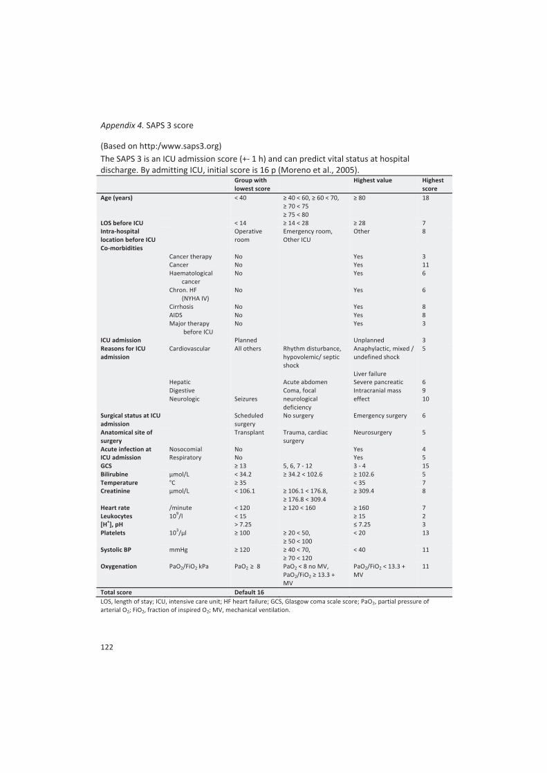
Deviations from treatment goals:definitions/criteriaThe data were registered by an intensive care nurseand were quality checked by a specialist in anaes-thesiology (K. S-M.). Simplified acute physiologyscore II (SAPS II) was calculated for the first 24 h.9
The Injury Severity Score (ISS) was registeredaccording to Baker et al.10 The frequencies of physi-
Intensive care and traumatic brain injury
47

ological values not within the recommended limitswere identified by reviewing all ICU charts. Mod-erate or severe deviations from treatment goals weredefined in Table 2. Blood gas analyses, including themeasurements of haemoglobin, blood glucose andelectrolytes serum concentration, were usually taken5–10 times daily depending on the patient’s clinicalcondition. Continuous intra-arterial blood pressuremeasurements and other vital parameters weredocumented hourly or as required. ICP was regis-tered hourly and at the time of ICP increases. Amoderate/severe deviation in ICP and CPP wasdefined as ICP 21–25 mmHg/ > 25 mmHg and CPP50–59 mmHg/ < 50 mmHg, respectively. For boththese variables, a deviation was registered if meas-ured � 3 times/day. However, if treatment was ter-minated due to no hope of survival, vital parametersincluding ICP after cessation of active treatmentwere not registered in the database.
Organ failure during ICU/HDU treatmentThe definitions of extracranial complications aregiven in Table 3. The incidence of organ failure was
identified by reviewing the patients’ records, chartsand radiologist consultants’ evaluation of X-rays,computer tomography (CT) and magnetic reso-nance imaging (MRI). Complications such asventilator-associated pneumonia (VAP), pneumoniadue to other causes, acute lung injury (ALI), acuterespiratory distress syndrome (ARDS) and severesepsis were noted.11,12 A sequential organ failureassessment (SOFA) score of three or four was usedto define renal failure, liver failure and a coagulationdisorder.13
Outcome assessmentA specialist in physical medicine and rehabilitation(T. S.) and a research nurse collected the 12-monthGlasgow Outcome Scale Extended (GOSE) score.14
GOSE score was not possible to score in the follow-ing cases: (1) patients who died of other causes thanthe head injury during the follow-up (n = 1), (2)patients who needed special care before the TBI(n = 3), (3) patients who got another injury duringthe follow-up study (n = 1) and (4) patients whowere lost to follow-up (n = 1). Of the six patients
Table 1
TBI treatment protocol for the 133 patients included.
Level Treatment Definitions Frequency% (n = 133)
1 Adequate sedation and analgesiaThiopental bolus During procedures 49.6%15–20° elevation of the headSurgical evacuation of haematoma if indicatedPaO2 > 13 kPa or oxygen saturation > 95%PaCO2 Around 4.5 kPaHaemoglobin (Hgb) > 100 and < 120 g/lNormovolemiaNormothermia Temp. � 37°CSerum sodium � 140 mmol/lSerum glucose 4.5–6.5 mmol/l*Cerebral perfusion pressure (CPP)** Adult � 60 mmHg
Children > 45–60 mmHg, depending on age2 Check level 1!
Consider a new CT scan.Hypertonic saline 2,9% (S-Na < 150 mmol/l) Adult 250 ml
50.4%Children 1–2.5 mmol/kg,If infusion 0.05–0.5 mmol/kg/h
Mannitol 150 mg/ml (S-Osm � 320 mosm/kg) Adult 200–300 ml 33.8%Children 0.25–1 g/kgCSF drainage if possibleModerate hyperventilation PaCO2 4.0–4.5 kPa 33.1%Moderate hypothermia (not in children) Temp 35–36°C 26.3%
3 Check level 1 and 2!Consider a new CT scan.Decompressive craniectomy (high priority) 6.0%Barbiturate coma Thiopental 1–5 mg/kg/h 11.3%Hyperventilation PaCO2 3.6–4.0 kPaHypothermia (not in children) Temp < 35°C 14.3%Lumbal catheter for CSF drainage 6.8%
*The desired S-Glu level was changed to 4.5–8 mmol/l after the study period.**Zero level of the arterial pressure is by the level of the heart. CSF, cerebrospinal fluid; CT, computer tomography.
K. Schirmer-Mikalsen et al.
48

where 12-month GOSE was not possible to score,three had a valid registered 6-month GOSE score,which was used in the analyses.
Statistical analysesThe descriptive data are given in numbers, percent-ages, median and ranges as appropriate. We usedordinal logistic regressions to explore the associa-tion between outcome and prognostic factors.Outcome was assessed by GOSE score as dependentvariable. The GOSE scale was reclassified into worseoutcome [GOSE score 1–4 (1 dead, 2–4 severe dis-ability)], moderate disability (GOSE score 5–6) and
good recovery (GOSE score 7–8), and the latter waschosen as reference category.
The following predictor variables were exploredusing univariable logistic regression: age (continu-ous variable), GCS score, pupil dilation during thefirst 24 h, hypotension (systolic BP < 90 mmHg),elevated ICP (21–25 mmHg and ICP > 25 mmHg),hypoventilation (pCO2 � 5 kPa), hypoxia (PaO2 �11 kPa), anaemia (Hgb � 100 g/l), hyperglycaemia(blood glucose � 8 mmol/l), hyponatremia (serumsodium � 135 mmol/l), hypoalbuminemia (serumalbumin < 25 g/l), hyperthermia (central bodytemperature � 38°C), pneumonia (including VAP),
Table 2
Recorded deviations from treatment goals.
Moderate/severedeviations*
Number of patients(n = 133)
Days median(range)
Patient datamissing**
Respiration PaCO2 5–6 kPa/ 99 (74.4%) 3 (1–15) 6PaCO2 > 6 kPa 21 (15.8%) 1 (1–9) 6PaO2 8–11 kPa/ 86 (64.7%) 3 (1–16) 6PaO2 < 8 kPa 4 (3.0%) 1 (1-1) 6
Circulation Systolic BP < 90 mmHg 27 (20.3%) 1 (1–10) 5Haemoglobin (Hgb) Hgb 80–100 g/l 101 (75.9%) 6 (1–27) 0
Hgb < 80 g/l 22 (16.5%) 1 (1–19) 0Blood glucose (S-Glu) S-Glu 8–10 mmol/l/ 52 (39.1%) 1 (1–4) 3
S-Glu > 10 mmol/l 12 (9.0%) 1 (1–3) 3Serum sodium (S-Na) S-Na 130–135 mmol/l/ 46 (34.6%) 2 (1–11) 0
S-Na < 130 mmol/l 4 (3.0%) 1,5 (1–4) 1Serum albumin Serum albumin < 25 g/l 41 (30.8%) 12Temperature (T) T 38–39°C/ 72 (54.1%) 3 (1–14) 2
T > 39°C 21 (15.8%) 2 (1–9) 2
Some patients had both severe and moderate deviations and are therefore counted twice.*PaCO2, PaO2, systolic BP, S-Glu, S-Na and T are registered as a moderate/severe deviation if deviations are measured � 3times/day. Hgb is registered as a moderate/severe deviation if deviation is measured once during the same day, while S-Alb isregistered as a deviation if measured low any time during the ICU stay.**Missing data as a result of insufficient/lacking chart for one or more days.
Table 3
Definition and frequencies of extracranial organ complications.
Complication Definition Frequencies(%)
Patientdatamissing*
Ventilator-associatedpneumonia (VAP)
Artificial ventilation > 48 h, infiltrates on chest X-ray, leucocytes > 10 or< 3 ¥ 109/l, temp. > 38.5°C or < 35°C and purulent tracheal secretions
47 (35.3%) 0
Pneumonia (other than VAP) As above but start of symptoms before 48 h of respiratory treatment 50 (37.6%) 0Acute lung injury (ALI) Acute respiratory failure, PaO2/FiO2 ratio < 39.9 kPa, bilateral infiltrates
on chest X-ray and no signs of heart failure19 (14.3%) 1
Acute respiratory distresssyndrome (ARDS)
Acute respiratory failure, PaO2/FiO2 ratio < 26.6 kPa, bilateral infiltrateson chest X-ray and no signs of heart failure
23 (17.3%) 1
Severe sepsis Sepsis with organ dysfunction, hypoperfusion or hypotension in spite ofadequate fluid therapy
4 (3.0%) 0
Renal failure Serum creatinine > 299 mmol/l or diuresis less than 500 ml/24 h (adults) 1 (0.8%) 0Bleeding disorder Platelets < 50 ¥ 109/l 6 (4.5%) 2Liver failure Serum bilirubin > 101 mmol/l 1 (0.8%) 3
*Missing data as a result of insufficient/lacking chart for one or more days.
Intensive care and traumatic brain injury
49

ALI/ARDS, sepsis, renal failure, bleeding disorderand liver failure. Significant Spearman correlationcoefficients were tested between the continuouspredictor variables, before they were implementedin the regression models. Variables with less thanthree cases in one of the GOSE categories were notanalysed. All variables reaching P � 0.10 in the uni-variable analyses were included in a multiple logis-tic regression model. In this model, we adjusted forthe covariates age, GCS score and pupil dilation.
The precision of the estimates was assessed with95% confidence intervals (CI). All tests were consid-ered statistically significant at a probability value< 0.05. The statistical software Statistical Package forthe Social Sciences version 18 (SPSS Inc., Chicago,IL, USA) or STATA/SE version 11.2 (StataCorp LP,College Station, Texas, USA) was used in all analyses.
EthicsThe study was done according to the principles ofthe Helsinki Declaration. Written informed consentwas obtained from the patient, or if incapacitatedfrom their next of kin. The Regional Committee forMedical Research Ethics approved the study.
Results
DemographicsOf the 133 included patients 79.7% were male andthe median age was 30 years (range 1–81 years;Table 4). Twelve of the patients were less than 15years (1–13.7 years). The majority of the TBIs werecaused by either a traffic accident or a fall injury. Themedian GCS score before sedation was 6. One fourthof the patients had uni- or bilateral pupil dilation onadmission. The initial CT findings often showed acombination of intracerebral pathologies with trau-matic subarachnoidal haemorrhage (tSAH), sub-dural haematoma (SDH) and multiple contusions asthe most frequent findings. In all seven patients withnegative findings on CT examinations, the MRIshowed diffuse axonal injury or cerebral contusions.Median ISS was 26 and median SAPS II in patients> 18 years was 43. One hundred and thirty of the 133patients received invasive mechanical ventilation(0.5–34 days, median 6.0 days). 43.6% of the patientswere operated for intracranial pathology and 43.6%were operated due to extracranial injuries.
Intracranial hypertension and treatmentaccording to the three-level TBI protocolICP was monitored in 105 patients (78.9%). Out ofthe 105 patients with ICP monitoring, 29 did not
have any episodes with elevated ICP, while 26patients had episodes of ICP between 21–25 mmHgand 47 patients had episodes of ICP > 25 mmHg(missing ICP and CPP information in three patients)(Table 5).
About 75% of the patients with ICP monitoringreceived level 2 treatment due to high ICP andaround 30% of the patients with ICP monitoringreceived level 3 treatment (all patients with level 3
Table 4
Patient demographics of the 133 included patients.
Number ofpatients n (%)
Median(range)
SexMale 106 (79.7%)Female 27 (20.3%)
Age (years) 30 (1–81)Cause of accident
Traffic 70 (52.6%)Fall 54 (40.6%)Other 7 (5.3%)
GCS score 131 6 (3–8)Pupil dilation on admission 34 (25.6%)
Unilateral 23Bilateral 11
Injury Severity Score (ISS) 133 26 (5–54)SAPS2 (> 18 years) 112 43 (21–91)Cerebral CT findings*
tSAH 97 (72.9%)SDH 81 (60.9%)EDH 24 (18.0%)Multiple contusions 70 (52.6%)Single contusion 7 (5.3%)Normal 7 (5.3%)
Surgery for intracranial injury 58 (43.6%)Surgery for extracranial injury 58 (43.6%)
*Many patients had more than one intracerebral pathology oncerebral CT.EDH, epidural haematoma; GCS, Glasgow Coma Scale;SDH, subdural haematoma; tSAH, traumatic subarachnoidalhaemorrhage.
Table 5
Registrated elevations in intracranial pressure (ICP) andreductions in cerebral perfusion pressure (CPP) in patients withICP monitoring (n = 105)*.
Number ofpatients(n = 105)
Patientdatamissing**
ICP 21–25 mmHg 26 (24.8%) 3ICP > 25 mmHg 47 (44.8%) 3CPP 60–69 mmHg 21 (20.0%) 3CPP 50–59 mmHg 55 (52.3%) 3CPP < 50 mmHg 20 (19.0%) 3
*Elevations in ICP and reductions in CPP are registered if meas-ured � 3 times/day.**Missing data as a result of insufficient/lacking chart for one ormore days.
K. Schirmer-Mikalsen et al.
50

treatment had also had level 2 treatment). Six of thepatients with ICP monitoring did not have any epi-sodes of CPP below 70 mmHg, while 21 patientshad the lowest registration of CPP 60–69 mmHgand 55 patients had CPP 50–59 mmHg. A cerebralperfusion pressure below 50 mmHg was measuredin 20 patients; three of them were children < 15 yearsold.
Deviations from other treatment goalsAlmost two thirds of the TBI patients (65.4%) hadperiods of PaO2 � 11 kPa during the ICU/HDU stay(Table 2). Hypotension was observed in 27 (20.3%)patients, 12 of them were children. Fifteen patientshad low blood pressure during the first 24 h inthe ICU/HDU. Anaemia (haemoglobin � 100 g/l)was observed in 77.4% of the patients. A mode-rate hyperglycaemia (8–10 mmol/l) was frequent(39.1%), but a blood glucose above 10 mmol/l wasrare (9.0%). One third (34.6%) of the patients hadan observed serum sodium (S-Na) level of� 135 mmol/l while 30.8% of the patients hadhypoalbuminemia. Elevated body temperature wasfound in more than half of the patients (55%).
Extracranial complications duringICU/HDU stayPulmonary complications were a frequent findingwith 73.7% of the patients having at least one ofthe following: VAP, pneumonia due to other causes,ALI or ARDS (Table 3). Pneumonia was the mostcommon complication in TBI patients during theICU/HDU stay. About one third (35.3%) of thepatients had a VAP, whereas 37.6% had pneumoniadue to other causes (one patient had both VAP andpneumonia due to aspiration).
ALI was diagnosed in 14.3% of the patients,whereas 17.3% met the definitions of ARDS. Most ofthe patients (83%) including all patients with ARDS,got vasoconstrictors during their ICU/HDU stay.Four patients (3%) had severe sepsis. One patient gota renal failure and needed continuous renal replace-ment therapy. Low platelets were found in sixpatients (4.5%). One of the patients had both lowplatelets and elevated bilirubin serum concentrationand was classified as having a liver failure.
Prediction of outcomeTwenty-six (19.5%) of the 133 patients included dieddue to the head injury within the first 12 months; allbut two died within the first month. Intracranialhypertension was the cause of death in 21 patients
while five patients died due to sequels from theirhead injury. A good recovery (GOSE scores 7–8) wasseen in 43 patients (32.3%), a moderate disability(GOSE scores 5–6) was seen in 34 patients (25.6%)and a worse outcome (GOSE score � 4) in 53patients (39.8%). One patient died of other causes 3weeks after the TBI.
In the univariable analyses age, uni- or bilateralpupil dilation on admission and low GCS scorewere all highly associated with a worse outcomecategory (Table 6). Additionally, among the ICU/HDU observations, ICP > 25 mmHg, PaO2 � 11 kPa,hyperglycaemia, pneumonia and ARDS were all sig-nificantly associated with a worse outcome. Hypo-tension (even if we excluded children � 15 years),anaemia, hyponatremia, hypoalbuminemia, ALIand elevated body temperature were not associatedto outcome.
In the multiple logistic regression with age, GCSscore and pupil dilation as covariates, high ISS,ICP > 25 mmHg, S-Glu � 8 mmol/l, and pneumo-nia were significantly related to a worse outcome.Intracranial hypertension with ICP less than25 mmHg was associated with better outcome.
DiscussionAfter the implementation of a three-level protocolfor treatment of patients with severe TBI, we foundthat deviations from the specific treatment goalswere frequent. Almost three out of four patients gota pulmonary complication during the ICU/HDUstay, whereas other extracranial complications wereinfrequent. High ISS, hyperglycaemia, pneumoniaand ICP > 25 mmHg predicted a worse outcomeeven after adjustment for the strong prognosticfactors age, GCS score and pupil dilation.
Intracranial hypertension and degreeof multi-traumaThe value of intracranial hypertension as a prognos-tic outcome factor is well established in earlier stud-ies.15,16 In our study, more than one third of thepatients had ICP measurements above 25 mmHg,which was significantly associated with a worseoutcome. However, for the patients with episodes ofslightly increased ICP levels, we found no increasedrisk of a worse outcome.
While a study by Stocchetti et al. found that thefrequency of surgical decompression and the use ofbarbiturates were restricted to less than 10% ofpatients with severe TBI, we found a relatively highincidence of level 3 treatment.17 This is mostly
Intensive care and traumatic brain injury
51

caused by the use of barbiturate infusion and hypo-thermia. The effect of decompressive craniectomy intreating cerebral oedema is controversial and only6% of the patients in our study had a hemi/craniectomy.18,19 In this study, nine patients receiveda lumbal drainage catheter, but four of the ninepatients received lumbal drainage because of CSFleakage. However, occasionally, lumbal drainage isused and reduces ICP.20 The treatment, still, is con-troversial due to the risk of cerebral herniation andthis treatment is not a routine intervention in TBIpatients.
The severity of the multi-trauma, measured by theISS, predicted outcome independently after adjust-ing for other well-known prognostic factors. Previ-ous studies of TBI patients have also shown that ISScould predict outcome.21,22
Deviations and outcomeDespite a desired blood sugar of 4.5–6.5 mmol/lduring the study period, almost half of the patients(43%) in our study had episodes of hyperglycaemia> 8 mmol/l. The association with hyperglycaemia toa poor outcome observed in our study is in accord-ance with earlier studies.23,24 However, the optimalblood glucose level in TBI patients is not establishedand several researchers advice against too strict
blood glucose control due to the concern ofhypoglycaemia, especially in the central nervoussystem.25–28 Also, it is uncertain whether it is theblood glucose itself that is harmful to the brain or ifthe blood glucose level reflect the severity of theTBI.29
Our study could not find any significant relation-ship between hypotension in the ICU/HDU and apoor outcome. Several other studies have shownthat early hypotension in patients with severe headinjury is related to a poor outcome.2,30 One explana-tion for this discrepancy can be that we addressedhypotension in the ICU/HDU and therefore did notinclude hypotension and hypoxia at the scene ofaccident or during the transportation to the hospital.Also, during established ICU treatment, somecauses of hypotension such as hypovolemia areusually corrected, and therefore, a low arterial bloodpressure is often not associated with decreasedcardiac output and lower oxygen delivery. Asoxygen delivery to the brain is a result of severalfactors, it may be that a combination of risk factors ismore important than the presence of each isolatedrisk factor. The IMPACT study observed that thecombination of early hypoxia and early hypotensionwas related to poorer outcome than if the patientshad only one of the events.30
Table 6
Association with outcome.*
Variable Univariate† Oddsratio (OR)
Confidenceinterval (CI)
Multiple logisticregression‡
Multiple logisticregression
Multiple logisticregression
P P OR CI
Age P < 0.001 1.04 1.02–1.06Pupil dilation P = 0.002 3.57 1.62–7.88GCS P < 0.001 0.71 0.59–0.86ISS P = 0.08 1.03 0.996–1.06 P = 0.006 1.05 1.02–1.09ICP21–25 mmHg P = 0.062 0.45 0.20–1.04 P = 0.021 0.33 0.13–0.84> 25 mmHg P < 0.001 7.20 3.14–16.49 P < 0.001 7.40 2.93–18.71PaCO2 � 5 kPa P = 0.37PaO2 � 11 kPa P = 0.002 3.23 1.54–6.81 P = 0.12Hypotension P = 0.84S-Na � 135 mmol/l P = 0.33Hgb � 100 g/l P = 0.13S-Glu � 8 mmol/l P < 0.001 4.04 2.03–8.06 P = 0.002 3.33 1.53–7.26Temp � 38°C P = 0.101 1.72 0.90–3.30S-Alb < 25 g/l P = 0.915 0.999 0.98–1.02 P = 0.71Pneumonia P = 0.004 2.99 1.43–6.28 P = 0.048 2.27 1.01–5.14ALI P = 0.34ARDS P = 0.030 2.59 1.096–6.12 P = 0.14
*The outcome ordinal variable = GOSE score 1–4 = poor outcome. OR greater than one indicates increased risk for worse outcomecategory, while OR less than one indicates decreased risk of worse outcome.†Odds ration (OR) and confidence interval (CI) are only registered for variables with a significance value P < 0.10.‡In the multiple logistic regression, age, GCS score and pupil dilation are covariates.GCS, Glasgow Coma Scale; ICP, intracranial pressure; ISS, Injury Severity Score.
K. Schirmer-Mikalsen et al.
52

While the use of hypothermia in TBI patients iscontroversial, there is a general agreement to avoidhyperthermia.8,31–33 A recent study from the Chinesehead trauma bank indicates that both degree andduration of early post-injury hyperthermia areclosely related to an unfavourable outcome.34
However, we did not observe any significant rela-tion between hyperthermia and a worse outcome.
We could not find any significant relationshipbetween anaemia and a poor outcome. A review byKramer and Zygun on anaemia and red cell trans-fusion in neurocritical care concluded that there areno randomised controlled trials related to the trans-fusion threshold among brain-injured patients.35
Thus, the optimal haemoglobin level in TBI patientsis not known. One third of the TBI patients had lowalbumin levels but in contrast to our earlier study,we did not observe that hypoalbuminemia wasrelated to a worse outcome.2 The importance of thealbumin level in patients with TBI is uncertain. Apost-hoc follow-up study from the SAFE (Saline orAlbumin for Fluid Resuscitation in Patients withTraumatic Brain Injury) study concluded that fluidresuscitation with albumin to TBI patients was asso-ciated with higher mortality than resuscitation withsaline.36 Contrary to this finding is that the Lundconcept use albumin as the main plasma volumeexpander and other studies that observe a negativerelationship between low albumin and survival inthe neurosurgical ICU patients.37,38
Extracranial complications and outcomePneumonia (including VAP) had a negative effect onoutcome in our study. This agrees with a study byPiek et al. where pneumonia was one of the predic-tors of increased morbidity and mortality in TBIpatients.39
Despite a high frequency of ARDS, only fourpatients had episodes of severe deviations in PaO2
during their ICU/HDU stay. This low incidence ofoxygenation failure suggests that the respiratorycomplication is usually possible to compensate withICU respiratory interventions. There was no associa-tion between ALI, ARDS, hypo oxygenation and aworse outcome category. Previous studies in TBIpatients are divergent; some studies have shownthat ALI and ARDS are associated with higher mor-tality in TBI patients, while other studies fail to rep-licate this association.40–43 The conventional TBItreatment includes the use of vasoconstrictors tokeep an optimal cerebral perfusion pressure. We didnot assess if the use of vasoconstrictor was due toincrease CPP, circulatory instability or to counteract
hypotension caused by sedation. It has been shownthat the use of vasoconstrictors is associated with ahigher incidence of ARDS.41 The fact that all patientswith ARDS in our study were given vasoconstrictorscould support this association, but this is highlyuncertain since most of the patients in our studyreceived vasoactive medications.
The importance of treatment protocol for TBIpatients in the ICU/HDUBefore the introduction of a treatment protocol forTBI patients, we performed a retrospective study onTBI patients in the ICU/HDU, including 133patients with severe TBI during a 5-year period(1998–2002).2 This study showed that hypotension,elevated blood sugar and hypoalbuminemia werepossible predictors of an unfavourable outcome. Theintroduction of a treatment protocol for TBI patientsresulted in a decrease in severe deviations of physi-ological parameters. For instance, a blood sugarlevel above 10 mmol/l was found in 26% in theretrospective study, while only 9% had severehyperglycaemia in the prospective study. However,the frequency of patients with low albumin andhypotension were similar in both studies. The dif-ferences between frequency of deviation in physi-ological variables before and after an introduction ofa treatment protocol suggest that the TBI treatmentinterventions that can be controlled and improvedby the personnel (e.g. regulate the blood glucosewith insulin infusion) are improved, while otherfactors may be more difficult to control due to thebrain injury itself (e.g. brain oedema) and is lessprone to be improved. As mentioned by Lingsmaet al., it may also be that deviations in physiologicalvariables are related to the severity of the braininjury, and that normalising the parameters does notnecessarily improve the outcome.29 This opinion issupported by Patel et al. who showed that patientswith TBI observed between 1996 and 2003 showedless improvement in the adjusted odds of deathsince 1989 compared to patients without headinjury.5
Limitations in the studyWe have not analysed interactions between the dif-ferent variables except for keeping the highly signifi-cant variables age, GCS and pupil dilation ascovariates. We also have not analysed factors relatedto the treatment at the scene of accident and duringthe transportation to the hospital. Due to the obser-vational nature of the study, the observed associa-tions are not evidence for certain cause and effect
Intensive care and traumatic brain injury
53

relationships. Finally, we have not taken intoaccount the duration of the deviations in the analy-ses of outcome predictors.
ConclusionOur study showed that deviations from specifictreatment goals according to the treatment protocolof TBI patients were frequent also after the introduc-tion of a TBI protocol. Pulmonary complicationswere common during the ICU/HDU stay, whereasother extracranial complications were infrequent.Age, GCS, pupil dilation, ISS, ICP > 25 mmHg,hyperglycaemia and pneumonia were all highly sig-nificant predictors of a worse outcome.
Acknowledgements
Funded by the St. Olav University Hospital.Conflicts of interest: The authors have no conflicts of interests.Funding: Kent Gøran Moen has, during the study period,
received a research grant from the Liaison Committee betweenthe Central Norway Regional Health Authority (RHA) and theNorwegian University of Science and Technology (NTNU).
References1. Maas AI, Dearden M, Teasdale GM, Braakman R, Cohadon
F, Iannotti F, Karimi A, Lapierre F, Murray G, Ohman J,Persson L, Servadei F, Stocchetti N, Unterberg A. EBIC-guidelines for management of severe head injury in adults.Acta Neurochir 1997; 139: 286–94.
2. Schirmer-Mikalsen K, Vik A, Gisvold SE, Skandsen T,Hynne H, Klepstad P. Severe head injury: control of physi-ological variables, organ failure and complications in theintensive care unit. Acta Anaesthesiol Scand 2007; 51: 1194–201.
3. Clayton TJ, Nelson RJ, Manara AR. Reduction in mortalityfrom severe head injury following introduction of a protocolfor intensive care management. Br J Anaesth 2004; 93: 761–7.
4. Patel HC, Menon DK, Tebbs S, Hawker R, Hutchinson PJ,Kirkpatrick PJ. Specialist neuroctritical care and outcomefrom head injury. Intensive Care Med 2002; 28: 547–53.
5. Patel HC, Bouamra O, Woodford M, King AT, Yates DW,Lecky FE., on behalf of the Trauma Audit and ResearchNetwork. Trends in head injury outcome from 1989 to 2003and the effect of neurosurgical care: an observational study.Lancet 2005; 366: 1538–44.
6. The Brain Trauma Foundation; American Association ofNeurological Surgeons; Congress of Neurological Surgeons;Joint Section on Neurotrauma and Critical Care, AANS/CNS, Bratton SL, Chestnut RM, Ghajar J, McConnellHammond FF, Harris OA, Hartl R, Manley GT, Nemecek A,Newell DW, Rosenthal G, Schouten J, Shutter L, TimmonsSD, Ullman JS, Videtta W, Wilberger JE, Wright DW. Guide-lines for the management of severe traumatic brain injury. JNeurotrauma 2007; 24 (Suppl 1): S1–95.
7. Adelson PD, Bratton SL, Carney NA, Chesnut RM, duCoudray HE, Goldstein B, Kochanek PM, Miller HC, Part-ington MP, Selden NR, Warden CR, Wright DW, AmericanAssociation for Surgery of Trauma; Child Neurology
Society; International Society for Pediatric Neurosurgery;International Trauma Anesthesia and Critical Care Society;Society of Critical Care Medicine; World Federation of Pedi-atric Intensive and Critical Care Societies. Guidelines for theacute medical management of severe traumatic brain injuryin infants, children and adolescents. Pediatr Crit Care Med2003; 4 (Suppl 3): S1–75.
8. Hutchison JS, Ward RE, Lacroix J, Hébert PC, Barnes MA,Bohn DJ, Dirks PB, Doucette S, Fergusson D, Gottesman R,Joffe AR, Kirpalani HM, Meyer PG, Morris KP, Moher D,Singh RN, Skippen PW., for the Hypothermia PediatricHead Injury Trial Investigators and the Canadian CriticalCare Trials Group. Hypothermia therapy after traumaticbrain injury in children. N Engl J Med 2008; 358: 2447–56.
9. Le Gall JR, Lemeshow S, Saulnier F. A new simplified acutephysiology score (SAPS II) based on a European/NorthAmerican multicenter study. JAMA 1993; 270: 2957–63.
10. Baker SP, O’Neill B, Haddon W, Long WB. The injury sever-ity score: a method for describing patients with multipleinjuries and evaluating emergency care. J Trauma 1974; 14:187–96.
11. Cook DJ, Walter SD, Cook RJ, Griffith LE, Guyatt GH, LeasaD, Jaeschke RZ, Brun-Buisson C, for the Canadian CriticalTrials Group. Incidence of and risk factors for ventilator-associated pneumonia in critically ill patients. Ann InternMed 1998; 129: 433–40.
12. Bernard GR, Artigas A, Brigham KL, Carlet J, Falke K,Hudson L, Lamy M, Legall JR, Morris A. Spragg R and theConsensus Committee. The American-European consensusconference on ARDS. Definitions, mechanisms, relevant out-comes, and clinical trial coordination. Am J Respir Crit CareMed 1994; 149: 818–24.
13. Janssens U, Graf C, Graf J, Radke PW, Königs B, Koch KC,Lepper W, vom Dahl J, Hanrath P. Evaluation of the SOFAscore: a single-center experience of a medical intensive careunit in 303 consecutive patients with predominantly cardio-vascular disorders. Intensive Care Med 2000; 26: 1037–45.
14. Nichol AD, Higgins AM, Gabbe BJ, Murray LJ, Cooper DJ,Cameron PA. Measuring functional and quality of life out-comes following major head injury: common scales andchecklists. Injury 2011; 42: 281–7.
15. Vik A, Nag T, Fredriksli OA, Skandsen T, Moen KG,Schirmer-Mikalsen K, Manley GT. Relationship of ‘dose’ ofintracranial hypertension to outcome in severe traumaticbrain injury. J Neurosurg 2008; 109: 678–84.
16. Stein DM, Hu PF, Brenner M, Sheth KN, Liu K, Xiong W,Aarabi B, Scalea TM. Brief episodes of intracranial hyperten-sion and cerebral hypoperfusion are associated with poorfunctional outcome after severe traumatic brain injury. JTrauma 2011; 71: 364–74.
17. Stocchetti N, Zanaboni C, Colombo A, Citero G, Beretta L,Ghisoni L, Zanier ER, Canavesi K. Refractory intracranialhypertension and ‘second-tier’ therapies in traumatic braininjury. Intensive Care Med 2008; 34: 461–7.
18. Taylor A, Butt W, Rosenfeld J, Shann F, Ditchfield M, LewisE, Klug G, Wallace D, Henning R, Tibballs J. A randomizedtrial of very early decompressive craniectomy in childrenwith traumatic brain injury and sustained intracranialhypertension. Childs Nerv Syst 2001; 17: 154–62.
19. Cooper DJ, Rosenfeld JV, Murray L, Arabi YM, Davies AR,D’Urso P, Kossmann T, Ponsford J, Seppelt I, Reilly P, WolfeR, DECRA Trial Investigators; Australian and New ZealandIntensive Care Society Clinical Trials Group. Decompressivecraniectomy in diffuse traumatic brain injury. N Eng J Med2011; 364: 1493–502.
20. Tuettenberg J, Czabanka M, Horn P, Woitzik J, Barth M,Thomé C, Vajkoczy P, Schmiedek P, Muench E. Clinical
K. Schirmer-Mikalsen et al.
54

evaluation of the safety and efficacy of lumbar cerebrospinalfluid drainage for the treatment of refractory increasedintracranial pressure. J Neurosurg 2009; 110: 1200–8.
21. Foreman BP, Caesar RR, Parks J, Madden C, Gentilello LM,Shafi S, Carlile MC, Harper CR, Diaz-Arrastia RR. Useful-ness of the abbreviated injury score and the injury severityscore in comparison to the Glasgow coma scale in predictingoutcome after traumatic brain injury. J Trauma 2007; 62:946–50.
22. Gennarelli TA, Champion HR, Copes WS, Sacco WJ. Com-parison of mortality, morbidity and severity of 59,713 headinjured patients with 114,447 patients with extracranial inju-ries. J Trauma 1994; 37: 962–8.
23. Rovlias A, Kotsou S. The influence of hyperglycemia on neu-rological outcome in patients with severe head injury. Neu-rosurgery 2000; 46: 335–51.
24. Salim A, Hadjizacharia P, Dubose J, Brown C, Inaba K, ChanLS, Margulies D. Persistent hyperglycemia in severe trau-matic brain injury: an independent predictor of outcome.Am Surg 2009; 75: 25–9.
25. Meier R, Bechir M, Ludwig S, Sommerfeld J, Keel M, SteigerP, Stocker R, Stover JF. Differential temporal profile oflowered blood glucose levels (3.5 to 6.5 mmol/L versus 5 to8 mmol/L) in patients with severe traumatic brain injury.Crit Care 2008; 12: 1–13.
26. Green DM, O’Phelan KH, Bassin SL, Chang CWJ, Stern TS,Asai SM. Intensive versus conventional insulin therapy incritically ill neurologic patients. Neurocrit Care 2010; 13:299–306.
27. Bilotta F, Rosa G. Glucose management in the neurosurgicalpatient: are we yet any closer? Curr Opin Anaesthesiol 2010;23: 539–43.
28. Coester A, Neumann CR, Schmidt MI. Intensive insulintherapy in severe traumatic brain injury: a randomized trial.J Trauma 2010; 68: 904–11.
29. Lingsma HF, Roozenbeek B, Steyerberg EW, Murray GD,Maas AIR. Early prognosis in traumatic brain injury: fromprophecies to predictions. Lancet Neurol 2010; 9: 543–54.
30. McHugh GS, Engel DC, Butcher I, Steyerberg EW, Lu J,Mushkudiani N, Hernandez AV, Marmarou A, Maas AIR,Murray GD. Prognostic value of secondary insults in trau-matic brain injury: results from the IMPACT study. J Neu-rotrauma 2007; 24: 287–93.
31. Clifton GL, Valadka A, Zygun D, Coffey CS, Drever P, Four-winds S, Janis LS, Wilde E, Taylor P, Harshman K, Conley A,Puccio A, Levin HS, McCauley SR, Bucholz RD, Smith KR,Schmidt JH, Scott JN, Yonas H, Okonkwo DO. Very earlyhypothermia induction in patients with severe brain injury(the National acute brain injury study: Hypothermia II): arandomised trial. Lancet Neurol 2011; 10: 131–9.
32. Polderman KH. Induced hypothermia and fever control forprevention and treatment of neurological injuries. Lancet2008; 371: 1955–69.
33. Grände PO, Reinstrup P, Romner B. Active cooling in trau-matic brain-injured patients: a questionable therapy? ActaAnaesthesiol Scand 2009; 53: 1233–8.
34. Li J, Jiang J. Chinese head trauma data bank: effect of hyper-thermia on the outcome of acute head trauma patients. JNeurotrauma 2012; 29: 96–100.
35. Kramer AH, Zygun DA. Anemia and red blood cell trans-fusion in neurocritical care. Crit Care 2009; 13: R89.
36. The SAFE Study Investigators; Australian and New ZealandIntensive Care Society Clinical Trials Group; Australian RedCross Blood Service; George Institute for InternationalHealth, Myburgh J, Cooper DJ, Finfer S, Bellomo R, NortonR, Bishop N, Kai Lo S, Vallance S. Saline or albumin for fluidresuscitation in patients with traumatic brain injury. N EnglJ Med 2007; 357: 874–84.
37. Grände PO. PRO: the ‘Lund concept’ for treatment ofpatients with severe traumatic brain injury. J NeurosurgAnesthesiol 2011; 23: 251–5.
38. Ramesh VJ, Umamaheswara Rao GS, Kandavel T, Kumaras-wamy SD, Iyyamanda UB, Chandromouli BA. Predictivemodel for survival among neurosurgical intensive carepatients. J Neurosurg Anesthesiol 2011; 23: 183–7.
39. Piek J, Chesnut RM, Marshall LF, van Berkum-Clark M,Klauber MR, Blunt BA, Eisenberg HM, Jane JA, MarmarouA, Foulkes MA. Extracranial complications of severe headinjury. J Neurosurg 1992; 77: 901–7.
40. Holland MC, Mackersie RC, Morabito D, Campbell AR,Kivett VA, Patel R, Erickson VR, Pittet JF. The developmentof acute lung injury is associated with worse neurologicoutcome in patients with severe traumatic brain injury. JTrauma 2003; 55: 106–11.
41. Contant CF, Valadka AB, Gopinath SP, Hannay HJ, Robert-son CS. Adult respiratory distress syndrome: a complicationof induced hypertension after severe head injury. J Neuro-surg 2001; 95: 560–8.
42. Mascia L, Sakr Y, Pasero D, Payen D, Reinhart K, Vincent JL,Sepsis Occurrence in Acutely ill Patients (SOAP) Investiga-tors. Extracranial complications in patients with acute braininjury: a post-hoc analysis of the SOAP study. Intensive CareMed 2008; 34: 720–7.
43. Treggiari MM, Hudson LD, Martin DP, Weiss NS, CaldwellE, Rubenfeld G. Effect of acute lung injury and acute respi-ratory distress syndrome on outcome in critically ill traumapatients. Crit Care Med 2004; 32: 327–31.
Address:Kari Schirmer-MikalsenDepartment of Anaesthesia and Acute MedicineSt. Olav University HospitalPostboks 3250 Sluppen7006 Trondheim, Norwaye-mail: [email protected]
Intensive care and traumatic brain injury
55

Paper III

Is not included due to copyright


A A Appendix 1-6