Embed Size (px)
Citation preview
The Trauma and Orthopaedic Research Unit (TORU) has capacity to undertake
clinical and laboratory studies in the field of musculoskeletal disease.
This includes clinical aspects of arthroplasty, tissue reconstruction and trauma,
fracture surveillance and management, medical imaging and joint kinematics.
TORU has established a laboratory facility at both Canberra Hospital and at the
John Curtin School of Medical Research at the ANU. This enables us to conduct
translational research within our own unit.
TORU’s mission is to conduct excellent research which meaningfully impacts on the clinical
practice of orthopaedics and the well-being of patients. This includes the development of novel
surgical modalities and therapeutic concepts by conducting translational medicine that bridges
basic science and clinical practice. We aim to create a rich research environment by promoting
active, multidisciplinary cooperation among orthopaedic surgeons, rheumatologists,
physiotherapists, engineers, pathologists, biologists, mathematicians and physicists. Fundamental
to our research endeavour is the provision of an educational hub for students, orthopaedic
registrars and researchers.
Message from the Director
November 2014
Volume 7, 2014
Trauma and Orthopaedic
Research Unit Newsletter
Inside this issue:
We would like to thank Dr Shyam
Rajagopalan for his support as a
clinical and research fellow over the
last 12 months
TORU has published 12 peer reviewed papers and over 40 conferences presentations over the last 12 months
We are very pleased to welcome Joe Lynch, who has come on board as a senior research officer. Joe has come to Canberra from
Canada via Sydney. He is a biomechanist with a special interest in knees who has been working with the SORI group in Sydney .
TORU has commenced a large multicentre trial looking at the survival and patient outcomes of the ATTUNE® Total Knee
Replacement System (Johnson and Johnson).
Version 1.0 of the Canberra Trauma Fracture Database has been launched at The Canberra Hospital. This tool allows clinicians
to keep a detailed record of treatments and outcomes for all patients who have sustained a fracture
The characterising of whiplash injuries study is progressing with the exciting development of a collaboration with both UNSW @
ADFA and the University of Canberra for image processing innovation.
We have commenced a collaborative project with University of Canberra and Prince of Wales Hospital on the impact of dementia
on access to, and outcomes from, rehabilitation following fracture related hospitalisation.
Mission Statement
Research Highlights
The Trauma and Othopaedic Research Unit has once again had a
productive year but it is surprising how quickly the year has gone. We
have invested a great deal of time and effort with the support of
Synthes in database development and have recently implemented
our fracture trauma database. This has been a long term project and
will provide the ACT Orthopaedic region with a wonderful resource for
the future. This web-based database will enable us to collect
longitudinal outcome data which has been specifically categorized by
AO fracture classification and coded for comorbidities at source. Our
ambition is to interest other centres in this format with the potential
of creating an Australia-wide virtual registry from which we can make more informed and cost
effective decisions.
The orthopaedic education and research world in Australia is changing. The Australian Orthopaedic
Association has embraced this evolution and research training together with technical surgical
training will be essential parts of the advanced Orthopaedic Surgical Training Program for the
future. Aspiring trainees are now required to conduct research prior to being included in the
program and also during their journey through the advanced programme. This emphasis on high
level training in research will undoubtedly strengthen the profile of Australian orthopaedics on the
world stage. With the infrastructure now in place, the ACT Orthopaedic Unit with TORU is in a great
position to support the research and training agenda.
Cover Page 1
TORU Laboratory and
Clinical Research
Report
2-3
TORU Staff and
Fellows
4-5
TORU Medical
Students
6-7
PhD Scholars 8-9
Project Highlights 10-11
Conferences 12-13
TORU Journal Articles 14-18
Funding Received 19
Collaborators 20-21
TORU Laboratory Research Report
Page 2 Volume 7, 2014
TORU Laboratory team bridges basic and
clinical sciences and facilitates
communication among TORU’s collaborative
institutes, universities and orthopaedic
industries.
TORU team presently investigates chronic
and complex bone diseases, some of which
cause life-long pain and disability. These
chronic conditions can be rare, such as
revision joint replacement and osteolysis or
can be remarkably common, such as
arthritis, trauma and osteoporotic fractures.
Combined, they afflict millions of
Australians and cause tremendous human
suffering, and cost million dollars in health
care.
The team utilizes a mix of conventional
molecular biology approaches as well as
global methods such as next generation
sequencing to study mRNA expression and
its regulation by non-coding RNA e.g.
microRNAs with an ultimate goal of
identifying novel molecules that regulate
bone resorption, formation, fracture repair
and bone homeostasis.
Key Research Areas
Osteoimmunology, microRNAs’ (miRNA)
regulation and genetic risk factors in
biomaterial related osteolysis in total joint
replacement.
This research has in part supported by AOA
Research Foundation. Building on the
foundation laid by the dendritic cells
involvement in osteolysis, the group is using
off-cut tissues from the cohorts of healthy,
primary and revision subjects of TJR for
characterization of wear particles and
identification of molecular and genetic risk
factors that contribute to the osteolysis. The
ultimate goals are to contribute to the
development of better predictive markers,
treatments, and prevention strategies.
The third generation of magnesium (Mg)-
based biomaterial development
This area addresses a need for translational
research to enhance treatment and improve
management of bone diseases and
disorders. To advance the understanding of
interaction at the interface of biomaterials
and biological systems, the team is studying
biocompatibility, biodegradability and
bioactivity on a series of magnesium (Mg)-
based biomaterials either on controlling
biodegradation or osteointegration.
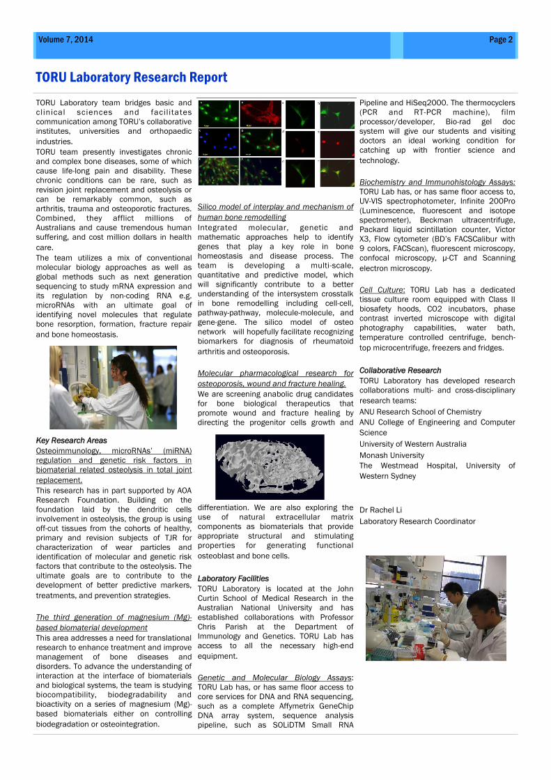
Silico model of interplay and mechanism of
human bone remodelling
Integrated molecular, genetic and
mathematic approaches help to identify
genes that play a key role in bone
homeostasis and disease process. The
team is developing a multi-scale,
quantitative and predictive model, which
will significantly contribute to a better
understanding of the intersystem crosstalk
in bone remodelling including cell-cell,
pathway-pathway, molecule-molecule, and
gene-gene. The silico model of osteo
network will hopefully facilitate recognizing
biomarkers for diagnosis of rheumatoid
arthritis and osteoporosis.
Molecular pharmacological research for
osteoporosis, wound and fracture healing.
We are screening anabolic drug candidates
for bone biological therapeutics that
promote wound and fracture healing by
directing the progenitor cells growth and
differentiation. We are also exploring the
use of natural extracellular matrix
components as biomaterials that provide
appropriate structural and stimulating
properties for generating functional
osteoblast and bone cells.
Laboratory Facilities
TORU Laboratory is located at the John
Curtin School of Medical Research in the
Australian National University and has
established collaborations with Professor
Chris Parish at the Department of
Immunology and Genetics. TORU Lab has
access to all the necessary high-end
equipment.
Genetic and Molecular Biology Assays:
TORU Lab has, or has same floor access to
core services for DNA and RNA sequencing,
such as a complete Affymetrix GeneChip
DNA array system, sequence analysis
pipeline, such as SOLiDTM Small RNA
Pipeline and HiSeq2000. The thermocyclers
(PCR and RT-PCR machine), film
processor/developer, Bio-rad gel doc
system will give our students and visiting
doctors an ideal working condition for
catching up with frontier science and
technology.
Biochemistry and Immunohistology Assays:
TORU Lab has, or has same floor access to,
UV-VIS spectrophotometer, Infinite 200Pro
(Luminescence, fluorescent and isotope
spectrometer), Beckman ultracentrifuge,
Packard liquid scintillation counter, Victor
X3, Flow cytometer (BD’s FACSCalibur with
9 colors, FACScan), fluorescent microscopy,
confocal microscopy, µ-CT and Scanning
electron microscopy.
Cell Culture: TORU Lab has a dedicated
tissue culture room equipped with Class II
biosafety hoods, CO2 incubators, phase
contrast inverted microscope with digital
photography capabilities, water bath,
temperature controlled centrifuge, bench-
top microcentrifuge, freezers and fridges.
Collaborative Research
TORU Laboratory has developed research
collaborations multi- and cross-disciplinary
research teams:
ANU Research School of Chemistry
ANU College of Engineering and Computer
Science
University of Western Australia
Monash University
The Westmead Hospital, University of
Western Sydney
Dr Rachel Li
Laboratory Research Coordinator
TORU Clinical Research Report
Page 3 Volume 7, 2014
The TORU clinical research unit has had a
busy and successful year. Our research
profile is broad and varied with topics rang-
ing between arthroplasty, trauma, materi-
als science and whiplash. Although suc-
cessful we face many future challenges
and opportunities. Attracting adequate
resources is always a challenge but creat-
ing more opportunities for collaboration
with other units such as ours is a rewarding
way to approach the problem. In this way
we hope to become more competitive with
respect to NHMRC funding which, as every-
one knows, is a difficult commodity to
come by in surgery.
We are enjoying increased engagement
from more the senior and the junior clini-
cians. Professor Smith, as our director, is
our main source of clinical expertise but if
Canberra is to achieve its potential and
become a centre of excellence for ortho-
paedics, we need to engage all of the clini-
cians in a research agenda.
Canberra has many advantages with re-
spect to research. We are small enough
that the orthopaedic surgeons are able to
act as a single unit across both the public
and the private sectors. This enhances the
opportunity for comprehensive thought
exchange and collaboration. Indeed we are
enjoying much more engagement with the
senior clinicians who are supervising re-
search projects in a number of areas this
year. This combined with our close prox-
imity to other large academic institutions
gives us a useful edge.
TORU also has advantages over many simi-
lar units. We are embedded and supported
by ACTH. At the same time our affiliation
with the ANU allows us to take part in many
educational opportunities and engage in
supervising post-graduate degree students.
We are currently supporting five MPhil stu-
dents and four PhD students. In addition,
six medical students per annum conduct
their research under our auspices and we
have projects being undertaken by interns
and registrars keen to make their mark in
research (and earn bonus points for their
careers). Of interest is a project by Dr Jen-
nifer Truong who is using the National
Health Cost Data Collection format to prop-
erly cost two-stage revision for infection in
arthroplasty which has been vastly under-
estimated in the past.
By virtue of our location, affiliations, and
collaborations we have a rich and produc-
tive network which has allowed us to ex-
plore ideas in new and creative ways. For
example our image registration technology
is due to a very productive collaboration
with Assoc. Prof Mark Pickering at UNSW.
This project has extended from the knee to
the hip and we are also examining ways of
automating our whiplash image data. We
have two other collaborations at UNSW
including the development of intrinsic bear-
ing sensor technologies with Dr Sean
O’Byrne and the exploration of trunnion
wear mechanics with Dr Krishna Shankar.
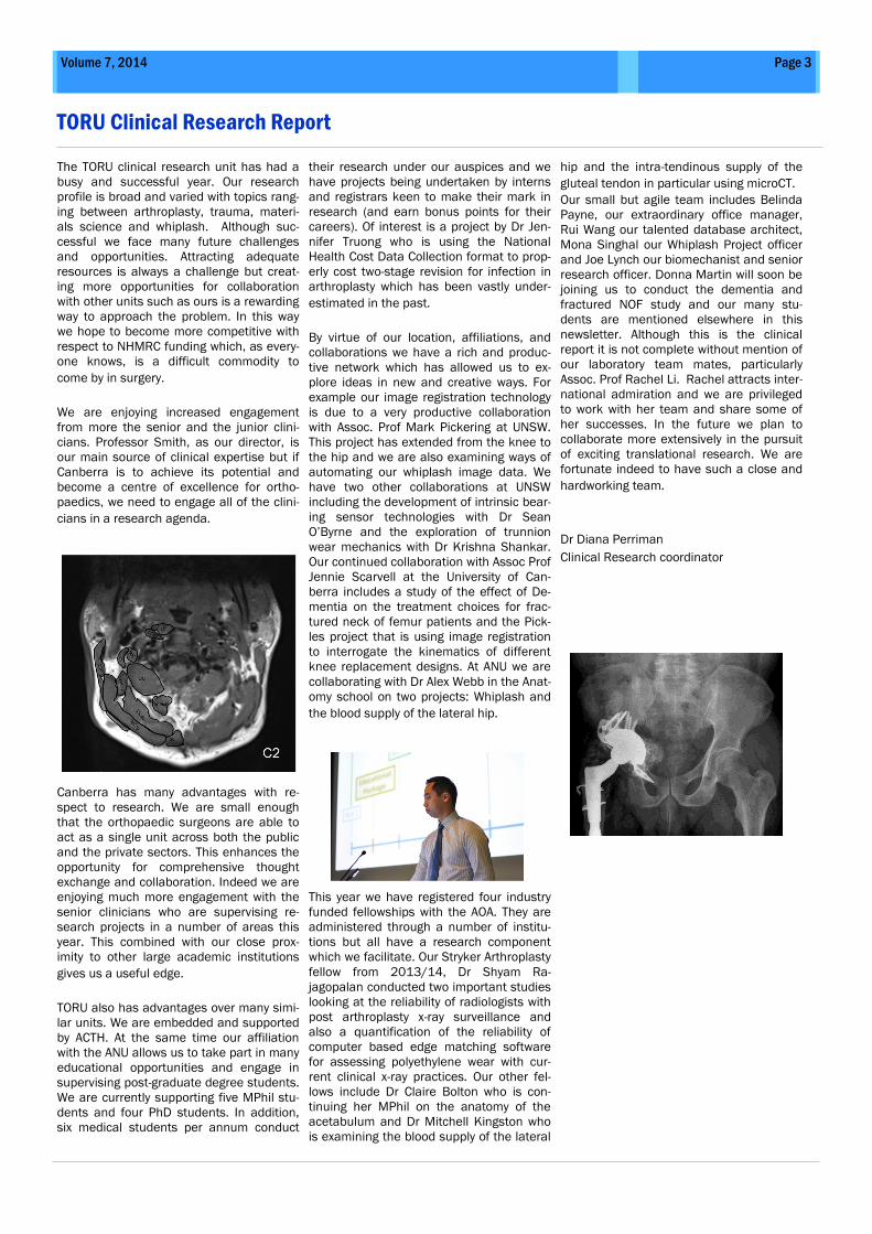
Our continued collaboration with Assoc Prof
Jennie Scarvell at the University of Can-
berra includes a study of the effect of De-
mentia on the treatment choices for frac-
tured neck of femur patients and the Pick-
les project that is using image registration
to interrogate the kinematics of different
knee replacement designs. At ANU we are
collaborating with Dr Alex Webb in the Anat-
omy school on two projects: Whiplash and
the blood supply of the lateral hip.
This year we have registered four industry
funded fellowships with the AOA. They are
administered through a number of institu-
tions but all have a research component
which we facilitate. Our Stryker Arthroplasty
fellow from 2013/14, Dr Shyam Ra-
jagopalan conducted two important studies
looking at the reliability of radiologists with
post arthroplasty x-ray surveillance and
also a quantification of the reliability of
computer based edge matching software
for assessing polyethylene wear with cur-
rent clinical x-ray practices. Our other fel-
lows include Dr Claire Bolton who is con-
tinuing her MPhil on the anatomy of the
acetabulum and Dr Mitchell Kingston who
is examining the blood supply of the lateral
hip and the intra-tendinous supply of the
gluteal tendon in particular using microCT.
Our small but agile team includes Belinda
Payne, our extraordinary office manager,
Rui Wang our talented database architect,
Mona Singhal our Whiplash Project officer
and Joe Lynch our biomechanist and senior
research officer. Donna Martin will soon be
joining us to conduct the dementia and
fractured NOF study and our many stu-
dents are mentioned elsewhere in this
newsletter. Although this is the clinical
report it is not complete without mention of
our laboratory team mates, particularly
Assoc. Prof Rachel Li. Rachel attracts inter-
national admiration and we are privileged
to work with her team and share some of
her successes. In the future we plan to
collaborate more extensively in the pursuit
of exciting translational research. We are
fortunate indeed to have such a close and
hardworking team.
Dr Diana Perriman
Clinical Research coordinator
Professor Smith is an ortho-
paedic surgeon at the Can-
berra Hospital and at Calvary
John James Hospital in Can-
berra. He is also Co-Director of
the Trauma and Orthopaedic
Research Unit at the Canberra
Hospital. Prof Smith is also
president of the Arthroplasty
Society of Australia, and Clini-
cal Director of Orthopaedic
surgery at the Canberra Hospi-
tal.
Prof Smith received his medi-
cal and surgical training in
Adelaide before specialising in
hip and knee joint reconstruc-
tive and replacement surgery.
He was a Royal Australasian
College of Surgeons Travelling
Fellow in 1996 and 1997 with
Fellowships in joint replace-
ment surgery at the University
of Western Ontario in Canada
and at The Princess Elizabeth
Orthopaedic Hospital in Eng-
land. He has been honoured
by The Knee Society, receiving
the inaugural John N Insall
Travelling Fellowship in knee
surgery and has been ap-
pointed as Professor of Ortho-
paedic Surgery at the ANU
Medical School. Prof Smith's
particular clinical interests are
in reconstruction and replace-
ment surgery of the hip and
knee, complex revision joint
replacement surgery and man-
agement of pelvic and
acetabular injuries.
Contact:
search Unit since returning
from the UK in 2003.
Her PhD research investigated
the thoracic spine and ky-
photic thoracic posture in ag-
ing., a suite of thoracic spine
biomechanical and imaging
studies culminating in a ran-
domized controlled trial of the
effect of conservative treat-
ment for thoracic kyphosis.
Dr Perriman has also been the
recipient of an NHMRC Dora
Lush scholarship for this re-
search. As clinical research
Diana Perriman, BAppSc
(USyd), MSc. (University of East
London), PhD (ANU). Dr Perri-
man is currently the clinical
research coordinator of TORU.
Dr Perriman is a physiothera-
pist who has completed her
PhD at the ANU.
Her clinical career has
spanned two decades in which
she worked in hospitals, the
community and private prac-
tice both in Australia and the
UK. She has worked at the
Trauma and Orthopaedic Re-
coordinator Dr Perriman’s re-
search interests lie in arthro-
plasty and fracture outcomes
in accordance with the main
focus of the Trauma and Or-
thopaedic Research Unit.
Contact:
Prof Paul Smith, BMBS FRACS (Ortho). Director
Dr Diana Perriman, PhD. Clinical Research Co-ordinator
cessfully transferred an intel-
lectual property to pharmaceu-
tical industry.
In 2003 Dr Li completed her
PhD in pharmacology at South-
ern Cross University and
gained her postdoctoral re-
search experience in molecu-
lar pharmacology in John A
Burns School of Medicine, at
University of Hawaii.
Dr Li returned to Australia in
2006 and established the
TORU Laboratory which pio-
neered basic orthopaedic re-
search at the ACT region. She
has made some major re-
search contributions to the
fields of osteoimmunology.
She has developed a range of
research strengths and capa-
bilities in orthopaedic research
and biomaterial testing. Along
with her focus on bone biology
and immunology, she has a
long-standing interest in the
biomedical markers and tar-
gets that are anabolic to bone
growth.
Contact:
Dr Rachel W Li, MD, PhD. Laboratory Research Co-ordinator
Dr Li is a molecular pharma-
cologist and osteoimmunolo-
gist with interests in under-
standing the processes that
control a ‘foreign body reaction
or response’ initiated by bio-
materials implanted into bone
or exposed to human cells.
Dr Li received a Bachelor of
Medicine and Master of Medi-
cine, and worked as a surgeon
and senior liver diseases spe-
cialist at China Medical Univer-
sity from 1982 to 1996. She
led a number of clinical trials
in anti-v iral and anti-
inflammatory drugs and suc-
TORU’s People
Page 4 Volume 7, 2014
Dr Donghai Zhang
Dr Zhang is a
C h i n e s e
Anaesthetist from
Shandong University
who travelled to
Canberra to join the
TORU lab team
working specifically
on ‘Biocompatibility of novel sensoring
materials for assessment of fracture
healing.‘ He has now returned to
China.
Dr Maoyuan Xin
Dr Maoyuan Xin is
a visiting 3rd year
PhD student from
the medical school
o f S h a n d o n g
University, located
in Jinan city of China. Xin’s work is
supervised by Prof Paul Smith and
A/Prof Rachel Li and focuses on novel
sensor materials for use in medicine
particularly in the field of orthopedics.
Dr Partha Palit
Dr Partha Palit won
a n E n d e a v o ur
P o s t d o c t o r a l
Fellowship 2014,
allowing him to join
TORU’s laboratory
research for 6
month. Endeavour
P o s t d o c t o r a l
Fe l lowsh ip i s in te rna t i ona l l y
competitive, merit-based fellowship
p r o v i d e d b y t h e A u s t r a l i a n
Government that support citizens of
the Asia-Pacific, the Middle East,
Europe and the Americas to undertake
study, research and professional
development programmes in Australia.
Since his arrival, Dr Palit has been
actively working on our research
project investigating the chemical
compounds for anti-inflammatory and
antiosteoporosis.
Dr Tom Ward PhD
Tom is an orthopaedic
registrar, PhD and
Rhodes scholar. His
current research
interests are in validating image
registration techniques for the hip and
FAI in particular.
TORU Staff and Fellows
Christ ine Hanrahan,
Database Manager
Christine Hanrahan is a
qualified nurse and has
worked with both the Red
Cross and the Therapeutic
Goods Administration.
Christine helped to
develop the Arthroplasty
surveillance database and continues to
manage the database which is based at
Orthopaedics ACT.
Ms Liz Abbott
L i z h a s b e e n a
physiotherapist in Canberra
for over 2 decades with a
s p e c i a l i n t e r e s t i n
o r t h o p a e d i c a n d
musculoskeletal injuries.
Currently Liz coordinates the explant retrieval
database.
Research Fellows
Dr Jennie Scarvell
B(App)Sc Physiotherapy
(Sydney), Grad Cert
Higher Ed, (Canberra)
Cert Health Economics
( M on ash ) Ph D
(Sydney), A/Professor
D e p a r t m e n t o f
Pysiotherapy (University of Canberra).
A career as clinical physiotherapist lead
Jennie to a PhD on knee kinematics and the
role of aberrant motion in degenerative
change using a model of ACL injury. Jennie is
an affiliate Senior Lecturer at University of
Canberra and at ANU.
Dr Claire Bolton
Claire is an orthopaedic
registrar now based in Adelaide.
She is conducting her MPhil on
the morphology of the
acetabulum in osteoarthritis
and hip dysplasia. She is supervised by Prof
Smith and Dr Perriman.
Dr Mitchell Kingston
Dr Kingston is an
orthopaedic registrar at
the Canberra Hospital
and Research Fellow. He
is currently undertaking
an MPhil at the ANU
looking at the anatomy of the circumflex
femoral arteries
Ms Belinda Payne
Belinda is TORU's Office
Manager, Belinda has
been with TORU since
2013 and can be
contacted at any time for
queries regarding the unit.
Belinda comes from a
clinical background in
nursing which gives her great insight into the
many different aspects of orthopaedic
research. Her role is diverse and
comprehensive including conference and
meeting organisation, financial management
and administrative duties
Mr Joe Lynch, Research
Officer
Joe joined the team in mid
2014. He completed his
Bachelor of Science in
Exercise Science, and a
Master of Science in
Biomechanics at the
University of Ottawa.
At present Joe is involved in the running of
various trials within the unit with his main
interest being in functional and imaging
analysis following injury and surgery
Ms Ruidang Wang,
Database Architect
Rui is a database
architect who is currently
designing the Fracture
Surveillance Database.
Rui has extensive IT
experience specialising in
applying database design,
analytic informatics, business intelligence
and online platform technologies to clinical
context. Rui is currently completing her PhD
at ANU.
Dr Mona Singhal, Research
Officer
Dr Singhal studied medicine
at Bangalore University,
India. Dr Singhal has just
completed the Australian
Medical Council Registration
Examination. She joined
TORU in 2013 to work as a Research Officer
on the Whiplash study.
Page 5 Volume 7, 2014
Page 6 Trauma and Orthopaedic Research Unit Newsletter
Medical Student Projects
Harrison Pickup Mathew Lim
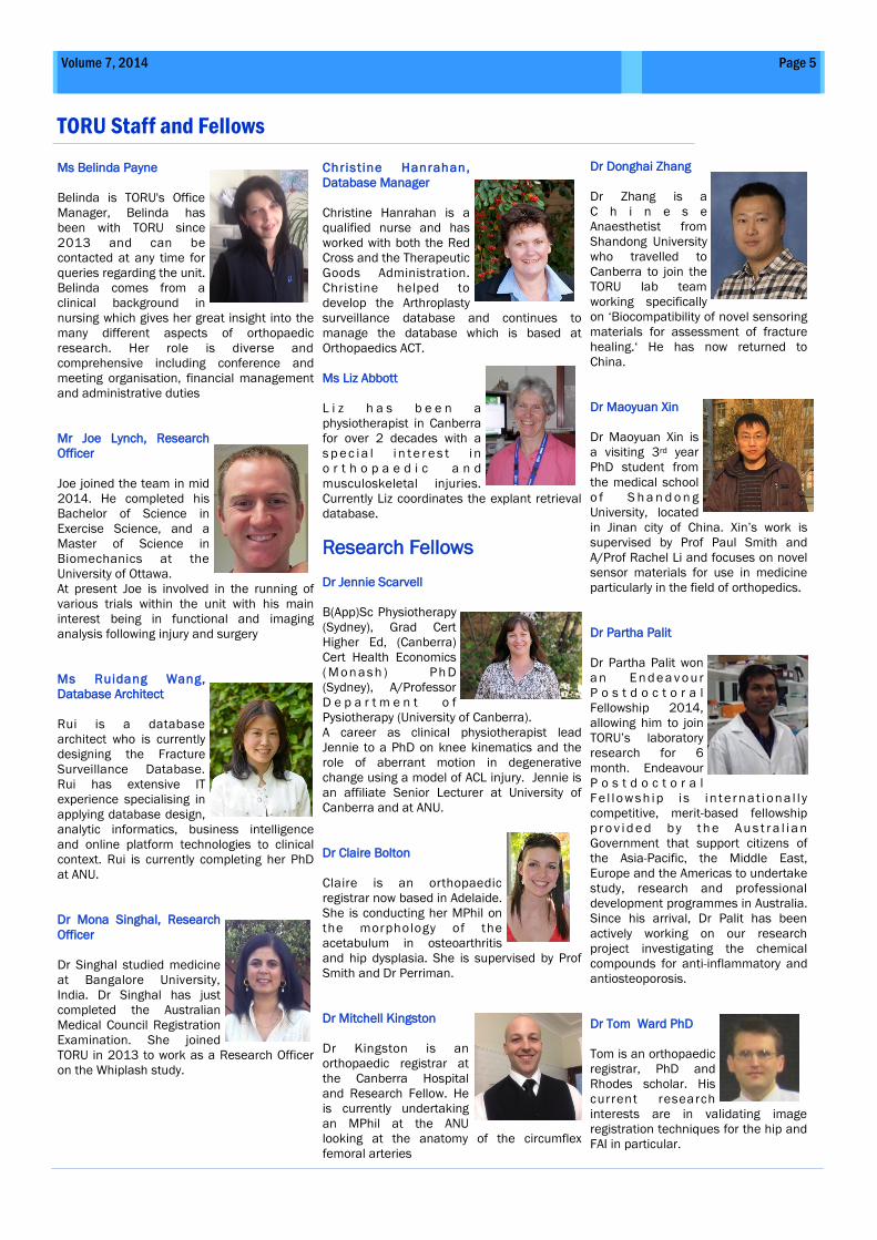
Do Volar plates lead to the best outcome in
patients with distal radius fractures?
Background
As the elderly population continues to grow, the incidence of
DRFs is increasing. The introduction of volar plates to the
repertoire of surgeons has led to an increase in their popularity
over other methods of fixation. This paper looks at the
functional outcomes of treating distal radius fractures in the
elderly with volar plates and other fixation methods.
Methods
A retrospective review of patients aged 65 years or older treated
for distal radius fracture during 2005, 2006, 2008 and 2009 at
The Canberra Hospital was conducted. Patient outcomes were
measured using the Disabilities of the Arm, Shoulder and Hand
(DASH), Assessment of Quality of Life (AQoL 6D) and a Visual
Analogue Scale (VAS) for patient satisfaction with wrist
performance and pain. Fractures were classified from patient x-
rays using the AO classification system. Fixation method, which
extremity and length of stay were obtained from patients
medical records.
Results
86 patients responded with a 38% response rate and a mean
age of 73 at time of fracture. A statistically and clinically
significant difference was found in the DASH scores among
those with volar plates and other fixation treatment in favour of
volar plates. Patients in the volar plate group had a better AQoL
score. This difference was clinically but not statistically
significant. In terms of satisfaction with pain and wrist
performance, patients in the volar plate group once again
displayed better scores than those in other treatment groups.
Conclusion
There was a significant difference in terms of functional
outcomes from patients treated with a volar plate compared to
other treatment methods. This finding indicates that the use of
volar plates for distal radius fractures in the elderly leads to an
overall superior outcome both in functional outcomes and
patients quality of life.
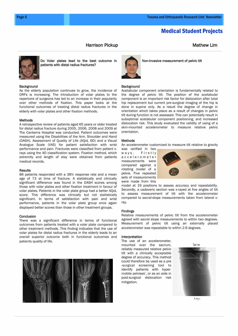
Non-invasive measurement of pelvic tilt
Background
Acetabular component orientation is fundamentally related to
the degree of pelvic tilt. The position of the acetabular
component is an important risk factor for dislocation after total
hip replacement but current pre-surgical imaging of the hip is
done in supine only. As a result the degree of change in
orientation which takes place as a result of changes in pelvic
tilt during function is not assessed. This can potentially result in
suboptimal acetabular component positioning, and increased
dislocation risk. This study evaluated the validity of using of a
skin-mounted accelerometer to measure relative pelvic
orientation.
Methods
An accelerometer customized to measure tilt relative to gravity
was verified in two
w a y s . F i r s t l y
a c c e l e r o m e t e r
measurements were
compared against a
rotating model of a
pelvis. Five repeated
sets of measurements
were made from this
model at 19 positions to assess accuracy and repeatability.
Secondly, a cadaveric section was x-rayed at five angles of tilt
to assess measurement of tilt with the accelerometer
compared to sacral-slope measurements taken from lateral x-
ray.
Findings
Relative measurements of pelvic tilt from the accelerometer
agreed with sacral slope measurements to within two degrees.
Measurement of pelvic tilt using an externally placed
accelerometer was repeatable to within 2.6 degrees.
Interpretation
The use of an accelerometer,
mounted over the sacrum,
reliably measured relative pelvic
tilt with a clinically acceptable
degree of accuracy. This method
could therefore be used as a pre
-surgical screening tool to
identify patients with hyper-
mobile pelvises’, or as an aide in
post-surgical dislocation risk
mitigation.
Page 7 Volume 7, 2014
Medical Student Projects
New 2014 Medical Students
Mike Van Alphen
Patient-reported outcomes
following mid-clavicular
fractures: Non-operative vs
Operative Management
To fix or not to fix – that is a
question which has caused some heated
exchange in our clinical meetings. This
project aims to ascertain whether the 5
year outcomes for patients who have had
surgical fixation for a displaced mid-
clavicular fracture are better than those
treated non-operatively. Mike is an ANU
medical student with a keen interest in
sports and orthopaedic injuries, having
come from a physiotherapy background.
Sarah-Jane Meresfield
Life After Arthroplasty –
when will I have to have
another joint replacement?
For 10 years TORU has been
collecting outcome data
from patients who have had their knees
and hips replaced in the ACT. This data is
now being plundered for a study which
aims to examine and describe the effect
of having an arthroplasty on the likelihood
of having another and how soon. This
data will enable both patients and
clinicians to better understand what their
orthopaedic future is likely to be. Sarah
Jane Merefield is former public servant/
Antarctic zoology student, and entered
medicine in 2014 to find out why my knee
always hurts.
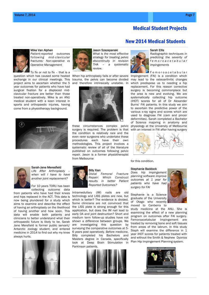
Jason Szezepanski
What is the most effective
strategy for treating pelvic
discontinuity in revision
THA – a systematic
review.
When hip arthroplasty fails or after severe
trauma, the pelvis can become divided
and therefore intrinsically unstable. In
these circumstances complex pelvic
surgery is required. The problem is that
the condition is relatively rare and the
even rarer surgeons who undertake these
procedures each have their own
methodologies. This project involves a
systematic review of all of the literature
published on outcomes following pelvic
repair. Jason is a former physiotherapist
from Melbourne
Billy Xian
Distal Femoral Fracture
Repair: Which Construct
results in better Patient
Reported Outcomes?
Intramedullary (IM) nails are old
technology and LISS plates are new, but
which is better? The evidence is divided.
Some clinicians are not convinced that
the LISS plate is strong enough for this
application, but does the IM nail lead to
early OA and joint destruction? Short and
medium term follow-up studies have not
shown a difference between groups. We
are investigating this question by
surveying the comparative outcomes at 5-
6 years post operatively. Before medicine,
Billy completed his Bachelors and
Masters degree in Canada, specifically
look at Deep Brain Stimulation in
Parkinson patients.
Sarah Ellis
Radiographic techniques in
predicting the severity of
f e m o r o a c e t a b u l a r
impingements
F e m o r o a c e t a b u l a r
Impingement (FAI) is a condition which
may lead to the osteoarthritic changes
which predispose us to needing a hip
replacement. For this reason corrective
surgery is becoming commonplace but
the area is new and evolving. We are
systematically collecting hip outcome
(iHOT) scores for all of Dr Alexander
Burns’ FAI patients. In this study we aim
to ascertain the predictive power of the
various x-ray signs and scores which are
used to diagnose FAI (cam and pincer
deformities). Sarah completed a Bachelor
of Science majoring in anatomy and
physiology at the University of Melbourne
with an interest in FAI after having surgery
for this condition.
Stephanie Baddock
Does hip impingement
planning software improve
outcomes at 1 year for
patients who have had
surgery for FAI
Stephanie is a Science
graduate of the University
of Otago who recently
moved to Canberra to
study medicine at the ANU. She is
examining the effect of a new planning
program on outcomes after FAI surgery.
Femoroacetabular Impingement are
treated by removing bone and soft tissues
from areas of the labrum. In this study
Steph will examine the difference in 1
year iHOT scores for patients treated with
and without the Smith & Nephew Dyonics
Plan Hip Impingement Planning system.
Page 8 Trauma and Orthopaedic Research Unit Newsletter
PhD Scholars
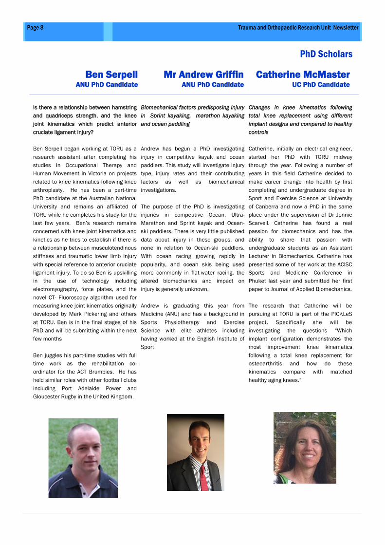
Ben Serpell ANU PhD Candidate
Mr Andrew Griffin ANU PhD Candidate
Catherine McMaster UC PhD Candidate
Is there a relationship between hamstring
and quadriceps strength, and the knee
joint kinematics which predict anterior
cruciate ligament injury?
Ben Serpell began working at TORU as a
research assistant after completing his
studies in Occupational Therapy and
Human Movement in Victoria on projects
related to knee kinematics following knee
arthroplasty. He has been a part-time
PhD candidate at the Australian National
University and remains an affiliated of
TORU while he completes his study for the
last few years. Ben’s research remains
concerned with knee joint kinematics and
kinetics as he tries to establish if there is
a relationship between musculotendinous
stiffness and traumatic lower limb injury
with special reference to anterior cruciate
ligament injury. To do so Ben is upskilling
in the use of technology including
electromyography, force plates, and the
novel CT- Fluoroscopy algorithm used for
measuring knee joint kinematics originally
developed by Mark Pickering and others
at TORU. Ben is in the final stages of his
PhD and will be submitting within the next
few months
Ben juggles his part-time studies with full
time work as the rehabilitation co-
ordinator for the ACT Brumbies. He has
held similar roles with other football clubs
including Port Adelaide Power and
Gloucester Rugby in the United Kingdom.
Biomechanical factors predisposing injury
in Sprint kayaking, marathon kayaking
and ocean paddling
Andrew has begun a PhD investigating
injury in competitive kayak and ocean
paddlers. This study will investigate injury
type, injury rates and their contributing
factors as well as biomechanical
investigations.
The purpose of the PhD is investigating
injuries in competitive Ocean, Ultra-
Marathon and Sprint kayak and Ocean-
ski paddlers. There is very little published
data about injury in these groups, and
none in relation to Ocean-ski paddlers.
With ocean racing growing rapidly in
popularity, and ocean skis being used
more commonly in flat-water racing, the
altered biomechanics and impact on
injury is generally unknown.
Andrew is graduating this year from
Medicine (ANU) and has a background in
Sports Physiotherapy and Exercise
Science with elite athletes including
having worked at the English Institute of
Sport
Changes in knee kinematics following
total knee replacement using different
implant designs and compared to healthy
controls
Catherine, initially an electrical engineer,
started her PhD with TORU midway
through the year. Following a number of
years in this field Catherine decided to
make career change into health by first
completing and undergraduate degree in
Sport and Exercise Science at University
of Canberra and now a PhD in the same
place under the supervision of Dr Jennie
Scarvell. Catherine has found a real
passion for biomechanics and has the
ability to share that passion with
undergraduate students as an Assistant
Lecturer in Biomechanics. Catherine has
presented some of her work at the ACISC
Sports and Medicine Conference in
Phuket last year and submitted her first
paper to Journal of Applied Biomechanics.
The research that Catherine will be
pursuing at TORU is part of the PICKLeS
project. Specifically she will be
investigating the questions “Which
implant configuration demonstrates the
most improvement knee kinematics
following a total knee replacement for
osteoarthritis and how do these
kinematics compare with matched
healthy aging knees.”
Page 9 Trauma and Orthopaedic Research Unit Newsletter
Use of heparanase as a potential bio-
marker in rheumatoid arthritis.
Olympia John as a Ph.D. student joined
TORU laboratory research team since
February 2014 under the supervision of
Dr Rachel Li, Professors Paul Smith and
Chris Parish. She obtained her Masters in
Pharmacy from C.U. Shah College of Phar-
macy, Shreemati Nathibhai Thackersey
(SNDT), Mumbai, India. She has also com-
pleted The Postgraduate Diploma in Intel-
lectual Property Management from Acad-
emy of Intellectual Property Studies
(AIPS), Mumbai, India. She began her
career as a Formulator in Cosmetics
(Haircare and Skincare) at Emami Ltd,
India. Her last assignment involved formu-
lating ANDA (abbreviated new drug appli-
cations) Generics for US and European
markets at Glenmark Generics Limited
from 2006 to 2010. She has over 6 years
of experience in pharmaceutical sciences.
She is currently working at TORU Labora-
tory located at John Curtin School of
Medical Research, Australian National
University. Her research involves hepara-
nase as a potential biomarker in rheuma-
toid arthritis.
A Bioinformatics approach to establish an
osteo-network: Osteomics
Song is a PhD student of ANU. Song
gained his bachelor of applied physics
from Shanghai JiaoTong University in
China and master of engineering from
ANU. He has a background in theoretical
physics and computational analysis of
engineering materials.
Song’s PhD project is to investigate inter-
actions at the interfaces among pathways
of multiple systems in bone remodelling.
His work is currently focusing on re-
building signalling pathways in os-
teoblasts and osteoclasts by mathemati-
cal description and proving this descrip-
tion by designing the experiment to treat
osteoblasts, osteoclasts and co-culture of
osteoblasts and osteoclasts under physi-
cal stimulus from low frequency electro-
magnetic field. Song’s PhD project is su-
pervised from both TORU and the college
of engineering and computer science in
ANU.
Simulation of wear in total knee replace-
ment using finite element analysis
Obinna is a PhD student at UNSW Can-
berra. He obtained his Masters degree in
2012 at the same university. His current
research interest is in the area of pros-
thetic devices for joint replacements.
His PhD research work is on the investiga-
tion of wear of total hip replacement at
the taper-trunnion junction. Recently, it’s
been identified that excessive fretting
wear at the taper-trunnion (head-neck)
junction potentially contributes to prema-
ture failure of some total hip replacement
procedures.
The project aims to develop novel meth-
ods for investigating, evaluating and
quantifying wear of total hip prostheses at
the taper-trunnion junction by employing
numerical methods via finite element
modelling. In a broader sense, the princi-
pal goal is to work toward the minimiza-
tion of wear debris produced in the hip
joint, thereby resulting in a longer pros-
thetic lifetime. This work is supported by
Global Orthopaedics.
PhD Scholars
Olympia Noronha ANU PhD Candidate
Song Chen ANU PhD Candidate
Obinna Ihulsior UNSW @ ADFA PhD Candidate
Page 10 Volume 7, 2014
Pickles Knee Study—A prospective imaging study of cruciate retaining and substituting
knee replacement, in osteoarthritis and healthy aging
Fracture Database Genetic Markers of OA Pressure Sensitive Carbon-
Nanotube Film Study
The fracture database is a unique tool with
the capacity to make every patient admitted
to the Canberra Hospital for fracture
management a study participant. This cloud
based tool enables remote surveillance and
evaluation of longitudinal outcomes in our
whole patient population. Of course
complete capture will not be possible but
this tool puts in place the capacity to track
outcomes using current technology while
providing the perfect test bed for new
technology surveillance. This database went
live in August of this year. After embedded
testing and editing we will investigate the
possibilities of making this tool available to
other jurisdictions. This exciting initiative is
a leap forward in data collection and patient
evaluation.
Nanoscale wear particle related osteolysis
in total joint replacement (TJR) and
genetic markers
This research has been in part supported by
AOA Research Foundation and discovered
that the dendritic cells is involved in
osteolysis. Current work now focuses on a
b e t t e r u n d e r s t a n d i n g o f t h e
osteoimmunology, human tissue’s
responses to implants, microRNAs’ (miRNA)
variant expression and miRNA sequencing
in the wear particle related osteolysis, as
well as the translation of this knowledge to
the clinical management of a variety of
diseases and conditions related to TJR.
TORU Laboratory has used off-cut tissues
from the cohorts of healthy, primary and
revision subjects of TJR for the
characterization of the size and shape of
wear particles and for the identification of
genetic and environmental risk factors that
contribute to the osteolysis. The ultimate
goals are to contribute to the development
of better predictive markers, treatments,
and prevention strategies of the osteolysis.
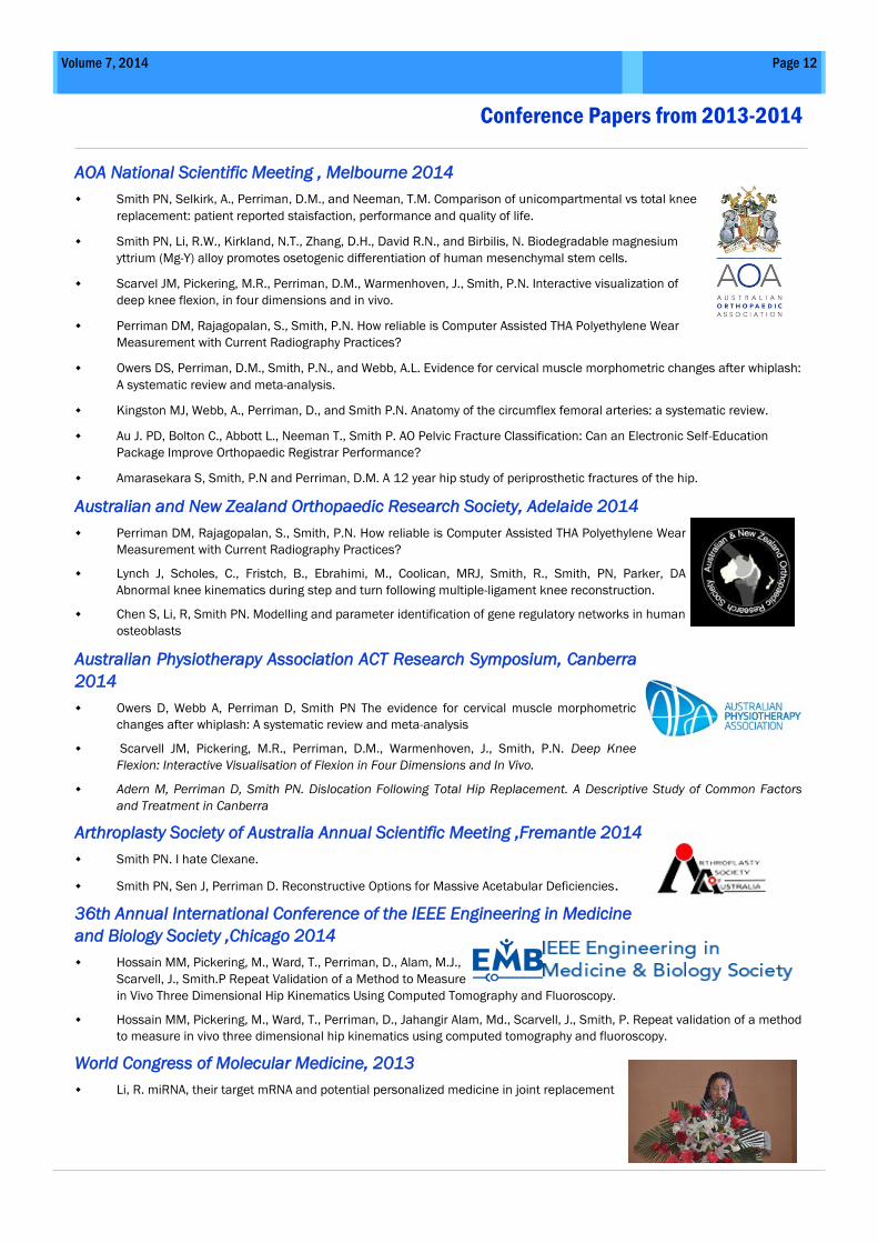
This project is the first stage in the
development of an innovative new pressure
sensing technology which will seamlessly
interface with ‘smart’ wireless data
acquisition.
The specific aims of this research are to:
1. Develop pressure-sensitive plastic
films doped with carbon nanotubes.
2. Test the potential for these films to be
used with embedded microcomputers
as sensors in knee replacements.
3. Test the applicability as shoe inserts
for mapping of load patterns in running
and jumping sports and in the military.
4. Secure funding with a view to
commercialisation of the product all
possible applications.
The project was just one of a small number
which were funded by ACT health in 2013-
14. Dr Sean O’Byrne at UNSW has
commenced the preliminary work involved
in fabrication and validation. TORU will be
involved in testing the sensor in its knee
simulator.
This study aims to examine knee kinematics
before and after knee arthroplasty and
compare those to the kinematics of knees
in a non-arthritic age-matched cohort. In the
past the only way of measuring knee
kinematics accurately in three planes was
to implant RSA beads or use of bone pins
These methods are both highly invasive. In
this study we aim to overcome this problem
by using an image registration technology
developed at TORU by Prof Smith, Assoc
Prof Jennie Scarvell and Assoc Prof Mark
Pickering by combining 3D CT and 2D video
fluoroscopy. This study is unique because,
for the first time, knee replacement patients
will have their knee kinematics accurately
measured both before and after surgery.
Recruitment is underway for normal and
osteoarthritic participants where their knee
is scanned while they perform a number of
loaded end-of-range activities. These
movements allow us to see how the knee
kinematics change following surgery. OA
patients are randomised to receive one of
three different design of implant. Testing is
being done by the TORU staff in
combination with the medical imaging
department at The Canberra Hospital.
Catherine McMaster has recently joined the
project and this will make up the bulk of her
PhD work.
This study has been funded by the Canberra
hospital Private Practice Fund, the
University of Canberra and Biomet.
Page 11 Trauma and Orthopaedic Research Unit Newsletter
Whiplash is a term describing a range of injuries caused by or related to sudden
distortion of the neck. It is commonly associated with motor vehicle accidents, usually
when the vehicle has been hit in the rear.
Injuries to the cervical spine following motor vehicle accidents (whiplash injury) are
one of the most common causes of neck pain and disability in the developed world
with significant numbers likely to develop long lasting symptoms. In the 2007/2008
financial year, the annual financial costs associated with whiplash injuries were
approximated to be $AUD 700 million in New South Wales alone. To date there are no
reliable physical markers with which to unequivocally diagnose whiplash injury or the
degree of severity.
Dr Alex Webb has recently developed methods for quantifying the morphometry of
the synovial folds in the cervical spine using MR imaging. These structures may represent structures of interest with respect to whiplash
diagnosis and prognostication.
The purpose of this study is to investigate the use of 3T magnetic resonance imaging (MRI) to detect structural damage to synovial folds
and surrounding structures in whiplash patients with neck pain caused by motor vehicle accident.
In this study, participants who have both acute and chronic neck pain following a motor vehicle accident will be compared with
participants who do not have neck pain using MRI as well as clinical tools which have previously been used to determine the severity
and prognosis of whiplash injury.
Dr Monah Singal has led the recruitment for this study and is currently looking for patients who have either acute or chronic whiplash
symptoms and healthy volunteers, We expect recruitment to finish in early 2015.
TORU has also partnered with Assoc Prof Mark Pickering from UNSW @ ADFA and Assoc Prof Girija Chetty from UC to develop a method
of automatically segmenting specific muscle groups that surround the cervical spine using 3D MRI.
Characterising whiplash injury using magnetic resonance imaging
Short, Medium and Long Term Survivorship of Attune Primary® Total Knee
Prostheses
Arthroplasty Database
The arthroplasty database is also an online tool which introduced in
the private sector in 2009 and we hope will be taken up by ACT
health in the future. The data provided by this tool serves to pro-
vide clinical surveillance reports and longitudinal outcome collec-
tion but also has some unique functions for management. The data
collected has the potential to facilitate waiting list categorization
and follow-up frequency management thereby improving efficiency
while remaining responsive. The database is currently being
streamlined.
TORU recently began a trial looking at the long term survivorship of the AT-
TUNE® cemented primary Total Knee Arthroplasty (TKA) system for 4 differ-
ent implant configurations. Data from this study will be used to support
post-market surveillance of the ATTUNE® Knee System for the 4 implant
configurations, which are (1) Cruciate retaining fixed bearing (2) Cruciate
retaining rotating platform (3) Posterior stabilized fixed bearing and (4) Pos-
terior stabilized rotating platform. The study is being run in collaboration
with Orthopaedic ACT, specifically, Dr Al Burns and Dr Damian Smith. Sub-
jects who are eligible will be evaluated in clinic prior to surgery, follow-up
clinic visits at less than 1 year, and at minimum 1, minimum 2, minimum 5,
minimum 10, and minimum 15 years post surgery. Visit evaluations include
clinical and radiographic evaluations, subject self assessment question-
naires and check for adverse events. This study is funded by Depuy Synthes.
Page 12 Volume 7, 2014
AOA National Scientific Meeting , Melbourne 2014
Smith PN, Selkirk, A., Perriman, D.M., and Neeman, T.M. Comparison of unicompartmental vs total knee
replacement: patient reported staisfaction, performance and quality of life.
Smith PN, Li, R.W., Kirkland, N.T., Zhang, D.H., David R.N., and Birbilis, N. Biodegradable magnesium
yttrium (Mg-Y) alloy promotes osetogenic differentiation of human mesenchymal stem cells.
Scarvel JM, Pickering, M.R., Perriman, D.M., Warmenhoven, J., Smith, P.N. Interactive visualization of
deep knee flexion, in four dimensions and in vivo.
Perriman DM, Rajagopalan, S., Smith, P.N. How reliable is Computer Assisted THA Polyethylene Wear
Measurement with Current Radiography Practices?
Owers DS, Perriman, D.M., Smith, P.N., and Webb, A.L. Evidence for cervical muscle morphometric changes after whiplash:
A systematic review and meta-analysis.
Kingston MJ, Webb, A., Perriman, D., and Smith P.N. Anatomy of the circumflex femoral arteries: a systematic review.
Au J. PD, Bolton C., Abbott L., Neeman T., Smith P. AO Pelvic Fracture Classification: Can an Electronic Self-Education
Package Improve Orthopaedic Registrar Performance?
Amarasekara S, Smith, P.N and Perriman, D.M. A 12 year hip study of periprosthetic fractures of the hip.
Australian and New Zealand Orthopaedic Research Society, Adelaide 2014
Perriman DM, Rajagopalan, S., Smith, P.N. How reliable is Computer Assisted THA Polyethylene Wear
Measurement with Current Radiography Practices?
Lynch J, Scholes, C., Fristch, B., Ebrahimi, M., Coolican, MRJ, Smith, R., Smith, PN, Parker, DA
Abnormal knee kinematics during step and turn following multiple-ligament knee reconstruction.
Chen S, Li, R, Smith PN. Modelling and parameter identification of gene regulatory networks in human
osteoblasts
Australian Physiotherapy Association ACT Research Symposium, Canberra
2014
Owers D, Webb A, Perriman D, Smith PN The evidence for cervical muscle morphometric
changes after whiplash: A systematic review and meta-analysis
Scarvell JM, Pickering, M.R., Perriman, D.M., Warmenhoven, J., Smith, P.N. Deep Knee
Flexion: Interactive Visualisation of Flexion in Four Dimensions and In Vivo.
Adern M, Perriman D, Smith PN. Dislocation Following Total Hip Replacement. A Descriptive Study of Common Factors
and Treatment in Canberra
Arthroplasty Society of Australia Annual Scientific Meeting ,Fremantle 2014
Smith PN. I hate Clexane.
Smith PN, Sen J, Perriman D. Reconstructive Options for Massive Acetabular Deficiencies.
36th Annual International Conference of the IEEE Engineering in Medicine
and Biology Society ,Chicago 2014
Hossain MM, Pickering, M., Ward, T., Perriman, D., Alam, M.J.,
Scarvell, J., Smith.P Repeat Validation of a Method to Measure
in Vivo Three Dimensional Hip Kinematics Using Computed Tomography and Fluoroscopy.
Hossain MM, Pickering, M., Ward, T., Perriman, D., Jahangir Alam, Md., Scarvell, J., Smith, P. Repeat validation of a method
to measure in vivo three dimensional hip kinematics using computed tomography and fluoroscopy.
World Congress of Molecular Medicine, 2013
Li, R. miRNA, their target mRNA and potential personalized medicine in joint replacement
Conference Papers from 2013-2014
Page 13 Trauma and Orthopaedic Research Unit Newsletter
Conference Papers from 2013-2014
International Congress for Joint Reconstruction Sydney 2014
Smith P. How to manage severe angular deformities when doing TKR
Smith P. Femoral Revision: What are the options? 2014
AOA ACT Branch Scientific Meeting, Canberra 2013
Ward T, Perriman, D., Hossain, M.M., Scarvell, J., Pickering, M., Smith, P. Repeat validation of a method to
measure in vivo three dimensional hip kinematics using computed tomography and fluoroscopy.
Amarasekara S, Thabet, A., Smith, P., Shankar, P., Perriman, D. A biomechanical comparison of femoral
fracture fixation methods.
Chen S, Qin, Q., Zhang, D., Smith, P., Li, R. The influence of extremely low frquency electromagnetic field on
human osteoblasts.
Rajagopalan S, Perriman, D., Neeman, T., Smith, P. Arthroplasty Surveillance: Are radiology reports useful?
O'Byrne S, Liu, M., Zhang, L., Perriman, D., Smith, P. Piezoelectric polymer sensors for orthopaedic application.
Avakian Z, Wardle, B., Perriman, D., Smith, P. Results after one year of experience using tranexamic acid for total knee
arthroplasty
McLellan M, Perriman, D., Smith, P. The Relationship Between Metallosis and Patient Quality of Life Following Birmingham
Hip Resurfacing.
Ardern M, Perriman, D., Smith. Dislocation following total hip replacement, a descriptive study of common factors and
treatment in Canberra.
AU J, Perriman, D., Bolton, C., Abbott, L., Neeman, T., Smith, P. Pelvic Fracture Classification: Can an Educational Package
Improve Orthopaedic Registrar Performance?
Perriman D, Wang, R., Smith, P. The Fracture Surveillance Database.
Owen D, Russell, N., Walter, W., Smith, P. Ceramic on ceramic total hip arthroplasty squeak: an estimation of the incidence
of squeak and revision surgery for squeak
Bolton C, Perriman, D., Griffin, A., Smith, P., Neeman, T. The Transverse Acetabular Ligament: Does age affect its
relationship with the acetabulum?
Selkirk A, Perriman, D., Smith, P.N. Comparison of Unicompartmental vs Total Knee Joint Replacement: Patient Reported
Satisfaction, Performance and Quality of Life.
European Hip Society 11th Congress, Stockholm 2014
Rajagopolan S, Perriman, D., Neeman, T., Smith, P. "Arthroplasty Surveillance: Are radiology reports
useful?"
Rajagopalan S. Arthroplasty Surveillance: Are radiology reports useful?
Canberra Health Annual Research Meeting, 2014
Owers DS, Perriman, D., Smith, P.N., Webb, A.L. The evidence for cervical muscle morphometric
changes after whiplash: A systematic review and meta-analysis. Poster
Scarvell JM, Pickering, M.R., Perriman, D.M., Warmenhoven, J., Smith, P.N.
Interactive visualisation of deep knee flexion, in four dimensions and in
vivo.
Li RW, Smith, P.N., Birbilis, N. Biodegradable Magnesium Yttrium (Mg-Y)
Alloy Promotes Osteogenic Differentiation of Human Mesenchymal Stem Cells (hMSCs).
Ardern MK, Perriman, D.M., Smith, P.N. Dislocation following total hip replacement, a descriptive study of common factors
and treatment in Canberra. CHARM 2014.
Perriman DM, Rajagopalan, S., Smith, P.N. How reliable is Computer Assisted THA Polyethylene Wear Measurement with
Current Radiography Practices?
Lynch J, Scholes, C., Fristch, B., Ebrahimi, M., Coolican, M.R.J., Smith, R., Smith, P.N., Parker.D.A. Abnormal knee
kinematics during step and turn following multiple-ligament knee reconstruction.
Page 14 Volume 7, 2014
Journal Articles 2013-2014
Robust initialisation for single-plane 3D
CT to 2D fluoroscopy image registra-
tion.
The Bonar score revisited: Region of
evaluation significantly influences the
standardized assessment of tendon
degeneration.
Standing or supine x-rays after Total
Hip Replacement - When is the safe
zone not safe?
Akter, M., Lambert, A.J., Pickering,
M.R., Scarvell, J.M. & P.N. Smith.
Computer Methods in Biomechanics and
Biomedical Engineering: Imaging &
Visualization
Gradient descent-based automatic image
registration algorithms typically fail when
the initial misalignment between objects
is large. This is a major limitation for
routine clinical applications. The
registration task is even more difficult for
multi-modal images because of the
nonlinear relationship between the pixel
intensities in the images to be aligned. In
this paper, we present a fast and
accurate multi-modal image registration
algorithm which successfully registers
three-dimensional (3D) computed
tomography to two-dimensional single-
plane fluoroscopy data for large initial
displacements between the images. Our
experimental results show that the
proposed approach can increase the
range of initial displacements up to
± 20 mm for all translations and up to
± 20° for rotation while maintaining high
precision and small bias in all six 3D rigid
body transform parameters.
Fearon, A., Dahlstrom, J. E., Twin, J., Cook,
J., & Scott, A
Journal of Science and Medicine in Sport
Background: Tendinopathy is a common,
costly condition affecting both sporting
and sedentary populations. Research into
tendinopathy frequently involves the
evaluation of tendinosis, a pathology
characterized by a lack of inflammatory
c e l l s , c o l l a g e n d i s r u p t i o n ,
neovascularisation, altered cell numbers
and morphology and increased
glycosaminoglycans. Evaluation of these
characteristics can be undertaken using
the Bonar histopathology score, but the
characteristics are heterogeneous
throughout tendon specimens with no
standardized method of determining the
area to be evaluated. The objective of this
study was to assess whether the Bonar
score varies depending on the criteria
used to define the area of evaluation.
Methods: Two independent assessors,
with a third to resolve disputes, evaluated
103 areas from 35 tendon specimens
using the Bonar score. Specimens were
scored once each in the area of worst
collagen disruption, degree of
vascularization, and cell morphological
changes. The inter-tester reliability of the
updated Bonar scale was good
(r(2)=0.71)
Results: The Bonar score was highest in
the areas of worst cell morphological (CM)
changes, followed by collagen disruption
(CD) and lowest for the area of most
extensive vascular proliferation (VS)
(regression: CD vs. CM, p=0.008, CM vs.
VS, p<0.001, CD vs. VS, p=0.013).
Suggested modifications to the Bonar
score include the addition of a cellularity
domain, specific definitions of hypo- and
hypercellularity, and changes to the
vascularity score to include pathological
avascularity.
Conclusions: The updated Bonar score
includes a standardized method of
selecting the area of evaluation, which
should provide increased reliability when
assessing the extent of tendon
degeneration.
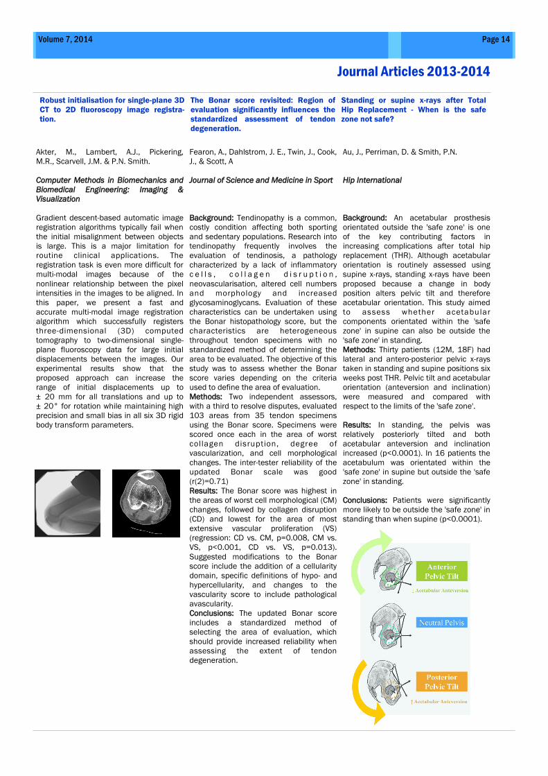
Au, J., Perriman, D. & Smith, P.N.
Hip International
Background: An acetabular prosthesis
orientated outside the 'safe zone' is one
of the key contributing factors in
increasing complications after total hip
replacement (THR). Although acetabular
orientation is routinely assessed using
supine x-rays, standing x-rays have been
proposed because a change in body
position alters pelvic tilt and therefore
acetabular orientation. This study aimed
to assess whether acetabular
components orientated within the 'safe
zone' in supine can also be outside the
'safe zone' in standing.
Methods: Thirty patients (12M, 18F) had
lateral and antero-posterior pelvic x-rays
taken in standing and supine positions six
weeks post THR. Pelvic tilt and acetabular
orientation (anteversion and inclination)
were measured and compared with
respect to the limits of the 'safe zone'.
Results: In standing, the pelvis was
relatively posteriorly tilted and both
acetabular anteversion and inclination
increased (p<0.0001). In 16 patients the
acetabulum was orientated within the
'safe zone' in supine but outside the 'safe
zone' in standing.
Conclusions: Patients were significantly
more likely to be outside the 'safe zone' in
standing than when supine (p<0.0001).
Page 15 Trauma and Orthopaedic Research Unit Newsletter
Journal Articles 2013-2014
Greater trochanteric pain syndrome
negatively affects work, physical activ-
ity and quality of life: a case control
study
Increased substance P expression in
the trochanteric bursa of patients with
greater trochanteric pain syndrome
Hip Fracture After Stroke in the Elderly:
Trends in the Beginning of the 21st
Century and Projections into the Future
in Australia
Fearon, A. M., Cook, J. L., Scarvell, J. M.,
Neeman, T., Cormick, W., & Smith, P. N.
Journal of Arthroplasty
Musculoskeletal injury causes pain and
when chronic can affect mental health,
employment and quality of life. This study
examined work participation, function and
quality of life in people with greater
trochanteric pain syndrome (GTPS, n=42),
severe hip osteoarthritis (OA, n=20) and
an asymptomatic group (ASC, n=23). No
differences were found between the
symptomatic groups on key measures,
both were more affected than the ASC
group, they had lower quality of life score
(p<0.001), Harris Hip Score (p<0.001)
and higher Oswestry Disability Index
(p<0.001). Participants with GTPS were
the least likely to be in fulltime work
(prob. GTPS=0.29; OA=0.52; and
ASC=0.68). GTPS appears to confer levels
of disability and quality of life similar to
levels associated with end stage hip OA. :
Fearon, A. M., Twin, J., Dahlstrom, J. E.,
Cook, J. L., Cormick, W., Smith, P. N.,
Scott A
Rheumatology International
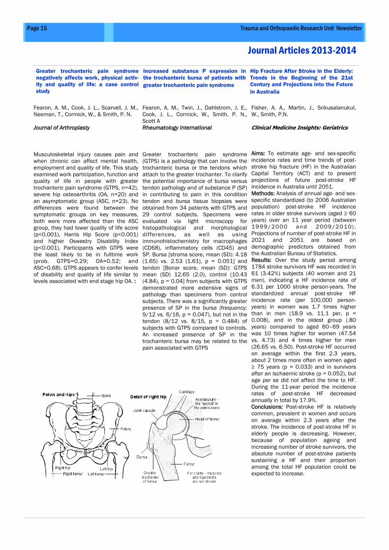
Greater trochanteric pain syndrome
(GTPS) is a pathology that can involve the
trochanteric bursa or the tendons which
attach to the greater trochanter. To clarify
the potential importance of bursa versus
tendon pathology and of substance P (SP)
in contributing to pain in this condition
tendon and bursa tissue biopsies were
obtained from 34 patients with GTPS and
29 control subjects. Specimens were
evaluated via light microscopy for
histopathological and morphological
differences, as well as using
immunohistochemistry for macrophages
(CD68), inflammatory cells (CD45) and
SP. Bursa [stroma score, mean (SD): 4.18
(1.65) vs. 2.53 (1.61), p = 0.051] and
tendon [Bonar score, mean (SD): GTPS
mean (SD) 12.65 (2.0), control (10.43
(4.84), p = 0.04] from subjects with GTPS
demonstrated more extensive signs of
pathology than specimens from control
subjects. There was a significantly greater
presence of SP in the bursa (frequency:
9/12 vs. 6/16, p = 0.047), but not in the
tendon (8/12 vs. 8/15, p = 0.484) of
subjects with GTPS compared to controls.
An increased presence of SP in the
trochanteric bursa may be related to the
pain associated with GTPS
Fisher, A. A., Martin, J., Srikusalanukul,
W., Smith, P.N.
Clinical Medicine Insights: Geriatrics
Aims: To estimate age- and sex-specific
incidence rates and time trends of post-
stroke hip fracture (HF) in the Australian
Capital Territory (ACT) and to present
projections of future post-stroke HF
incidence in Australia until 2051.
Methods: Analysis of annual age- and sex-
specific standardized (to 2006 Australian
population) post-stroke HF incidence
rates in older stroke survivors (aged ≥ 60
years) over an 11 year period (between
1999/2000 and 2009/2010) .
Projections of number of post-stroke HF in
2021 and 2051 are based on
demographic predictors obtained from
the Australian Bureau of Statistics.
Results: Over the study period among
1784 stroke survivors HF was recorded in
61 (3.42%) subjects (40 women and 21
men), indicating a HF incidence rate of
6.31 per 1000 stroke person-years. The
standardized annual post-stroke HF
incidence rate (per 100,000 person-
years) in women was 1.7 times higher
than in men (18.9 vs. 11.1 per, p =
0.008), and in the oldest group (.80
years) compared to aged 60–69 years
was 10 times higher for women (47.54
vs. 4.73) and 4 times higher for men
(26.65 vs. 6.50). Post-stroke HF occurred
on average within the first 2.3 years,
about 2 times more often in women aged
≥ 75 years (p = 0.033) and in survivors
after an ischaemic stroke (p = 0.052), but
age per se did not affect the time to HF.
During the 11-year period the incidence
rates of post-stroke HF decreased
annually in total by 17.9%.
Conclusions: Post-stroke HF is relatively
common, prevalent in women and occurs
on average within 2.3 years after the
stroke. The incidence of post-stroke HF in
elderly people is decreasing. However,
because of population ageing and
increasing number of stroke survivors, the
absolute number of post-stroke patients
sustaining a HF and their proportion
among the total HF population could be
expected to increase.
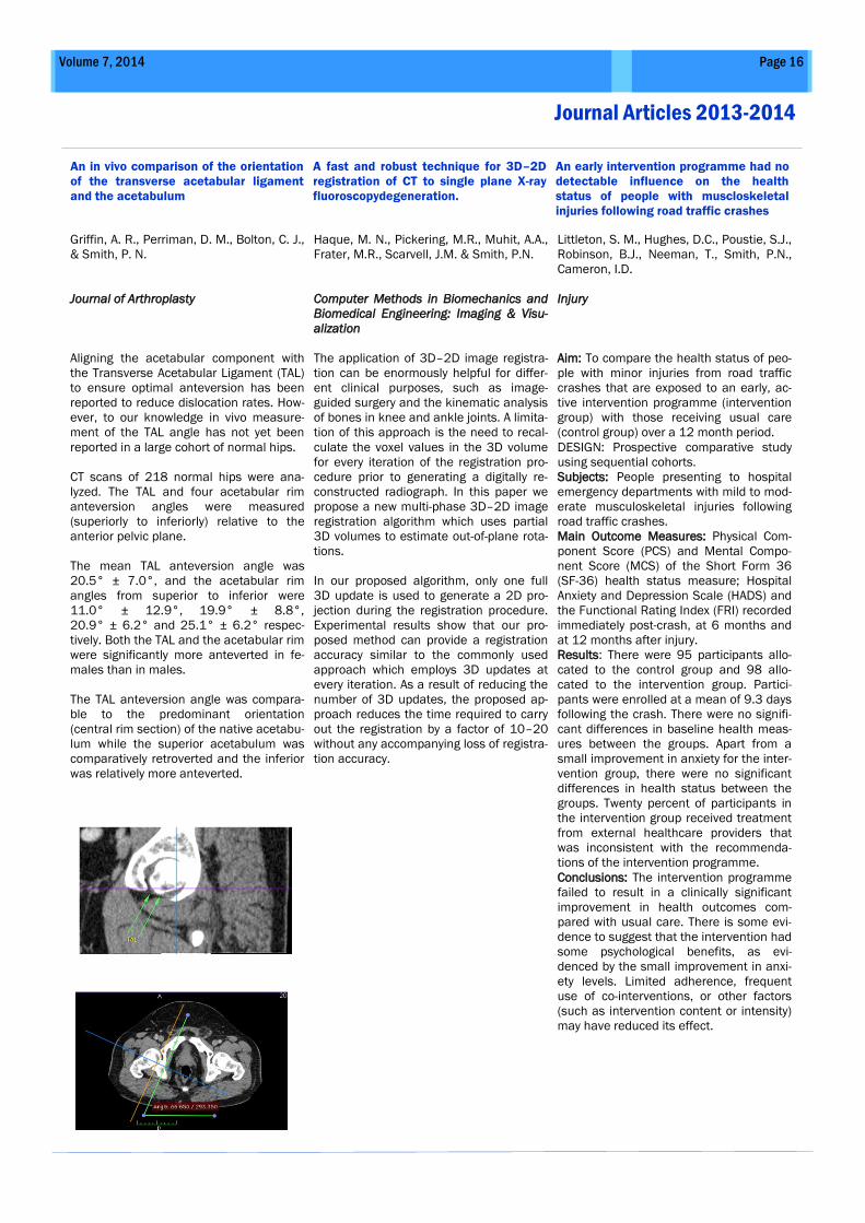
Griffin, A. R., Perriman, D. M., Bolton, C. J.,
& Smith, P. N.
Journal of Arthroplasty
Aligning the acetabular component with
the Transverse Acetabular Ligament (TAL)
to ensure optimal anteversion has been
reported to reduce dislocation rates. How-
ever, to our knowledge in vivo measure-
ment of the TAL angle has not yet been
reported in a large cohort of normal hips.
CT scans of 218 normal hips were ana-
lyzed. The TAL and four acetabular rim
anteversion angles were measured
(superiorly to inferiorly) relative to the
anterior pelvic plane.
The mean TAL anteversion angle was
20.5° ± 7.0°, and the acetabular rim
angles from superior to inferior were
11.0° ± 12.9°, 19.9° ± 8.8°,
20.9° ± 6.2° and 25.1° ± 6.2° respec-
tively. Both the TAL and the acetabular rim
were significantly more anteverted in fe-
males than in males.
The TAL anteversion angle was compara-
ble to the predominant orientation
(central rim section) of the native acetabu-
lum while the superior acetabulum was
comparatively retroverted and the inferior
was relatively more anteverted.
Haque, M. N., Pickering, M.R., Muhit, A.A.,
Frater, M.R., Scarvell, J.M. & Smith, P.N.
Computer Methods in Biomechanics and
Biomedical Engineering: Imaging & Visu-
alization
The application of 3D–2D image registra-
tion can be enormously helpful for differ-
ent clinical purposes, such as image-
guided surgery and the kinematic analysis
of bones in knee and ankle joints. A limita-
tion of this approach is the need to recal-
culate the voxel values in the 3D volume
for every iteration of the registration pro-
cedure prior to generating a digitally re-
constructed radiograph. In this paper we
propose a new multi-phase 3D–2D image
registration algorithm which uses partial
3D volumes to estimate out-of-plane rota-
tions.
In our proposed algorithm, only one full
3D update is used to generate a 2D pro-
jection during the registration procedure.
Experimental results show that our pro-
posed method can provide a registration
accuracy similar to the commonly used
approach which employs 3D updates at
every iteration. As a result of reducing the
number of 3D updates, the proposed ap-
proach reduces the time required to carry
out the registration by a factor of 10–20
without any accompanying loss of registra-
tion accuracy.
Littleton, S. M., Hughes, D.C., Poustie, S.J.,
Robinson, B.J., Neeman, T., Smith, P.N.,
Cameron, I.D.
Injury
Aim: To compare the health status of peo-
ple with minor injuries from road traffic
crashes that are exposed to an early, ac-
tive intervention programme (intervention
group) with those receiving usual care
(control group) over a 12 month period.
DESIGN: Prospective comparative study
using sequential cohorts.
Subjects: People presenting to hospital
emergency departments with mild to mod-
erate musculoskeletal injuries following
road traffic crashes.
Main Outcome Measures: Physical Com-
ponent Score (PCS) and Mental Compo-
nent Score (MCS) of the Short Form 36
(SF-36) health status measure; Hospital
Anxiety and Depression Scale (HADS) and
the Functional Rating Index (FRI) recorded
immediately post-crash, at 6 months and
at 12 months after injury.
Results: There were 95 participants allo-
cated to the control group and 98 allo-
cated to the intervention group. Partici-
pants were enrolled at a mean of 9.3 days
following the crash. There were no signifi-
cant differences in baseline health meas-
ures between the groups. Apart from a
small improvement in anxiety for the inter-
vention group, there were no significant
differences in health status between the
groups. Twenty percent of participants in
the intervention group received treatment
from external healthcare providers that
was inconsistent with the recommenda-
tions of the intervention programme.
Conclusions: The intervention programme
failed to result in a clinically significant
improvement in health outcomes com-
pared with usual care. There is some evi-
dence to suggest that the intervention had
some psychological benefits, as evi-
denced by the small improvement in anxi-
ety levels. Limited adherence, frequent
use of co-interventions, or other factors
(such as intervention content or intensity)
may have reduced its effect.
Page 16 Volume 7, 2014
Journal Articles 2013-2014
An in vivo comparison of the orientation
of the transverse acetabular ligament
and the acetabulum
A fast and robust technique for 3D–2D
registration of CT to single plane X-ray
fluoroscopydegeneration.
An early intervention programme had no
detectable influence on the health
status of people with muscloskeletal
injuries following road traffic crashes
Opar, D. A., & Serpell, B. G.
Archives of Physical Medicine and Reha-
bilitation
Hamstring strain injuries (HSIs) are the
most prevalent injury in a number of
sports, and while ACL injuries are less
common, they are far more severe and
have long-term implications. Given the
high incidence and severity of these inju-
ries, they are key targets of injury preven-
tive programs in elite sport. Evidence has
shown that a previous knee injury
(including ACL injury) increases the risk of
HSI; however, whether the functional defi-
cits that occur after HSI result in an in-
creased risk of ACL injury has yet to be
considered. In this clinical commentary,
we present evidence that suggests that
the link between previous HSI and in-
creased risk of ACL injury requires further
investigation by drawing parallels between
deficits in hamstring function after HSI
and in women athletes, who are more
prone to ACL injury than men athletes.
Comparisons between the neuromuscular
function of the male and female hamstring
has shown that women display lower ham-
string-to-quadriceps strength ratios during
isokinetic knee flexion and extension,
increased activation of the quadriceps
compared with the hamstrings during a
stop-jump landing task, a greater time
required to reach maximal isokinetic ham-
string torque, and lower integrated
myoelectrical hamstring activity during a
sidestep cutting maneuver. Somewhat
similarly, in athletes with a history of HSI,
the previously injured limb, compared with
the uninjured limb, displays lower eccen-
tric knee flexor strength, a lower ham-
strings-to-quadriceps strength ratio, lower
voluntary myoelectrical activity during
maximal knee flexor eccentric contraction,
a lower knee flexor eccentric rate of
torque development, and lower voluntary
myoelectrical activity during the initial
portion of eccentric contraction. Given that
the medial and lateral hamstrings have
different actions at the knee joint in the
coronal plane, which hamstring head is
previously injured might also be expected
to influence the likelihood of future ACL.
Whether the deficits in function after HSI,
as seen in laboratory-based studies, trans-
late to deficits in hamstring function dur-
ing typical injurious tasks for ACL injury
has yet to be determined but should be a
consideration for future work.
Owen, D. H., Russell, N. C., Smith, P. N., &
Walter, W. L.
Bone and Joint Journal
Squeaking arising from a ceramic-on-
ceramic (CoC) total hip replacement (THR)
may cause patient concern and in some
cases causes patients to seek revision
surgery. We performed a meta-analysis to
determine the incidence of squeaking and
the incidence of revision surgery for
squeaking. A total of 43 studies including
16,828 CoC THR that reported squeaking,
or revision for squeaking, were entered
into the analysis. The incidence of squeak-
ing was 4.2% and the incidence of revision
for squeaking was 0.2%. The incidence of
squeaking in patients receiving the Acco-
lade femoral stem was 8.3%, and the inci-
dence of revision for squeaking in these
patients was 1.3%.
Serpell, B. G., Scarvell, J.M., Ball, N.B
& Smith, P.N.
Journal of Sports Sciences
The purpose of this study was to establish
if vertical stiffness was greater in profes-
sional Australian rules footballers who
sustained a lower limb skeletal muscle
strain compared to those who did not, and
to establish if a relationship between age,
or training history, and vertical stiffness
existed.
Thirty-one participants underwent weekly
rebound jump testing on a force platform
over two seasons. Vertical stiffness was
calculated for injured players and the un-
injured cohort 1 and 3 weeks prior to sus-
taining an injury and at the end of pre-
season. Eighteen athletes were in the
"uninjured" cohort and 13 in the "injured"
cohort.
No significant difference in vertical stiff-
ness was observed between groups
(P = 0.18 for absolute stiffness; P = 0.08
for stiffness relative to body mass), within
groups (P = 0.83 and P = 0.88, respec-
tively) or for a time*cohort interaction
(P = 0.77 and P = 0.80, respectively). No
relationship between age and vertical
stiffness existed (r = -0.06 for absolute
and relative stiffness), or training history
and vertical stiffness (r = -0.01 and 0.00
for absolute and relative stiffness, respec-
tively) existed.
These results and others lend to suggest
that vertical stiffness is not related to
lower limb muscle strain injury.
Page 17 Volume 7, 2014
Journal Articles 2013-2014
Is There a Potential Relationship Be-
tween Prior Hamstring Strain Injury and
Increased Risk for Future Anterior Cruci-
ate Ligament Injury?
An estimation of the incidence of squeak-
ing and revision surgery for squeaking in
ceramic-on-ceramic THR: a meta-analysis
and report from the AOA NJRR
Vertical stiffness and muscle strain in
professional Australian football
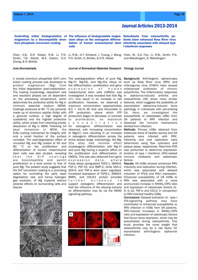
Page 18 Volume 7, 2014
Journal Articles 2013-2014
Controlling initial biodegradation of
magnesium by a biocompatible stron-
tium phosphate conversion coating
The influence of biodegradable magne-
sium alloys on the osteogenic differen-
tiation of human mesenchymal stem
cells
Osteoblasts from osteoarthritis pa-
tients show enhanced Ross River virus
infectivity associated with delayed type
I interferon responses
Chen, X.B., D.R. Nisbet, R.W. Li, P.N.
Smith, T.B. Abbott, M.A. Easton, D.H.
Zhang, & N. Birbilis
Acta Biomaterialia
A_simple strontium phosphate (SrP) conv
ersion coating process was developed to
protect magnesium (Mg) from
the initial degradation post-implantation.
The coating morphology, deposition rate
and resultant phases are all dependent
on the processing temperature, which
determines the protective ability for Mg in
minimum essential medium (MEM).
Coatings produced at 80 °C are primarily
made up of strontium apatite (SrAp) with
a granular surface, a high degree of
crystallinity and the highest protective
ability, which arises from retarding anodic
dissolution of Mg in MEM. Following 14
days' immersion in MEM, the
SrAp coating maintained its integrity with
only a small fraction of the surface
corroded. The post-degradation effect of
uncoated Mg and Mg coated at 40 and
80 °C on the proliferation and
differentiation of human mesenchymal
stem cells was also studied, revealing
t h a t t h e S r P c o a t i n g s
are biocompatible and permit
proliferation to a level similar to that of
pure Mg. The present study suggests that
the SrP conversion coating is a promising
option for controlling the early rapid
degradation rate, and hence hydrogen
gas evolution, of Mg implants without
adverse effects on surrounding cells and
tissues
Li, R.W., N.T. Kirkland, J. Truong, J. Wang,
P.N. Smith, N. Birbilis, & D.R. Nisbet
Journal of Biomedical Materials Research
The postdegradation effect of pure Mg,
Mg-1Y, Mg-5Al, and Mg-2Ca alloys on
the differentiation, proliferation and gene
e x p r e s s i o n o f h u m a n
mesenchymal stem cells (hMSCs) was
investigated. It was revealed that that Mg
(2+) ions result in an increase in cell
proliferation. However, we observed a
maximum concentration (approximately
8.0 × 10(-4) M) that was favourable to
ATP production, above which ATP
production began to decrease. In contrast
to prol i feration, no maximum
c o n c e n t r a t i o n
for osteogenic differentiation was
observed, with increasing concentration
of Mg(2+) ions resulting in an increase
in osteogenic differentiation across the
entire tested range. Interestingly, the Mg-
2Ca alloy had minimal effect
onosteogenic differentiation, with Mg-1Y
and pure Mg having a superior effect on
the proliferation and differentiation of
hMSCs. This was also observed from gene
e x p r e s s i o n d a t a , w h e r e
these alloys upregulated TGFβ-1, SMAD4,
FGF-2, FGF-10, and BMP-2, while SOX-2,
SOX-9, and TNF-α were down regulated.
Increased expression of TGFβ-1, SMAD4,
BMPs, and COLIA1 protein provided
f u r t h e r e v i d e n c e t o
support osteogenic differentiation and
that the influence of the alloying extracts
on differentiation may be via the SMAD
signalling pathway.
Chen, W., S.S. Foo., Li, R.W., Smith, P.N.
and Mahalingam, S. Mahalingam
Virology Journal
Background: Arthritogenic alphaviruses
such as Ross River virus (RRV) and
chikungunya virus (CHIKV) have caused
widespread outbreaks of chronic
polyarthritis. The inflammatory responses
in alphavirus-induced arthritis and
osteoarthritis (OA) share many similar
features, which suggests the possibility of
exacerbated alphavirus-induced bone
pathology in individuals with pre-existing
OA. Here, we investigated the
susceptibility of osteoblasts (OBs) from
OA patients to RRV infection and
dissected the immune mechanisms
elicited from infection.
Methods: Primary hOBs obtained from
trabecular bone of healthy donors and OA
patients were infected with RRV.
Infectivity and viral replication were
determined using flow cytometry and
plaque assay, respectively. Real-time PCR
was performed to determine expression
kinetics of type I interferon (IFN)-related
immune mediators and osteotropic
factors.
Results: OA hOBs showed enhanced RRV
infectivity and replication during infection,
which was associated with delayed
induction of IFN-β and RIG-I expression.
Enhanced susceptibility of OA hOBs to
RRV was associated with a more
pronounced increase in RANKL/OPG ratio
and expression of osteotropic factors (IL-
6, IL-1β, TNF-α and CCL2) in comparison
to RRV-infected healthy hOBs.
Conclusions: Delayed activation of - type I
IFN-signalling pathway may have
contributed to enhanced susceptibility to
RRV infection in hOBs from OA patients.
RRV-induced increases in RANKL/OPG
ratio and expression of osteotropic factors
that favour bone resorption, which may be
exacerbated during osteoarthritis. This
study provides the novel insight that
osteoarthritis may be a risk factor for
exacerbated arthritogenic alphaviral
infection.
Page 19 Trauma and Orthopaedic Research Unit Newsletter
Funding Awarded 2013-2014
Awarding Body ACT Health
Project Title Thin-film Carbon Nanotube Load Sensors for Application to Community Health and
Sport
Recipient Strategic Opportunities Fund
Amount $77,000
Awarding Body The Canberra Hospital Private Practice Fund
Project Title The Arterial Anatomy of the Gluteal Tendons: a comparison of CT, microCT and Histol-
ogy
Recipient Dr Mitali Fadia
Amount $9,044
Awarding Body CSC (Chinese Scholarship Council) PhD Scholarship Grant
Project Title Bioinformatics approach for establishing an osteo-network.
Recipient Mr. Song Chen
Amount $88,000
Awarding Body Partnership Fund, People’s Hospital of Changle County
Project Title Research into healthy bone
Recipient Dr Rachel Li
Amount $250,000
Awarding Body Australian government Department of Education-Endeavour Research Fellowship
Project Title Structure and Activity Relationship Study of Amaryllidaceae Alkaloids and Discovery
of Novel Leads for Anti-inflammatory Drug Design
Recipient Dr Partha Palit
Amount $30,000
Awarding Body ANU Postgraduate Scholarship Grant
Project Title Heparanase as a biomarker in Rheumatoid arthritis.
Recipient Ms Olympia John
Amount $75,000
2013 – 2014 Collaborative Associates
Page 20 Volume 7, 2014
A/Prof Jennie Scarvell Dr Nick Ball Head of Physiotherapy, University of Canberra Head of Sport Science, University of Canberra
A/Prof Mark Pickering, Dr Sean O’Byrne, School of Engineering and Information Technology School of Engineering and Information Technology Dr Krishna Shankar A/ Prof Heiko Timmers School of Engineering and Information Technology School of Physical and Mathematical Sciences
Prof Chris Parish Prof Graham Buirski Department of Immunology, John Curtin School of Medical Re- Director of Medical Imaging Dr Alexandra Webb Prof Jan Provis School of Anatomy Director, School of Anatomy Prof Peter Hickman Prof Christian J Lueck Director Chemical Pathology Head, Department of Neurology Prof Qinghua Qin A/Prof Alex Fisher College of Engineering and Computer Science Geriatric Medicine Dr Teresa Neeman Prof Jane Dahlstrom Statistical Consulting Unit Sub Dean Canberra Hospital Campus; Anatomical Pathology A/Prof Yun Liu Prof Thomas Bruessel Head of the Functional Materials Research Group, Research Director of Anaesthesiology Dr Steven Weiss A/Prof Shankar Kalyanasundaram College of Medicine, Biology and Environment College of Engineering and Computer Science Dr Claudio Mastronardi Dr David Nisbet Pharmacogenomics Group, John Curtin School of Medical Re-
search College of Engineering and Computer Science
Dr Bryan Ashman Dr Bruce Shadbolt Director of Surgery and Orthopaedic Surgeon, Canberra Hospital Centre for Clinical Epidemiology, Canberra Hospital Di Lane, Amy Krause Adrian Meyer and Linda Crawford Medical Imaging, Radiography, Canberra Hospital
Page 21 Trauma and Orthopaedic Research Unit Newsletter
Prof Dongsheng Zhou Prof Jiake Xu
Department of Trauma and Orthopaedics, Shandong University,
Jinan, China.
School of Pathology and Laboratory Medicine and Head of mo-
lecular laboratory, UWA
Prof Julio Licinio Prof Ma-Li Wong Flinders University Adelaide Deputy Director for Translational
Medicine and Mind and Brain Flinders University Adelaide
Prof Brett Kirk Dr Jian-Ping Wu Associate Deputy Vice-Chancellor, Research office of Research
and Development, Supervisor of 3D imaging and bioengineering
Deputy supervisor of 3D imaging and bioengineering lab, Depart-
ment of Mechanical Engineering
Dr Alexander Burns Dr Damian Smith Consultant orthopaedic Surgeon Consultant orthopaedic Surgeon
Prof Bill Walsh A/Prof Alex Scott UNSW Orthopaedic Research Institute Dept of Physiotherapy, University of British Colombia
Prof Shen Lu, Prof Xiaomin Wang, Department of Pathology, Dalian Medical University, Dalian China Capital Medical University, Beijing, China
Professor Nicholas Birilis Dr Xiaobo Chen Dean of the Dept of Materials Engineering, Monash University Dept of Materials Engineering, Monash University
Prof David Little A/Prof Peng Wang The Kids Research Institute, The Children's Hospital Westmead Beijing Union University, Beijing, China
Trauma and Orthopaedic Research Unit
Building 6, level 1
Canberra Hospital
PO Box 11
Woden, ACT 2606, Australia
Phone: 6244 3858
Fax: 6205 2157
E-mail: [email protected]
www.health.act.gov.au/research
TORU is affiliated with the Australian National University and works closely
with UNSW@ADFA and University of Canberra as well. Prospective higher
degree students are encouraged to consider possible research
opportunities at TORU in 2015. Contact TORU or prepare a 1-2 page
research proposal for TORU to consider. TORU is committed to progressing
research in trauma and orthopaedics, and to developing young
researchers.
Interested in Studying with TORU in 2015?