Embed Size (px)
Citation preview
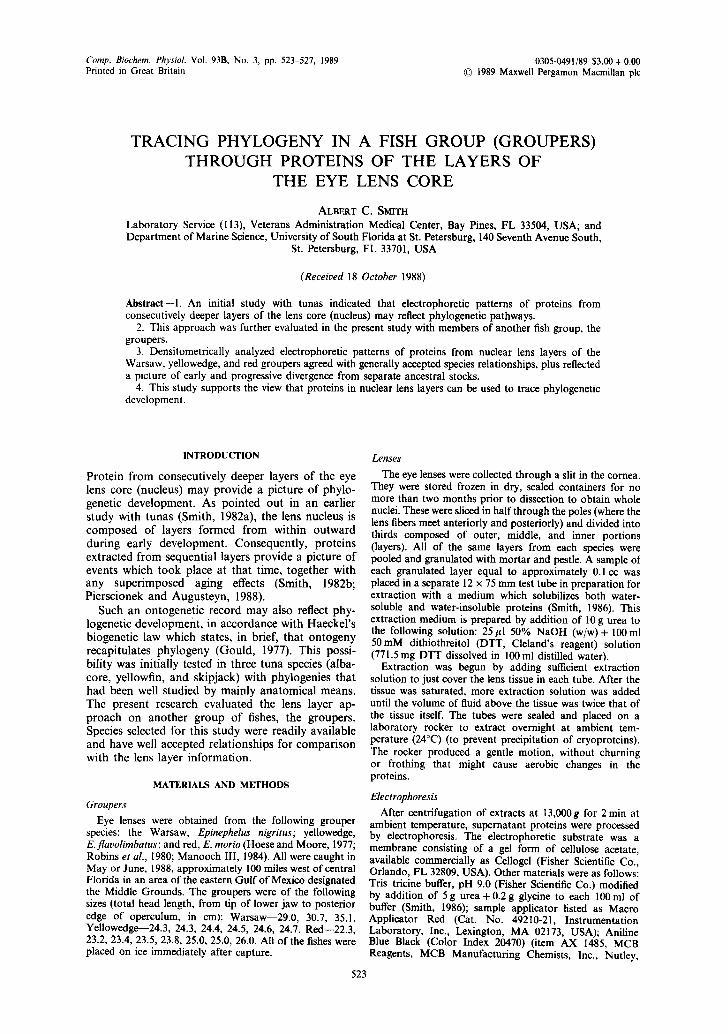
Comp. Biochem. Physiol. Vol. 93B, No. 3, pp. 523-527, 1989 0305-0491/89 $3.00 + 0.00 Printed in Great Britain © 1989 Maxwell Pergamon Macmillan plc
TRACING PHYLOGENY IN A FISH GROUP (GROUPERS) THROUGH PROTEINS OF THE LAYERS OF
THE EYE LENS CORE
ALBERT C. SMITH Laboratory Service (113), Veterans Administration Medical Center, Bay Pines, FL 33504, USA; and Department of Marine Science, University of South Florida at St. Petersburg, 140 Seventh Avenue South,
St. Petersburg, FL 33701, USA
(Received 18 October 1988)
Abstract--1. An initial study with tunas indicated that electrophoretic patterns of proteins from consecutively deeper layers of the lens core (nucleus) may reflect phylogenetic pathways.
2. This approach was further evaluated in the present study with members of another fish group, the groupers.
3. Densitometrically analyzed electrophoretic patterns of proteins from nuclear lens layers of the Warsaw, yellowedge, and red groupers agreed with generally accepted species relationships, plus reflected a picture of early and progressive divergence from separate ancestral stocks.
4. This study supports the view that proteins in nuclear lens layers can be used to trace phylogenetic development.
INTRODUCTION
Protein from consecutively deeper layers of the eye lens core (nucleus) may provide a picture of phylo- genetic development. As pointed out in an earlier study with tunas (Smith, 1982a), the lens nucleus is composed of layers formed from within outward during early development. Consequently, proteins extracted from sequential layers provide a picture of events which took place at that time, together with any superimposed aging effects (Smith, 1982b; Pierscionek and Augusteyn, 1988).
Such an ontogenetic record may also reflect phy- logenetic development, in accordance with Haeckel 's biogenetic law which states, in brief, that ontogeny recapitulates phylogeny (Gould, 1977). This possi- bility was initially tested in three tuna species (alba- core, yellowfin, and skipjack) with phylogenies that had been well studied by mainly anatomical means. The present research evaluated the lens layer ap- proach on another group of fishes, the groupers. Species selected for this study were readily available and have well accepted relationships for comparison with the lens layer information.
MATERIALS AND METHODS
Groupers
Eye lenses were obtained from the following grouper species: the Warsaw, Epinephelus nigritus; yellowedge, E.flavolimbatus; and red, E. morio (Hoese and Moore, 1977; Robins et al., 1980; Manooch III, 1984). All were caught in May or June, 1988, approximately 100 miles west of central Florida in an area of the eastern Gulf of Mexico designated the Middle Grounds. The groupers were of the following sizes (total head length, from tip of lower jaw to posterior edge of operculum, in cm): Warsaw--29.0, 30.7, 35.1. Yellowedge--24.3, 24.3, 24.4, 24.5, 24.6, 24.7. Red--22.3, 23.2, 23A, 23.5, 23.8, 25.0, 25.0, 26.0. All of the fishes were placed on ice immediately after capture.
Lenses
The eye lenses were collected through a slit in the cornea. They were stored frozen in dry, sealed containers for no more than two months prior to dissection to obtain whole nuclei. These were sliced in half through the poles (where the lens fibers meet anteriorly and posteriorly) and divided into thirds composed of outer, middle, and inner portions (layers). All of the same layers from each species were pooled and granulated with mortar and pestle. A sample of each granulated layer equal to approximately 0.1 cc was placed in a separate 12 x 75 mm test tube in preparation for extraction with a medium which solubilizes both water- soluble and water-insoluble proteins (Smith, 1986). This extraction medium is prepared by addition of 10 g urea to the following solution: 25gl 50% NaOH (w/w)+ 100ml 50mM dithiothreitol (DTT, Cleland's reagent) solution (771.5 mg DTT dissolved in 100 ml distilled water).
Extraction was begun by adding sufficient extraction solution to just cover the lens tissue in each tube. After the tissue was saturated, more extraction solution was added until the volume of fluid above the tissue was twice that of the tissue itself. The tubes were sealed and placed on a laboratory rocker to extract overnight at ambient tem- perature (24°C) (to prevent precipitation of cryoproteins). The rocker produced a gentle motion, without churning or frothing that might cause aerobic changes in the proteins.
Electrophoresis
After centrifugation of extracts at 13,000g for 2 min at ambient temperature, supernatant proteins were processed by electrophoresis. The electrophoretic substrate was a membrane consisting of a gel form of cellulose acetate, available commercially as Cellogel (Fisher Scientific Co., Orlando, FL 32809, USA). Other materials were as follows: Tris-tricine buffer, pH 9.0 (Fisher Scientific Co.) modified by addition of 5 g urea + 0.2 g glycine to each I00 ml of buffer (Smith, 1986); sample applicator listed as Macro Applicator Red (Cat. No. 49210-21, Instrumentation Laboratory, Inc., Lexington, MA 02173, USA); Aniline Blue Black (Color Index 20470) (item AX 1485, MCB Reagents, MCB Manufacturing Chemists, Inc., Nutley,
523

524 ALBERT C. SMITH
NJ 08027, USA), 5g dissolved in 1000ml of 10% acetic acid; and Cliniscan Scanning Densitometer (Helena Laboratories, Beaumont, TX 77704, USA).
Lens extracts were applied twice to the same position at the cathodal end of membranes, and electrophoresis was performed at 200V for 55min at ambient temperature. Proteins were then fixed in position on the membranes by immersion for 10 min in 6% trichloroacetic acid (TCA), and stained by immersion for 10min in thoroughly mixed Aniline Blue Black. A brief rinse in tap water, followed by several rinses first in 5% acetic acid and then absolute methanol, removed background stain. Finally, after several minutes immersion in distilled water containing a small amount of preservative TCA, the finished membranes with protein patterns were scanned on the densitometer at a wavelength of 595 nm, visible slit setting of 9, and neutral
density filter setting of V-20D. The strips were stored under refrigeration in an airtight box on filter paper saturated with the distilled water/TCA solution.
RESULTS
The study, conducted in duplicate, produced a set of electrophoretic pat terns of proteins from the nuclear lens layers, as shown densitometrically in Figs 1-3.
Densitometric tracings A - C are of proteins from the outer nuclear lens layer. The tracings of the Warsaw grouper (A) and red grouper (C) are most complex. The tracing of the yellowedge grouper (B)
A
B
30 1 2 3 4 5 6 7 8 9 10 I1 12 13 14 15 ' lr6 17 1
i ! I : q I ~ ! ] i '
i ;
! : , = ~ ! , ! ! [ ,
: ~ - : i i ? 4~ ' t
v . . . . . . . ' - . . . . . l g 9 •
B
¢ 6 ,
17 18 19 20 21 ~Z 23 2~1 2S 26 27 28 ;9 }0 I 2
6
5 . . . .
I 4 . . . . . . .
Fig. 1. Outer nuclear lens layer group, A~S. Densitometric tracings are from the Warsaw grouper, yellowedge grouper, and red grouper, respectively. Arrow at bottom left of tracings indicates the zone
of sample application. Cathode is to the left.

Eye lens proteins 525
ml r.~'r, IdO. I04t + 14t+dl~ L+IIO~'IOIIIU I :0~+ ~ , ~'ed~l PJ+T, '~0. + 04 | e ~ U t I O A A ~ (:Ore'. O~amlO
i~ . . . . I - : - 9 + + . -
J ! ! a- ? 7 . . . . i
i I . , ii+~ i
: ~ ! ; ! / i 1 ! ! - ! X ! + i : l r' i I . . . . . +,,,' + i i L u , + , .
' iiii++!i + iil ii l)I ! + ~ N l I l i t i+ l~"7"" ' q . - - -~ , , . ~ l t I
Fig. 2. Middle nuclear lens layer group, D-F. Tracing sequence and other information as in Fig. 1.
is relatively simple, consisting mainly of four well resolved components.
Tracings D--F are of proteins from the middle nuclear lens layers. The tracings of the Warsaw grouper (D) and yellowedge grouper (E) show mostly quantitative changes, with corresponding com- ponents from the outer nuclear layer tracing either increased (e.g., the first distinct peak in the Warsaw tracing) or, more commonly, decreased (e.g., the last three peaks in the yellowedge tracing). The middle nuclear layer tracing of the red grouper clearly shows the latter trend of peak reduction, but additionally shows an apparent fusion of the first two peaks in the outer layer tracing.
Tracings G-I are of proteins from the inner nuclear lens layer. The tracings from all three grouper species
are greatly simplified to essentially a major com- ponent and some minor variations. In the Warsaw and yellowedge tracings, minor components consist of several low peaks which are most clearly resolved in the yellowedge tracing. The red grouper tracing shows a unique shoulder, apparently representing a lower quantity of the protein forming the second of the fused peaks in the middle layer tracing. The absence of multiple minor components seen in the tracings of the other two grouper species further sets apart the red grouper tracing.
D I S C U S S I O N A N D C O N C L U S I O N S
The densitometric tracings of electrophoretically separated proteins from the inner nuclear lens layer

G
H
vzx i l e~£ , ~ . 1a4~ #v~Ja ,~ LU lO*A~ ~ , Wt4UUOW~, ~XAS e~X NO 104Z O ~ J L~ma~AXO~.Z COrdp. IWJ
] T "1 : F . . . . . . ~ - ~ ' " 5 ! ;
• i ~ ! -
5 2 6 A L B E R T C . S M I T H
. . . . ° ~ . . . . . . . . . . . , ~ . . . . . . . . . L~A LA~ . ,~ .~ ~ .~ OL .~N* ' tAAS ca r NO 1~2 , ~LE~A
9
! J * i it - ' x ~
" i 2 !
[_ ._ ' n n u I - - . I ~ r B 9 10 T1 12 13 14 I~t 16 I 1 10 19 20 21 22 23
Fig. 3. Inner nuclear lens layer group, G-I. Tracing sequence and other information as in Figs 1 and 2.
in all three grouper species show slight differences. With increasing distance from the inner lens layer, the tracings change, becoming progressively more different. The overall picture is, thus, of similar tracings (inner nuclear lens layer) representing closely related ancestral stocks which, in turn, may have evolved from a common ancestor. The tracings then rapidly diverge (middle and outer nuclear lens layers), representing the subsequent evolution of the groupers into three well differentiated species.
The concept of a linked, but not a direct common, ancestral relationship among these grouper species is consistent with published phylogenetic relationships (Smith, 1971) that are generally considered definitive at the species level (Randall, 1987). Further, the
nuclear lens layer findings agree with (1) assignment of a closer relationship between the more derived Warsaw and yellowedge groupers than between either of these species and the more primitive red grouper, and (2) reports of species specificity of lens proteins from other members of the Family Serranidae to which the groupers belong (Siau and Bouain, 1982; Bouain and Siau, 1983).
In conclusion, I found that in the three grouper species studied, the information derived from the nuclear lens layers correlates well with the published studies. In addition, the grouper study indicated that (1) generalized fishes near the base of a diverse fish order--typified by the groupers as they are very generalized (Randall, 1987) and belong to the largest

Eye lens proteins 527
and most diverse of fish orders (Perciformes) (Nelson, 1984)--can produce complex lens protein patterns, (2) less derived species (red grouper) can produce nuclear lens protein patterns as complex or more complex than patterns from more advanced species (Warsaw and yellowedge groupers, respectively), and (3) relationships may show up more clearly in simpler protein patterns from the inner nuclear lens layer than in patterns that are highly divergent and complex from middle and outer nuclear lens layers.
This grouper study supports the view that phyloge- netic information can be derived from electrophoretic analysis of proteins in sequential layers of the eye lens nucleus.
Acknowledgements--I wish to thank Mr Joe Henson, St Petersburg Junior College, Florida, for excellent tech- nical assistance; Mr Tim Nachman, Hubbard Enterprises, St. Petersburg, Florida, for the lenses; and Mr David Gratz and the Medical Media Production Service, and Dr Clif Jones, Laboratory Service, Veterans Administration Medical Center, Bay Pines, Florida, for much-appreciated photographic contributions and critical reading of the manuscript, respectively.
REFERENCES
Bouain A. and Siau Y. (1981) Etude 61ectrophoretique des protrines cristalliniennes de serrans (G. serranus) et de loups (G. dicentrarchus) des ~otes orientales de la Tunisie (golfe de Gabrs). Archs Inst. Pasteur Tunis 58, 465-475.
Gould S. J. (1977) Ontogeny and Phylogeny. The Belknap Press of Harvard University, Cambridge, MA.
Hoese H. D. and Moore R. H. (1977) Fishes of the Gulf of Mexico, Texas, Louisiana, and Adjacent Waters, 1st ed. Natural History Series, no. 1, Texas A&M, College Station.
Manooch III C. S. (1984) Fisherman's Guide to the Fishes oJ the Southeastern United States. North Carolina State Museum of Natural History, Raleigh.
Nelson J. S. (1984) Fishes of the World, 2nd ed. Wiley, New York.
Pierscionek B. and Augusteyn R. C. (1988) Protein distribu- tion patterns in concentric layers from single bovine lenses: changes with development and ageing. Curr. Eye Res. 7, 11-23.
Randall J. E. (1987) A preliminary synopsis of the groupers (Perciformes: Serranidae: Epinephelinae) of the Indo- Pacific region. In Tropical Snappers and Groupers (Edited by Polovina J. J. and Ralston S.), Ch. 3, pp. 89-188. Westview Press, Boulder, CO.
Robins C. R., Bailey R. M., Bond C. E., Brooker J. R., Lachner E. A., Lea R. N. and Scott W. B. (1980) A List of Common and Scientific Names of Fishes from the United States and Canada, 4th ed. Special Publication no. 12, American Fisheries Society, Bethesda, MD.
Siau Y. and Bouain A. (1982) Etude ~lectrophor&ique des prot6ins cristalliniennes de m~rous, genres Epinephelus et Mycteroperca (Pisces, Serranidae). Archs Inst. Pasteur Tunis 59, 269-281.
Smith A. C. (1982a) Tracing phylogeny through proteins of the layers of the eye lens nucleus. Comp. Biochem. Physiol. 71B, 723-726.
Smith A. C. (1982b) Electrophoretic study of proteins from solubilized eye lens nuclei of fishes. Comp. Biochem. Physiol. 71B, 337-343.
Smith A. C. (1985) New medium for solubilizing proteins of the eye lens core (nucleus). Comp. Biochem. Physiol. 80B, 377-380.
Smith A. C. (1986) Some advances in electrophoretic sepa- ration of proteins from fish eye lens cores (nuclei): a brief report. J. Aquaric. Aquatic Sci. IV, 94.
Smith C. L. (1971) A revision of the American groupers: Epinephelus and allied genera. Bull. Am. Mus. Nat. Hist. 146, 67-242.