Embed Size (px)
Citation preview


-3-
Toxicity Review for Benzylnbutyl Phthalate (Benzyl Butyl Phthalate or BBP)
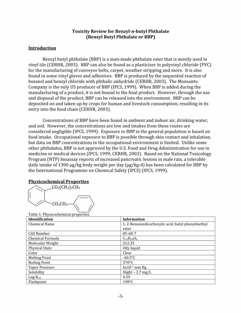
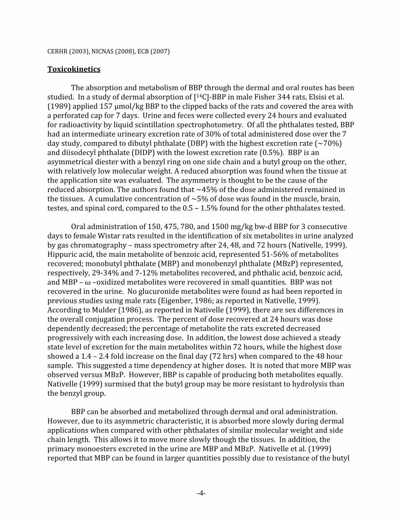
Introduction Benzyl butyl phthalate (BBP) is a man‐made phthalate ester that is mostly used in vinyl tile (CERHR, 2003). BBP can also be found as a plasticizer in polyvinyl chloride (PVC) for the manufacturing of conveyor belts, carpet, weather stripping and more. It is also found in some vinyl gloves and adhesives. BBP is produced by the sequential reaction of butanol and benzyl chloride with phthalic anhydride (CERHR, 2003). The Monsanto Company is the only US producer of BBP (IPCS, 1999). When BBP is added during the manufacturing of a product, it is not bound to the final product. However, through the use and disposal of the product, BBP can be released into the environment. BBP can be deposited on and taken up by crops for human and livestock consumption, resulting in its entry into the food chain (CERHR, 2003). Concentrations of BBP have been found in ambient and indoor air, drinking water, and soil. However, the concentrations are low and intakes from these routes are considered negligible (IPCS, 1999). Exposure to BBP in the general population is based on food intake. Occupational exposure to BBP is possible through skin contact and inhalation, but data on BBP concentrations in the occupational environment is limited. Unlike some other phthalates, BBP is not approved by the U.S. Food and Drug Administration for use in medicine or medical devices (IPCS, 1999; CERHR, 2003). Based on the National Toxicology Program (NTP) bioassay reports of increased pancreatic lesions in male rats, a tolerable daily intake of 1300 µg/kg body weight per day (µg/kg‐d) has been calculated for BBP by the International Programme on Chemical Safety (IPCS) (IPCS, 1999). Physicochemical Properties CO2(CH2)3CH3 CO2CH2 Table 1: Physicochemical properties Identification Information Chemical Name 1, 2‐Benzenedicarboxylic acid, butyl phenylmethyl
ester CAS Number 85‐68‐7Chemical Formula C19H20O4Molecular Weight 312.35Physical State Oily liquidColor ClearMelting Point ‐40.5°CBoiling Point 370°CVapor Pressure 6x10‐7 mm HgSolubility Slight – 2.7 mg/LLog Kow 4.59Flashpoint 198°C
-4-
CERHR (2003), NICNAS (2008), ECB (2007) Toxicokinetics The absorption and metabolism of BBP through the dermal and oral routes has been studied. In a study of dermal absorption of [14C]‐BBP in male Fisher 344 rats, Elsisi et al. (1989) applied 157 µmol/kg BBP to the clipped backs of the rats and covered the area with a perforated cap for 7 days. Urine and feces were collected every 24 hours and evaluated for radioactivity by liquid scintillation spectrophotometry. Of all the phthalates tested, BBP had an intermediate urineary excretion rate of 30% of total administered dose over the 7 day study, compared to dibutyl phthalate (DBP) with the highest excretion rate (~70%) and diisodecyl phthalate (DIDP) with the lowest excretion rate (0.5%). BBP is an asymmetrical diester with a benzyl ring on one side chain and a butyl group on the other, with relatively low molecular weight. A reduced absorption was found when the tissue at the application site was evaluated. The asymmetry is thought to be the cause of the reduced absorption. The authors found that ~45% of the dose administered remained in the tissues. A cumulative concentration of ~5% of dose was found in the muscle, brain, testes, and spinal cord, compared to the 0.5 – 1.5% found for the other phthalates tested. Oral administration of 150, 475, 780, and 1500 mg/kg bw‐d BBP for 3 consecutive days to female Wistar rats resulted in the identification of six metabolites in urine analyzed by gas chromatography – mass spectrometry after 24, 48, and 72 hours (Nativelle, 1999). Hippuric acid, the main metabolite of benzoic acid, represented 51‐56% of metabolites recovered; monobutyl phthalate (MBP) and monobenzyl phthalate (MBzP) represented, respectively, 29‐34% and 7‐12% metabolites recovered, and phthalic acid, benzoic acid, and MBP – ω –oxidized metabolites were recovered in small quantities. BBP was not recovered in the urine. No glucuronide metabolites were found as had been reported in previous studies using male rats (Eigenber, 1986; as reported in Nativelle, 1999). According to Mulder (1986), as reported in Nativelle (1999), there are sex differences in the overall conjugation process. The percent of dose recovered at 24 hours was dose dependently decreased; the percentage of metabolite the rats excreted decreased progressively with each increasing dose. In addition, the lowest dose achieved a steady state level of excretion for the main metabolites within 72 hours, while the highest dose showed a 1.4 – 2.4 fold increase on the final day (72 hrs) when compared to the 48 hour sample. This suggested a time dependency at higher doses. It is noted that more MBP was observed versus MBzP. However, BBP is capable of producing both metabolites equally. Nativelle (1999) surmised that the butyl group may be more resistant to hydrolysis than the benzyl group.
BBP can be absorbed and metabolized through dermal and oral administration. However, due to its asymmetric characteristic, it is absorbed more slowly during dermal applications when compared with other phthalates of similar molecular weight and side chain length. This allows it to move more slowly though the tissues. In addition, the primary monoesters excreted in the urine are MBP and MBzP. Nativelle et al. (1999) reported that MBP can be found in larger quantities possibly due to resistance of the butyl

-5-
group to hydrolysis. This would lead to a more rapid break down of the benzyl ring in MBzP compared to the butyl group of MBP, and a reduced concentration of MBzP. Exposure According to IPCS (1999) the primary source of exposure to BBP is food intake. However, BBP and/or its metabolites can also be found in indoor air and consumer products. As reported in a review by Schettler (2006), contaminated food is likely the largest single source of exposure with an estimated daily intake of 0.11 – 0.29 µg/kg/d (MAFF, 1996). Inhalation of indoor air and household dust may be an important pathway of exposure for low molecular weight molecules such as BBP. Otake et al. (2004) collected indoor air samples from 27 homes in the Tokyo Metropolitan area and evaluated the concentration of phthalates found in the samples. The mean concentration of BBP was 0.02±0.03 µg/m3 in the indoor air samples, which was below the concentration of the other phthalates tested. Similar concentrations of BBP were found by Adibi et al. (2003) when they collected 48 hour personal air samples from women in New York City and Krakow, Poland (0.10±0.15 and 0.04±0.04 µg/m3 respectively). MBP and MBzP, the primary metabolites of BBP, were found at 54.4±24.5 and 26.0±28.2 µg/g creatinine in the collected urine samples. Creatinine is produced by the body at a consistent rate based on muscle mass. A significant correlation (r=0.65, p<0.01) was found between BBP in the air samples and MBzP in the urine samples. However, the demographics of this study did not represent the general population. The women from Krakow were considered to be in a higher socioeconomic status than the general population of their area, while the women of New York were considered to be at a lower socioeconomic status. One or both may be disproportionately exposed to products and goods containing BBP and other phthalates. In addition, product formulations in Europe may be different than the United States. Therefore these conclusions cannot be generalized to all women. Koch (2005) hypothesized that if children and adults took up the same amount of phthalates, children would have an increased amount of phthalate metabolite in their urine and have increased exposure compared to the adult due to body weight differences. To address this question, a voluntary study was conducted in a southern Germany nursery school. Morning urine voids were taken from children, school teachers, and parents. Urine samples were analyzed for metabolites using a multi‐dimensional liquid chromatography tandem mass spectrometry method. MBP and MBzP concentrations were higher in the children when compared with the adults. The median urinary MBP concentration for the children was about 1.5 times higher than for the adults. The concentration for MBzP was about 5 times more than MBP and about twice as high as concentrations found in adults. This is different than what was previously reported by Nativelle et al. (1999). However, the metabolic pathway may be different between rats and human. The authors estimate that an infant would receive a seven times greater dose than an adult with respect to body weight. Since creatinine excretion is approximately proportional to muscle mass, the authors think creatinine adjustment could compensate for the differences in children and

-6-
adults. With this adjustment, the internal exposure of children to MBP and MBzP was enhanced about two fold, and was significantly (p<0.0001) higher than the adults. The maximum values for internal exposure, after removing a child on medication that may contain a coating with DBP, was 517 µg/g for the children and 149 µg/g for the adults. The maximum concentration for children is about 3.5 times the maximum concentration of the adults. A significant correlation between the excretion of MBP and MBzP was found (r=0.723; p<0.001). After reviewing the answers from a questionnaire regarding use of skin care or body care products, the authors concluded that the use of skin care or body care products may have a significant influence on BBP exposure of children, which may contribute to their MBP exposure. However, it must be noted that MBP is also the primary metabolite of DBP. Therefore, exposures to MBP due to personal care products may also be due to another phthalate. There was no correlation between personal care products and MBzP exposure. BBP can be found in vinyl flooring. Koch (2005) suggested that children may be exposed to BBP through abrasions in the floor or household dust particles, which may contribute to their MBzP exposure. Sørensen et al. (2006) collected milk and milk products from around the world, including raw milk, pasteurized and homogenized milk, yogurt with fruit, reconstituted infant formula from different parts of the world, and liquid infant formula from Europe. They found that these milk products contained <4 µg/kg (4 µg/kg is the limit of detection) when tested by pneumatically assisted electro‐spray ionization. No studies were reviewed on occupational exposure of BBP. BBP has been identified in cosmetic products (Koo, 2004; Hubinger, 2006). The Cosmetic Ingredient Review (CIR) Expert Panel has determined that BBP in cosmetics is safe at concentrations <1% (CIR, 1992; as reported in Hubinger, 2006). The data is insufficient to determine the extent of exposure through occupational exposure or use of cosmetic products. Hazard Identification
In evaluating toxicity data, staff applies the definition for toxicity in the regulations
(16 CFR 1500.3 (c)(2)(ii)) and chronic hazard guidelines (CPSC, 1992) promulgated under the FHSA (15 U.S.C. 1261‐1278). A substance or mixture is classified as “known to be toxic” in humans only if there is sufficient evidence in humans, and is regarded as “probably toxic” if there is either limited evidence in humans, or sufficient evidence in animals (Table 2). If a substance is “known to be toxic” or “probably toxic” in humans it is considered “toxic” under the FHSA. If a substance is “possibly toxic”, it would not be considered “toxic” under the FHSA. Table 2: Classification of Chronic Hazards under the FHSA Evidence Human studies Animal studies Sufficient evidence Known a Probable a Limited evidence Probable a Possible Inadequate evidence Possible ‐‐‐ a Considered “toxic” under the FHSA

-7-
Acceptable daily intake values (ADI’s) are calculated when a given chemical is
considered “toxic” due to chronic effects and sufficient toxicity information is available. The ADI is the amount of a chemical that a person may be exposed to on a daily basis without the chemical posing a significant risk of adverse health effects. In some cases insufficient data is available to calculate an ADI. Systemic Effects Several studies have been reviewed for systemic effects of BBP. The main effects seen are decreases in body weights and increased organ weights.
In an NTP study, 6‐week old male Fischer 344/N rats were fed a diet with 0, 300, 900, 2800, 8300, or 25,000 ppm BBP (calculated/estimated doses of 0, 30, 60, 180, 550, or 1650 mg/kg body weight/day) for 26 weeks (NTP, 1997). NTP reported effects starting at the 550 mg/kg‐day dose. There were increased liver to body weight ratios and an increase in the mean cell hemoglobin for experimental days 60 – 180. NTP suggested that this may be associated with macrocytic anemia, which is found at the next dosing level on days 30 – 180. At the highest dose, 1650 mg/kg‐day, a decrease in total body weight was observed, presumably due to a reduction in food consumption. This reduction in actual food consumption made it difficult for the dose to be calculated. Therefore, the 1650 mg/kg‐day dose is estimated from the intake levels of the lower doses. Since the dose is based on the amount of food intake, the results seen may be due to a lower dose than what was calculated. In addition, an increase in liver and kidney to body weight ratios, and a decrease in testes, seminal vesicle, and epididymis weights were noted. Upon histological examination, testicular and epididymal degeneration, seminiferous tubule atrophy, and reduced sperm counts were found. The no observed adverse effect level (NOAEL) was 180 mg/kg‐day, based on the increased liver weights found at the next dosing level, and the lowest observed adverse effect level (LOAEL) was 550 mg/kg‐day, due to the observed increase in mean cell hemoglobin found on days 60 – 180 (NTP, 1997). In a chronic study by NTP, 60 male Fischer 344/N rats per group were fed a diet with concentrations of BBP of 0, 3000, 6000, or 12,000 ppm (calculated doses of 0, 120, 240, or 500 mg/kg‐day) and 60 female rats per group were fed a diet with concentrations of BBP of 0, 6000, 12,000, or 24,000ppm (calculated doses of 0, 300, 600, and 1200 mg/kg‐day) for 2 years starting at 6 weeks of age. Tissue and blood samples were analyzed at 6, 8, 15, and 24 months. In the male rats, kidney weights increased starting at 120 mg/kg‐day, epididymis weights increased starting at 240 mg/kg‐day, and liver weights increased at 500 mg/kg‐day. At the highest dose, a decrease in total body weight was observed. Histopathology revealed renal tubule pigmentation, hepatic granulomas, and focal pancreatic hyperplasia, with some evidence of pancreatic carcinogenicity due to acinar cell adenoma and adenoma or carcinoma. Testicular changes were not seen, but there were decreases in red blood cell counts, an increase in the mean cell hemoglobin, and skin lesions (NTP, 1997).

-8-
In the female rats, at the end of the 2 year exposure, nephropathy was seen at all dosing levels. At the highest dose, 1200 mg/kg‐day, a decrease in total body weight, renal tubule pigmentation, and increased microcytic anemia, were noted as well as evidence of pancreatic and urinary bladder carcinogenicity, based on pancreatic acinar cell adenoma and urinary bladder transitional cell epithelial papillomas. Triiodo‐thyronine was decreased from months 6 – 15. Based on kidney organ weights in males and nephropathy in females, the LOAEL for general toxicity effects was determined to be 120 mg/kg‐day for males and 300 mg/kg‐day for females (NTP, 1997). Table 3: NTP’s 2‐year study endpoint for Fisher 344/N rats exposed to BBP in diet*
Dose (mg/kgday)
0 120 240 500
# of Males 60 60 60 60 ↑kidney wt ↑kidney wt
↑epid. wt ↑kidney wt ↑epid. wt ↑liver wt Renal tubule pigmentation Hepatic granuloma Pancreatic hyperplasia w/ evidence of carcinogenicity
0 300 600 1200 # of Females 60 60 60 60 Nephropathy Nephropathy Nephropathy
Renal tubule pigmentation Evidence of pancreatic carcinogenicity
(modified from CERHR, 2003) *Significant changes reported In a recent two generation study by Aso et al. (2005), male and female Crj:CD
Sprague‐Dawley IGS rats were administered BBP by gavage at doses of 0, 100, 200, or 400 mg/kg‐day. BBP was administered starting at 5 weeks of age for the F0 parents and 3 weeks of age for the F1 parents for 10 weeks prior to mating, and continued through weaning. In the F0 parents, no altered body weights or weight gains, and no significant changes in serum hormone levels at 400 mg/kg‐day were found when compared to the controls (100 and 200 mg/kg‐day doses were not reported, except for FSH in males showing no significant change). A significant (p<0.05) increase in renal weights starting at 200 mg/kg‐d in the male rats, and increased liver weight (p<0.01), as well as decreased epididymal weights (p<0.05) at 400 mg/kg‐d in males were reported. The females showed significantly increased liver and kidney weights (p<0.01) starting at the 200 mg/kg‐d dose, while a reduced relative uterine weight (p<0.01) was seen only for the 200 mg/kg‐d dose. At autopsy, no abnormal findings were seen. Histopathology showed an increase in hyperplasia of Leydig cells of the testes and a decrease in spermatozoa in the lumina of the epididymes for males receiving a dose of 400 mg/kg‐day. The F1 male rats showed significantly (p‐value not found) lower body weights starting at 100 mg/kg‐day. There were no body weight changes in the female rats;

-9-
however an increase in the anogenital distance (AGD) in females was noted by the authors, and splenic weights of the males were reduced. No other gross abnormalities were seen at birth. The F1 rats, at later stages, did not show any changes in weight, weight gain, or serum hormone levels. The males did show lowered epididymal weights and increased liver weight starting at 200 mg/kg‐day. Males also showed significantly (p<0.05) reduced seminal vesicle weights and increased thyroid weights, while the females had increased liver weights at 400 mg/kg‐day. Autopsy revealed aplasia and/or dysplasia and small epididymes and testes at 400 mg/kg, and softening testes starting at 100 mg/kg. Histological changes were seen starting at 100 mg/kg, including atrophy of testicular seminiferous tubules, decreased spermatozoa and residual germ cells in epididymal lumina. Atrophy of seminiferous tubules and hyperplasia of Leydig cells in the testes were significant (p<0.05) in the 400 mg/kg males. The F2 male offspring showed a decrease in AGD at the 100 mg/kg dose and higher, decreased body weights in the males and females at birth, and reduced male splenic weights. These findings, in addition to reproductive findings, lead to a NOEL/NOAEL for the parental animals of less than 100 mg/kg‐day, and a NOEL/NOAEL for the development and growth of offspring also of less than 100 mg/kg‐day (Aso, 2005). The findings of the Aso et al. study confirm a previous two generation study by Tyl et al. (2004). In this study, CD rats were given BBP concentrations of 0, 750, 3750, and 11,250 ppm (0, 50, 250, and 750 mg/kg‐day) in their diet for 10 weeks prior to mating and through weaning of F2 generation pups. Similar to the Aso et al. findings, they found reduced AGD in F1 and F2 males, reduced body weights, increased liver and kidney weights, liver lesions, as well as some indications of reproductive toxicity. The NOAEL for parental systemic toxicity was 250 mg/kg‐day, and the NOEL for offspring toxicity was 50 mg/kg‐day.
In an inhalation study, Hammond et al. (1987; as reported in CERHR, 2003; NICNAS,
2008) showed that BBP causes increased liver and kidney weights, as well as decreased serum glucose in male rats exposed to 789mg/m3 BBP in a mist. Six to eight week old Spague Dawley rats inhaled mists of BBP at 50, 218, or 789 mg/m3 (9.2, 39.4, or 143 mg/kg‐day for males and 9.8, 42, or 152 mg/kg‐d for females) for 6 hours/day, 5 days a week for 13 weeks. There were no effects on body weight, histopathology, or hematology, and effects on organ weights and blood chemistry were seen only at the highest dose. The NOAEL was 39.4 mg/kg‐d for males and 42 mg/kg‐d for females. The LOAEL was 143 mg/kg‐d for males and 152 mg/kg‐d for females (as reported in CERHR, 2003; NICNAS, 2008). These studies show that BBP can cause systemic toxic effects. Each study showed an increase in liver weights and kidney weights. These changes in organ weights were seen at the low doses. The two generation studies and the chronic study also showed decreases in body weights. Irritation/Allergic Response

-10-
There have been some studies correlating BBP exposure and allergic response and pulmonary function. Using the third National Health and Nutrition Examination Survey (NHANES), Hoppin et al. (2004) determined that in males, but not females, an increase in MBP, the primary metabolite of BBP, correlated to a decrease in pulmonary function. The study consisted of 140 women and 100 men with urine samples, pulmonary function data, and medical and smoking histories. Forced vital capacity (FVC), forced expiratory volume at 1 second (FEV1), peak expiratory flow (PEF), and maximum mid‐expiratory flow (MMEF) were used to determine pulmonary function. Statistically significant (p<0.05) decreases in FVC, FEV1, and PEF were associated with an increase in MBP. Removing male smokers sustained the significant decrease in FVC. With no adjustment for creatinine, the association between MBP and PEF became statistically insignificant. BBP is in indoor air and household dust, and is associated with allergic symptoms of the airway, nose, and skin. In 2008, Kolarik et al. examined the association between phthalates in dust and allergic disease among Bulgarian children. They collected dust samples from the bedrooms of 102 children between the ages of 2 and 7 years with symptoms of wheezing, rhinitis, and/or eczema within the previous 12 months. Eighty‐two non‐symptomatic children were used as controls. BBP was found in most samples that were collected. BBP was found to be higher in the homes of children with wheezing and eczema, although not statistically significantly (p=0.305; p=0.207, respectively). The authors did not find an association between BBP and allergic disease in their sample population. Bornehag et al. (2004) conducted a nested case control study where they collected samples from the homes of 198 individuals diagnosed with persistent allergic symptoms and 202 controls from a cohort. They found that the geometric mean concentrations of BBP were higher in bedrooms with PVC flooring when compared with those without PVC flooring. There were more doctor confirmed diagnoses of rhinitis or eczema found in individuals whose bedrooms had higher concentrations of BBP found in the dust compared to controls. A dose response relationship was found between BBP in dust and doctor confirmed diagnosed cases of rhinitis and eczema. Using the Mann‐Whitney U‐test, there was a significant association between BBP and rhinitis and eczema (p=0.007; p=0.001, respectively). Both the Bornehag et al. and Kolarik et al. studies found higher BBP levels in dust in homes of individuals with allergic symptoms compared to controls. However, only the Bornehag study found that the association was statistically significant. Some of the factors that may account for the lack of a significant association between BBP and allergic disease in the Kolarik study are: the use of the word “balatum” for both linoleum and PVC in Bulgaria; a low response rate compared to that of the Swedish (Bornehag et al.) study; and refusal of some participants to allow inspections and/or dust sampling even though they had given prior consent. The Kolarik study did evaluate the concentration of phthalates in the “balatum” flooring compared to other flooring and found no correlation. The fact that some of the flooring included as “balatum” flooring was actually linoleum could have diluted the presence of phthalate. According to Bornehag et al. (2004), previous studies show phthalate concentrations associated with the presence of PVC flooring, and that the

-11-
PVC floor, in turn, is associated with the allergic symptoms. Although unresponsiveness to questionnaires is not novel to epidemiologic studies, this condition raises concerns about representativeness of the sample since those most likely to respond to surveys/questionnaires may be more prone to allergic disease. This phenomenon may or may not introduce bias. These studies provide evidence that BBP might have an effect on respiratory function and allergic response. These findings are strengthened by the authors measuring BBP levels in the environment and correlating the levels to the presence of allergic response. In the Hoppin et al. study on lung function, their correlations are with MBP, a metabolite of BBP. However, the other studies show an increase in the number of children with allergic responses, such as wheezing, rhinitis, and eczema, when there was increasing presence of BBP in the environment. Although Kolarik et al. found an increasing incidence of allergic response in children with higher levels of BBP in their environment, these associations were not significant. BBP may cause sensitization; however, the current evidence is insufficient to determine if BBP meets the criteria for a strong sensitizer in humans under the FHSA. Endocrine Effects Previously, several studies have been conducted evaluating BBP effects on hormone expression and receptor activation, particularly peroxisome proliferator activated receptor (PPAR). PPAR heterodimerizes with retinoid X receptor and binds DNA of target gene sequences. More recently, investigations of the effects of BBP on the pituitary‐gonadal and thyroid hormones have been conducted. It has been found that BBP increases PPAR activity and pituitary‐gonodal hormones, while decreasing thyroid hormones. In addition, it is thought that BBP may activate other receptors, such as the estrogen receptor, in vitro.
Main et al. (2006) did a study to look at phthalates in human breast milk and its
association with altered endogenous reproductive hormones in 3 month old infants (reviewed in the developmental effects section of this review). Using samples from a prospective Danish‐Finnish cohort study on cryptorchidism from 1997‐2001, they found no correlation between monoester and cryptorchidism. There was a significant positive correlation between MBP and the sex hormone binding globulin (SHBG) and MBP and LH:free testosterone ratio. They also found a significant negative correlation (‐15%) between free testosterone and MBP. Urine samples from adult men recruited from Massachusetts General Hospital between 1999 and 2003 were tested for phthalate metabolites, including MBP, and reproductive hormones (Duty et al. 2005). It was found that the concentrations for testosterone and inhibin B, a member of the transforming growth factor‐β family secreted by the Sertoli cells in males (Bernard et al., 2001; Skinner et al., 1989), closely approximated normality. However, there was a slight positive association between those with elevated MBP and elevated inhibin B when linear regression models were applied. The range of concentration of follicle stimulating hormone (FSH) was increased compared to the reference ranges for the hospital. Ninety percent of those men with increased FSH

-12-
had at least one abnormal sperm parameter. Overall, 25% of the men had sperm concentrations <20x106/ml (previously associated with altered inhibin B and FSH (Jensen et al., 1997)), 50% had motility below the WHO reference range, and 35% had <4% sperm with normal morphology. These findings are unexpected since for both men and women, in general, inhibins inhibit FSH. This would lead to a reduction of testosterone in men (Skinner et al., 1989). The authors do admit that the changes in hormones did not show the expected pattern, and it is unclear if these changes are physiologically relevant or the product of conducting multiple comparisons (Duty et al. 2005). In another study, serum and spot urine samples were collected from Taiwanese pregnant women in their second trimester to investigate associations between thyroid hormones and phthalate monoesters (Huang et al. 2007). Although reference ranges for estrodiol and thyroid hormones for pregnant women were not reported, the authors reported that more than 90% of the women had triiodothyronine (T3), thyroxine (T4), and thyroid‐stimulating hormone (TSH) thyroid hormones within the reference values for the general population. The authors found that the median level of FreeT4 (FT4) for the pregnant women were at the lowest level of the general population, suggesting that approximately 50% of the participating women had a slight insufficiency in T4 hormone levels, hypothyroidism. Analysis of the data using Spearman correlation coefficients resulted in significant (p<0.05) positive associations between E2 and progesterone, T3 and T4, and T4 and FT4. Significant (p<0.05) negative correlations were found between T4 and MBP, and FT4 and MBP. Other correlations found were increased age and decreased T3 and T4 levels, increased BMI and increased urinary MBP (Huang et al. 2007). Several studies have shown that some BBP binds to the estrogen receptor (Harris, 1997; Zacharewski, 1998). Using estrogen receptor (ER) competitive ligand binding and mammalian and yeast based gene expression assays, Zacharewski et al. (1998) showed that BBP weakly competed with estradiol (E2) for the ER. BBP also showed 46% activity in a transiently transfected MCF‐7 Gal‐4 human ER construct at 10µM, where E2 is 100% at 10nM. In addition, BBP significantly (p<0.05) induced stably transfected HeLa cells to 34% at 10µg. BBP did not show any estrogenic activity in vivo when uterine wet weights and vaginal cell conification of ovariectomized Sprague‐Dawley rats orally treated with 20, 200, or 2000 mg/kg BBP dose were assessed. Lampen et al. (2003) found that BBP can act like hormones by activating peroxisome proliferation activated receptors (PPAR). To determine this they utilized a F9 teratocarcinoma cell differentiation assay and a PPAR ligand binding domain assay in Chinese hamster ovary (CHO) reporter cells. In the F9 cell differentiation assay, cells were exposed for 20 hours to concentrations of different phthalate esters, including BBP, at 0.0625, 0.125, 0.25, 0.5 0.75, 1 mM, plus a positive and negative control. It was found that both BBP and MBzP induced cell differentiation. Based on the results from the CHO cell PPAR ligand binding assay, it was concluded that BBP interacted with all three PPAR isoforms (α, β/δ, and γ). MBzP was a potent activator of PPAR α and γ, and a moderate activator of PPAR β/δ. PPAR β/δ can be found in a variety of tissues, including colon, intestines, and liver, and has been implicated, along with PPAR α and γ, in the development of chronic diseases such as obesity, diabetes, and cancer in animals. PPAR β/δ, specifically,

-13-
has been linked to colorectal cancer using a human intestinal cell line and human colorectal tissue samples (Takayama et al., 2006). Lampen et al. went on to compare mPPAR and hPPAR activation. They found that the BBP and its monoester MBzP also activated hPPAR β/δ. There are differences in the effects of binding to the various PPARs in animals and humans. Peroxisome proliferators increase the levels of enzymes in the peroxisomal fatty acid β‐oxidation system leading to the generation of hydrogen peroxide. This peroxiosome proliferation is thought to be hepatocarcinogenic in rats. There is no evidence for the relevancy of the hepatocarcinogenic processes in humans. These studies have provided limited evidence that there is an association between BBP and hormone expression. Some scientists believe that alterations in the expression of the hormones can lead to deficiencies and malformations. The human data does show some consistency with animal data giving evidence of decreased testosterone levels and increased FSH in male subjects. Duty et al. (2005) notes that their results may be due to multiple comparisons, therefore significance is questionable, and Main et al. (reviewed in the developmental section of this review) was not able to show a correlation between phthalates and cryptorchidism or cryptorchidism and hormone level. Increased MBP correlated to a decrease in FT4. However, there is no reference range for pregnant women and thyroid hormones. It is unclear if this is a normal range during pregnancy. It was found that BBP and/or MBzP were able to bind and activate ER and three PPAR isotypes. However, no in vivo studies were reviewed showing estrogenic or other hormone activity directed by BBP. At this time it cannot be concluded that BBP caused detrimental endocrine effects. Reproductive Effects Human Data Phthalates have been associated with reproductive toxicity in laboratory animals. Concerns that they may also cause similar toxic effects in humans have led to investigations of phthalates, their metabolites, and their effect on the human reproductive system. No studies were found showing a relationship between BBP and reproductive function in humans. However, some correlations were found between female endometriosis and BBP. The effects of BBP or its metabolites on male reproductive function are inconsistent.
Men presenting at the Massachusetts General Hospital Andrology Laboratory, who
were part of a subfertile couple, were recruited by Duty et al. (2003) for semen analysis from January 2000 to April 2001. They measured phthalate metabolites in their urine by high‐performance liquid chromatography and tandem mass spectrometry, and used the 1999 World Health Organization’s reference values for sperm concentration and motility, and Tygerberg Strict criteria for morphology to analyze the sperm parameters. Both monobutyl phthalate (MBP) and monobenzyl phthalate (MBzP) are considered primary metabolites of BBP (Ema, 1995). Duty et al. found correlations between both metabolites and the sperm parameters. Those men who had MBP levels above the median were 2.4 times more likely to have sperm motility below the reference value. Odds ratios were increased for the associations of MBP and MBzP to sperm concentration and morphology,

-14-
as well as motility for MBzP. For sperm concentration, there was a dose response relationship for both phthalate metabolites. The authors feel that their results are consistent with those found in animal studies showing impaired sperm quality with increased phthalate/metabolite exposure. Significance was not evaluated in this study. Findings in a study by Jönsson et al. (2005) did not confirm the effects found in the Duty et al. (2003) study. Using urine, serum, and semen samples collected from 234 young Swedish men undergoing medical examination before entering into the military, Jönsson et al. evaluated semen volume, sperm concentration and motility, sperm chromatin integrity, and biochemical markers for epididymal and prostate function between monophthalate (MBP and MBzP) concentration quartiles. The authors found no convincing evidence that the phthalate BBP negatively affect male reproductive function. The concentrations of the monoesters found in the urine for the 95th percentile were, for creatinine adjusted (nmol/mmol creatinine) MBP 81 and MBzP 19, and for unadjusted (ng/mL) MBP 330 and MBzP 74. Differences between this study and the Duty et al. study that might account for different findings are the medical status and age of the subjects; healthy young men age 18‐21 (Sweden) versus potentially subfertile men age 18‐54 (United States). In addition, intake concentrations of phthalates can be different in different countries. This can be due to the extent of use of personal care products and certain home finishing materials, such as flooring.
One study was reviewed on the effects on BBP on female reproduction. This study used blood samples collected from infertile women with endometriosis and those without endometriosis, but having other causes of infertility (Reddy, 2006). In addition, blood samples were collected from fertile women with no history of gynecological disorders. Gas chromatography, revealed a strong and significant correlation between increased BBP concentration in women with endometriosis (r=+0.78, p<0.0001), when compared to infertile women without endometriosis and fertile women; however there was no significant difference in the BBP concentration between the infertile women without endometriosis and the fertile women. The author concluded that higher phthalate serum concentrations may be associated with increased endometriosis in women. Animal Data Several animal studies have associated BBP exposure, and/or its monophthalate, to toxic effects on the reproductive system. Male animals showed decreased spermatozoa and decreased sex organ weights and/or degeneration. These effects are magnified when doses are extended across generations. Female animals also showed sex organ weight changes, as well as reduced pregnancies and live births.
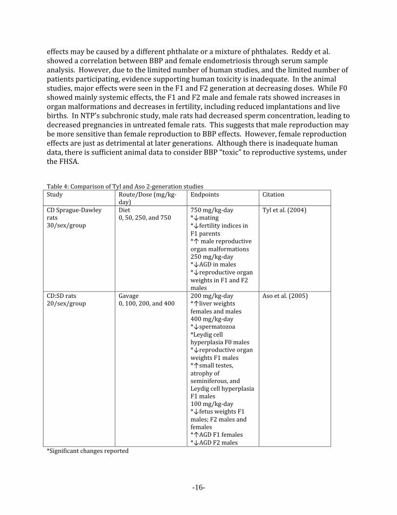
In Tyl et al.’s (2004) two generation study, BBP was administered in the diet of CD rats at concentrations of 0, 750, 3750, and 11,250 ppm (0, 50, 250, and 750 mg/kg‐day) for 10 weeks prior to mating and through weaning of the F2 generation pups, at which time reproductive toxicity was evaluated. Not only were there systemic effects, such as reduced body weights and increased organ weights, but for the F0 females there were reduced ovarian and uterine weights at the highest dose. No structural or functional effects were
-15-
seen in the F0 males. The F1 generation, exposed since their inception at the 750 mg/kg‐day dose, showed reduced mating and fertility indices, and reduced testes, epididymides, seminal vesicles, coagulating gland, and prostate weights. In addition, there were reproductive tract malformations, such as hypospadia, missing organs, and abnormal organ size and/or shape. The males also showed reduced epididymal sperm number, motility, progressive motility, and increased histopathologic effects in the testis and epididymis. The females also had reduced mating and fertility indices, reduced uterine implantations, reduced total and live pups, reduced number of litters, reduced ovarian weights, and increased uterine weights. There were no histopathologic lesions in the reproductive tract organs. In addition, the F2 generation, showed similar reproductive organ weight changes, as well as other developmental effects, such as decreased AGD in males starting at 250 mg/kg‐day (along with the F1 males) and increased nipple/areolae retention at 750 mg/kg‐day in males. There were no treatment related increases in reproductive tract malformations in the F1 or F2 generations. The NOAEL for F0 and F1 parental reproductive toxicity was 250 mg/kg‐day, the NOAEL for F1 and F2 offspring reproductive toxicity was also 250 mg/kg‐day, and the NOEL for F1 and F2 offspring males was 50 mg/kg‐day due to the decreases in AGD. Findings in a two generation study by Aso et al. (2005) (previously reviewed in Systemic Effects section) were in agreement with Tyl et al. Using Crj:CD Sprague‐Dawley IGS rats, Aso et al. (2005) reported reduced F0 reproductive organ weights; and reduced reproductive organ weights, softened testes, aplasia of the epididymis, hyperplasia of the Leydig cells, and reduced spermatozoa and germ cells in F1 males. Reproductive capacity was also impaired, with a reduction in fertility indices in the F1 males. In addition, the F1 females had increased AGD, while the F2 males had decreased AGD. Aso et al. (2005) determined that the NOEL/NOAEL for the parental animals and for offspring growth and development was less than 100 mg/kg‐day. In a 10 week, sub‐chronic study by NTP, male F344/N rats were given 0, 300, 2800 or 25,000 ppm (0, 20, 200, or 2200 mg/kg‐day) in their diet. Two days after the 10 week exposure, male rats were each mated to 2 untreated females for 7 days, then sacrificed and examined. The female rats were sacrified 13 days after mating and evaluated. Starting at the 200 mg/kg‐day dose there were decreased epididymal spermatozoa. With the 2200 mg/kg‐day dose, sperm concentration was decreased by >99%; prostate, testes, epididymis, and seminal vesicle weights were decreased; there was evidence of testicular and epididymal degeneration; body weight gain was down by 29%; liver and thymus weights were increased; and the animals had a mild macrocytic anemic response. When the females that mated with the males treated with 2200 mg/kg‐day BBP were examined, 10 of the 13 females showed evidence of mating and no pregnancies were found. The no observed effect level (NOEL) was determined to be 20 mg/kg‐day, while the NOAEL was 200 mg/kg‐day (NTP, 1997). In summary, in the human studies reviewed, authors made correlations between sperm quality and MBP and/or MBzP, as well as, female endometriosis and BBP. However, the reduction in sperm quality was inconsistent. These correlations were based on increases in metabolites MBP and MBzP, and since MBP is not exclusive to BBP, these
-16-
effects may be caused by a different phthalate or a mixture of phthalates. Reddy et al. showed a correlation between BBP and female endometriosis through serum sample analysis. However, due to the limited number of human studies, and the limited number of patients participating, evidence supporting human toxicity is inadequate. In the animal studies, major effects were seen in the F1 and F2 generation at decreasing doses. While F0 showed mainly systemic effects, the F1 and F2 male and female rats showed increases in organ malformations and decreases in fertility, including reduced implantations and live births. In NTP’s subchronic study, male rats had decreased sperm concentration, leading to decreased pregnancies in untreated female rats. This suggests that male reproduction may be more sensitive than female reproduction to BBP effects. However, female reproduction effects are just as detrimental at later generations. Although there is inadequate human data, there is sufficient animal data to consider BBP “toxic” to reproductive systems, under the FHSA. Table 4: Comparison of Tyl and Aso 2‐generation studies Study Route/Dose (mg/kg‐
day) Endpoints Citation
CD Sprague‐Dawley rats 30/sex/group
Diet 0, 50, 250, and 750
750 mg/kg‐day*↓mating *↓fertility indices in F1 parents *↑ male reproductive organ malformations 250 mg/kg‐day *↓AGD in males *↓reproductive organ weights in F1 and F2 males
Tyl et al. (2004)
CD:SD rats 20/sex/group
Gavage 0, 100, 200, and 400
200 mg/kg‐day*↑liver weights females and males 400 mg/kg‐day *↓spermatozoa *Leydig cell hyperplasia F0 males *↓reproductive organ weights F1 males *↑small testes, atrophy of seminiferous, and Leydig cell hyperplasia F1 males 100 mg/kg‐day *↓fetus weights F1 males; F2 males and females *↑AGD F1 females *↓AGD F2 males
Aso et al. (2005)
*Significant changes reported

-17-
Developmental Effects
The developmental effects of the o‐DAP’s have been well‐studied in animals. A thorough review of the developmental effects of o‐DAP’s in general is beyond the scope of this review. Briefly, perinatal exposure to certain phthalates is associated with the “phthalate syndrome” in rats, which encompasses a range of effects on the development of the male genitourinary system including reduced anogenital distance (AGD), nipple retention, undescended testes, testicular atrophy, testicular histopathology, underdeveloped gubernacular cords, and hypospadias (reviewed in Foster et al. 2001; Foster 2006; Howdeshell et al. 2008). These effects persist into adulthood, even in the absence of further exposure (Barlow et al. 2004; compare McIntyre et al. 2001). The effects are mainly due to the inhibition of testosterone synthesis (Mylchreest et al. 1998; Foster et al. 2001; Gray et al. 2000; Parks et al. 2000), along with reduced expression of insulin‐like hormone 3 gene (insl3) (Wilson et al. 2004). The specific cellular and molecular targets of o‐DAP’s are unknown (Howdeshell et al. 2008). Human Data BBP has been associated with altered male reproductive system development in animals. However, there are fewer human studies. The following BBP studies conclude that the metabolites MBP and/or MBzP can cause shortened AGD and impaired testicular descent in male infants, and reduced Leydig cell development and function with perinatal exposure.
One study was reviewed on the association of prenatal phthalate exposure, including BBP, and the decrease of male infant anogenital distance (AGD). Swan et al. (2005) used data from the Study of Future Families from 1999‐2002. The study participants were pregnant women at least 18 years of age who were seen at one of the four study clinics. Their pregnancies ended in a live birth, and their babies were 2‐36 months of age at the time of examination. Urine samples were collected from the mothers during mid‐gestation, and physical examinations were done on the infants, as well as post natal maternal and infant urine samples collected. There were no obvious genital malformations or grossly abnormal parameters. Of all the male infants tested, 86.6% of them had normal or normal‐retractile testes. Eighty‐five, out of 214 mothers, were then tested for phthalate metabolites, including MBP, one of the main metabolites for BBP. Prenatal, post‐natal and infant urine samples were evaluated in these 85 mothers and infants. There was a significant inverse relationship between MBP and the male anogenital index (AGI), a modification of AGD where AGD is divided by the weight of the child. Swan further grouped the examined infants based on their AGI; short AGI, those who fell below the 25th percentile, long AGI, those who were at or above the 75th percentile, and those in between were considered intermediate. Those in the short AGI group, on average, had an AGI 18.3% shorter than predicted by the regression model used. In addition, there was a significant increase of incompletely descended testicles (20, 9.5, and 5.9% for short, intermediate, and long ADI, respectively) when short ADI was compared to all other boys. Other significant correlations were the proportion of boys with scrotum categorized as small or indistinguishable from surrounding tissue, penile volume, and penile
-18-
volume/weight. The authors concluded that the AGD is shortened and testicular descent impaired by prenatal maternal exposure to phthalates.
The Swan et al. study has raised questions about the non‐conventional use of the
AGI in humans and statistical methods used. There is no standardized range for a normal AGD or AGI. Several scientists have reviewed the data presented by Swan et al. and have not been able to follow statistical logic or reproduce significance and associations presented. National Institutes of Health (NIH) and Center for the Evaluation of Risks to Human Reproduction (CERHR) (draft dated October 1, 2005) were unable to validate the findings reported (Butterworth, 2005). In addition, BASF (June, 2005) outlined nine specific questions regarding methodology, the results, and interpretation of data. Due to these questions and concerns, CPSC staff is unable to draw conclusions from this study. In another study, breast milk samples were obtained from a joint Danish and Finnish prospective, longitudinal cohort study conducted from 1997‐2001 on cryptorchidism (also known as undescended testes). The samples were analyzed for phthalate monoesters, including MBP and MBzP (Main, 2006). Serum samples from infant boys were analyzed for gonadotropins, sex‐hormone binding globulin (SHBG), testosterone, and inhibin B. The breast milk samples were additive aliquots of 1‐3 months postnatally collected breast milk. There was no correlation between cryptorchidism and either phthalate monoester concentration (p =0.440–0.823) in the breast milk. The authors did, however, find a significant positive correlation between MBP and SHBG (r=0.272, p=0.01), as well as a positive correlation between MBzP and SHBG that did not reach significance. MBP was significantly correlated with an increase in the LH:free testosterone ratio (p=0.0005). MBP was positively correlated with an increase in LH:testosterone ratios (p=0.008) and negatively correlated with free testosterone (p=0.004). These correlations were found when analyzing breast milk and serum samples from mothers and boys 3 months of age with and without cryptorchidism. The authors estimated that the median individual intake of phthalate monoesters MBP and MBzP were 3.46 and 0.70 µg/day for Denmark and 9.77 and 1.13 for Finland, respectively. In addition, the authors found that the concentration of MBP in the breast milk was significantly increased when a breast pump was used (p = 0.02). However, they could not determine the cause of this increase. No apparent leaching of BBP was seen during the use of a commonly used breast pump, and it is not thought to be linked with the association between breast milk phthalate concentrations and hormone levels since mothers in these countries are typically still on maternity leave when their babies are 3 months old and would not need to use a breast pump. It was concluded that the human Leydig cell development and function may also be vulnerable to perinatal exposure to phthalates (Main et al., 2006). SHBG, LH, and testosterone are considered markers for Leydig cell function. Alterations in these hormones can alter the pituitary‐gonadal axis and can lead to gonadotropin deficiencies or malformed testes. The authors did not find a correlation between the phthalates and cryptorchidism, or cryptorchidism and hormone levels. Animal Data

-19-
There are many studies showing that BBP and/or its metabolites MBP and MBzP cause developmental effects in animals. The effects most commonly seen were reduced AGD in males, undescended testes, reproductive tract malformations, male nipple/areolae retention, and reduced fertility.
Shono et al. (2000) showed that a brief exposure to MBP during fetal development
can inhibit the transabdominal migration of the testis and reduce testosterone content in the rat. MBP (0.3 g/day) was administered by stomach tube to three groups of pregnant rats. They were treated on gd 7‐10, or gd 11‐14, or gd 15‐18. The control group was treated with vehicle only (sesame oil) on gd 7‐18. Caesarean section was done at gd 20 and fetuses were evaluated for development. They reported that the mean transabdominal testicular migration values for groups 2 and 3 were significantly higher than the control group, meaning the testes of groups 2 and 3 were found higher in the abdominal cavity than the control group 4. Histology showed that the MBP treated groups had poorly developed epididymis, small thin ductus deferens, and the mean testicular testosterone level for the treated fetuses, when compared to the control testes, was significantly (p<0.001) decreased (50.9 pg/testis versus 852 pg/testis). Ema et al. (2003) found a decreased AGD and an increased incidence of undescended testes in fetuses when pregnant rats were given 167, 250, or 375 mg/kg MBzP on gd 15‐17. The fetuses were collected and examined on gd 21. The male and female fetal weights were significantly reduced with treatment of 375 mg/kg MBzP. Similar to Shono et al., the incidence of undescended testes and the degree of transabdominal testicular ascent were significantly (p<0.05) increased at 250 mg/kg and higher. The AGD and the AGD:cubed root of body weight were both significantly (p<0.05) decreased at 250 and 375 mg/kg treatments in the male fetuses when compared to the control group. The NOAEL for offspring was 250 mg/kg. However, this is significantly higher than the lowest NOAEL for BBP of 20 mg/kg by Nagao et al. (2000; as reported in Ema, 2003). Saillenfait et al. (2003) compared the toxic effects of BBP, MBP, and MBzP on embryos of Crl:OF1 (outbred) mice and Sprague‐Dawley rats in an in vivo and in vitro study. In the in vivo study, pregnant mice and rats were given a single oral dose of 280, 560, 1120, and 1690 mg BBP/kg, 200, 400, 800, and 1200 mg MBP/kg, or 230, 460, 920, and 1380 mg MBzP/kg on gestational days 8 for mice or 10 for rats. Mice and rats were euthanized on gd 18 or 21, respectively, and uteri and fetuses evaluated. In the mouse in vivo studies, they found a significant (p<0.05) increase in the percentage of resorption sites at doses greater than or equal to 560 mg/kg for BBP, 400 mg/kg MBP, and 1380 mg/kg MBzP. At the highest doses this reached 74, 81, and 46% for BBP, MBP, and MBzP respectively. External fetal malformations were also seen. The percentages of malformations were 43, 35, and 23% at the highest doses for BBP, MBP, and MBzP respectively. These external malformations typically consisted of exencephaly and imperforate anus, although other malformations were seen as well. For BBP and MBP, fetal body weights were significantly (p<0.05) reduced with the highest dose. In the rat in vivo study, there were no significant effects on implantation, resorption, number of live fetuses, and post implantation loss with the administration of MBP or MBzP. There were no

-20-
significant increases in resorptions or decreases in fetal body weight for BBP at 1120 and 1690 mg/kg. Five cases of exencephaly were found in the 1690 mg BBP/kg group.
In the in vitro mouse and rat studies, embryos were harvested for use in whole embryo cultures on gd 8 and 10 respectively. The embryos were exposed to MBP or MBzP concentrations ranging from 0.5 to 3 or 5 mM for 46 hours. Rat embryos were only exposed to MBzP. Open cranial neural folds were seen in 80% of the embryos exposed to 5 mM MBP. One of the same malformations was seen in embryos exposed to 1 and 2 mM MBP. MBzP caused a significant (p<0.05) reduction in crown rump length, head length, and number of somites at concentrations greater than or equal to 1 mM. Open cranial neural folds were found in 18 and 80% of embryos exposed to 2 and 3 mM MBzP respectively. Viable embryos were slightly reduced with 5 mM MBzP with all surviving showing poor and abnormal development. In the rat embryos, a statistically significant (p<0.05) reduction in head length and morphological scoring was seen at 1 mM MBzP. All the parameters were significantly (p<0.05) decreased at 2 mM. Twenty three percent of live embryos exposed to 2 mM MBzP showed an open cranial neural tube. There was also a decrease in the number of viable embryos at 2 and 3 mM, with those surviving at 3 mM having altered features of development (Saillenfait et al., 2003).
Previously, Ema et al. (1995) found that with a daily dose of 1250 mg/kg‐day BBP
administered to Wistar rats by gastric intubation given on gd 7‐9, complete resorption of all implanted embryos was seen. Those receiving 750 and 1000 mg/kg‐day BBP showed significantly (p<0.01) lowered weights for both female and male fetuses. A significant (p<0.01) increase in the incidence of skeletal malformations was seen with treatment given on gd 7‐9 in all groups. However, there were no significant increases in internal malformation for any of the treatment groups receiving treatment on gd 7‐9. Those groups receiving treatment on gd 10‐12 showed a significantly (p<0.01) lower rate of viability with 1000 mg/kg‐day BBP treatment and no viable litters were found with 1250 mg/kg‐day BBP treatment. In addition, a significant (p<0.01) increase in post implantation loss was seen at these doses as well as at 750 mg/kg‐day BBP. A significant (p<0.05) difference was seen in the sex ratio of live fetuses ((male/female) control 68/87 versus BBP treated 20/12) and reduced body weights in the 1000 mg/kg‐day BBP group. No significant changes were seen, compared to control groups, in the incidence of external, skeletal, and internal malformations. With 1250 mg/kg‐day BBP treatment on gd 13‐15, complete resorption of all implanted embryos was found. The sex ratio of live fetuses in the 750 mg/kg‐day BBP group was significantly (p<0.05) different when compared to controls ((male/female) control 68/87versus BBP treated 51/39), and there were increased incidents of external malformations in all groups treated with BBP on gd 13‐15. The incidents of skeletal malformations were also found in all groups treated with BBP on gd 13‐15. Animals received a fixed dose regardless of weight changes and were terminated on gd20. Table 5: Ema et al. (1995) Gestational Day and Dose Effects Dose (mg/kgday) 750 1000 1250 Exposure Time Frame (gd)

-21-
gd 79 *↓fetal body weight *↑internal malformations
*↓fetal body weight *↑internal malformations
Complete resorption
1012 *↑post implantation loss
*Reduced viability*↑post implantation loss *altered sex ratio *↓fetal body weight
Complete resorption
1315 *altered sex ratio*external malformation
*external malformation
Complete resorption
*Significant changes reported In Tyl et al.’s two generation study (see Table 4), they administered BBP in the diet
of CD rats at concentrations of 0, 750, 3750, and 11,250 ppm (0, 50, 250, and 750 mg/kg‐day) for 10 weeks prior to mating and through weaning of F2 generation pups. The F1 generations, which were exposed since their conception, at the dose of 750 mg/kg‐day, showed reduced testes, epididymides, seminal vesicles, coagulating gland, and prostate weights. In addition, there were reproductive tract malformations, such as hypospadia, missing organs, and abnormal organ size and/or shape. The males also showed reduced epididymal sperm number, motility, progressive motility, and increased histopathologic effects in the testis and epididymis. The females also had reduced mating and fertility indices, reduced uterine implantations, reduced total and live pups, reduced number of litters, reduced ovarian weights, and increased uterine weights. There were no histopathologic lesions in the reproductive tract. In addition, the F2 generation showed similar reproductive organ weight changes, as well as other developmental effects, such as decreased AGD starting at 250 mg/kg‐day (along with the F1 males) and increased nipple/areolae retention at 750 mg/kg‐day. The NOAEL for the F0 and F1 parental reproductive toxicity was 250 mg/kg‐day; the NOAEL for the F1 and F2 offspring reproductive toxicity was also 250 mg/kg‐day; and the NOEL for the F1 and F2 offspring males was 50 mg/kg‐day due to the decreases in AGD.
The human studies provide some evidence that phthalates may cause detrimental developmental effects. However, these data rely on exposure to the metabolite MBP or MBzP. Due to the ubiquitous nature of phthalates in consumer products, it cannot be certain that the metabolite (MBP) in these studies is due to the metabolism of BBP. There is an extensive amount of data showing developmental effects in animals. In two generation studies, these effects tend to be more severe. The studies reviewed showed decreased testicular descent, decreased AGD, retention of nipple/areolae in males, as well as organ and skeletal malformations. It is thought that the male reproductive system is more sensitive than the female reproductive system. However, malformations were also seen in females. Based on sufficient animal data demonstrating adverse developmental effects, BBP can be considered probably “toxic” under the FHSA. Genotoxicity and Carcinogenicity

-22-
Studies have investigated the potential of BBP to alter genes and gene expression, and increase carcinogenesis. Moral et al. (2007) showed that BBP can affect the gene expression profile of the mammary gland in rats. They exposed Sprague‐Dawley CD rats to 500 mg BBP/kg bw on postnatal days 2‐20. After weaning, female offspring were assessed on pnd 21, 35, 50 and 100. They did not find any significant change in the epithelial structures of the mammary gland. When the proliferative index of the mammary gland was evaluated, they found that the terminal end buds from the BBP exposed rats had a significantly (p<0.05) higher proliferation index by day 35 when compared to controls. Using microarray analysis, gene expression was assessed. At day 21, 151 genes were up‐regulated, including those involved in morphogenesis and cell differentiation, transcription factors, cell proliferation, response to stress, signal transduction, metabolism, and transport and cell organization. The number of up‐regulated genes was decreased at each evaluation point thereafter. One gene was found to be down‐regulated, Gad 1. Gad 1 codes for the protein Gad 67, glutamic acid decarboxylase, which catalyzes the production of the neurotransmitter GABA. Validation was done by RT‐PCR of some genes differentially expressed in the BBP treated rats. Genes Hfh1, a transcription factor, and Ahr, involved in stress response, were significantly (p<0.05) up regulated; while the other genes tested, Foxg1, involved in morphogenesis, organogenesis, and cell differentiation, and Nfyc, a transcription factor, were close to significant. The authors estimated that the dose received by the offspring was 0.5‐5mg BBP/kg ‐d, and puts it near the EPA estimated safe exposure for human adults of 0.2 mg/kg‐day and above the estimated exposure of human adults of 2 µg/kg ‐d. They further explain that the terminal end buds are the most susceptible epithelial structures to malignant transformation, and that it cannot be ruled out that the modifications caused by BBP may have an effect on the mammary gland’s susceptibility to carcinogenesis. NTP conducted a carcinogenicity study where they treated male F344/N rats by diet with 120, 240, and 500 mg/kg bw, and female rats with 300, 600, and 1200 mg/kg bw BBP for 24 months. Throughout the treatment, they found no clinical signs of toxicity and only sporadic changes in blood cell counts and chemistries in both male and female rats. The incidents of pancreatic acinar cell adenoma and adenoma and carcinoma combined were significantly (p=0.014) increased over controls in the 500 mg/kg bw treatment group for male rats. One carcinoma was found in one male rat at 500 mg/kg bw, and two adenomas were found in the 1200 mg/kg bw group of female rats. In the males at the highest dose, there was an increase in the number of focal hyperplasia of the pancreatic acinar cell seen. In the females at the highest dose, a significant (p≤0.05) increase in transitional epithelial hyperplasia in the urinary bladder was seen, as well as two females showing transitional epithelial papillomas. Based on this data, NTP’s conclusions were that there was evidence of carcinogenic activity of BBP in male F344/N rats due to the increased incidents of pancreatic acinar cell adenoma and acinar cell adenoma and carcinoma. There was minimal evidence of carcinogenic activity of BBP in female F344/N rats based on the marginal increase of pancreatic acinar adenoma and transitional epithelial papilloma of the urinary bladder (NTP, 1997).

-23-
Table 6: Neoplasms and Non‐neoplastic Lesions in the NTP 2‐year F344/N Rat Study of BBP in Feed (modified from CERHR, 2003) Dose (mg/kg bw) 0 120 240 500 Males Pancreas Acinus Hyperplasia 4/50 7/49 9/50 12/50 Acinus Adenoma 3/50 2/49 3/50 10/50 Acinus Carcinoma 0/50 0/49 0/50 1/50 Acinus Adenoma or Carcinoma
3/50 2/50 3/50 11/50*
Kidney Nephropathy 48/50 47/50 50/50 48/50 Renal Tubule Pigmentation 49/50 48/50 50/50 50/50 Hyperplasia 6/50 10/50 6/50 1/50
Skin Acanthosis 0/50 2/50 2/50 10/50** Hyperkeratosis 0/50 2/50 3/50 13/50**
Preputial Gland Hyperplasia 1/50 2/50 2/50 1/50 Adenoma 4/50 3/50 1/50 0/50 Adenoma or Carcinoma 5/50 8/50 2/50 0/50 0 300 600 1200 FemalesPancreas Acinus Hyperplasia 1/50 4/50 2/50 0/50 Acinus Adenoma 0/50 0/50 0/50 2/50
Bladder Hyperplasia 4/50 0/50 1/50 10/50* Papilloma 1/50 0/50 0/50 2/50
Kidney Nephropathy 34/50 47/50** 43/50* 45/50** Renal Tubule Pigmentation 49/50 49/50 49/50 47/50 Hyperplasia 0/50 3/50 7/50** 4/50
Mammary Gland Fibroadenoma 28/50 30/50 31/50 11/50 (*p≤0.05, significantly different from control; **p≤0.01, significantly different from control) In a similar NTP study (1982) (as reported by NICNAS, 2008), B6C3F1 mice (50/sex/group) were treated by diet with 6000 or 12,000 ppm BBP (equivalent to 840 and 1680 mg/kg‐d) for 2 years. No increases in tumor incidents were associated with BBP. Study was not repeated along with rat studies in 1997. NTP (1997) conducted a study to evaluate peroxisome proliferation in response to BBP in female F344/N rats. The rats were fed 0, 6000, 12,000, or 24,000 ppm BBP (equivalent to 300, 600, and 1200 mg/kg‐d) for one month (n=5/group) or one year (n=10/group). Using Palmitoyl CoA Oxidase (PCoA) and Carnitine Acetyl Transferase (CAT) as markers of peroxisome proliferation, CAT was significantly (p≤0.05) increased starting at 300 mg/kg‐d and PCoA was significantly (p≤0.05) increased starting at 600 mg/kg‐d in both the one month and one year groups. Using di(2‐ethylhexyl) phthalate (DEHP) as the positive control, NTP concluded that compared to DEHP, BBP is a mild peroxisome proliferator. Similar findings for BBP were also reported in Barber et al.’s

-24-
(1987) study on peroxisome proliferation induced by phthalates (not discussed in this review). These studies provide strong data showing BBP can alter the expression of genes and have carcinogenic activity. In addition, BBP was shown to activate PPARs, which have been associated with tumorigenesis (see Endocrine section of this review) However, although the NTP study shows significant data on the carcinogenicity of BBP, the data showed carcinogenicity in one sex of one species. Therefore, the data is considered limited, and is not sufficient to consider BBP carcinogenic under the FHSA. Discussion BBP is a phthalate ester that is ubiquitous in the environment. It can be absorbed orally, dermally, and through inhalation. It is thought that the main source of BBP is from food intake. It has been shown that BBP can cause systemic toxicity effects. Chronic, subchronic, and two generation studies with BBP show increases in liver and kidney weights at the lower doses, which can lead to decreased body weights. Gavage and doses administered in the diet during the two‐generation studies, and the chronic study, show that these effects increase over time. Inhalation of BBP showed similar effects. NTP also showed systemic effects in both a chronic and a subchronic study. Due to the consistency of these effects and the kidney effects seen at the lowest dose, using the NTP LOAEL of 120 mg/kg‐d, an ADI of 1.2 mg/kg was calculated for BBP (calculated from the LOAEL divided by a safety factor of 100 for species difference and sensitive population).
The studies reviewed have provided evidence that BBP can be considered toxic to the reproductive system under the FHSA. The human data was inadequate to correlate BBP exposure to reduced sperm quality in men. The data was inconsistent and the correlations made were to the metabolites MBP and MBzP. Correlations were made between serum BBP levels and endometriosis in women. Due to the lack of further evidence, it could not be determined if BBP causes reproductive effects in humans. The animal data showed strong evidence of BBP reducing fertility and sperm quality. It also showed reduced implantations and live births. NTP’s subchronic study showed reduced pregnancies due to reduced sperm quality in treated male rats. These studies, in conjunction with the two two‐generation studies showing increased detrimental effects in the next generation, and the consistency of effects between studies, are sufficient evidence to conclude that BBP is toxic to the reproductive system under the FHSA. The studies show that BBP causes increased developmental malformations in animals. The human studies raise many questions, and no conclusions were made. In addition, MBP is not exclusive to BBP. However, the animal data showed not only reduced AGD and testicular descent, but retention of nipples/areola in males, underdeveloped and malformed reproductive organs, and skeletal and external malformations. Saillenfait et al. used both mouse and rat studies to show that as much as 43% of pups showed malformations with BBP treatment. They also used in vitro studies and showed that up to 80% of embryos showed neural fold malformations, and those embryos surviving treatment were underdeveloped and damaged. The two‐generation studies showed that

-25-
malformations were evident in later generations and were increasingly severe. These results were sufficient enough to consider BBP developmentally toxic under the FHSA. In addition, using the NOAEL of 200 mg/kg‐d reported by NTP (1997), CPSC staff has calculated a reproductive/developmental ADI of 2.0 mg/kg‐d (calculated from the NOAEL divided by a safety factor of 100 for species difference and sensitive population). It has been questioned if phthalates have their effect through altering hormone activation. BBP and its metabolite, MBP, have been linked to altered expression of testosterone, LH, FSH, estrodiol, thyroid hormones, inhibin B, as well as, activating ER and PPAR receptors. Main et al. found a negative correlation with the monoester MBP and free testosterone in 3 month old infants. Without reference ranges for estrodiol and thyroid hormones in pregnant women, the negative correlation found by Huang et al. between MBP and T4 or FT4 is inadequate. BBP binds to the ER receptor and weakly competes with estrodiol. However, BBP did not show estrogenic activity in vivo. In addition, Lampen et al. (2003) found that BBP can bind to and activate murine PPARs and human PPARs. MBzP was a potent activator of the α and γ isotypes. Phthalates are found in indoor air and household dust. They have been associated with allergic symptoms and airway function changes. In one study, an increase in MBP was correlated to a decrease in the respiratory function parameters. When smokers were removed from the data set, lung FVC (forced vital capacity) remained significantly decreased. There was a non‐significant correlation between increased BBP in a home environment with children diagnosed with wheezing and eczema within the year prior to the study. In one study, children with persistent allergic symptoms had more BBP in the dust in their bedrooms then children without the persistent symptoms. In addition, a higher concentration of BBP was found in the bedrooms that contained PVC flooring, and the children using these bedrooms had more diagnosed cases of rhinitis or eczema. However, due to the limited data, BBP cannot be considered a strong sensitizer under the FHSA. BBP can alter the expression of genes, and may lead to increased susceptibility to carcinogenesis. Genes involved in morphogenesis and cell differentiation, transcription factors, cell proliferation, response to stress, signal transduction, metabolism, and transport and cell organization were among the genes found to be altered after BBP treatment. NTP’s carcinogenicity study provided limited evidence of carcinogenic activity. They found pancreatic adenomas and carcinomas in male rats treated with BBP, as well as increased incidence of focal hyperplasia. In female rats they found epithelial hyperplasia in the bladder, and papillomas. However, no liver tumors were found to be associated with BBP treatment, as previously associated with other phthalates. In addition, induction of peroxisome proliferation was found to be minimal when compared to the control DEHP. The animal studies for carcinogenicity are limited, and do not support a conclusion that BBP is carcinogenic under the FHSA. To be considered a “hazardous substance” under the FHSA, the substance must first present one or more of the following hazards: it must be toxic, corrosive, flammable, an irritant or a strong sensitizer, or generate pressure through decomposition, heat, or in

-26-
other ways; and second it must have the potential to cause substantial personal injury or illness during or as a result of any customary or reasonably foreseeable handling or use, including reasonably foreseeable ingestion by a child.
Following the definitions set forth by the FHSA, BBP can be considered toxic based on the available scientific data supporting kidney effects. Products that contain BBP would be considered ‘hazardous’ under the FHSA if oral exposure during ‘reasonably foreseeable handling and use’ were to exceed the ADI of 1.2 mg/kg‐d BBP.

-27-
Works Cited Adibi, J. J., Perera, F. P., Jedrychowski, W., Camann, D. E., Barr, D., Jacek, R., et al. (2003). Prenatal exposure to phthalates among women in New York and Krakow, Poland. Environmental Health Perspectives , 111 (14), 1719‐1722. Aso, S., Ehara, H., Miyata, K., Hosyuyama, S., Shiraishi, K., Umano, T., et al. (2005). A two generation reproductive toxicity study of butyl benzyl phthalate in rats. The Journal of Toxicological Sciences , 30, 39‐58. Barber, E., Astill, B., Moran, E., Schneider, B., Gray, T., & Lake, B. (1987). Peroxisome induction of seven phthalate esters. Toxicology and Industrial Health , 2, 7‐22. Barlow, N., McIntyre, B., & Foster, P. (2004). Male reproductive tract lesions at 6, 12, and 18 months of age following in utero exposure to di(n‐butyl) phthalate. Toxicologic Pathology , 32, 79‐90. Bernard, D. J., Chapman, S. C., & Woodruff, T. K. (2001). Mechanisms of inhibin signal transduction. Recent Progress in Hormone Research , 56 (1), 417‐450. Bornehag, C.‐G., Sundell, J., Weschler, C. J., Sigsgaard, T., Lundgren, B., Hasselgren, M., et al. (2004). The association between asthma and allergic symptoms in children and phthalates in house dust: A nested case control study . Environmental Health Perspectives , 112 (14), 1393‐1397. Butterworth, T. (2005, October 17). NIH panel unable to validate key finding in Swan phthalate baby study. Retrieved May 6, 2009, from Stats.org: www.stats.org/stories/NIH%20Panel_Swant_oct17_05.htm Carruthers, C. M., & Foster, P. M. (2005). Critical window of male reproductive tract development in rats following gestational exposure to di‐n‐butyl phthalate. Birth Defects Research (Part B) , 74, 277‐285. Duty, S. M., Calafat, A. M., Silva, M. J., Ryan, L., & Hauser, R. (2005). Phthalate exposure and reproductive hormones in adult men. Human reproduction , 20 (3), 604‐610. Duty, S. M., Silva, M. J., Barr, D. B., Brock, J. W., Ryan, L., Chen, Z., et al. (2003). Phthalate exposure and human semen parameters. Epidemiology , 14, 269‐277. Elsisi, A. E., Carter, D. E., & Sipes, I. G. (1989). Dermal absorption of phthalate diesters in rats. Fundamental and Applied Toxicology , 12, 70‐77.

-28-
Ema, M., Kurosaka, R., Amano, H., & Ogawa, Y. (1995). Comparative developmental toxicity of n‐butyl benzyl phthalate and di‐n‐butyl phthalate in rats. Archives of Environmental Contamination and Toxicology , 28, 223‐228. Ema, M., Miyawaki, E., Hirose, A., & Kamata, E. (2003). Decreased anogenital distance and increased incidence of undescended testes in fetuses of rats given monobenzyl phthalate, a major metabolite of butyl benzyl phthalate. Reproductive Toxicology , 17, 407‐412. European Chemicals Bureau. (2007). European Union Risk Assessment Report Benzyl Butyl Phthalate (BBP). Foster, P. (2006). Disruption of reproductive development in male rat offspring following in utero exposrue to phthalate esters. International Journal of Andrology , 29, 140‐147. Foster, P., Mylchreest, E., Gaido, K., & Sar, M. (2001). Effects of phthalate esters on the developing reproductive tract of male rats. Human Reproductive Update , 7, 231‐235. Gray, L., Ostby, J., Furr, J., Price, M., Veeramachaneni, D., & Parks, L. (2000). Perinatal exposure to phthalates DEHP, BBP, and DINP, but not DEP, DMP or DOTP, alters sexual differentiation of the male rat. Toxicological Sciences , 58, 350‐365. Harris, C. A., Henttu, P., Parker, M. G., & Sumpter, J. P. (1997). The estrogenic activity of phthalate esters in vitro. Environmental Health Perspectives , 105, 802‐811. Hoppin, J. A., Ulmer, R., & London, S. J. (2004). Phthalate exposure and pulmonary function. Environmental Health Perspectives , 112 (5), 571‐574. Howdeshell, K., Wilson, V., Furr, J., Lambright, C., Rider, C., Blystone, C., et al. (2008). A mixture of five phthalate esters inhibits fetal testicular testosterone production in the Sprague‐Dawley rat in a cumulative, dose additive manner. Toxicological Sciences , 105, 153‐165. Huang, P.‐C., Kuo, P.‐L., Gue, Y.‐L., Liao, P.‐C., & Lee, C.‐C. (2007). Associations between urinary phthalate monoesters and thyroid hormones in pregnant women. Human Reproduction , 22 (10), 2715‐2722. Hubinger, J. C., & Havery, D. C. (2006). Analysis of consumer cosmetic products for phthalate esters. Journal of Cosmetic Science , 57, 127‐137. International Programme on Chemical Safety. (1999). Butyl Benzyl Phthalate. International Programme on Chemical Safety. (1999). Butyl Benzyl Phthalate. Jonsson, B. A., Richthoff, J., Rylander, L., Giwercman, A., & Hagmar, L. (2005). Urinary phthalate metabolites and biomarkers of reproductive function in young men. Epidemiology , 16 (4), 487‐493.

-29-
Koch, H. M., Preuss, R., Drexler, H., & Angerer, J. (2005). Exposure of nursery school children and their parents and teachers to di‐n‐butylphthalate and butylbenzylphthalate. International Archieves of Occupational and Environmental Health , 78, 223‐229. Koo, H. J., & Lee, B. M. (2004). Estimated exposure to phthalates in cosmetics and risk assessment. Journal of Toxicology and Environmental health, Part A , 67, 1901‐1914. Kolarik, B., Naydenov, K., Larsson, M., Bornehag, C.‐G., & Sundell, J. (2008). The association between phthalates in dust and allergic diseases among Bulgarian children. Environmental Health Perspectives , 116 (1), 98‐103. Lampen, A., Zimnik, S., & Nau, H. (2003). Teratogenic phthalate esters and metabolites activate the nuclear receptors PPARs and induce differentiation of F9 cells. Toxicology and Applied Pharmacology , 188, 14‐23. Main, K. M., Mortensen, G. K., Kaleva, M. M., Boisen, K. A., Damgaard, I. N., Chellakooty, M., et al. (2006). Human breast milk contamination with phthalates and alterations of endogenous reproductive hormones in infants three months of age. Environmental Health Perspectives , 114 (2), 270‐276. McIntyre, B., Barlow, N., & Foster, P. (2001). Androgen mediated development in male rat offspring exposed to flutamide in utero: permanence and correlation of early postnatal changes in anogenital distance and nipple retention with malformations in androgen dependent tissues. Toxicological Sciences , 62, 236‐249. Moral, R., Wang, R., Russo, I. H., Mailo, D. A., Lamartiniere, C. A., & Russo, J. (2007). The plasticizer butyl benzyl phthalate induces genomic changes in rat mammary gland after neonatal/prepubertal exposure. BMC Genomics , 8, 453. Mylchreest, E., Cattley, R., & Foster, P. (1998). Male reproductive tract malformations in rats following gestational and lactational exposure to di(n‐butyl) phthalate: an antiandrogenic mechanism? Toxicological Sciences , 43, 47‐60. National Industrial Chemicals Notification and Assessment Scheme. (2008). Existing Chemical Hazards Assessment Report Butylbenzyl Phthalate. National Toxicology Program. (2003). NTPCERHR Monograph on the Potential Human Reproductive and Developmental Effects of Butyl Benzyl Phthalate (BBP). National Toxicology Program. (1997). Benzyl Butyl Phthalate Feed Studies. Nativelle, C., Picard, K., Valentin, I., Lhuguenot, J. C., & Chagnon, M. C. (1999). Metabolism of N‐butyl benzyl phthalate in the female Wistar rat. Identification of new metabolites. Food and Chemical Toxicology , 37, 905‐917. Otake, T., Yoshinaga, J., & Yanagisawa, Y. (2004). Exposure to phthalate esters from indoor environment. Journal of Exposure Analysis and Environmental Epidemiology , 14, 524‐528.

-30-
Parks, L., Ostby, J., Lambright, C., Abbott, B., Klinefelter, G., Barlow, N., et al. (2000). The plasticizer diethylhexyl phthalate induces malformations by decreasing fetal testosterone synthesis during sexual differentiation in the male rat. Toxicological Sciences , 58, 339‐349. Reddy, B., Rozati, R., Reddy, B., & Raman, N. (2006). Association of phthalate esters with endometriosis in Indian women. International Journal of Obstetrics and Gynaecology , 113, 515‐520. Saillenfait, A.‐M., Sabate, J.‐P., & Gallissot, F. (2003). Comparative embryotoxicities of butyl benzyl phthalate, mono‐n‐butyl phthalate and mono‐benzyl phthalate in mice and rats: in vivo and in vitro observations. Reproductive Toxicology , 17, 575‐583. Schettler, T. (2006). Human exposure to phthalates via consumer products. International Journal of Andrology , 29, 134‐139. Shono, T., Kai, H., Suita, s., & Nawata, H. (2000). Time specific effects of mono‐n‐butyl phhalate on the transabdominal descent of the testis in rat fetuses. BJU International , 86, 121‐125. Skinner, M., McLachlan, R., & Bremner, W. (1989). Stimulation of Sertoli cell inhibin secretion by the testicular paracrine factor PModS. Molecular and Cellular Endocrinology , 66 (2), 239‐249. Sorensen, L. K. (2006). Determination of phthalates in milk and milk products by liquid chromatography/tandem mass spectrometry. Rapid Communications in Mass Spectrometry , 20, 1135‐1143. Swan, S. H., Main, K. M., Liu, F., Stewart, S. L., Kruse, R. L., Calafat, A. M., et al. (2005). Decrease in anogenital distance among male infants with prenatal phthalate exposure. Environmental Health Perspectives , 113 (8), 1056‐1061. Takayama, O., Yamamoto, H., Damdinsuren, B., Sugita, Y., Ngan, C., Xu, X., et al. (2006). Expression of PPAR delta in multistage carcinogenesis of the colorectum: implications of malignant cancer morphology. British Journal of Cancer , 95, 889‐895. Tyl, R. W., Myers, C. B., Marr, M. C., Fail, P. A., Seely, J. C., Brine, D. R., et al. (2004). Reproductive toxicity evaluation of dietary butyl benzyl phthalate (BBP) in rats. Reproductive Toxicology , 18, 241‐264. U.S. Consumer Product Safety Commission. (1992). Federal Hazardous Substances Act Regulation. 16 CFR 1500.135. Zacharewski, T., Meek, M., Clemons, J., Wu, Z., Fielden, M., & Matthews, J. (1998). Examination of the in vitro and in vivo estrogenic activities of eight commerial phthalate esters. Toxicological Sciences , 46, 282‐293.

-31-
Appendixes Appendix 1: NOAEL and LOAEL w/endpoints Citation Model Route/Dose NOAEL LOAEL & Endpoint Ema et al. (2003)
Wistar rats Gastric intubation0, 167, 250, 375mg/kg Gd15‐17
250mg/kg
Tyl et al. (2004)
CD Sprague‐Dawley rats
Diet 0, 50, 250, 750mg/kg‐day
(M)250mg/kg‐day 50mg/kg‐day
(M)750mg/kg‐day *organ wt changes *liver histology 250mg/kg‐day *↓AGD *reproductive organ wt changes
Nagao et al. (2000)
Sprague‐Dawley rats
Gavage0, 20, 100, 500mg/kg‐day
(M)100mg/kg‐day 20mg/kg‐day
(M)500mg/kg‐day *↓bw gain *↑liver wt *↓testes, epididymis & seminal vesicle wt *↓#germ cells & sperm 100mg/kg‐day *↓birth wt
Aso et al. (2005)
CD:SD rats Gavage0, 100, 200, 400mg/kg‐day
(M)100mg/kg‐day 200mg/kg‐day
(M)200mg/kg‐day *↑liver wt 400mg/kg‐day (fert.) *↓spermatozoa *Leydig cell hyperplasia *small testes 100mg/kg‐day *↓fetal bw *↑AGD(f) *↓AGD(m)
NTP (1997) Fisher 344 rats Diet 0, 20, 200, 2200mg/kg‐day 10 weeks
200mg/kg‐day 2200mg/kg‐day *↓spermatozoa conc. *↓bw *↓sex organ wt *no preg.
NTP (1997) Fisher 344 rats Diet 0, 30, 180, 550, 1660mg/kg‐day 26 weeks
550mg/kg‐day 1660mg/kg‐day *↓fertility *↓spermatozoa conc. *↓sex organ wt
Piersma et al. (1999)
Cpb‐WU rats Gavage0, 270, 350, 450, 580, 750, 970, 1250, 1600, 2100mg/kg‐day 28 weeks
350mg/kg‐day 450mg/kg‐day *↓testosterone level

-32-
Agarwal et al. (1985)
Fisher 344 rats Diet 0, 312, 625, 1250, 2500mg/kg‐day 14 days
625mg/kg‐day 1250mg/kg‐day *↓bw *↓sex organ wt *histological changes in testes, prostate, & seminal vesicle *immature sperm *tubular epithelium necrosis ↑LH & FSH levels ↓progesterone levels
Piersma et al. (1995)
RIVM‐bred WU rats
Gavage0, 250, 500, 1000mg/kg‐day 14 days
(M)500mg/kg‐day 250mg/kg‐day
(M)1000mg/kg‐day *↓bw *↓preg. rate, live pups, epididymis wt, test. deg. 500mg/kg‐day ↓pup wt
Lake et al. (1978)
Sprague‐Dawley rats
Gavage0, 160, 480, 1600mg/kg‐day 14 days
160mg/kg‐day 480mg/kg‐day *histological changes in testes 1600mg/kg‐day *↓testes wt & testicular atrophy
Lake et al. (1978)
Wistar rats Sprague‐Dawley rats
Gavage0, 480, 1600mg/kg‐day 14 days
(W)480mg/kg‐day (SD) NE
480mg/kg‐day *testicular atrophy (SD) 1600mg/kg‐day *↓testes wt & testicular atrophy
Lake et al. (1978)
Sprague‐Dawley rats
Gavage0, 800, 1600 mg/kg‐day 4 days
800mg/kg‐day *↓testes wt & testicular atrophy
Hammond et al. (1987)
Sprague‐Dawley rats
Diet 0, 500, 1000, 1500, 2000, 3000, 4000mg/kg‐day 4 weeks
1000mg/kg‐day 1500mg/kg‐day *testicular atrophy *↓bw
Monsanto (1996)
Alkp:ApfSD rats
Gavage0, 56, 280, 1120, 2240mg/kg‐day
2240mg/kg‐day NE
Zacharewski et al. (1998)
Alkp:ApfSD rats
Subcutaneous0, 0.5, 5, 50, 500, 5000mg/kg‐day
5000mg/kg‐day NE
Zacharewski et al. (1998)
OXV Sprague‐Dawley rats
Gavage0, 20, 200, 2000mg/kg‐day
2000mg/kg‐day NE
Monsanto (1993)
Wistar rats Water0, 0.2, 0.4, 0.8%
(M)0.4%0.8% ((m)418 (f)446mg/kg‐day)
(M)0.8% *↓bw gain *↓food consumption *↑liver wt

-33-
NTP (1990) Swiss DC‐1 mice
Diet 0, 182, 910, 2330, 4121mg/kg‐day Gd6‐15
(M)182mg/kg‐day
(M)910mg/kg‐day *↓wt gain *↑prenatal mortality *↑malformations
NTP (1989) Sprague‐Dawley rats
Diet 0, 419, 1102, 1641mg/kg‐day
(M)419mg/kg‐day
(M)1102mg/kg‐day *↓wt gain *↓fetus wt gain *↑resorption & malformations
Ema et al. (1990)
Wistar rats Diet 0, 185, 375, 654, 974mg/kg‐day Gd0‐20
(M)375mg/kg‐day 185mg/kg‐day
(M)654mg/kg‐day *↓wt gain 375mg/kg‐day *↓#fetus/litter
Ema et al. (1992)
Wistar rats Gavage0, 500, 750, 1000mg/kg‐day Gd7‐15
(M)500mg/kg‐day
(M)750mg/kg‐day *↓food consumption and bw gain *resorptions in some *↓fetus wt *↑malformations *↓bw gain
Ema et al. (1993)
Wistar rats Gavage0, 600, 750, 1000mg/kg‐day Gd7‐9, 10‐12, 13‐15
600mg/kg‐day 750mg/kg‐day *↓fetus wt gd7‐9 *↑malformations gd7‐9, 13‐15
Ema et al. (1998)
Wistar rats Gavage0, 250, 500, 750, 1000mg/kg‐day Gd0‐8
250mg/kg‐day (M)250mg/kg‐day *↓bw gain 500mg/kg‐day *↓fetus wt
Keen (1998) Wistar rats Gavage0, 250, 1000, 1500, 2000mg/kg‐day Gd11, 12, 13
(M)250mg/kg‐day
1000mg/kg‐day *skeletal anomalies
Gray et al. (2000)
Sprague‐Dawley rats
Gavage0, 750mg/kg‐day Gd14‐pnd3
750mg/kg‐day *testes malformation *↓AGD *↓sex organ wts pnd2 *male areolas pnd13
Ema et al. (1991)
Wistar rats Diet 0, 974mg/kg‐day Gd0‐20
(M)974mg/kg‐day *↓bw *↑post implantation loss
Ema et al. (1992)
Wistar rats Diet 0, 974mg/kg‐day Gd0‐20, 0‐7, 7‐16, 16‐20
974mg/kg‐day *↑post implantation loss gd0‐20, 0‐7, 7‐16 *teratogenicity gd16‐20
Ema et al. (1992)
Wistar rats Diet 0, 974mg/kg‐day 0‐20, 0‐11, 11‐20
(M)974mg/kg‐day *↓bw gain *↑complet resorptions gd0‐20, 0‐11 *teratogenic effects gd11‐20

-34-
Ema et al. (1994)
Wistar rats Diet 0, 974mg/kg‐day Gd7, 9, 11
(M)974mg/kg‐day
974mg/kg‐day *↑post implantation loss gd11 *↓ovarian and uterine wts *↓serum progesterone all days
Hotchkiss et al. (2004)
Sprague‐Dawley rats
Gavage0, 75 linuron, 500 BBP, combomg/kg‐day
(M)500mg/kg‐day
500mg/kg‐day *↓testosterone *↓AGD (m) *↑reproductive organ malformations *↓reproductive organ wts
Saillenfait et al. (2003)
OF1 mice Gavage0, 280, 560, 1120, 1690mg/kg‐day Gd8
(M)560mg/kg‐day 280mg/kg‐day
(M)1120mg/kg‐day *maternal deaths *↓body wt gain 560mg/kg‐day *↑resorptions *↑malformations *↑post implantation loss
Saillenfait et al. (2003)
Sprague‐Dawley rats
Gavage0, 560, 1120, 1690mg/kg‐day
(M)1120mg/kg‐day
(M)1690mg/kg‐day *maternal deaths *↑malformations
Ema et al. (1995)
Wistar rats
Gavage (MBP)0, 250, 500, 625mg/kg‐day Gd7‐15
(M)250mg/kg‐day
(M)500mg/kg‐day *↓wt gain *↑prenatal mortality *↓fetal wt *↑malformations *↑visceral variations
Ema et al. (1996)
Wistar rats Gavage (MBzP)0, 250, 313, 375, 438, 500mg/kg‐day Gd7‐15
250mg/kg‐day (M)250mg/kg‐day *↓food consumption 313mg/kg‐day *skeletal malformations
Price et al. (1990)
CD‐1 mice Dietary0, 182, 910, 2330mg/kg‐day Gd6‐15
(M)182mg/kg‐day
(M)910mg/kg‐day *↓wt gain *↑prenatal mortality *malformations
European Council (1998)
Wistar rats Water0, 0.012, 0.140, 0.385mg/kg‐day
(M)0.385mg/kg‐day 0.14mg/kg‐day
0.385mg/kg‐day *↑pup deaths pnd1‐4
Bayer (1998) Wistar rats Water and Diet(d)0, 0.11‐0.16, 0.34‐0.49 (w)0, 0.17‐0.24, 0.54‐0.80
(M)(d)0.34‐0.49(w)0.54‐0.80
NE
*data collected from CERHR (2000), NICNAS (2008), ECB (2007) and referenced articles

-35-
Appendix 2: Reproductive and Developmental Critical Effects Citation Model/Dose Critical Effects Reproductive Reddy et al. (2006) Infertile human women ↑endometriosis Duty et al. (2003) Male human patients ↓sperm motility,
concentration, morphology Duty et al. (2005) Male human patients ↑abnormal sperm parametersEma et al. (1995) Wistar rats – gastric intubation
0.75, 1.0, 1.25g/kg Gd7‐9, 10‐12, 13‐15
↑post implantation loss ↑fetal malformations
Saillenfait et al. (2003) OF1 mice & Sprague‐Dawley rats – single oral dose 0, 0.9, 1.8, 3.6, 5.4mmol/kg Gd8 & 10
↑resorptionMaternally lethal
Aso et al. (2005) Sprague‐Dawley rats – oral0, 100, 200, 400mg/kg‐d 10 wk prebreed to weaning
↓fertility index ↓spermatozoa ↓complete preputial separation
Piersma et al. (1995) WU rats – gavage0, 250, 500, 1000mg/kg‐day 2 wk prebreed +29 days (m) or pnd 6 (f)
↓fertility↓litter size Testicular lesions
TNO Wistar rats – dietary(m)0, 108, 206, 418 10wk premating (f)0, 106, 217, 446mg/kg‐day 2wk premating 0, 116, 235, 458mg/kg‐day gestation 0, 252, 580, 1078mg/kg‐day lactation
No structural or functional effects
NTP (1997) F344 rats – diet0, 20, 200, 2200mg/kg‐day 10 wk prebreed
↓sperm count ↓fertility Testicular and epididymal lesions ↓sex organ wts
Tyl et al. (2004) CD Sprague‐Dawley rats –dietary 0, 50, 250, 750mg/kg‐day 2 generations
↓mating↓fertility
Nagao et al. (2000) CD Sprague‐Dawley rats –gavage 0, 20, 100, 500 mg/kg‐day 2 generation
↓body wt gain ↑kidney and liver wts ↓testosterone & ↑FSH in males
Agrawal et al. (1985) Fisher 344 rats – diet312, 625, 1250, 2500mg/kg‐day 14 days
↓organ and sex organ wts Histological changes in sex organs Immature sperm General toxicosis ↑LH and FSH ↓progesterone

-36-
Piersma et al. (2000) Cpb‐WU rats – gavage0, 270, 350, 450, 580, 750, 970, 1250, 1600, 2100 mg/kg‐day 28 days
↑liver wt↓testicular wts w/ atrophy ↓testosterone
Monsanto (1993) Wistar rats – diet0.2, 0.4, 0.8% One generation/2 litters
↓body wt gain
Lake et al. (1978) Wistar and Sprague‐Dawley rats – gastric intubation 160, 480, 1600mg/kg‐day 14 days Sprague‐Dawley rats – gastric intubation 800, 1600 BBP, 855 MBP, 985 MBzP mg/kg‐day 4 days
Histological changes in testes (SD) ↓testes wt w/testicular atrophy (W&SD) ↓testes wt w/testicular atrophy (all)
Hammond et al. (1987) Sprague‐Dawley rats – dietary500, 1000, 1500, 2000, 3000, 4000mg/kg‐day 4 weeks
↓body wt↑testicular atrophy
Hammond et al. (1987) Sprague‐Dawley rats – dietary188, 375, 750, 1125, 1500mg/kg‐day 3 months
↑kidney and liver wts
Hammond et al. (1987) Wistar rats – dietary151, 381, 960mg/kg‐day 3 months
↑kidney and liver wts ↓body wt Anaemia Histological changes in liver and pancreas
Developmental Shono et al. (2002) Wistar King A rats – stomach
tube 0.3g/d Gd7‐10, 11‐14, 15‐18
↓transabdominal testicular migration ↓epididymal development
Swan et al. (2005) Paired human male infants and mothers
↓AGI↓testicular decent
Ema et al. (2003) Wistar rats – gastric intubation167, 250, 375mg/kg‐d Gd16‐17
↑undecended testes ↑degree transabdominal testicular ascent ↓AGD
Saillenfait et al. (2003) OF1 mice & Sprague‐Dawley rats – single oral dose 0, 0.9, 1.8, 3.6, 5.4mmol/kg Gd8 & 10
↑malformations
Field et al. (1989) Sprague‐Dawley rats – dietary0, 420, 1100, 1640mg/kg‐day Gd6‐15
↑prenatal mortality ↓fetal wt ↑malformations
Ema et al. (1990) Wistar rats – dietary0, 185, 375, 654, 974mg/kg‐dayGd0‐20
↑prenatal mortality ↓fetal wt

-37-
Ema et al. (1992) Wistar rats – gavage0, 500, 750, 1000mg/kg‐day Gd7‐15
↑prenatal mortality ↓fetal wt ↑malformations ↑complete mortality
Ema et al. (1995) Wistar rats – gavage0, 250, 500, 625mg MBP/kg ‐d Gd7‐15
↑prenatal mortality ↓fetal wt ↑malformations
Ema et al. (1996) Wistar rats – gavage0, 313, 375, 438, 500mg/kg‐dayGd7‐15
↑prenatal mortality ↑skeletal malformations
Price et al. (1990) CD‐1 mice – dietary0, 182, 910, 2330mg/kg‐day Gd6‐15
↑prenatal mortality ↓fetal wt ↑malformations
Tyl et al. (2004) CD Sprague‐Dawley rats –dietary 0, 50, 250, 750mg/kg‐day 2 generations
↓AGD↓sex organ wts ↑reproductive organ lesions in males
*data collected from CERHR (2000), NICNAS (2008), ECB (2007) and referenced articles

-38-
Appendix 3: LD50
Model/Route LD50 Citation
Rat – oral 20400 mg/kg Hammond et al. 1987
Rat – oral 20400 mg/kg Monsanto 1976
Rat – oral 2330 mg/kg NTP 1982
Guinea pig – oral >13750 mg/kg bw RTECS 2005
Mouse – oral 6160 mg/kg (m) 4170 mg/kg (f)
NTP 1982
Rat – oral >4000 mg/kg Mallette et al. 1952
Rat – IP >1800 mg/kg Mallette et al. 1952
Mouse – IP 4000<LD50<5000mg/kg Monsanto 1983
Mouse – IP 4000<LD50<5000mg/kg Robinson et al. 1985
Mouse – IP 3160 mg/kg Calley et al. 1966
Rat – dermal 6700 mg/kg Statsek 1974
Rabbit – dermal >10000 mg/kg Hammond et al. 1987
Rabbit – dermal >10000 mg/kg Monsanto 1976
*modified from NICNAS (2008); ECB (2007)


![MASTERARBEITAIBN 2,2´-azo bisisobutyronitile BARF sodium tetrakis[3,5-bis(trifluoromethyl)phenyl]borate Bn benzyl Binol 1,1´-bi-2,2´-naphthol BOC t-butoxycarbonyl Bu (nBu) butyl](https://img.dokumen.tips/doc/110x75/5e7ce5a4bb305342984bda2d/masterarbeit-aibn-22-azo-bisisobutyronitile-barf-sodium-tetrakis35-bistrifluoromethylphenylborate.jpg)