Embed Size (px)
Citation preview
1
Title: The biaxial biomechanical behaviour of abdominal aortic aneurysm tissue
Abbreviated Title: Biaxial behaviour of abdominal aortic aneurysm
Siobhan O’Leary1, Donagh Healey
2, Eamon Kavanagh
2, Michael Walsh
1, Tim
McGloughlin1,3
and Barry Doyle4,5
.
1. Centre for Applied Biomedical Engineering Research, Materials and Surfaces Science
Institute, Mechanical, Aeronautical and Biomedical Engineering Department, University
of Limerick, Limerick, Ireland
2. Department of Vascular Surgery, University Hospital, Limerick, Ireland
3. Department of Biomedical Engineering, Khalifa University of Science, Technology &
Research (KUSTAR), P.O. Box: 127788, Abu Dhabi, UAE
4. Vascular Engineering, Intelligent Systems for Medicine Laboratory, School of
Mechanical and Chemical Engineering, The University of Western Australia, Perth WA
6009, Australia
5. Centre for Cardiovascular Science, The University of Edinburgh, United Kingdom
Corresponding Author address:
Barry Doyle,
The University of Western Australia,
35 Stirling Highway (M050),
Perth, WA 6009,
Australia.
Tel: +61 8 64881054
Email: [email protected]
2
ABSTRACT
Background
Rupture of the Abdominal Aortic Aneurysm (AAA) occurs when the local wall stress
exceeds the local wall strength. Knowledge of AAA wall mechanics plays a fundamental
role in the development and advancement of AAA rupture risk assessment tools. Therefore,
the aim of this study is to evaluate the biaxial mechanical properties of AAA tissue.
Methods
Multiple biaxial test protocols were performed on AAA samples harvested from 28 patients
undergoing open surgical repair. Both the Tangential Modulus (TM) and stretch ratio (λ)
were recorded and compared in both the circumferential (ϴ) and longitudinal (L) directions at
physiologically relevant stress levels, the influence of patient specific factors such as sex, age
AAA diameter and status were examined. The biomechanical response was also fit to a
hyperplastic material model.
Results
The AAA tissue was found to be anisotropic with a greater tendency to stiffen in the
circumferential direction compared to the longitudinal direction. An anisotropic hyperelastic
constitutive model represented the data well and the properties were not influenced by the
investigated patient specific factors however, a future study utilising a larger cohort of
patients is warranted to confirm these findings.
Conclusions
This work provides further insights on the biomechanical behaviour of AAA and may be
useful in the development of more reliable rupture risk assessment tools.
Key terms: Mechanical Properties, AAA, Anisotropy
3
INTRODUCTION
Abdominal aortic aneurysm (AAA) is a primarily asymptomatic, degenerative disease
defined as a localised and irreversible dilatation of the distal aortic wall by more than 50%42
.
The prevalence of AAA increases with age and in the United States the estimated total
number of people, aged between 50 and 84 years old, with AAA is 1.1 million (prevalence
1.4%)13
. If untreated, AAA progressively expands until it ruptures. Rupture of AAA is
responsible for approximately 6,000 to 10,000 deaths per year in the USA29, 33
. Considering,
individual AAAs are treated based on the assessment of rupture risk, it is imperative that this
assessment is accurate. Currently, preventative surgical treatment is recommended when the
maximum diameter is greater than 5.5cm (5cm in women) and the growth rate is greater than
1cmyear-1. However, reports of ruptured smaller AAAs22
(1.6%) and nonruptured larger
AAAs7 (2.0%) have questioned the reliability of this criterion. Based on the hypothesis that
rupture occurs when the induced wall stress exceeds the wall strength, many researchers have
advocated that the finite element method (FEM) (determining the peak wall stress
computationally) may aid in the prediction of AAA rupture10
.
Considering the AAA material model chosen to describe the mechanics of the AAA wall in
vivo is integral to the validity of wall stress computations21
, it seems of crucial importance to
fully understand, evaluate and describe the mechanics of the AAA wall accurately. The
development of AAA is associated with a degradation of structurally important connective
tissues such as elastin and collagen5 and therefore has a direct impact on the tissue’s
mechanical properties. Previous assessments of AAA biomechanics utilising uniaxial
mechanical test methods have found that the AAA tissue is weaker and stiffer than non-
aneurysmal aorta24, 32
, has regionally varying properties23, 34
and can vary significantly from
patient to patient9. Also, some studies have reported similar properties in both the
4
circumferential and longitudinal directions24, 26
, while another found that the tissue tended to
be more stiff in the circumferential than the longitudinal direction34
.
Considering AAA tissue is subjected to multi axial loading in vivo, biaxial testing is
recommended. This mode of testing is essential for detecting anisotropy and determining the
material coefficients for nonlinear constitutive models where multiple test protocols,
spanning the entire range of physiological stresses, are required to ensure accuracy28
. These
factors have been shown to have a direct influence on computationally derived AAA rupture
risk assessment21, 39
. Despite this, there have been few studies which have investigated the
biaxial properties of AAA tissue. Vande Geest et al.38
reported that the AAA was anisotropic
with a reduction in AAA extensibility and an increase in AAA stiffness in the circumferential
direction compared to the abdominal aorta. In addition, this study demonstrated the
differences in the strain energy derived from uniaxial and biaxial data, further underlining the
superiority of the biaxial test mode. While in basic agreement with the previous work, Tong
et al.35
concluded that the age of the intraluminal thrombus (ILT) increased the mechanical
anisotropy and decreased the strength of the underlying AAA wall.
Investigating the influence of patient specific factors such as sex, age, AAA diameter and
AAA status, i.e. elective or emergency repair, on the AAA biomechanical properties may
contribute to the understanding of which patients are more susceptible to rupture. Although
some of these factors have been linked to AAA failure properties previously8, 25, 26, 32, 37
, their
influence on the biaxial properties has not been well documented36
.
Knowledge of AAA wall mechanics plays a fundamental role in the development and
advancement of AAA rupture risk assessment tools. However, considering endovascular
repair (EVAR) is becoming increasingly popular, opportunities to harvest and conduct
experimental analyses on these tissues may become rare. Therefore, although there already
exists studies detailing the biaxial mechanical response of these tissue it is of crucial

5
importance that this database is expanded to include the properties of more patients due to the
high inherent inter and intra patient variability, while the tissue is still available, and also to
investigate if common patient–specific factors influence AAA biaxial mechanical properties.
Thus, the aim of this study is to (a) evaluate the biaxial mechanical properties of AAA tissue,
(b) estimate the material coefficients of a suitable constitutive model capable of describing
the tissue’s biomechanical response and finally, for the first time (c) investigate the influence
of various patient specific factors on the biaxial mechanical properties of AAA.
MATERIALS AND METHODS
Tissue Specimens
This study was approved by the University Hospital Limerick and the University of Limerick
Ethics Committees, patient consent was obtained in all cases and this work adheres to the
Declaration of Helsinki. Tissue from the anterior region of the AAA wall was harvested from
28 patients undergoing open AAA repair at the University Hospital Limerick. All tissue was
excised such that the longitudinal dimension was always larger than the circumferential
dimension. Upon excision, all samples were stored frozen in isotonic saline at -20°C until
further analysis19
.
Tissue Preparation
AAA tissue samples were thawed at 4°C overnight, allowed to equilibrate at room
temperature (~20°C), and then immersed in warm (37°C) isotonic saline. To prepare the
samples for tensile testing the loose connective tissue was carefully removed from the
abluminal side of the tissue. At least one 14 mm square sample was cut from each tissue
piece using a custom made die orientated such that the longitudinal axis was parallel to the
blood-flow. Care was taken to avoid atherosclerotic or calcified regions when cutting the
samples. Using a digital thickness gauge (Mitutoyo, Series 547), the thickness of all samples
was measured at five locations, repeated three times and averaged as described in a recent

6
study18
. A light mist of white paint (enamel, rustoleum) was then applied to the surface of the
tissue for use with the optical strain tracking system. Samples were allowed to dry (~5mins)
and then immersed in isotonic saline (37°C) to avoid any adverse effects of tissue
dehydration.
Biaxial Test Method
Specific details of the biaxial tensile test methods have been recently reported18-20
. Briefly,
the square samples (14 x 14 mm) were mounted in a BioTester 5000 test system (Cellscale,
Biomaterials Testing, Waterloo, Canada) using a set of four BioRakes (one rake per side).
Each BioRake consists of five evenly spaced tungsten tines used to pierce and grip adjacent
sides of the square tissue sample. Each specimen was mounted so that it was stretched in the
circumferential and longitudinal directions. All samples were submerged in 37°C isotonic
saline and tested using a series of circumferential to longitudinal displacement controlled
ratios, i.e. λϴ:λL = 1:1, 0.75:1, 1:0.75, 0.5:1, 1:0.5, 0.25:1, 1:0.25, 1:1. The 1:1 ratio was
repeated at the end of the regime to ensure the tissue was not mechanically compromised as a
result of test protocols. The BioRakes were set to displace by a maximum of 15% of the
gripped area (8.9 mm x 8.9 mm) at a rate of 0.2 mms-1. Therefore, a prescribed ratio of for
example λϴ:λL = 0.75:1 was equivalent to a circumferential stretch of 11.25% and a
longitudinal stretch of 15%. Samples were preconditioned for 10 cycles for all protocols, with
data from the final cycle used for subsequent analysis. As the tissue was stretched, the force
was measured continuously by a 5N load cell (accurate to 10mN) and images of the tissue
were taken by a charge coupled device (CCD) camera (1280 x 960 pixels at 15 frames per
second). All strain measurements were computed using the particle tracking software
provided with the BioTester and analysed from the central 25% of the tissues dimensions to
avoid important edge effects. This method is fully described in O’Leary et al.18
and the
associated Supplementary Material (Appendix B).

7
Data Analysis
The Cauchy stress was determined as follows: σϴϴ=Fϴλϴ/TXL and σLL=FLλL/TXϴ, where, F
is the measured load, λ is the stretch, X is the initial width, T is the averaged initial thickness
of the tissue sample, subscripts ϴ and L represent the circumferential and longitudinal
directions, respectively and shear was considered negligible. Transverse forces due to tine
deflection were found to be small compared to the axial forces and therefore have been
neglected in these analyses. Stretch was measured within the central region and was
calculated as follows: λϴ = xϴ/Xϴ and λL = xL/XL, where, x represents the final length in each
direction.
Mechanical Parameters
The stretch and Tangential Modulus (TM) were recorded at a Cauchy stress of 80kPa and
150kPa for each tissue sample in both the circumferential and longitudinal directions. These
values represent a range of stresses that the AAA is potentially exposed to in vivo and have
previously been utilised by similar studies 35, 36, 39
. The Tangential Modulus (TM) was
calculated by determining the slope of the Cauchy stress-stretch curve at each stress level.
The stretch and TM were compared in the circumferential and longitudinal directions, at both
80kPa and 150kPa, in order to identify and quantify anisotropy as a function of stress. To
compare the degree of anisotropy present within the experimental data to that predicted by
the constitutive model (described later), the following anisotropic index (AI) was calculated
at 150kPa as follows: AIexp = λL/λϴ, where, subscript ‘exp’ relates to the experimental data
and the material is considered isotropic when AI =1.
Constitutive Model
The AAA tissue was assumed to be a homogeneous30
, incompressible6, anisotropic
35, 38,
hyperelastic material undergoing finite deformations. Based on these assumptions, a
constitutive model originally proposed by Choi and Vito4, for canine pericardium, employed

8
by Sacks and Choung27
for chemically modified bovine pericardium and most recently by
Vande Geest et al.38
for both aneurysmal and non-aneurysmal abdominal aortic tissue was
chosen to fit the data presented here. The proposed strain energy function (SEF) takes the
following form:
𝑾 = 𝒃𝒐 [𝒆𝒃𝟏
𝟏𝟐
(𝝀𝜭
𝟐−𝟏𝟐
)
𝟐
+ 𝒆𝒃𝟐
𝟏𝟐
(𝝀𝑳
𝟐−𝟏𝟐
)
𝟐
+ 𝒆𝒃𝟑(
𝝀𝜭𝟐−𝟏𝟐
)(𝝀𝑳
𝟐−𝟏𝟐
)]
Where, W is the strain energy and b0, b1, b2 and b3 are the material coefficients to be
determined. Considering, σi = λi(dw/dλi), the expressions for Cauchy stress are as follows:
𝝈𝜭 = 𝒃𝟎 [𝝀𝜭𝟐𝒃𝟏 (
𝝀𝜭𝟐 − 𝟏
𝟐) 𝒆
𝒃𝟏𝟏𝟐
(𝝀𝜭
𝟐−𝟏𝟐
)
𝟐
+ 𝝀𝜭𝟐𝒃𝟑 (
𝝀𝑳𝟐 − 𝟏
𝟐) 𝒆
𝒃𝟑(𝝀𝜭
𝟐−𝟏𝟐
)(𝝀𝑳
𝟐−𝟏𝟐
) ]
and
𝝈𝑳 = 𝒃𝟎 [𝝀𝑳𝟐𝒃𝟐 (
𝝀𝑳𝟐 − 𝟏
𝟐) 𝒆
𝒃𝟐𝟏𝟐
(𝝀𝜭
𝟐−𝟏𝟐
)
𝟐
+ 𝝀𝑳𝟐𝒃𝟑 (
𝝀𝜭𝟐 − 𝟏
𝟐) 𝒆
𝒃𝟑(𝝀𝜭
𝟐−𝟏𝟐
)(𝝀𝑳
𝟐−𝟏𝟐
) ]
The data from the test protocols 1:1, 0.75:1, 1:0.75, 0.5:1, 1:0.5 for each sample in the
circumferential and longitudinal direction were then simultaneously fit to this model using a
Marquart-Levenberg least-squares optimisation technique27
and the material coefficients were
obtained for individual samples. As previously suggested by Vande Geest et al.38
, constraints
i.e. b1>0, b2 >0 and b3>0 were imposed to ensure that the parameters were physically
sensible. In order to obtain a single set of material constants which represent the group of
samples, data from each protocol was averaged to obtain a single dataset to which the
material model could then be fitted to. Additional test protocols, λϴ:λL = 0.25:1 and 1:0.25
were then used to assess the predictability of the model based on the derived material
coefficients.

9
Based on the relative contribution of both material coefficients b1 and b2 to the strain energy,
the following measure of anisotropy was calculated4, 27, 38
as follows: AImod = √b1/b2, where,
subscript ‘mod’ relates to the material model and as before, the material is considered
isotropic when AI =1.
Statistical Analysis
All measurements are presented as a mean ± standard deviation. The Shapiro-Wilk normality
test was performed prior to statistical tests which assumed a normally distributed sample.
Paired t-tests were employed to detect differences between the mechanical properties in the
circumferential and longitudinal direction of each tissue sample. Independent t-tests were
utilised to assess differences in the tissue’s properties grouped by patient specific factors, i.e.
sex, age, AAA diameter and AAA status. Pearson’s correlation coefficient was used to
determine if a relationship existed between AIexp and AImod. A two-sided p-value less than
0.05 indicated statistical significance for all paired and independent t-tests and also when
determining a Pearson’s correlation coefficient. All analyses were performed using IBM
SPSS Statistics (IBM Corp. Released 2011. IBM SPSS Statistics for Windows, Version 20.0.
Armonk, NY: IBM Corp).
RESULTS
Sample Characteristics
To summarise patient sample characteristics; 22/28 of patients were male, 18/28 were
elective repair, the average maximum AAA diameter, ɸ, was 6.65 ± 1.57 cm and the average
patient age was 71.60 ± 6.67 years. In total, from the AAA wall tissue samples of 28 patients,
34 specimens were tested successfully. Samples which underwent structural damage (n=7)
during testing evidenced by the final 1:1 testing ratio were excluded from the results. The
main reason for structural damage was attributed to the tines pulling through the tissue when
experiencing high force. Where more than one sample was tested from the same tissue piece
10
(patient no. = 5), the average mechanical properties of the specimens were chosen to
represent the overall mechanical response for that patient.
Equibiaxial Response
As shown in Figure 1, the AAA tissue was found to be anisotropic, with higher average TM
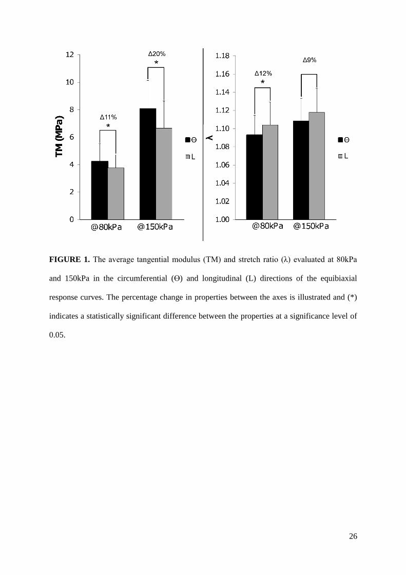
values and lower average λ values in the circumferential than in the longitudinal direction
when evaluated at both 80kPa (i.e. TMϴ vs TML: 4.2 ± 1.3 vs 3.7 ± 0.9 (p = .04) and λϴ vs λL:
1.09 ± 0.02 vs. 1.10 ± 0.02 (p = .02)) and 150kPa (i.e. TMϴ vs TML: 8.1 ± 3.2 vs 6.6 ± 1.2 (p
= .01) and λϴ vs λL: 1.11 ± 0.03 vs. 1.12 ± 0.03 (p = .06)). Therefore, in general the tissue is
found to be stiffer and less extensible in the circumferential than in the longitudinal direction.
The percentage difference between TM measured in each direction is shown to increase (9%)
when measured at higher stresses while, the percentage difference between λ is shown to
decrease (3%) at higher stresses.
Constitutive Model
From the average Cauchy stress-stretch response plots of the multiple test protocols in both
the circumferential and longitudinal directions illustrated in Figure 2, it can be seen that the
material model provided an excellent fit for the averaged group data (R2 = 0.98) and also fit
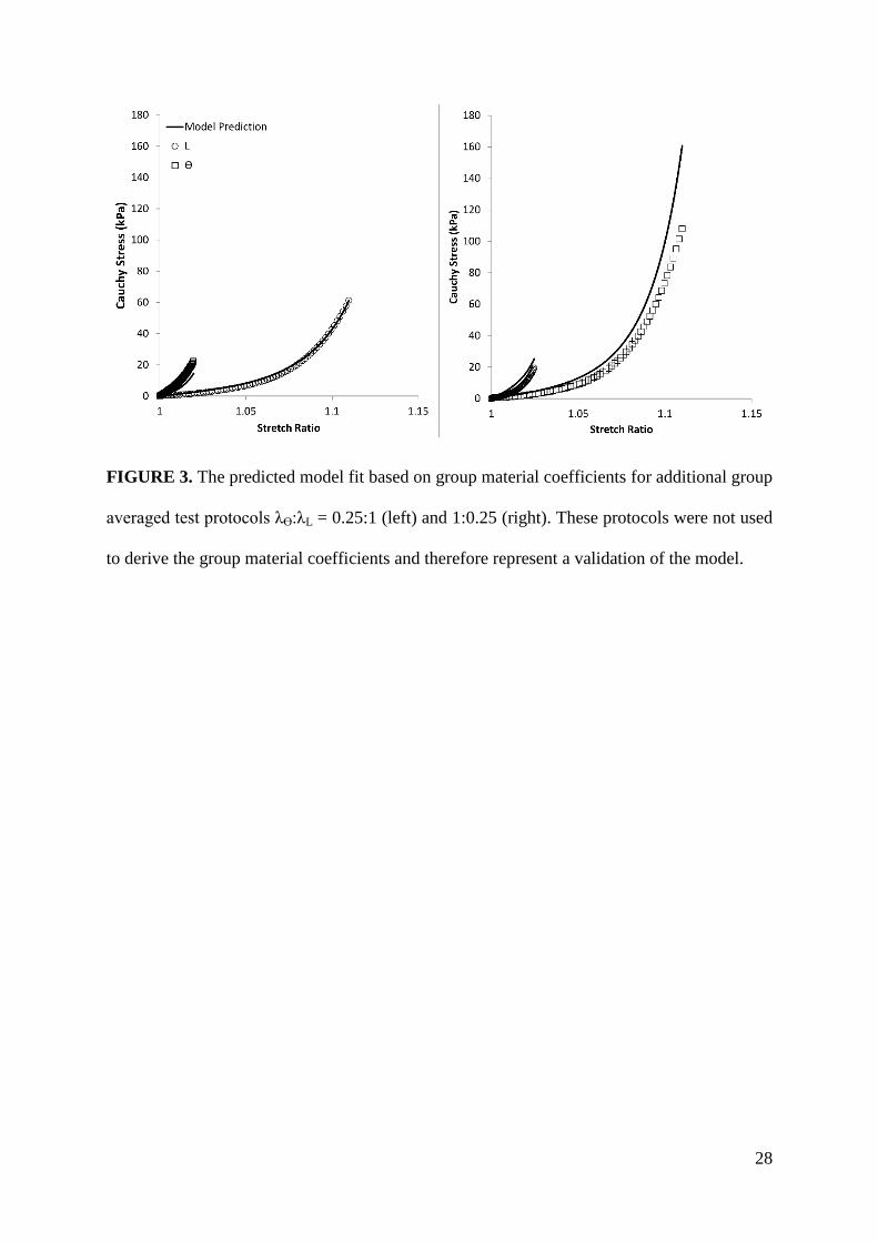
the individual samples very well (R2 = 0.96 ± 0.03). Validation of the models predicative
capabilities based on the group material coefficients and using test protocols, 0.25:1 and
1:0.25 was also successful and the agreement between the model and the experimental data
was high (R2 = 0.88).
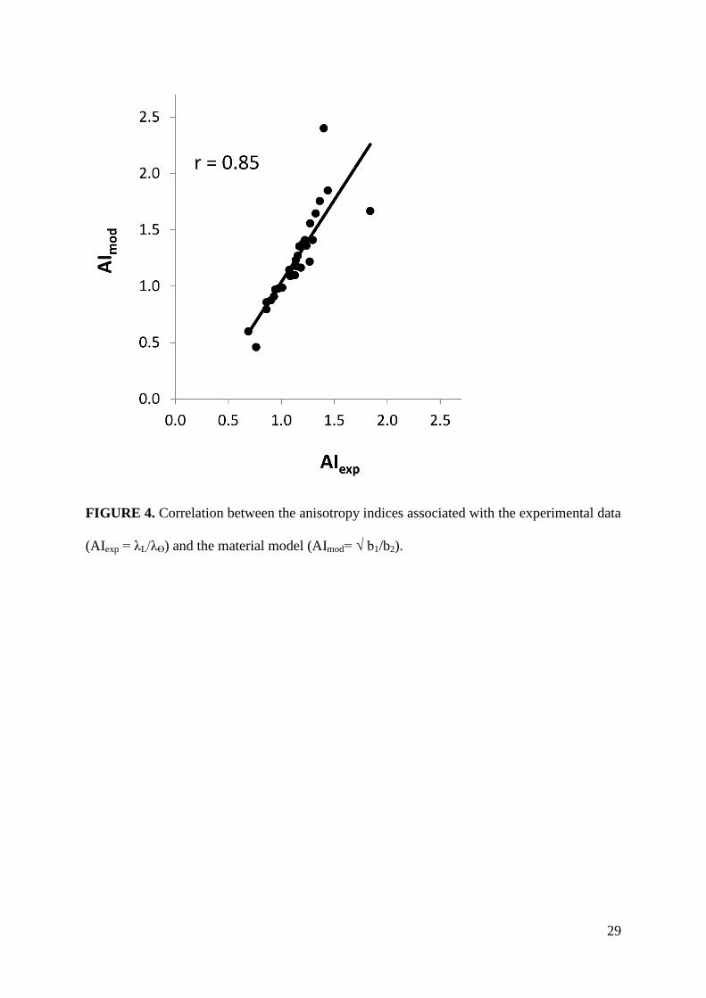
A positive correlation was found between the AIexp and AImod values (r = .85, p < .01) which
again shows good agreement between the model and the experimental data and also implies
that either index can be employed to assess the degree of anisotropy (Figure 4). The AImod
values indicate that the relative contribution of b1 to the strain energy is higher than b2 for

11
19/28 patients, lower than b2 for 6/28 patients and approximately equal for the remaining 3
patients (0.95 ≤ AI ≤ 1.05).
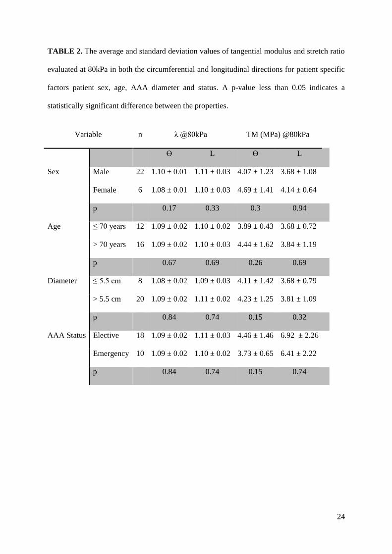
Patient Specific Factors
Table 2 and Table 3 compare the measured mechanical properties based on various groupings
such as male vs. female, AAA diameter: <5.5 cm vs. >5.5 cm, AAA status: elective vs.
emergency repair (ruptured or highly symptomatic) and patient age: <70 years vs. >70 years.
There was no significant difference found between the mechanical parameters measured for
any of these groups. In addition, all groups were well matched for thickness (Supplementary
Data).
DISCUSSION
In this study, the biaxial mechanical properties of AAA tissue from 28 patients undergoing
open AAA repair were evaluated. In general, the AAA tissue was found to be anisotropic
with a tendency to be stiffer in the circumferential than in the longitudinal direction. The
constitutive relationship chosen to model the biomechanical response provided an excellent
fit for the data and the material coefficients derived from the averaged model were shown to
be capable of predicting physiologically relevant loading regimes accurately. A significant
difference in mechanical properties as result of sex, age, AAA diameter or AAA status was
not detected.
Equibiaxial Response
Comparing our results to the literature, our study concurs that AAA is anisotropic with
preferential stiffening in the circumferential direction35, 38
. While, our reported values of TM
at 150kPa are comparable to those of Tong et al.35
at the same stress level, i.e. 8.1 ± 3.2 MPa
and 6.6 ± 1.2 MPa vs. 8.7 ± 1.7 MPa and 6.6 ± 2.7 MPa in the circumferential and
longitudinal directions respectively, the magnitude of the differences between our measured
material properties in each direction is lower, i.e. 20% vs. 27%. Vande Geest et al. 38
reported
12
average values for TM in each direction that were larger than either of these studies, i.e. 11.7
± 1.9 and 8.3 ± 1.2 MPa and also had more pronounced anisotropy (34% difference between
TM in the circumferential and longitudinal direction).
Interestingly, larger deviances were reported for the values of stretch in both directions
compared to previous studies, i.e. λϴ = 1.07 ± 0.03 and λL = 1.11 ± 0.0435
, λϴ = 1.07 ± 0.01
and λL = 1.09 ± 0.0138
vs. λϴ = 1.11 ± 0.03 and λL = 1.12 ± 0.03 (current study @150kPa) and
also the average percentage difference between these directions is larger i.e. 44%35
and 25%38
vs. 9% (current study @150kPa). Considering our reported values for TM in the stiff region
of the Cauchy stress–stretch response plots are similar, differences in the reported values of
stretch indicate that there was an increase in extensibility in the toe region of the Cauchy
stress–stretch response plot prior to stiffening compared to these studies. In a more recent
publication by Polzer et al.21
, who performed similar tests on the AAA tissue of a smaller
group of patients (n=7), a similar trend was observed and these differences were potentially
attributed to the use of different reference configurations i.e. stress-free (Polzer et al., 21) vs.
pre-stressed (Vande Geest et al.38
). Similar to this study, a pre stress was not prescribed here
and therefore it is possible that these subtle differences in protocols may contribute to the
differences in extensibility values reported in the literature.
There are a number of other possible reasons for deviations in the biomechanical parameters
found between these studies. For example, the presence of high inherent inter and intra-tissue
variability is always influential when comparing mechanical properties between different
groups of patients. Furthermore, the presence of intraluminal thrombus is thought to be
responsible for the proteolytic31
and hypoxic41
degradation of the wall resulting in a thinner
wall of diminished strength. In addition, increasing thrombus age has also been associated
with an increasing degree of anisotropy of the underlying AAA wall35
. Considering data
detailing the thrombus age is unavailable for the current study and it is not clear whether the

13
AAA wall tested by both Vande Geest et al.38
and Polzer et al.21
had previously been covered
by thrombus, it is possible that the variation in stiffness, extensibility and degree of
anisotropy may be associated with the presence, absence or age of the intraluminal thrombus.
Constitutive Model
Although we found that the degree of anisotropy (differences between the TM and λ in the
circumferential and longitudinal direction) to be small at lower stresses (80kPa), it was shown
to increase with stress (150kPa). Therefore, our decision to choose an anisotropic model to
represent the biomechanical response of AAA tissue in vivo is justified. The anisotropic index
associated with the constitutive model also corroborates the findings from the measured
mechanical properties which indicated that the circumferential direction has a tendency to be
stiffer than the longitudinal direction. Although the majority of the tissue specimens
displayed preferential stiffening in the circumferential direction, variations in the preferred
direction also existed within the test data. Due to the changes of the wall’s structural
components such as elastin and collagen as a result of aneurysm progression, anisotropy can
be affected which has previously been observed in aortic aneurysmal tissues38
and most other
arteries12
. Comparing our material coefficients to those acquired in a similar study of AAA
by Vande Geest et al.38
, our group values are comparable but lower, i.e. b0, b1, b2 and b3 =
0.44, 297.0, 219.9 and 184.3 vs. 0.14, 477.0, 416.4 and 408.3, which is not surprising
considering the previously discussed differences in the mechanical properties between
studies.
Patient Specific Factors
Sex
A recent biaxial study35
and a previous uniaxial study38
of the AAA biomechanical properties
have concluded that men have AAA tissue that is stiffer, less extensible and has a higher
tensile strength than women, which may explain why although, the occurrence of AAA is less

14
common in women14
, the relative incidence of rupture is greater than their male
counterparts1. However, the current study, for which the ratio of male to females is
comparable to these previous studies, i.e. 21:635
, 24:1038
and 22:6 (current study), did not
find a significant difference between the AAA mechanical properties of men and women and
on average the female properties were higher than males. This suggests that the difference in
rupture risk observed between men and women may not be significantly influenced by their
biomechanical properties and other reported factors such as an increased AAA growth rate in
females16
may play a more dominant role.
Patient Age
It has been reported previously that the stiffness of the non-aneurysmal aorta increases with
age 17
, which is thought to be linked to a decrease in the amount of elastin and an increase in
the amount of collagen present in the aortic wall as it ages11
. It is also well known that the
prevalence of AAA increases with age40
. Although no statistically significant difference was
found between the measured properties of patients over and under 70 years of age the average
stiffness was increased and extensibility remained unchanged for the older group, which
indicates that the aortic wall even in the presence of AAA may increase in stiffness as it ages.
AAA Diameter
A larger diameter AAA is associated with an increase in rupture risk3, 15
. However, in the
largest uniaxial study and histological assessment of AAA tissue to date32
, the AAA tissue of
diameter > 5.5cm (n=65) was found to be have a higher failure strength and extensibility than
the AAA tissue of diameter < 5.5cm (n=25). In contrast, this study which examined the
biaxial properties of a smaller amount of patients (> 5.5cm = 20 vs. < 5.5cm = 8), although
proportionally similar, did not detect a significant difference in the properties between these
two groups. Reasons for increases in mechanical strength as the aneurysm increases in size
have been attributed to wall remodelling which results in an increase in the absolute mass of
15
the collagen present. However, the exact pathogenesis of AAA is still not fully understood
and the exact rate at which the AAA remodels in not known. This reason and possibly as a
result of lower patients numbers, may help explain why this study did not detect a significant
difference between the two groups.
AAA Status
It is hypothesised that AAA rupture occurs when the wall stress exceeds the wall strength,
however, there have been no known studies reporting the difference in biaxial properties
between the ruptured and nonruptured AAA tissue, presumably due to the life threatening
nature of AAA rupture repair surgery. Interestingly our study did not detect a statistically
significant difference in the mechanical properties between the two groups. Although not
directly comparable due to differing test modes, the findings support the most recent uniaxial
study of the AAA biomechanics of entire AAA structures where it was concluded that the
regional properties of ruptured AAAs are not globally weaker than nonruptured AAAs25
. In a
similar study8, it was found that the stiffness was unaffected by AAA status however, the
failure stress was significantly decreased for ruptured AAAs vs. nonruptured AAAs.
Future Perspective
Patient specific factors such as sex, age, AAA diameter and AAA status, i.e. elective or
emergency repair, have not been found to significantly influence AAA biomechanical
properties and therefore, it may not be possible to differentiate those AAAs which are more
susceptible to rupture than others, based on these factors. Although these findings need to be
confirmed by examining their influence amongst a larger cohort of patients, it is likely that
they do not play a key role in determining AAA rupture risk. Although other factors such as
the presence of calcification have been shown to correlate with increased AAA rupture risk2,
their influence on AAA biomechanical properties has not been well documented and has
rarely been included in computational rupture risk assessments. Therefore, future
16
examinations of the influence of calcification on AAA properties may further the
understanding of AAA rupture risk.
Limitations
Due to the nature of AAA repair surgery, excised samples for the purposes of this study were
restricted to the anterior region of the AAA sac. This is limitative, considering, AAA
mechanical properties have been found to change regionally25, 34
and for the ruptured or
highly symptomatic patient group, assessment of properties was not at the actual rupture site.
Furthermore, considering the tissue specimens had to be at least 14mm x 14mm for
successful biaxial testing, the size of the excised tissue limited the number of samples that
could be prepared per patient. Unfortunately, excision of the complete AAA structure is
extremely rare and can only be attained post mortem. As previously mentioned, at high
stresses the tines had a tendency to pull through the AAA tissue sample; therefore, failure
properties of the AAA tissue could not be assessed. Furthermore, considering that the
displacement of the tines was controlled and not the strain of the tissue, the strain measured at
the centre of the specimen did not always reach the exact amount of prescribed stretch,
presumably due to the inability of the fibrous structure (which is only loaded at discrete
points) to translate the entire load. Substantial effort was made to eliminate the effect of the
gripping method on the derived mechanical properties and yet the response of the small tissue
pieces may not fully represent the response of the complete AAA structure. Additionally,
residual stresses and strains, if present, were not accounted for in this analysis. Furthermore,
the evaluation of tensile stress may be subject to minor errors as a result of the instrument
used to measure the sample thickness18
.
CONCLUSION
The aim of this study was to both confirm the findings of similar studies utilising a separate
patient sample and increase our understanding of the role of patient-specific factors in AAA

17
biomechanics. In general, biaxial tests confirmed that the AAA tissue is anisotropic with a
greater tendency to stiffen in the circumferential direction compared to the longitudinal
direction. An anisotropic hyperelastic constitutive model was found to represent the data well
(although the average estimated material parameters were lower than a previous study) and
implementation of this model in future finite element studies of AAA tissue may provide a
more realistic assessment of AAA wall stress and rupture risk. No significant difference was
found between any of the patient groups based on sex, age, AAA diameter and AAA status
and their associated mechanical properties. Although these findings need to be confirmed by
examining their influence amongst a larger cohort of patients, it may be likely that they do
not play a key role in determining AAA rupture risk. It is possible that other factors such as
the presence of calcification may have a greater influence on the AAA rupture risk (this will
be reported in an upcoming publication).
ACKNOWLEDGEMENTS
The authors would like to thank the Irish Research Council ‘EMBARK Initiative’ (Grant No.
IDRS/2010/2941) for funding this study. BJD would like to acknowledge The University of
Western Australia Research Fellowship and NHMRC Project Grant APP1063986. This work
was supported by the Irish Government’s Programme for Research in Third Level Institutions
Cycle 5, with the assistance of the European Regional Development Fund. The authors would
also like to thank the Department of Vascular Surgery, University Hospital Limerick, Ireland,
in particular Dr. Peter Coyle, for their help in harvesting and collecting the AAA tissue and
also Caleb Horst for providing technical support for the Cellscale Biotester.
CONFLICT OF INTEREST STATEMENT
There are no conflicts of interest.

18
REFERENCES
1Brown P. M., D. T. Zelt, B. Sobolev. The risk of rupture in untreated aneurysms: The impact
of size, gender, and expansion rate. J. Vasc. Surg. 37:280-4, 2003.
2Buijs R., T. Willems, R. Tio, H. Boersma, I. Tielliu, R. Slart, et al. Calcification as a risk
factor for rupture of abdominal aortic aneurysm. Eur. J. Vasc. Endovasc. Surg. 46:542-8,
2013.
3Chaikof E. L., D. C. Brewster, R. L. Dalman, M. S. Makaroun, K. A. Illig, G. A. Sicard, et
al. The care of patients with an abdominal aortic aneurysm: The Society for Vascular Surgery
practice guidelines. J. Vasc. Surg. 50:S2-S49, 2009.
4Choi H. S., R. P. Vito. Two-Dimensional Stress-Strain Relationship for Canine Pericardium.
J. Biomech. Eng. 112:153-9, 1990.
5Choke E., G. Cockerill, W. R. W. Wilson, S. Sayed, J. Dawson, I. Loftus, et al. A Review of
Biological Factors Implicated in Abdominal Aortic Aneurysm Rupture. Eur. J. Vasc.
Endovasc. Surg. 30:227-44, 2005.
6Chuong C. J., Y. C. Fung. Compressibility and constitutive equation of arterial wall in radial
compression experiments. J. Biomech. 17:35-40, 1984.
7Darling R., C. Messina, D. Brewster. Autospsy of unoperated aortic aneurysms. The case of
early resection. Circ. 56:164-, 1977.
8Di Martino E. S., A. Bohra, J. P. Vande Geest, N. Gupta, M. S. Makaroun, D. A. Vorp.
Biomechanical properties of ruptured versus electively repaired abdominal aortic aneurysm
wall tissue. J. Vasc. Surg. 43:570-6, 2006.
9Doyle B., A. Callanan, P. Grace, E. Kavanagh. On the influence of patient-specific material
properties in computational simulations: A case study of a large ruptured abdominal aortic
aneurysm. Int. J. Num. Meth. Biomed. Eng. 29:150-64, 2013.

19
10Doyle B., T. McGloughlin, K. Miller, J. Powell, P. Norman. Regions of High Wall Stress
Can Predict the Future Location of Rupture of Abdominal Aortic Aneurysm. Cardiovasc.
Intervent. Radiol. 29:1-4, 2014.
11Jacob M. P. Extracellular matrix remodeling and matrix metalloproteinases in the vascular
wall during aging and in pathological conditions. Biomed. Pharmacother. 57:195-202, 2003.
12Kamenskiy A., Y. Dzenis, S. J. Kazmi, M. Pemberton, I. Pipinos, N. Phillips, et al. Biaxial
mechanical properties of the human thoracic and abdominal aorta, common carotid,
subclavian, renal and common iliac arteries. Biomech Model Mechanobiol. 1-19, 2014.
13Kent K. C., R. M. Zwolak, N. N. Egorova, T. S. Riles, A. Manganaro, A. J. Moskowitz, et
al. Analysis of risk factors for abdominal aortic aneurysm in a cohort of more than 3 million
individuals. J. Vasc. Surg. 52:539-48, 2010.
14Lo R., R. Bensley, A. Hamdan, M. Wyers, J. Adams, M. Schermerhorn. Gender differences
in abdominal aortic aneurysm presentation, repair, and mortality in the Vascular Study Group
of New England. J. Vasc. Surg. 57:1261-8, 2013.
15Maier A., M. Gee, C. Reeps, J. Pongratz, H. H. Eckstein, W. Wall. A Comparison of
Diameter, Wall Stress, and Rupture Potential Index for Abdominal Aortic Aneurysm Rupture
Risk Prediction. Ann. Biomed. Eng. 38:3124-34, 2010.
16Mofidi R., V. Goldie, J. Kelman, A. Dawson, J. Murie, R. Chalmers. Influence of sex on
expansion rate of abdominal aortic aneurysms. Br. J. Surg. 94:310-4, 2007.
17Newman D. L., R. C. Lallemand. The effect of age on the distensibility of the abdominal
aorta of man. Surg. Gynecol. Obstet. 147:211-4, 1978.
18O’Leary S., B. Doyle, T. McGloughlin. Comparison of methods used to measure the
thickness of soft tissues and their influence on the evaluation of tensile stress. J. Biomech.
46:1955-60, 2013.

20
19O’Leary S., B. Doyle, T. McGloughlin. The impact of long term freezing on the mechanical
properties of porcine aortic tissue. J. Mech. Beh. Biomed. Accepted for Publication, 2014.
20O’Leary S., E. Kavanagh, P. Grace, T. McGloughlin, B. Doyle. The Biaxial Mechanical
Behaviour of Abdominal Aortic Aneurysm Intraluminal Thrombus: Classification of
Morphology and the Determination of Layer and Region Specific Properties. J. Biomech.
47:1430, 2014.
21Polzer S., T. Christian Gasser, J. Bursa, R. Staffa, R. Vlachovsky, V. Man, et al. Importance
of material model in wall stress prediction in abdominal aortic aneurysms. Med. Eng. Phys.
35:1282-9, 2013.
22Thompson S. G., L. C. Brown, M. J. Sweeting, M. J. Bown, L. G. Kim, M. J. Glover, et al.
Systematic review and meta-analysis of the growth and rupture rates of small abdominal
aortic aneurysms: implications for surveillance intervals and their cost-effectiveness. Health
Technol. Assess. 17:1-118, 2013.
23Raghavan M., J. Kratzberg, E. Castro de Tolosa, M. Hanaoka, P. Walker, E. da Silva.
Regional distribution of wall thickness and failure properties of human abdominal aortic
aneurysm. J. Biomech. 39:3010-6, 2006.
24Raghavan M., M. Webster, D. Vorp. Ex vivo biomechanical behavior of abdominal aortic
aneurysm: Assessment using a new mathematical model. Ann. Biomed. Eng. 24:573-82,
1996.
25Raghavan M. L., M. M. Hanaoka, J. A. Kratzberg, M. d. L. Higuchi, E. S. da Silva.
Biomechanical failure properties and microstructural content of ruptured and unruptured
abdominal aortic aneurysms. J. Biomech. 44:2501-7, 2011.
26Reeps C., A. Maier, J. Pelisek, F. Härtl, V. Grabher-Meier, W. A. Wall, et al. Measuring
and modeling patient-specific distributions of material properties in abdominal aortic
aneurysm wall. Biomech Model Mechanobiol. 12:717-33, 2013.
21
27Sacks M., C. Chuong. Orthotropic Mechanical Properties of Chemically Treated Bovine
Pericardium. Ann. Biomed. Eng. 26:892-902, 1998.
28Sacks M. S., W. Sun. Multiaxial Mechanical Behavior of Biological Materials. Ann. Rev.
Biomed Eng. 5:251-84, 2003.
29Schlosser F. J., I. Vaartjes, G. J. van der Heijden, F. L. Moll, H. J. Verhagen, B. E. Muhs, et
al. Mortality after elective abdominal aortic aneurysm repair. Ann. Surg. 251:158-64, 2010.
30Stergiopulos N., S. Vulliémoz, A. Rachev, J. J. Meister, S. E. Greenwald. Assessing the
Homogeneity of the Elastic Properties and Composition of the Pig Aortic Media. J. Vasc.
Res. 38:237-46, 2001.
31Swedenborg J., P. E. R. Eriksson. The Intraluminal Thrombus as a Source of Proteolytic
Activity. Ann. N. Y. Acad. Sci. 1085:133-8, 2006.
32Tavares Monteiro J., E. Simão da Silva, M. Raghavan, P. Puech-Leão, M. Higuchi, J.
Otoch. Histologic, histochemical, and biomechanical properties of fragments isolated from
the anterior wall of abdominal aortic aneurysms. J. Vasc. Surg., 2013.
33Thompson M. M. Controlling the expansion of abdominal aortic aneurysms. Br. J. Surg.
90:897-8, 2003.
34Thubrikar M., M. Labrosse, F. Robicsek, J. Al-Soudi, F. B. Mechanical properties of
abdominal aortic aneurysm wall. J. Med. Eng. Technol. 25:133-42, 2001.
35Tong J., T. Cohnert, P. Regitnig, G. Holzapfel. Effects of Age on the Elastic Properties of
the Intraluminal Thrombus and the Thrombus-covered Wall in Abdominal Aortic
Aneurysms: Biaxial Extension Behaviour and Material Modelling. Eur. J. Vasc. Endovasc.
Surg. 42:207-19, 2011.
36Tong J., A. J. Schriefl, T. Cohnert, G. A. Holzapfel. Gender Differences in Biomechanical
Properties, Thrombus Age, Mass Fraction and Clinical Factors of Abdominal Aortic
Aneurysms. Eur. J. Vasc. Endovasc. Surg. 45:364-72, 2013.
22
37Vande Geest J., E. Dillavou, E. Di Martino, M. Oberdier, A. Bohra, M. Makaroun, et al.
Gender-Related Differences in the Tensile Strength of Abdominal Aortic Aneurysm. Ann. N.
Y. Acad. Sci. 1085:400-2, 2006.
38Vande Geest J., M. S. Sacks, D. Vorp. The effects of aneurysm on the biaxial mechanical
behavior of human abdominal aorta. J. Biomech. 39:1324-34, 2006.
39Vande Geest J., D. Schmidt, M. Sacks, D. Vorp. The Effects of Anisotropy on the Stress
Analyses of Patient-Specific Abdominal Aortic Aneurysms. Ann. Biomed. Eng. 36:921-32,
2008.
40Vardulaki K. A., T. C. Prevost, N. M. Walker, N. E. Day, A. B. M. Wilmink, C. R. G.
Quick, et al. Growth rates and risk of rupture of abdominal aortic aneurysms. Br. J. Surg.
85:1674-80, 1998.
41Vorp, P. C. Lee, D. H. J. Wang, M. S. Makaroun, E. M. Nemoto, S. Ogawa, et al.
Association of intraluminal thrombus in abdominal aortic aneurysm with local hypoxia and
wall weakening. J. Vasc. Surg. 34:291-9, 2001.
42Wanhainen A., R. Themudo, H. Ahlstrom, L. Lind, L. Johansson. Thoracic and abdominal
aortic dimension in 70-year-old men and women--a population-based whole-body magnetic
resonance imaging (MRI) study. J. Vasc. Surg. 47:504-12, 2008.

23
TABLE 1. Material parameters derived from the previously described material model,
calculated anisotropic index values and the material model correlation coefficients associated
with individual patient’s fit and also the averaged group fit.
Patient b0(kPa) b1 b2 b3 AImod AIexp R2 1 0.27 86.96 241.06 126.04 0.60 0.69 0.96 2 0.72 175.48 277.16 158.23 0.80 0.86 0.96 3 0.56 156.81 57.85 96.05 1.65 1.32 0.98
4 0.59 387.36 320.61 198.02 1.10 1.13 0.98 5 0.12 528.78 204.54 224.17 1.67 1.84 0.97 6 0.40 327.72 241.17 163.24 1.17 1.18 0.98 7 0.62 432.08 216.93 251.22 1.41 1.29 0.95 8 0.25 269.44 281.00 190.57 0.98 0.97 0.94 9 0.78 187.14 253.54 159.42 0.86 0.86 0.96
10 0.47 329.87 106.88 193.23 1.76 1.36 0.95 11 0.32 124.67 162.23 109.56 0.88 0.90 0.98 12 0.30 255.51 158.10 114.04 1.27 1.15 0.99 13 0.26 291.19 156.46 169.83 1.36 1.17 0.94 14 0.34 345.08 305.69 225.18 1.15 1.07 0.92
15 0.11 537.99 290.85 414.95 1.36 1.24 0.89 16 0.38 290.21 84.73 110.23 1.85 1.44 0.99 17 0.48 396.95 68.85 157.59 2.40 1.40 0.90 18 0.51 204.43 217.80 145.33 0.97 0.94 0.95 19 0.47 188.07 190.84 129.27 0.99 1.01 0.95 20 1.33 230.43 155.24 87.72 1.22 1.27 0.98 21 0.23 281.27 339.74 204.32 0.91 0.93 0.92 22 0.20 268.70 176.39 129.37 1.23 1.14 0.98 23 0.63 405.74 292.17 261.57 1.18 1.14 0.94 24 0.59 182.92 96.91 98.95 1.37 1.20 0.98 25 0.35 207.08 85.27 99.49 1.56 1.27 0.98
26 0.41 64.11 303.00 248.35 0.46 0.76 0.97 27 0.16 263.16 221.22 127.60 1.09 1.08 0.99
28 0.95 435.34 218.80 374.57 1.41 1.22 0.93
Group 0.44 306.83 213.34 218.71 1.20 1.09 0.98
24
TABLE 2. The average and standard deviation values of tangential modulus and stretch ratio
evaluated at 80kPa in both the circumferential and longitudinal directions for patient specific
factors patient sex, age, AAA diameter and status. A p-value less than 0.05 indicates a
statistically significant difference between the properties.
Variable n λ @80kPa TM (MPa) @80kPa
ϴ L ϴ L
Sex Male 22 1.10 ± 0.01 1.11 ± 0.03 4.07 ± 1.23 3.68 ± 1.08
Female 6 1.08 ± 0.01 1.10 ± 0.03 4.69 ± 1.41 4.14 ± 0.64
p 0.17 0.33 0.3 0.94
Age ≤ 70 years 12 1.09 ± 0.02 1.10 ± 0.02 3.89 ± 0.43 3.68 ± 0.72
> 70 years 16 1.09 ± 0.02 1.10 ± 0.03 4.44 ± 1.62 3.84 ± 1.19
p 0.67 0.69 0.26 0.69
Diameter ≤ 5.5 cm 8 1.08 ± 0.02 1.09 ± 0.03 4.11 ± 1.42 3.68 ± 0.79
> 5.5 cm 20 1.09 ± 0.02 1.11 ± 0.02 4.23 ± 1.25 3.81 ± 1.09
p 0.84 0.74 0.15 0.32
AAA Status Elective 18 1.09 ± 0.02 1.11 ± 0.03 4.46 ± 1.46 6.92 ± 2.26
Emergency 10 1.09 ± 0.02 1.10 ± 0.02 3.73 ± 0.65 6.41 ± 2.22
p 0.84 0.74 0.15 0.74

25
TABLE 3. The average and standard deviation values of tangential modulus and stretch ratio
evaluated at 150kPa in both the circumferential and longitudinal directions for patient
specific factors patient sex, age, AAA diameter and status. A p-value less than 0.05 indicates
a statistically significant difference between the properties.
Variable n λ @150kPa TM (MPa) @150kPa
ϴ L ϴ L
Sex Male 22 1.11 ± 0.03 1.12 ± 0.03 7.74 ± 3.04 6.43 ± 2.15
Female 6 1.10 ± 0.02 1.11 ± 0.03 8.96 ± 3.19 7.85 ± 2.29
p 0.24 0.54 0.39 0.17
Age ≤ 70 years 12 1.11 ± 0.02 1.12 ± 0.02 7.82 ± 2.71 6.42 ± 1.39
> 70 years 16 1.11 ± 0.03 1.12± 0.03 8.15 ± 3.37 6.98 ± 2.70
p 0.97 0.52 0.78 0.52
Diameter ≤ 5.5 cm 8 1.10 ± 0.02 1.11 ± 0.03 7.15 ± 2.97 6.85 ± 2.19
> 5.5 cm 20 1.11 ± 0.03 1.12 ± 0.03 8.35 ± 3.09 6.69 ± 2.28
p 0.53 0.58 0.45 0.93
AAA Status Elective 18 1.11 ± 0.02 1.12 ± 0.03 8.34 ± 3.15 6.92 ± 2.26
Emergency 10 1.11 ± 0.03 1.12 ± 0.02 7.41 ± 2.92 6.41 ± 2.22
p 0.54 0.61 0.45 0.57
26
FIGURE 1. The average tangential modulus (TM) and stretch ratio (λ) evaluated at 80kPa
and 150kPa in the circumferential (ϴ) and longitudinal (L) directions of the equibiaxial
response curves. The percentage change in properties between the axes is illustrated and (*)
indicates a statistically significant difference between the properties at a significance level of
0.05.

27
FIGURE 2. The averaged Cauchy stress-stretch response curves for the prescribed multiple
biaxial test regimes (λϴ:λL = 1:1, 0.75:1, 1:0.75, 0.5:1, 1:0.5) in both the circumferential (left)
and longitudinal (right) directions and the material model fit (solid lines).
28
FIGURE 3. The predicted model fit based on group material coefficients for additional group
averaged test protocols λϴ:λL = 0.25:1 (left) and 1:0.25 (right). These protocols were not used
to derive the group material coefficients and therefore represent a validation of the model.
29
FIGURE 4. Correlation between the anisotropy indices associated with the experimental data
(AIexp = λL/λϴ) and the material model (AImod= √ b1/b2).


![Best power point evar! [autosaved]](https://img.dokumen.tips/doc/110x75/548bdd07b479592b788b460b/best-power-point-evar-autosaved.jpg)