Embed Size (px)
Citation preview

334 VOLUME 15 NUMBER 4 APRIL 2008 NATURE STRUCTURAL & MOLECULAR BIOLOGY
during DSB repair21. INO80 could also act behind the fork to stabilize or position nucleosomes assembled by histone chaperones and thus might counteract fork regression or back-up, which could ultimately lead to DSBs and genome instability1,2. Another possibility is that INO80 remodels nucleosomes behind the fork to expose sites for histone modifications that are in turn required for fork stabilization. One such modification might be the acetylation of H3 at lysine 56 by Asf1 and Rtt109, as mutations that prevent this mark contribute to genome instability instigated by replicative stress22,23. Lastly, INO80 might use other activities besides nucleosome remodeling to restart stalled forks. INO80 contains two RuvB-like DNA helicase subunits that have been connected to branch migration during recombination in bacteria6,8,24. Unrepaired RFs can collapse into structures that form Holliday junctions, recombination intermediates that can be
processed by bacterial RuvB to reconstitute intact RFs and restore active replisomes24,25. Perhaps the RuvB-like activities in INO80 perform a similar function as an auxiliary role to nucleosome remodeling to restart stalled replication forks. Whatever the mechanism(s), it is clear that INO80 is an important new player in DNA replication to help safeguard the integrity of the genome.
1. Branzei, D. & Foiani, M. DNA Repair (Amst.) 6, 994–1003 (2007).
2. Aguilera, A. & Gomez-Gonzalez, B. Nat. Rev. Genet. 9, 204–217 (2008).
3. Linger, J.G. & Tyler, J.K. Mutat. Res. 618, 52–64 (2007).
4. Nelson, D.M. et al. Mol. Cell. Biol. 22, 7459–7472 (2002).
5. Papamichos-Chronakis, M. & Peterson, C.L. Nat. Struct. Mol. Biol. 15, 338–345 (2008).
6. Shen, X., Mizuguchi, G., Hamiche, A. & Wu, C. Nature 406, 541–544 (2000).
7. Jin, J. et al. J. Biol. Chem. 280, 41207–41212 (2005).8. Jonsson, Z.O., Jha, S., Wohlschlegel, J.A. & Dutta, A.
Mol. Cell 16, 465–477 (2004).9. Shen, X., Ranallo, R., Choi, E. & Wu, C. Mol. Cell 12,
147–155 (2003).
10. Morrison, A.J. et al. Cell 130, 499–511 (2007).11. Branzei, D. & Foiani, M. Nat. Rev. Mol. Cell Biol.
Advance online publication, doi:10.1038/nrm2351 (20 February 2008).
12. Papamichos-Chronakis, M., Krebs, J.E. & Peterson, C.L. Genes Dev. 20, 2437–2449 (2006).
13. Morrison, A.J. et al. Cell 119, 767–775 (2004).14. Downs, J.A. et al. Mol. Cell 16, 979–990 (2004).15. van Attikum, H., Fritsch, O., Hohn, B. & Gasser, S.M.
Cell 119, 777–788 (2004).16. Osley, M.A. & Shen, X. Trends Genet. 22, 671–677
(2006).17. Downs, J.A., Lowndes, N.F. & Jackson, S.P. Nature
408, 1001–1004 (2000).18. Franco, A.A., Lam, W.M., Burgers, P.M. & Kaufman, P.D.
Genes Dev. 19, 1365–1375 (2005).19. Moldovan, G.L., Pfander, B. & Jentsch, S. Cell 129,
665–679 (2007).20. Yu, E.Y. et al. Mol. Cell. Biol. 27, 5639–5649
(2007).21. Tsukuda, T., Fleming, A.B., Nickoloff, J.A. & Osley, M.A.
Nature 438, 379–383 (2005).22. Driscoll, R., Hudson, A. & Jackson, S.P. Science 315,
649–652 (2007).23. Han, J., Zhou, H., Li, Z., Xu, R.M. & Zhang, Z. J. Biol.
Chem. 282, 28587–28596 (2007).24. Donaldson, J.R., Courcelle, C.T. & Courcelle, J. J. Biol.
Chem. 281, 28811–28821 (2006).25. Eggleston, A.K. & West, S.C. J. Biol. Chem. 275,
26467–26476 (2000).
The discovery of microRNAs 15 years ago has produced a revolution in biology and medicine. The genesis of these molecules and their functions have been discussed many times1; what remains particularly enigmatic is not what miRNAs do, but how they do it. How do these small ~21-nucleotide RNAs induce the destruction or translational silencing of mRNAs, among other activities? Is there one general principle that elucidates the mechanism by which miRNAs repress mRNA translation? On page 346 of this issue, Eulalio and colleagues2 do not so much tell us how miRNAs control translation, but instead provide a cautionary tale that a consensus for miRNA activity is not yet at hand (Fig. 1).
miRNAs incompletely base-pair to sequences usually found in 3′ untranslated regions (UTRs) of specific mRNAs; by doing so, the Argonaute proteins, which interact with the miRNAs, directly or indirectly induce mRNA deadenylation, destruction or translational inhibition. Results from several studies indicate that miRNAs and Argonaute modulate translation at either initiation or post-initiation steps. That is, some miRNA-inhibited mRNAs sediment with polysomes in sucrose gradients and are nearly indistinguishable from bona fide translating mRNAs in their sensitivity to puromycin, an agent that induces premature polypeptide release and polysome disassembly3,4. Additional results show that miRNAs inhibit the translation of internal ribosome entry site (IRES)-containing RNAs, indicating that the 5′-terminal 7mG cap structure, and by extension the eIF4F cap binding complex and the early steps in initiation, are dispensable for miRNA-mediated translational inhibition3,5.
In contrast, other experiments show that miRNA-inhibited mRNAs sediment in sucrose gradient fractions that are lighter than polysomes, suggesting a block at initiation6. Moreover, some studies show that miRNAs do not inhibit the translation of IRES-containing mRNAs, thus indicating a requirement for the cap and for cap-binding factors6,7.
How can one make sense of these disparate results, which are largely based on the transfection of reporter-encoding plasmids into cells? One way forward would seem to involve the generation of in vitro systems that support miRNA-mediated translational inhibition. Three such systems, two derived from mammalian cells8,9 and one derived from Drosophila melanogaster embryos10, all suggest that the cap and/or cap-binding factors are required for miRNA-mediated inhibition. But what might the biochemical basis for such inhibition be? One possibility can be inferred from the results of Chendrimada and colleagues11,
Joel D. Richter is at the Program in Molecular Medicine, University of Massachusetts Medical School, Biotech 2, Suite 204, 373 Plantation Street, Worcester, Massachusetts 01605, USA. e-mail: [email protected]
Think you know how miRNAs work? Think again.Joel D Richter
Two proposed mechanisms for how microRNAs (miRNAs) and their associated Argonaute proteins inhibit translation in mammals do not seem to operate in Drosophila melanogaster cells, suggesting that insights into important miRNA functions remain elusive. However, the interaction between Argonaute and the P-body factor GW182 may help in elucidating the biochemical basis of translational control by miRNAs.
N E W S A N D V I E W S©
2008
Nat
ure
Pub
lishi
ng G
roup
ht
tp://
ww
w.n
atur
e.co
m/n
smb
NATURE STRUCTURAL & MOLECULAR BIOLOGY VOLUME 15 NUMBER 4 APRIL 2008 335
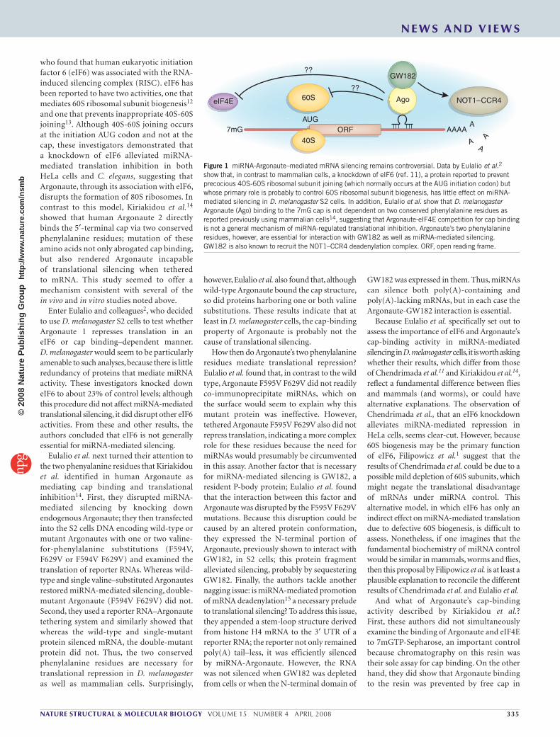
who found that human eukaryotic initiation factor 6 (eIF6) was associated with the RNA-induced silencing complex (RISC). eIF6 has been reported to have two activities, one that mediates 60S ribosomal subunit biogenesis12 and one that prevents inappropriate 40S-60S joining13. Although 40S-60S joining occurs at the initiation AUG codon and not at the cap, these investigators demonstrated that a knockdown of eIF6 alleviated miRNA-mediated translation inhibition in both HeLa cells and C. elegans, suggesting that Argonaute, through its association with eIF6, disrupts the formation of 80S ribosomes. In contrast to this model, Kiriakidou et al.14 showed that human Argonaute 2 directly binds the 5′-terminal cap via two conserved phenylalanine residues; mutation of these amino acids not only abrogated cap binding, but also rendered Argonaute incapable of translational silencing when tethered to mRNA. This study seemed to offer a mechanism consistent with several of the in vivo and in vitro studies noted above.
Enter Eulalio and colleagues2, who decided to use D. melanogaster S2 cells to test whether Argonaute 1 represses translation in an eIF6 or cap binding–dependent manner. D. melanogaster would seem to be particularly amenable to such analyses, because there is little redundancy of proteins that mediate miRNA activity. These investigators knocked down eIF6 to about 23% of control levels; although this procedure did not affect miRNA-mediated translational silencing, it did disrupt other eIF6 activities. From these and other results, the authors concluded that eIF6 is not generally essential for miRNA-mediated silencing.
Eulalio et al. next turned their attention to the two phenyalanine residues that Kiriakidou et al. identified in human Argonaute as mediating cap binding and translational inhibition14. First, they disrupted miRNA-mediated silencing by knocking down endogenous Argonaute; they then transfected into the S2 cells DNA encoding wild-type or mutant Argonautes with one or two valine-for-phenylalanine substitutions (F594V, F629V or F594V F629V) and examined the translation of reporter RNAs. Whereas wild-type and single valine–substituted Argonautes restored miRNA-mediated silencing, double-mutant Argonaute (F594V F629V) did not. Second, they used a reporter RNA–Argonaute tethering system and similarly showed that whereas the wild-type and single-mutant protein silenced mRNA, the double-mutant protein did not. Thus, the two conserved phenylalanine residues are necessary for translational repression in D. melanogaster as well as mammalian cells. Surprisingly,
however, Eulalio et al. also found that, although wild-type Argonaute bound the cap structure, so did proteins harboring one or both valine substitutions. These results indicate that at least in D. melanogaster cells, the cap-binding property of Argonaute is probably not the cause of translational silencing.
How then do Argonaute’s two phenylalanine residues mediate translational repression? Eulalio et al. found that, in contrast to the wild type, Argonaute F595V F629V did not readily co-immunoprecipitate miRNAs, which on the surface would seem to explain why this mutant protein was ineffective. However, tethered Argonaute F595V F629V also did not repress translation, indicating a more complex role for these residues because the need for miRNAs would presumably be circumvented in this assay. Another factor that is necessary for miRNA-mediated silencing is GW182, a resident P-body protein; Eulalio et al. found that the interaction between this factor and Argonaute was disrupted by the F595V F629V mutations. Because this disruption could be caused by an altered protein conformation, they expressed the N-terminal portion of Argonaute, previously shown to interact with GW182, in S2 cells; this protein fragment alleviated silencing, probably by sequestering GW182. Finally, the authors tackle another nagging issue: is miRNA-mediated promotion of mRNA deadenylation15 a necessary prelude to translational silencing? To address this issue, they appended a stem-loop structure derived from histone H4 mRNA to the 3′ UTR of a reporter RNA; the reporter not only remained poly(A) tail–less, it was efficiently silenced by miRNA-Argonaute. However, the RNA was not silenced when GW182 was depleted from cells or when the N-terminal domain of
GW182 was expressed in them. Thus, miRNAs can silence both poly(A)-containing and poly(A)-lacking mRNAs, but in each case the Argonaute-GW182 interaction is essential.
Because Eulalio et al. specifically set out to assess the importance of eIF6 and Argonaute’s cap-binding activity in miRNA-mediated silencing in D. melanogaster cells, it is worth asking whether their results, which differ from those of Chendrimada et al.11 and Kiriakidou et al.14, reflect a fundamental difference between flies and mammals (and worms), or could have alternative explanations. The observation of Chendrimada et al., that an eIF6 knockdown alleviates miRNA-mediated repression in HeLa cells, seems clear-cut. However, because 60S biogenesis may be the primary function of eIF6, Filipowicz et al.1 suggest that the results of Chendrimada et al. could be due to a possible mild depletion of 60S subunits, which might negate the translational disadvantage of mRNAs under miRNA control. This alternative model, in which eIF6 has only an indirect effect on miRNA-mediated translation due to defective 60S biogenesis, is difficult to assess. Nonetheless, if one imagines that the fundamental biochemistry of miRNA control would be similar in mammals, worms and flies, then this proposal by Filipowicz et al. is at least a plausible explanation to reconcile the different results of Chendrimada et al. and Eulalio et al.
And what of Argonaute’s cap-binding activity described by Kiriakidou et al.? First, these authors did not simultaneously examine the binding of Argonaute and eIF4E to 7mGTP-Sepharose, an important control because chromatography on this resin was their sole assay for cap binding. On the other hand, they did show that Argonaute binding to the resin was prevented by free cap in
Figure 1 miRNA-Argonaute–mediated mRNA silencing remains controversial. Data by Eulalio et al.2 show that, in contrast to mammalian cells, a knockdown of eIF6 (ref. 11), a protein reported to prevent precocious 40S-60S ribosomal subunit joining (which normally occurs at the AUG initiation codon) but whose primary role is probably to control 60S ribosomal subunit biogenesis, has little effect on miRNA-mediated silencing in D. melanogaster S2 cells. In addition, Eulalio et al. show that D. melanogaster Argonaute (Ago) binding to the 7mG cap is not dependent on two conserved phenylalanine residues as reported previously using mammalian cells14, suggesting that Argonaute-eIF4E competition for cap binding is not a general mechanism of miRNA-regulated translational inhibition. Argonaute’s two phenylalanine residues, however, are essential for interaction with GW182 as well as miRNA-mediated silencing. GW182 is also known to recruit the NOT1–CCR4 deadenylation complex. ORF, open reading frame.
eIF4E Ago
GW182
60S
AUG
??
??
NOT1–CCR4
7mG AAAAA
AA
A40S
ORF
N E W S A N D V I E W S©
2008
Nat
ure
Pub
lishi
ng G
roup
ht
tp://
ww
w.n
atur
e.co
m/n
smb
336 VOLUME 15 NUMBER 4 APRIL 2008 NATURE STRUCTURAL & MOLECULAR BIOLOGY
the binding buffer, a standard and essential control. Moreover, an Argonaute with the two valine for phenylalanine substitutions showed no detectable cap binding. It seems that approximately 5% of the wild-type Argonaute input was retained on the m7GTP-Sepharose, but without comparison to a control cap-binding protein, such as eIF4E, it is hard to know whether this represents moderate or even low levels of binding. Finally, Kiriakidou et al. used tagged immunopurified Argonaute from transfected 293T cells for their studies; thus, it is possible that another mammalian protein not present in Drosophila S2 cells might have aided Argonaute’s interaction with m7GTP-Sepharose.
Given the results of this study by Eulalio et al., it seems that more consideration should be given to GW182 as a key factor regulating the translational activities of miRNA. The observation that GW182 promotes mRNA deadenylation and destruction16,17 obviously complicates analysis of translation; nonetheless, as long as the mechanism(s) by which miRNAs control gene expression remain(s) controversial (Fig. 1), it is essential that new approaches with additional reagents be brought to bear on this issue of overriding importance.
1. Filipowicz, W., Bhattacharyya, S.N. & Sonenberg, N. Nat. Rev. Genet. 9, 102–114 (2008).
2. Eulalio, A., Huntzinger, E. & Izaurralde, E. Nat. Struct. Mol. Biol. 15, 346–353 (2008).
3. Petersen, C.P. et al. Mol. Cell 21, 533–542 (2006).4. Nottrott, S., Simard, M.J. & Richter, J.D. Nat. Struct.
Mol. Biol. 13, 1108–1114 (2006).5. Lytle, J.R., Yario, T.A. & Steitz, J.A. Proc. Natl. Acad.
Sci. USA 104, 9667–9672 (2007).6. Pillai, R.S. et al. Science 309, 1573–1576 (2005).7. Humphreys, D.T. et al. Proc. Natl. Acad. Sci. USA 102,
16961–16966 (2005).8. Wakiyama, M. et al. Genes Dev. 21, 1857–1862
(2007).9. Mathonnet, G. et al. Science 317, 1764–1767 (2007).10. Thermann, R. & Hentze, M.W. Nature 447, 875–878
(2007).11. Chendrimada, T.P. et al. Nature 447, 823–828 (2007).12. Basu, U. et al. Mol. Cell. Biol. 21, 1453–1462 (2001).13. Ceci, M. et al. Nature 426, 579–584 (2003).14. Kiriakidou, M. et al. Cell 129, 1141–1151 (2007).15. Wu, L., Fan, J. & Belasco, J.G. Proc. Natl. Acad.
Sci. USA 103, 4034–4039 (2006).16. Rehwinkel, J. et al. RNA 11, 1640–1647 (2005).17. Behm-Ansmant, I. et al. Genes Dev. 20, 1885–1898
(2006).
No SIRT6
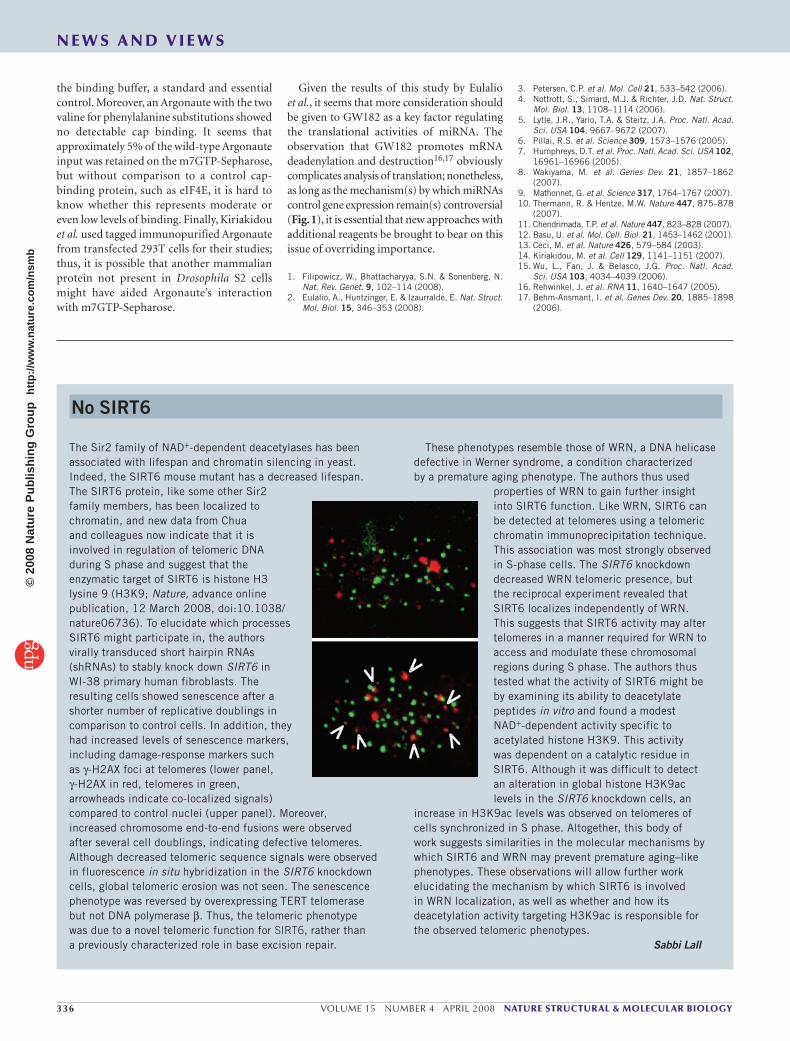
The Sir2 family of NAD+-dependent deacetylases has been associated with lifespan and chromatin silencing in yeast.Indeed, the SIRT6 mouse mutant has a decreased lifespan. The SIRT6 protein, like some other Sir2 family members, has been localized to chromatin, and new data from Chua and colleagues now indicate that it is involved in regulation of telomeric DNA during S phase and suggest that the enzymatic target of SIRT6 is histone H3 lysine 9 (H3K9; Nature, advance online publication, 12 March 2008, doi:10.1038/nature06736). To elucidate which processes SIRT6 might participate in, the authors virally transduced short hairpin RNAs (shRNAs) to stably knock down SIRT6 in WI-38 primary human fibroblasts. The resulting cells showed senescence after a shorter number of replicative doublings in comparison to control cells. In addition, they had increased levels of senescence markers, including damage-response markers such as γ-H2AX foci at telomeres (lower panel, γ-H2AX in red, telomeres in green, arrowheads indicate co-localized signals) compared to control nuclei (upper panel). Moreover, increased chromosome end-to-end fusions were observed after several cell doublings, indicating defective telomeres. Although decreased telomeric sequence signals were observed in fluorescence in situ hybridization in the SIRT6 knockdown cells, global telomeric erosion was not seen. The senescence phenotype was reversed by overexpressing TERT telomerase but not DNA polymerase β. Thus, the telomeric phenotype was due to a novel telomeric function for SIRT6, rather than a previously characterized role in base excision repair.
These phenotypes resemble those of WRN, a DNA helicase defective in Werner syndrome, a condition characterized by a premature aging phenotype. The authors thus used
properties of WRN to gain further insight into SIRT6 function. Like WRN, SIRT6 can be detected at telomeres using a telomeric chromatin immunoprecipitation technique. This association was most strongly observed in S-phase cells. The SIRT6 knockdown decreased WRN telomeric presence, but the reciprocal experiment revealed that SIRT6 localizes independently of WRN. This suggests that SIRT6 activity may alter telomeres in a manner required for WRN to access and modulate these chromosomal regions during S phase. The authors thus tested what the activity of SIRT6 might be by examining its ability to deacetylate peptides in vitro and found a modest NAD+-dependent activity specific to acetylated histone H3K9. This activity was dependent on a catalytic residue in SIRT6. Although it was difficult to detect an alteration in global histone H3K9ac levels in the SIRT6 knockdown cells, an
increase in H3K9ac levels was observed on telomeres of cells synchronized in S phase. Altogether, this body of work suggests similarities in the molecular mechanisms by which SIRT6 and WRN may prevent premature aging–like phenotypes. These observations will allow further work elucidating the mechanism by which SIRT6 is involved in WRN localization, as well as whether and how its deacetylation activity targeting H3K9ac is responsible for the observed telomeric phenotypes.
Sabbi Lall
N E W S A N D V I E W S©
2008
Nat
ure
Pub
lishi
ng G
roup
ht
tp://
ww
w.n
atur
e.co
m/n
smb