Embed Size (px)
Citation preview
The Trigeminal and Facial Nerves The Facial and Blink
Introduction – We commonly perform nerve conduction studies on three cranial nerves. Two of these, the trigeminal nerve (CN V) and the facial nerve (CN VII) are both mixed nerves, that is; they carry both motor and sensory fibers. In the EMG lab, lesions of the facial nerve are fairly common, thus requiring quality studies of the facial nerve. In addition, acquiring superior Blink Reflex studies give information about the trigeminal and facial nerve.
This paper will look at the anatomy of the trigeminal and facial nerves, common disorders of both nerves, specifics of NCS testing and case studies.
Anatomy of the Trigeminal Nerve – For convenience, anatomy of the trigeminal nerve will be divided into three segments: brainstem, preganglionic (including the trigeminal ganglion) and postganglionic. There are a variety of conditions, which may involve the different segments of the trigeminal nerve. Knowledge of its anatomic course allows an understanding of disorders involving the brainstem and adjacent skull base.
Brainstem
There are three sensory and one motor nuclei in the pons. The sensory components include the nucleus of the spinal tract, main sensory nucleus, the mesencephalic nucleus.
a. The spinal tract comes from the sensory root in the pons and proceeds downward into the upper cervical cord. This nucleus and tract receive pain and temperature sensation.
b. The main sensory nucleus lies lateral to the entering trigeminal root and receives sensation of light touch.
c. The mesencephalic trigeminal nucleus is near the lateral margin of the central gray matter anterior to the upper fourth ventricle and aqueduct. Fibers from the mesencephalic nucleus transmit proprioception from the teeth, hard palate, and temporomandibular joint and convey impulses that control mastication and the force of a bite.
d. The motor fibers of the trigeminal nerve emerge from the lateral portion of the pons alongside the main sensory nucleus to eventually innervate the muscles of mastication.
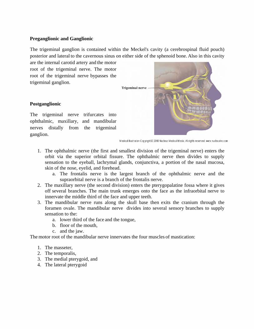
Preganglionic and Ganglionic
The trigeminal ganglion is contained within the Meckel's cavity (a cerebrospinal fluid pouch) posterior and lateral to the cavernous sinus on either side of the sphenoid bone. Also in this cavity are the internal carotid artery and the motor root of the trigeminal nerve. The motor root of the trigeminal nerve bypasses the trigeminal ganglion.
Postganglionic
The trigeminal nerve trifurcates into ophthalmic, maxillary, and mandibular nerves distally from the trigeminal ganglion.
1. The ophthalmic nerve (the first and smallest division of the trigeminal nerve) enters the
orbit via the superior orbital fissure. The ophthalmic nerve then divides to supply
sensation to the eyeball, lachrymal glands, conjunctiva, a portion of the nasal mucosa, skin of the nose, eyelid, and forehead.
a. The frontalis nerve is the largest branch of the ophthalmic nerve and the supraorbital nerve is a branch of the frontalis nerve.
2. The maxillary nerve (the second division) enters the pterygopalatine fossa where it gives off several branches. The main trunk emerges onto the face as the infraorbital nerve to innervate the middle third of the face and upper teeth.
3. The mandibular nerve runs along the skull base then exits the cranium through the foramen ovale. The mandibular nerve divides into several sensory branches to supply sensation to the:
a. lower third of the face and the tongue, b. floor of the mouth, c. and the jaw.
The motor root of the mandibular nerve innervates the four muscles of mastication:
1. The masseter, 2. The temporalis, 3. The medial pterygoid, and 4. The lateral pterygoid
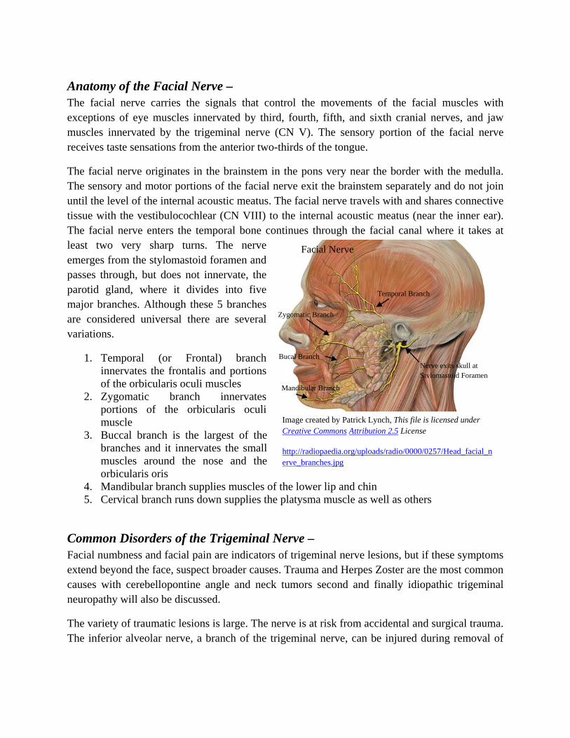
Anatomy of the Facial Nerve – The facial nerve carries the signals that control the movements of the facial muscles with exceptions of eye muscles innervated by third, fourth, fifth, and sixth cranial nerves, and jaw muscles innervated by the trigeminal nerve (CN V). The sensory portion of the facial nerve receives taste sensations from the anterior two-thirds of the tongue.
The facial nerve originates in the brainstem in the pons very near the border with the medulla. The sensory and motor portions of the facial nerve exit the brainstem separately and do not join until the level of the internal acoustic meatus. The facial nerve travels with and shares connective tissue with the vestibulocochlear (CN VIII) to the internal acoustic meatus (near the inner ear). The facial nerve enters the temporal bone continues through the facial canal where it takes at least two very sharp turns. The nerve emerges from the stylomastoid foramen and passes through, but does not innervate, the parotid gland, where it divides into five major branches. Although these 5 branches are considered universal there are several variations.
1. Temporal (or Frontal) branch innervates the frontalis and portions of the orbicularis oculi muscles
2. Zygomatic branch innervates portions of the orbicularis oculi muscle
3. Buccal branch is the largest of the branches and it innervates the small muscles around the nose and the orbicularis oris
4. Mandibular branch supplies muscles of the lower lip and chin 5. Cervical branch runs down supplies the platysma muscle as well as others
Common Disorders of the Trigeminal Nerve – Facial numbness and facial pain are indicators of trigeminal nerve lesions, but if these symptoms extend beyond the face, suspect broader causes. Trauma and Herpes Zoster are the most common causes with cerebellopontine angle and neck tumors second and finally idiopathic trigeminal neuropathy will also be discussed.
The variety of traumatic lesions is large. The nerve is at risk from accidental and surgical trauma. The inferior alveolar nerve, a branch of the trigeminal nerve, can be injured during removal of
Image created by Patrick Lynch, This file is licensed under Creative Commons Attribution 2.5 License
http://radiopaedia.org/uploads/radio/0000/0257/Head_facial_nerve_branches.jpg
Facial Nerve
Nerve exits skull at Stylomastoid Foramen
Temporal Branch
Zygomatic Branch
Bucal Branch
Mandibular Branch
impacted molar teeth. Persistent numbness and pain around the teeth is possible. The numb chin syndrome can occur after mandibular fracture.
Trigeminal Schwannomas arise from the trigeminal ganglion and cause numbness and paresthesias are the most common symptoms from these tumors, but pain and crawling sensations are sometimes present. Other tumors and perineurial spread of other cancers in the region of the trigeminal ganglion are rare, but can cause variable clinical symptoms.
The diagnosis of Idiopathic trigeminal neuropathy is made after excluding other causes of facial sensory changes. Idiopathic motor involvement of the trigeminal is even rarer.
Trigeminal neuralgia is characterized by short paroxysms of severe pain in one or more of the trigeminal nerve territories. It is almost always unilateral and seldom spreads to other territories after onset. Each episode of severe pain is brief, from just a few seconds to a minute. The spasms of trigeminal neuralgia are triggered by things like eating or talking. It is believed the cause is arterial loops causing compression at the trigeminal sensory roots.
Common Disorders of the Facial Nerve – The most common disorder of the facial nerve is idiopathic facial nerve paralysis, or Bell’s palsy. Bell’s palsy usually has an acute onset with unilateral facial weakness. Some patients report ipsilateral tearing or taste and ear pain. While 96% of patients without underlying risk factors (i.e. hypertension and cranial pain) experience spontaneous recovery, 10-15% has some residual facial weakness. Patients that do not show some facial muscle recovery after 6 months show a more guarded prognosis for spontaneous recovery. Of this group, prolonged facial muscle weakness, synkinesis, muscle spasm and abnormal tearing are common sequelae. Pathology that may give rise to Bell’s palsy includes herpes simplex virus (HSV) and Lyme’s disease to name only two.
Other disorders that may give rise to acute facial paralysis include Granulomatous disease (bilateral involvement), Amyloidosis, trauma, Connective tissue disease, Diabetes Mellitus, HIV, tumors and even pregnancy.
Hemifacial spasm consists of spontaneous, clonic, repetitive, involuntary twitching of the facial muscles as a result of blood vessels constricting the seventh cranial nerve. The typical patient is middle-aged and female. It is rarely bilateral.
Specific Nerve Conduction Studies-
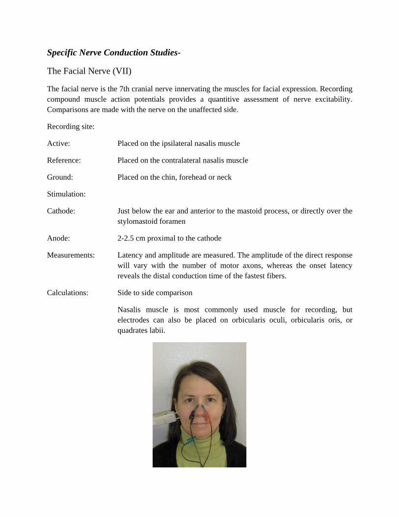
The Facial Nerve (VII)
The facial nerve is the 7th cranial nerve innervating the muscles for facial expression. Recording compound muscle action potentials provides a quantitive assessment of nerve excitability. Comparisons are made with the nerve on the unaffected side.
Recording site:
Active: Placed on the ipsilateral nasalis muscle
Reference: Placed on the contralateral nasalis muscle
Ground: Placed on the chin, forehead or neck
Stimulation:
Cathode: Just below the ear and anterior to the mastoid process, or directly over the stylomastoid foramen
Anode: 2-2.5 cm proximal to the cathode
Measurements: Latency and amplitude are measured. The amplitude of the direct response will vary with the number of motor axons, whereas the onset latency reveals the distal conduction time of the fastest fibers.
Calculations: Side to side comparison
Nasalis muscle is most commonly used muscle for recording, but electrodes can also be placed on orbicularis oculi, orbicularis oris, or quadrates labii.
To Assess a Lesion of the Facial Nerve-
To assess a lesion of the facial nerve, amplitude provides more information than onset latency of the direct response. The amplitude of the direct response varies with the amount of functioning motor axons, while the latency shows the distal conduction time of the fastest fibers. Onset latency tends to be normal or may only increase slightly. The amplitude will determine the prognosis of recovery by what degree of axonal loss has occurred. Normal values for facial nerve latencies (mean ± SD) in adults range from 3.4 ± 0.8 to 4.0 ± 0.5 ms (Kimura 413). The comparison of the nerve from side to side provides a more accurate assessment than the absolute value, which also varies from one patient to another.
Multiple studies are the best way to show the progression of axonal loss. For the first few days after injury, distal stimulation may show a normal response. From days following the onset to a week the amplitude will drop showing progression of injury and nerve degeneration. A prognosis is good if the direct response is still intact during the first week of injury. To prevent activating the masseter muscle, use caution making sure shocks of high intensity are not given. A larger response recorded from this muscle would appear to be a good prognosis when actually the facial nerve has already degenerated. Place hand over masseter to feel contractions or visual inspection for results in question.
Recording compound muscle action potentials provides an assessment of nerve excitability, better than visual inspection. Stimulating the facial nerve just below the ear, anterior to the mastoid process, or directly over the stylomastoid foramen recording distally along the branch of the facial nerve will result in selective activation of the muscles.

The Blink Reflex (Trigeminal V Facial VII)-
Stimulation of the trigeminal nerve activates a reflex pathway along the brainstem, resulting in a contraction of the orbicularis oculi. The Blink Reflex reflects the integrity of the afferent and efferent pathways including the proximal segment of the facial nerve.
Recording Sites: 2 Channel Study
Active: Placed over the inferior portion of the orbicularis oculi near the inner canthus
Reference: Placed 2 cm laterally
Ground: Placed on the chin or forehead
Stimulation: Ipsilateral same side of stimulation
Contralateral opposite side of stimulation
Cathode: Supraorbital notch
Anode: 2-2.5cm directly above cathode on forehead
(rotation of anode around cathode helps establish the best position of stimulating electrode and reduce shock artifact)
Measurements: Onset latency of R1 response-ipsilateral to the side of stimulation only
Onset latency of R2 response ipsilateral and contralateral to the side of stimulation
Calculations: Normal lab values side to side R1 and R2 comparison/difference
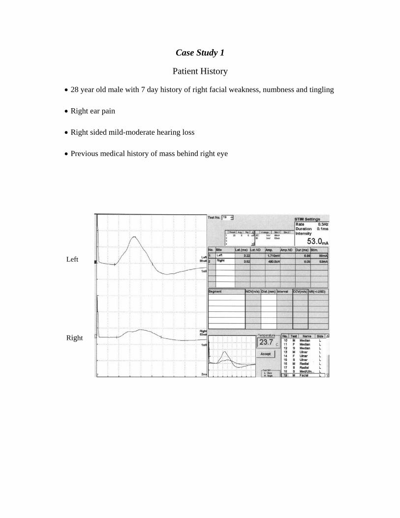
Case Study 1
Patient History
• 28 year old male with 7 day history of right facial weakness, numbness and tingling • Right ear pain
• Right sided mild-moderate hearing loss • Previous medical history of mass behind right eye
Right
Left
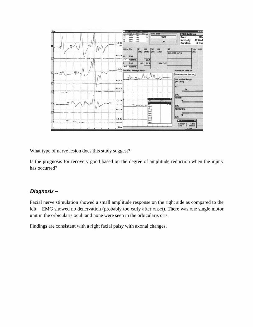
What type of nerve lesion does this study suggest?
Is the prognosis for recovery good based on the degree of amplitude reduction when the injury has occurred?
Diagnosis –
Facial nerve stimulation showed a small amplitude response on the right side as compared to the left. EMG showed no denervation (probably too early after onset). There was one single motor unit in the orbicularis oculi and none were seen in the orbicularis oris.
Findings are consistent with a right facial palsy with axonal changes.

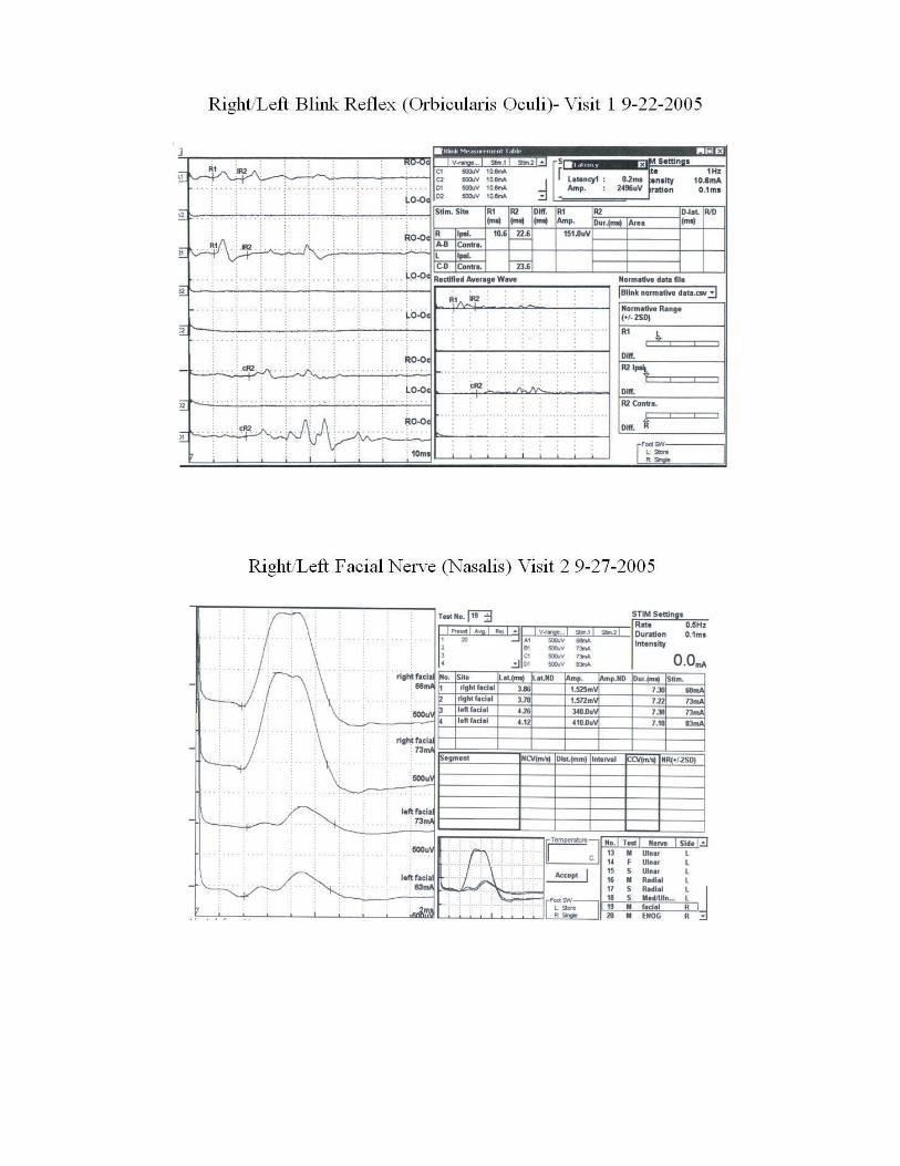
Case Study 2
Patient History
• 52 year old white male
• Fell and hit head at 8:00 am, seemed to be doing well
• By 11:00 pm he was unable to close mouth completely with noticeable drooling
• The next day after fall he could not close his left eye completely, and had no tone in the left side of his face
• Facial sensation and hearing intact
• Gag reflex present and tongue midline
• Past medical history unremarkable except for surgical fusion
Plan
• Facial nerve and blink reflex study to be repeated every few days to assess the progression of nerve injury
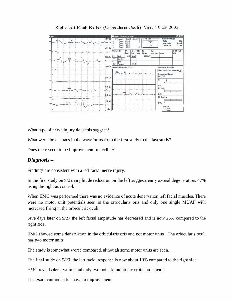
What type of nerve injury does this suggest?
What were the changes in the waveforms from the first study to the last study?
Does there seem to be improvement or decline?
Diagnosis –
Findings are consistent with a left facial nerve injury.
In the first study on 9/22 amplitude reduction on the left suggests early axonal degeneration. 47% using the right as control.
When EMG was performed there was no evidence of acute denervation left facial muscles. There were no motor unit potentials seen in the orbicularis oris and only one single MUAP with increased firing in the orbicularis oculi.
Five days later on 9/27 the left facial amplitude has decreased and is now 25% compared to the right side.
EMG showed some denervation in the orbicularis oris and not motor units. The orbicularis oculi has two motor units.
The study is somewhat worse compared, although some motor units are seen.
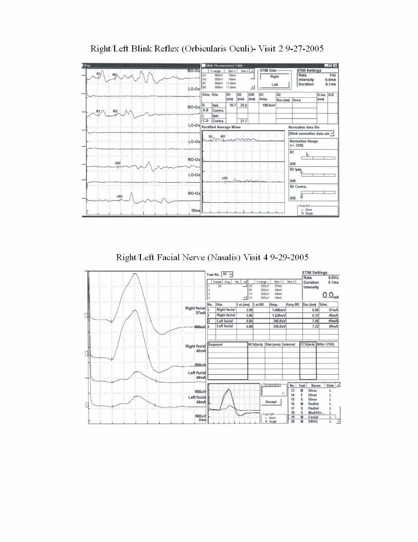
The final study on 9/29, the left facial response is now about 10% compared to the right side.
EMG reveals denervation and only two units found in the orbicularis oculi.
The exam continued to show no improvement.
Case Study 3
Patient History
• 80 year old female, onset of symptoms four weeks ago
• Frontal Headache, shingles around left eye
• Red and Swollen left eye she could not open
• Left droopy eyelid
• Blurry and double vision
• Past history of stroke several years ago causing left sided weakness and double vision
• Coronary artery disease, hypertension and diabetes
Physical Exam
• Sensation on jaw and facial expression muscles were symmetric
• Hearing preserved bilaterally
• Palate elevated symmetrically, tongue midline
• Shoulder shrug and head turning symmetric
• On the left there was decreased sensation on scalp in occipital area and to a lesser degree on cheek
Eye Examination
• Normal optic discs with intact visual fields
• Findings for the left eye showed limited eye movements
• no corneal sensation or reflex
• Pupil 4mm and not reactive
• Visual acuity 20/60
• Findings for the right eye showed eye movements intact
• Pupil 3mm and reactive
• Visual acuity 20/25
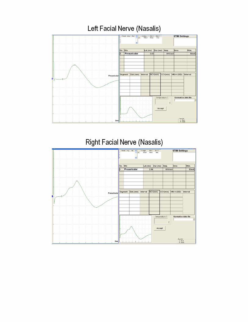

What type of nerve injury does this suggest?
What was most helpful in determining this outcome?
Diagnosis –
Findings are suggestive of left trigeminal (VI) lesion.
Clinical: Incomplete palsy of cranial nerves III and VI.

Stimulation of the Trigeminal Nerve-
Stimulation of the trigeminal nerve will elicit a reflex contraction of the orbicularis oculi. The blink reflex assesses the integrity of the afferent and efferent pathways which include the proximal segment of the facial nerve.
Stimulation applied to the supraorbital nerve will evoke two separate responses, the R1 and the R2. The R1 represents the conduction time along the facial and trigeminal nerves and pontine relay. The R2 represents the excitability of interneurons, delay for synaptic transmission time as well as the axonal conduction time. The variability in latency makes R2 less reliable for diagnostic purposes. Optimal stimulation intensity ranges from 5 to 10mA using duration of 0.1 ms. This will elicit a fairly stable R1 response in repeated attempts. At times shocks of appropriate intensity may not elicit a stable R1 response on either side of stimulation and mild voluntary contraction may be the only way to facilitate a response. A possibility of this occurrence may be due to the patient habituating to the shocks. Several trials on each side must be recorded to ensure the shortest latency measurements.
Upper limits of normal values are defined as the mean latency plus 3 standard deviation. In most labs the R1 response should not exceed 12.6 ms. With stimulation of the supraorbital nerve the R2 should not exceed 40 ms and the contralateral side 41 ms. The latency difference from side to side should not exceed 1.2 ms for electrically stimulated R1 response. The ipsilateral and contralateral R2 latency on the side being stimulated should not vary more than 5 ms (Kimura 419).
Analysis of the R1-
An increase in the latency of R1 response will usually imply demyelination of the peripheral pathway along the trigeminal or facial nerve, or the central reflex pathway of the pons. A posterior fossa tumor may also affect the R1 by compressing the cranial nerves or by involving the brainstem.
Analysis of the R2-
Changes in latency may imply lesions directly affecting the reflex pathway. The R2 helps determine if the abnormality is afferent or efferent when the R1 is delayed.
Conclusion-
There are many disorders that affect the facial and trigeminal nerves. This paper discussed specific techniques used to perform these studies, the anatomy and case studies to help better understand the components of these nerves.

References –
Blum Andrew S. and Seward B. Rutkove, eds. The Clinical Neurophysiology Primer. Humana Press Inc. Totowa, New Jersey. 2007.
Daube, Jasper, ed. Clinical Neurophysiology. F.A. Davis Company, Philadelphia. 1996.
Dumitru, Daniel, Anthony Amato and Machiel Zwarts. Electrodiagnostic Medicine, 2nd Ed. Hanley and Belfus, Philadalephia. 2002.
Hughes, Richard A.C. “Diseases of the Fifth Cranial Nerve.” Peripheral Neuropathy. Ed. Dyck, Peter J. and P. K. Thomas. 4th Ed. Elsevier Saunders. Philadelphia. 2005.
Kimura, Jun. Electrodiagnosis in Diseases of Nerve and Muscle: Principles and Practice. 3rd Ed. Oxford University Press, New York. 2001.
Klein, Caroline M. “Diseases of the Seventh Cranial Nerve.” Peripheral Neuropathy. Ed. Dyck, Peter J. and P. K. Thomas. 4th Ed. Elsevier Saunders. Philadelphia. 2005.
Oh, Shin. Clinical Electromyography: Nerve Conduction Studies. 2nd Ed. Williams and Wilkins, Baltimore. 1993.
Preston, David and Barbara Shapiro. Electromyography and Neuromuscular Disorders; Clinical-Electrophysiologic Correlations. 2nd Ed. Elsevier, Philadelphia. 2005.