Embed Size (px)
Citation preview
at SciVerse ScienceDirect
Biomaterials 33 (2012) 1162e1169
Contents lists available
Biomaterials
journal homepage: www.elsevier .com/locate/biomateria ls
The therapeutic efficacy of camptothecin-encapsulated supramolecularnanoparticles
Kuan-Ju Chen a,1, Li Tang b,1, Mitch André Garcia a, Hao Wang a, Hua Lu b, Wei-Yu Lin a, Shuang Hou a,Qian Yin b, Clifton K.-F. Shen a, Jianjun Cheng b,*, Hsian-Rong Tseng a,**
aDepartment of Molecular and Medical Pharmacology, California NanoSystems Institute (CNSI), Crump Institute for Molecular Imaging (CIMI), University of California,Los Angeles, 570 Westwood Plaza, Building 114, Los Angeles, CA 90095-1770, USAbDepartment of Materials Science and Engineering, University of Illinois at Urbana-Champaign-Urbana, 1304 West Green Street, Urbana, IL 61801, USA
a r t i c l e i n f o
Article history:Received 3 September 2011Accepted 16 October 2011Available online 8 November 2011
Keywords:Supramolecular assemblyNanoparticlesDrug deliveryPositron emission tomographyCancer therapeutics
* Corresponding author. Tel.: þ1 217 244 3924; fax** Corresponding author. Department of Medical aUniversity of California, Los Angeles, 570 WestwoAngeles, CA 90095-1770, USA. Tel.: þ1 310 794 1977;
E-mail addresses: [email protected] (J. Cheng(H.-R. Tseng).
URLs: http://cheng.mse.uiuc.edu/index.htm, http:/tsenglab/
1 These authors contributed equally to the work.
0142-9612/$ e see front matter � 2011 Elsevier Ltd.doi:10.1016/j.biomaterials.2011.10.044
a b s t r a c t
Nanomaterials have been increasingly employed as drug(s)-incorporated vectors for drug delivery due totheir potential of maximizing therapeutic efficacy while minimizing systemic side effects. However, therehave been twomain challenges for these vectors: (i) the existing synthetic approaches are cumbersome andincapable of achieving precise control of their structural properties, which will affect their biodistributionand therapeutic efficacies, and (ii) lack of an early checkpoint to quickly predictwhich drug(s)-incorporatedvectors exhibit optimal therapeutic outcomes. In this work, we utilized a new rational developmentalapproach to rapidly screen nanoparticle (NP)-based cancer therapeutic agents containing a built-incompanion diagnostic utility for optimal therapeutic efficacy. The approach leverages the advantages ofa self-assembly synthetic method for preparation of two different sizes of drug-incorporated supramo-lecular nanoparticles (SNPs), and a positron emission tomography (PET) imaging-based biodistributionstudy to quickly evaluate the accumulation of SNPs at a tumor site in vivo and select the favorable SNPs forin vivo therapeutic study. Finally, the enhanced in vivoanti-tumorefficacyof the selected SNPswas validatedby tumor reduction/inhibition studies.We foreseeour rational developmental approachproviding a generalstrategy in the search of optimal therapeutic agents among the diversity of NP-based therapeutic agents.
� 2011 Elsevier Ltd. All rights reserved.
1. Introduction
Nanoparticles (NPs) have been regarded as promising vectorsfor controlled delivery of anti-cancer drug(s), providing anemerging therapeutic strategy with enhanced anti-tumor efficacyand reduced systemic side effects [1e10]. However, even afterdecades of development, there are few successful examples thathave reached clinical usage [2,11]. One of the major bottlenecks canbe attributed to the expensive, time-consuming and labor-intensivepipeline adopted for the development of drug(s)-incorporated NPs,
: þ1 217 333 2736.nd Molecular Pharmacology,od Plaza, Building 114, Losfax: þ1 310 206 8975.), [email protected]
/labs.pharmacology.ucla.edu/
All rights reserved.
by which numerous optimization/evaluation cycles, includingmolecular design, multistep syntheses and in vitro/in vivo assays arerepeated. Such a developmental pipeline does not guaranteegenerating drug(s)-incorporated NPs that exhibit improved thera-peutic efficacy in clinic.
Herein, we utilize a new rational developmental approach torapidly screen NP-based cancer therapeutic agents containinga built-in companion diagnostic utility for optimal therapeuticefficacy. The approach leverages the advantages of a self-assemblysynthetic method for preparation of supramolecular nanoparticles(SNPs), and a positron emission tomography (PET) imaging-basedbiodistribution study to quickly evaluate the accumulation ofSNPs at a tumor site in vivo. The self-assembly synthetic methodenables a convenient, flexible and modular production of a smallcollection of drug-incorporated NPs with precisely controlled sizesranging from 37 to 104 nm, below the threshold of NP size (200 nm)that allows the efficient extravasation of NPs into the leaky tumorvasculatures and accumulation of the NPs in the tumor tissues viaenhanced permeability and retention (EPR) effect [12e17].
K.-J. Chen et al. / Biomaterials 33 (2012) 1162e1169 1163
Subsequently, PET imaging-based biodistribution studies areemployed to correlate the sizes of drug-incorporated NPs with theirdelivery performances. Thus, a specific size of drug-incorporatedNPs that exhibit optimal accumulation and prolonged retention intumor tissue can be readily identified, resulting in potentially thelead modality with improved therapeutic efficacy and reduced sideeffect that is subject to comprehensive pre-clinical studies.
Previously, we demonstrated a convenient, flexible, andmodular self-assembly synthetic method for crafting SNPs asoutstanding delivery vectors for highly efficient delivery of genes,[18,19] proteins [20] and inorganic nanoparticles [21]. Given theversatile utility of such a self-assembly synthetic method, weattempted to explore the use of SNP-based vectors for delivering ananti-cancer drug. In this case, camptothecin (CPT), a cytotoxicquinoline alkaloid that inhibits the topoisomerase I, was selectedfor the proof-of-concept demonstration. On the other hand, wehave already demonstrated the incorporation of radioisotopes anda contrast agent into SNPs for PET [22] and magnetic resonance(MR) imaging, [23] respectively. PET imaging [24e26] is a sensitive,non-invasive technology that can be utilized to determine thebiodistribution profiles of small molecules, polymers or NPs ata whole body level. The resulting biodistribution information [27]can be utilized to facilitate the implementation of pre-clinicalstudies. Moreover, once the drug-incorporated NPs with optimaltherapeutic efficacy are identified, they will contain a built-incompanion diagnostic utility that can be readily utilized if desired.
2. Materials and methods
2.1. General
Camptothecin (CPT) and other chemicals were purchased from SigmaeAldrich(St. Louis, MO) and used as received without further purification unless otherwisenoted. 1-Adamantanamine (Ad) hydrochloride and b-cyclodextrin (b-CD) werepurchased from TCI America (San Francisco, CA). N-hydroxysuccinimide (SCM) and
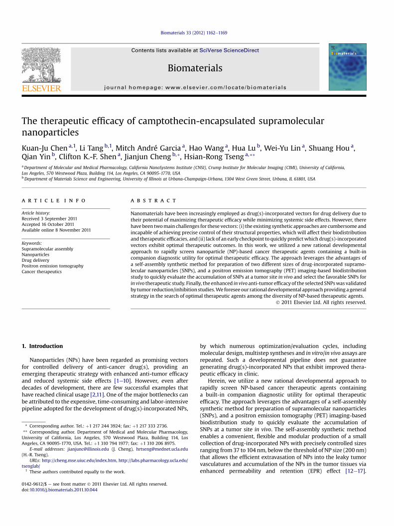
Scheme 1. Schematic representations of the self-assembly synthetic method for the producfrom the respective molecular building blocks and CPT-PGA (camptothecin-grafted poly(L-g
maleimido (MAL) hetero-functionalized poly(ethylene glycol) (SCM-PEG-MAL,MW ¼ 5 kD) were obtained from NANOCS Inc (New York, NY). CD-grafted branchedpoly(ethylenimine) (CD-PEI), Ad-grafted poly(ethylene glycol) (Ad-PEG) and DOTA-grafted CD-PEI (CD-PEI-DOTA) were prepared via themethod previously reported byour group [22,23]. Phosphate-Buffered Saline (PBS), Dulbecco’s Modified EagleMedium (DMEM), Eagle’s Minimum Essential Medium (EMEM), and penicillin/streptomycin were obtained from Invitrogen (Carlsbad, CA). MCF7 breast cancer cellline and Lewis Lung Carcinoma (LLC) cell line were purchased from American TypeCulture Collection (Manassas, VA). Fetal Bovine Serum (FBS) was obtained fromLonza Walkerrsville Inc (Walkerrsville, MD). 96 well BD Falcon culture plates werepurchased from Fisher Scientific. CellTiter-Blue� Cell Viability Assay was purchasedfrom Promega Corporation (Madison, WI). Human serum was purchased fromSigmaeAldrich.
The molecular weight of the PBLG were determined on a gel permeationchromatograph (GPC, also called size-exclusion chromatography (SEC)) equippedwith an isocratic pump (Model 1100, Agilent Technology, Santa Clara, CA), a DAWNHELEOS 18-angle laser light scattering detector (Wyatt Technology, Santa Barbara,CA), and an Optilab rEX refractive index detector (Wyatt Technology, Santa Barbara,CA). The wavelength of the HELEOS detector was set at 658 nm. The size-exclusioncolumns used were serially connected on the GPC (Phenogel columns 100 Å, 500 Å,103 Å and 104 Å, 5 mm, 300 � 7.8 mm, Phenomenex, Torrance, CA). DMF (HPLCgrade) was used as the mobile phase for GPC. HPLC analysis was performed ona Beckman Gold system (Beckman Coulter, Fullerton, CA) equipped with a 126Psolvent module, a System Gold 128 UV detector and an analytical C18 column (LunaC18, 250 � 4.6 mm, 5 m, Phenomenex, Torrance, CA). NMR analyses were conductedon a Varian U500, VXR500 or UI500NB (500 MHz).
2.2. Synthesis of Poly(L-glutamic acid) (PGA)
Poly(g-benzyl-L-glutamate) (PBLG50) was synthesized according to the proce-dure previously published. [28,29] The N-terminus of PBLG was capped by a carbo-benzyloxy (Cbz) group. The Mn was 12,600 g/mol and the MW distributions(MWD¼Mw/Mn) was 1.05 as determined by GPC (Fig. S1 in Supporting Information).The deprotection of PBLG50 was performed using standard HBr condition asdescribed below: PBLG50 (500 mg, 2.28 mmol glutamate residues) was dissolved inTFA (15 mL) in an ice bath. HBr (33 wt% in HOAc, 4 mL) was added dropwise intostirred solution. The reactionmixturewas stirred in the ice bath for an additional 2 hand then poured into cold ether (60mL) in two 50-mL centrifuge tubes. The polymerprecipitate was collected by centrifuge and washed with ether (30 mL � 3). Thepolymer was dried under vacuum to give the crude product. The polymer wasdissolved in NaOH (2 M � 10 mL) and was stirred at room temperature (rt)
tion of CPT-grafted PGA encapsulated Supramolecular NanoParticles (CPT-PGA3SNPs)lutamic acid)).
K.-J. Chen et al. / Biomaterials 33 (2012) 1162e11691164
overnight. The clear solution was acidified by 2 M HCl to pH 2. The product waspurified by dialysis against DI water and dried by lyophilization to give a whitepowder. 1H NMR (D2O, 500 MHz): d 4.86 (1H), 2.68 (2H), 2.34 (1H), 2.19 (1H).
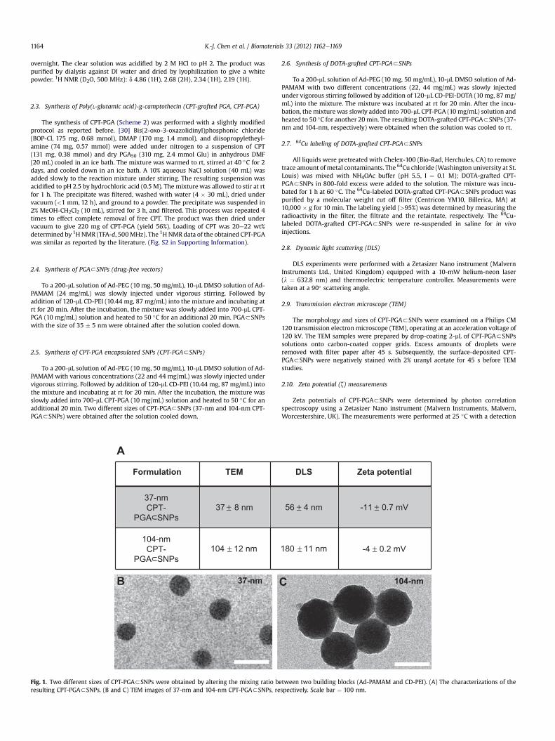
2.3. Synthesis of Poly(L-glutamic acid)-g-camptothecin (CPT-grafted PGA, CPT-PGA)
The synthesis of CPT-PGA (Scheme 2) was performed with a slightly modifiedprotocol as reported before. [30] Bis(2-oxo-3-oxazolidinyl)phosphonic chloride(BOP-Cl, 175 mg, 0.68 mmol), DMAP (170 mg, 1.4 mmol), and diisopropyletheyl-amine (74 mg, 0.57 mmol) were added under nitrogen to a suspension of CPT(131 mg, 0.38 mmol) and dry PGA50 (310 mg, 2.4 mmol Glu) in anhydrous DMF(20 mL) cooled in an ice bath. The mixture was warmed to rt, stirred at 40 �C for 2days, and cooled down in an ice bath. A 10% aqueous NaCl solution (40 mL) wasadded slowly to the reaction mixture under stirring. The resulting suspension wasacidified to pH 2.5 by hydrochloric acid (0.5 M). The mixture was allowed to stir at rtfor 1 h. The precipitate was filtered, washed with water (4 � 30 mL), dried undervacuum (<1 mm, 12 h), and ground to a powder. The precipitate was suspended in2% MeOH-CH2Cl2 (10 mL), stirred for 3 h, and filtered. This process was repeated 4times to effect complete removal of free CPT. The product was then dried undervacuum to give 220 mg of CPT-PGA (yield 56%). Loading of CPT was 20e22 wt%determined by 1HNMR (TFA-d, 500MHz). The 1H NMR data of the obtained CPT-PGAwas similar as reported by the literature. (Fig. S2 in Supporting Information).
2.4. Synthesis of PGA3SNPs (drug-free vectors)
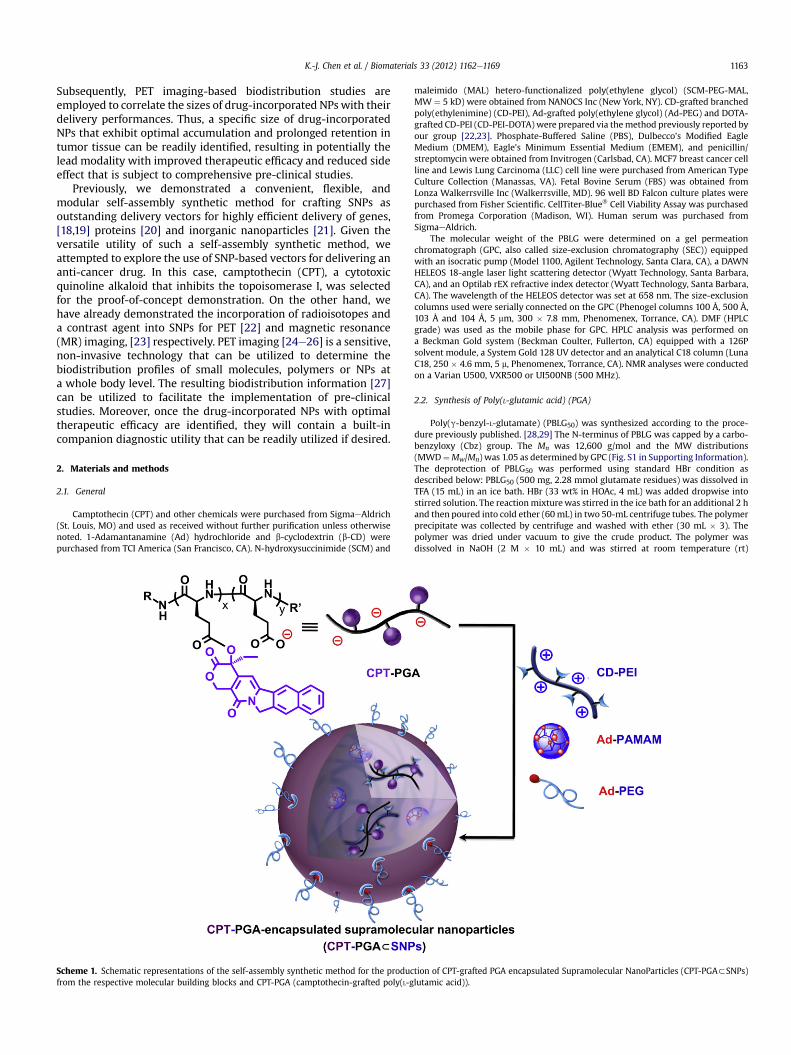
To a 200-mL solution of Ad-PEG (10 mg, 50 mg/mL), 10-mL DMSO solution of Ad-PAMAM (24 mg/mL) was slowly injected under vigorous stirring. Followed byaddition of 120-mL CD-PEI (10.44 mg, 87 mg/mL) into the mixture and incubating atrt for 20 min. After the incubation, the mixture was slowly added into 700-mL CPT-PGA (10 mg/mL) solution and heated to 50 �C for an additional 20 min. PGA3SNPswith the size of 35 � 5 nm were obtained after the solution cooled down.
2.5. Synthesis of CPT-PGA encapsulated SNPs (CPT-PGA3SNPs)
To a 200-mL solution of Ad-PEG (10 mg, 50 mg/mL), 10-mL DMSO solution of Ad-PAMAM with various concentrations (22 and 44 mg/mL) was slowly injected undervigorous stirring. Followed by addition of 120-mL CD-PEI (10.44 mg, 87 mg/mL) intothe mixture and incubating at rt for 20 min. After the incubation, the mixture wasslowly added into 700-mL CPT-PGA (10 mg/mL) solution and heated to 50 �C for anadditional 20 min. Two different sizes of CPT-PGA3SNPs (37-nm and 104-nm CPT-PGA3SNPs) were obtained after the solution cooled down.
A
B 37-nm
Formulation TEM
104-nmCPT-
PGA SNPs104 12 nm
37-nmCPT-
PGA SNPs37 8 nm±
±
Fig. 1. Two different sizes of CPT-PGA3SNPs were obtained by altering the mixing ratio beresulting CPT-PGA3SNPs. (B and C) TEM images of 37-nm and 104-nm CPT-PGA3SNPs, re
2.6. Synthesis of DOTA-grafted CPT-PGA3SNPs
To a 200-mL solution of Ad-PEG (10 mg, 50 mg/mL), 10-mL DMSO solution of Ad-PAMAM with two different concentrations (22, 44 mg/mL) was slowly injectedunder vigorous stirring followed by addition of 120-mL CD-PEI-DOTA (10 mg, 87 mg/mL) into the mixture. The mixture was incubated at rt for 20 min. After the incu-bation, the mixture was slowly added into 700-mL CPT-PGA (10 mg/mL) solution andheated to 50 �C for another 20min. The resulting DOTA-grafted CPT-PGA3SNPs (37-nm and 104-nm, respectively) were obtained when the solution was cooled to rt.
2.7. 64Cu labeling of DOTA-grafted CPT-PGA3SNPs
All liquids were pretreated with Chelex-100 (Bio-Rad, Herchules, CA) to removetrace amount of metal contaminants. The 64Cu chloride (Washington university at St.Louis) was mixed with NH4OAc buffer (pH 5.5, I ¼ 0.1 M); DOTA-grafted CPT-PGA3SNPs in 800-fold excess were added to the solution. The mixture was incu-bated for 1 h at 60 �C. The 64Cu-labeled DOTA-grafted CPT-PGA3SNPs product waspurified by a molecular weight cut off filter (Centricon YM10, Billerica, MA) at10,000 � g for 10 min. The labeling yield (>95%) was determined by measuring theradioactivity in the filter, the filtrate and the retaintate, respectively. The 64Cu-labeled DOTA-grafted CPT-PGA3SNPs were re-suspended in saline for in vivoinjections.
2.8. Dynamic light scattering (DLS)
DLS experiments were performed with a Zetasizer Nano instrument (MalvernInstruments Ltd., United Kingdom) equipped with a 10-mW helium-neon laser(l ¼ 632.8 nm) and thermoelectric temperature controller. Measurements weretaken at a 90� scattering angle.
2.9. Transmission electron microscope (TEM)
The morphology and sizes of CPT-PGA3SNPs were examined on a Philips CM120 transmission electron microscope (TEM), operating at an acceleration voltage of120 kV. The TEM samples were prepared by drop-coating 2-mL of CPT-PGA3SNPssolutions onto carbon-coated copper grids. Excess amounts of droplets wereremoved with filter paper after 45 s. Subsequently, the surface-deposited CPT-PGA3SNPs were negatively stained with 2% uranyl acetate for 45 s before TEMstudies.
2.10. Zeta potential (z) measurements
Zeta potentials of CPT-PGA3SNPs were determined by photon correlationspectroscopy using a Zetasizer Nano instrument (Malvern Instruments, Malvern,Worcestershire, UK). The measurements were performed at 25 �C with a detection
104-nmC
DLS Zeta potential
180 11 nm -4 0.2 mV
56 4 nm -11 0.7 mV± ±
±±
tween two building blocks (Ad-PAMAM and CD-PEI). (A) The characterizations of thespectively. Scale bar ¼ 100 nm.
K.-J. Chen et al. / Biomaterials 33 (2012) 1162e1169 1165
angle of 90� , and the raw data were subsequently correlated to Z average mean sizeusing a cumulative analysis by the Zetasizer software package.
2.11. Drug encapsulation efficiency
Free CPT was removed from CPT-PGA3SNPs by centrifugation of CPT-PGA3SNPs solution at 1300 rpm for 30 min using centrifugal filter devices (3000NMWL). After recovering the filtrate containing free CPT, CPT concentration wasanalyzed by ultraviolet absorption at a wavelength of 370 nm. The measurementswere performed in triplicate. The amount of the CPT encapsulated in the SNPs wasthen calculated by the total loading amount of CPT subtracts the free CPT in thefiltrate.
2.12. Time-dependent stability study
To ensure the in vivo stability of the resulting CPT-PGA3SNPs, it is critical toexamine their size variation under a physiological ionic strength. Following theprocedure described above, the 37-nm and 104-nm CPT-PGA3SNPs were preparedin PBS solutions (pH ¼ 7.2, containing 1.5 mM KH2PO4, 155 mM NaCl and 2.7 mMNa2HPO4). After mixing the three molecular building blocks in their respectiveratios, we employed real-time DLSmeasurements tomonitor the hydrodynamic sizevariation of the 37-nm and 104-nm CPT-PGA3SNPs at different times. The sizes ofCPT-PGA3SNPs were recorded up to 6 days.
2.13. Drug release profile
CPT-PGA (0.249mg/mL) or 37-nm CPT-PGA3SNPs (0.977mg/mL) was dispersedin 50% human serum (human serum:1 � PBS ¼ 1:1, v/v) and equally distributed to20 vials with 1 mL solution per vial, and then incubated at 37 �C. At selected timeintervals, one selected vial of each groupwas taken out of the incubator. The solutionwas mixed with an equal volume of methanol (1 mL) and centrifuged at 15,000 rpmfor 10 min. The supernatant (1 mL) was transferred to an eppendorf tube withoutdisturbing the precipitates and brought to pH 2 with phosphoric acid (85%, 100 mL).The resulting solutionwas directly injected into an HPLC equippedwith an analyticalC18 column. A mixture of acetonitrile and water (containing 0.1% TFA) at a volumeratio of 1:3 was used as the mobile phase. The flow rate was set at 1 mL/min. Thearea of the HPLC peak of the released CPT (labs ¼ 370 nm) was intergraded for thequantification of CPT as compared to a standard curve of free CPT prepared sepa-rately. The accumulative release of CPT is shown in Fig. 2B.
Scheme 2. Schematic representation of the synthesis o
2.14. In vitro cell viability
Cell viability was measured by the MTT assay. After incubating MCF-7 breastcancer cell line with 100-mL 37-nm CPT-PGA3SNPs, 104-nm CPT-PGA3SNPs, freeCPT, CPT-PGA and PGA3SNPs with respective concentrations in 96 well-plates for48 h, the solutions were removed and washed with PBS three times. Fresh mediumcontaining 20-mL CellTiter-Blue reagent was added to each well, followed by 3 hincubation at 37 �C. The cell viability results were quantified using fluorescent plate-reader.
2.15. Micro-PET imaging
C57BL/6 mice were purchased from DLAM Breeding Colony Services (LosAngeles, CA). All animal manipulations were performed with sterile technique andwere approved by the University of California at Los Angeles Animal ResearchCommittee (ARC protocol# 2006-135-12). C57BL/6 mice were injected subcutane-ously in the right flank with LLC cells suspended in a 1:1 mixture of PBS buffer andmatrigel (BD Biosciences, Franklin Lakes, NJ, USA). Prior to use in tumor induction,LLC cells were cultured in DMEM medium. Tumors were grown for 9 days and then100-mL 64Cu (150 mCi) labeled DOTA-grafted CPT-PGA3SNPs solution was injectedvia tail vein while the animal was anesthetized. Micro-PET imaging of the miceoccurred 24 h post injection and was performed with a micro-PET FOCUS 220 PETscanner (Siemens, Malvern, PA). The mice were anesthetized by using 1.5e2% iso-flurane in a heated (30 �C) induction chamber 15 min prior to imaging. The micewere then transferred to a heated isolation/imaging chamber for imaging. Staticmicro-PET scans (10 min) were then acquired. To determine the supramolecularnanoparticle concentration in various tissues, ellipsoid regions of interest wereplaced in the region that exhibited the highest 64Cu activity as determined by visualinspection using the Amide software. Relative intensity is expressed as thepercentage of activity in the organ of interest to that of the sum of all activity in theorgans listed.
2.16. In vivo study
C57BL/6 mice (female) were purchased from Charles River, USA. Feed and waterwas available ad libitum. The animal study protocol was reviewed and approved bythe Animal Care and Use Committee of University of Illinois at Urbana-Champaign.Female C57Bl/6 mice, 4e5 week old, were anesthetized, shaved, and prepared forimplantation of the tumor cells. LLC cells were collected from culture, and 3 � 105
cells suspended in a 1:1 mixture of PBS buffer and matrigel were then injectedsubcutaneously into rightflankof amouse. After 6 dayswhen tumors reachedaround
f poly(L-glutamic acid)-g-camptothecin (CPT-PGA).
K.-J. Chen et al. / Biomaterials 33 (2012) 1162e11691166
60e90mm3 in size, micewere divided into 4 groups of fivemice, minimizing weightand tumor size difference. Tumor-bearingmicewere treated by intravenous injectionof PBS, PGA3SNPs, CPT-PGA3SNPs (13.6 mg/kg CPT) or intraperitoneal injection ofCPT (13.6 mg/kg). Three doses were administrated with 5-day interval, i.e. at day 1,day6 andday11, respectively (except for CPTgroup,which only receiveda single doseat day 1 due to emerging toxicity [31]). After injections, the animals were monitoredclosely, and measurements of the tumor size and body weight for each animal wereperformed at regular intervals using calipers without knowledge of which injectioneach animal had received. The tumor volume for each time point was calculatedaccording to the formula, (length)� (width)2/2, where the long axis is the length, theshort axis is thewidth. Tumor density is assumed as 1mg/mm3. If bodyweight loss isbeyond 20% of pre-dosing weight, the animals were euthanized. When the tumorload reached 1500 mm3 or the animal had become moribund, the mouse wassacrificed. The statistical analysis was undertaken using a Student’s t-test (two-tailed), and p-values < 0.05 were considered statistically significant, p < 0.01 wereconsidered highly statistically significant. Median tumor growth curves prepared foreach group depicted the median tumor size as a function of time (Fig. 4A).
3. Results and discussion
Similar to the self-assembly preparation of DNA encapsulatedSNPs, [18,19] which used the coulombic interactions between thenegatively charged DNA plasmid with the positively charged SNPvector, 5 KD anionic poly(L-glutamic acid) (PGA) [28,29] wasemployed as a carrier to covalently link with CPT molecules,enabling encapsulation into SNP vectors. Approximately five CPTmolecules were conjugated to each PGA polymer chain (via esterbond formation) to give CPT-grafted PGA, denoted as CPT-PGA. [30]It is noteworthy that the connecting ester bonds can be degradedvia esterase-mediated hydrolysis, which allows controlled releaseof CPT under physiological conditions. The encapsulation of CPT-
100
200
Hyd
ro
dyn
am
ic S
ize (n
m)
0 2 4 6
Days
37-nm CPT-PGA SNPs
104-nm CPT-PGA SNPs
Ce
ll V
ia
bility
(%
)
A B
C
0
50
100
10
CPT (log nM)
1 100
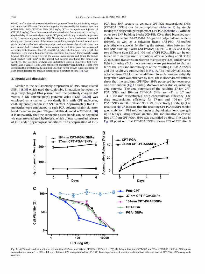
Fig. 2. (A) Time-dependent studies on the stability of 37-nm and 104-nm CPT-PGA3SNPsserum (human serum:1 � PBS ¼ 1:1, v/v). Released CPT was quantified by HPLC. (C) Dosecontrols.
PGA into SNP vectors to generate CPT-PGA encapsulated SNPs(CPT-PGA3SNPs) can be accomplished (Scheme 1) by simplymixing the drug conjugated polymer, CPT-PGA (Scheme 2), with theother two SNP building blocks (CD-PEI: CD-grafted branched pol-yethylenimine and Ad-PAMAM: Ad-grafted polyamidoamine den-drimer), as well as a solvation ligand (Ad-PEG: Ad-graftedpoly(ethylene glycol)). By altering the mixing ratios between thetwo SNP building blocks (Ad-PAMAM/CD-PEI ¼ 0.125 and 0.25),two different sizes (37 and 104 nm) of CPT-PGA3SNPs can be ob-tained with narrow size distributions after annealing at 50 �C for20 min. Both transmission electron microscopy (TEM) and dynamiclight scattering (DLS) measurements were performed to charac-terize the sizes and morphologies of the resulting CPT-PGA3SNPsand the results are summarized in Fig. 1A. The hydrodynamic sizesobtained from DLS for the two different formulations were slightlylarger thanwhatwas observed by TEM. These size characterizationsshow that the resulting CPT-PGA3SNPs possessed homogeneoussize distributions (Fig. 1B and C). Moreover, other studies, includingzeta potential (The zeta potentials of the resulting 37-nm CPT-PGA3SNPs and 104-nm CPT-PGA3SNPs are �11 � 0.7 and�4 � 0.2 mV, respectively.), drug encapsulation efficiency (Thedrug encapsulation efficiency for 37-nm and 104-nm CPT-PGA3SNPs are 90 � 3% and 95 � 2%, respectively.), stability (Theresults in Fig. 2A indicate that the resulting CPT-PGA3SNPs exhibitgood stability in PBS solution under a physiological ionic strengthup to 6 days.), drug release kinetics (The accumulative release offree CPT from CPT-PGA3SNPs was quantified by HPLC. The data inFig. 2B point out that CPT-PGA3SNPs release 20% of CPT after 6
Days
0 2 4 6
Ac
cu
mu
la
tiv
e R
ele
as
e (%
)
37-nm CPT-PGA SNPs
CPT-PGA
0
20
40
Free CPT
37-nm CPT-PGA SNPs
104-nm CPT-PGA SNPs
CPT-PGA
PGA SNPs
000
in 1 � PBS. (B) Release kinetics of CPT-PGA and 37-nm CPT-PGA3SNPs in 50% human-dependent cell viability studies of two different sizes of CPT-PGA3SNPs along with
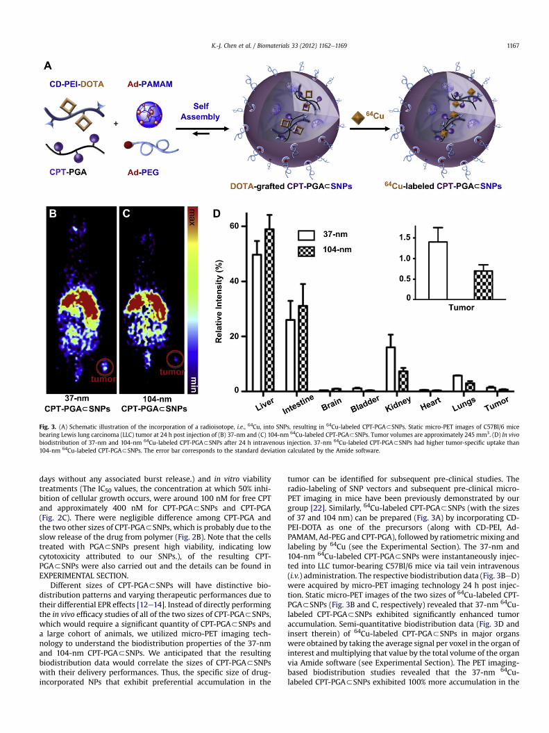
Fig. 3. (A) Schematic illustration of the incorporation of a radioisotope, i.e., 64Cu, into SNPs, resulting in 64Cu-labeled CPT-PGA3SNPs. Static micro-PET images of C57Bl/6 micebearing Lewis lung carcinoma (LLC) tumor at 24 h post injection of (B) 37-nm and (C) 104-nm 64Cu-labeled CPT-PGA3SNPs. Tumor volumes are approximately 245 mm3. (D) In vivobiodistribution of 37-nm and 104-nm 64Cu-labeled CPT-PGA3SNPs after 24 h intravenous injection. 37-nm 64Cu-labeled CPT-PGA3SNPs had higher tumor-specific uptake than104-nm 64Cu-labeled CPT-PGA3SNPs. The error bar corresponds to the standard deviation calculated by the Amide software.
K.-J. Chen et al. / Biomaterials 33 (2012) 1162e1169 1167
days without any associated burst release.) and in vitro viabilitytreatments (The IC50 values, the concentration at which 50% inhi-bition of cellular growth occurs, were around 100 nM for free CPTand approximately 400 nM for CPT-PGA3SNPs and CPT-PGA(Fig. 2C). There were negligible difference among CPT-PGA andthe two other sizes of CPT-PGA3SNPs, which is probably due to theslow release of the drug from polymer (Fig. 2B). Note that the cellstreated with PGA3SNPs present high viability, indicating lowcytotoxicity attributed to our SNPs.), of the resulting CPT-PGA3SNPs were also carried out and the details can be found inEXPERIMENTAL SECTION.
Different sizes of CPT-PGA3SNPs will have distinctive bio-distribution patterns and varying therapeutic performances due totheir differential EPR effects [12e14]. Instead of directly performingthe in vivo efficacy studies of all of the two sizes of CPT-PGA3SNPs,which would require a significant quantity of CPT-PGA3SNPs anda large cohort of animals, we utilized micro-PET imaging tech-nology to understand the biodistribution properties of the 37-nmand 104-nm CPT-PGA3SNPs. We anticipated that the resultingbiodistribution data would correlate the sizes of CPT-PGA3SNPswith their delivery performances. Thus, the specific size of drug-incorporated NPs that exhibit preferential accumulation in the
tumor can be identified for subsequent pre-clinical studies. Theradio-labeling of SNP vectors and subsequent pre-clinical micro-PET imaging in mice have been previously demonstrated by ourgroup [22]. Similarly, 64Cu-labeled CPT-PGA3SNPs (with the sizesof 37 and 104 nm) can be prepared (Fig. 3A) by incorporating CD-PEI-DOTA as one of the precursors (along with CD-PEI, Ad-PAMAM, Ad-PEG and CPT-PGA), followed by ratiometric mixing andlabeling by 64Cu (see the Experimental Section). The 37-nm and104-nm 64Cu-labeled CPT-PGA3SNPs were instantaneously injec-ted into LLC tumor-bearing C57Bl/6 mice via tail vein intravenous(i.v.) administration. The respective biodistribution data (Fig. 3BeD)were acquired by micro-PET imaging technology 24 h post injec-tion. Static micro-PET images of the two sizes of 64Cu-labeled CPT-PGA3SNPs (Fig. 3B and C, respectively) revealed that 37-nm 64Cu-labeled CPT-PGA3SNPs exhibited significantly enhanced tumoraccumulation. Semi-quantitative biodistribution data (Fig. 3D andinsert therein) of 64Cu-labeled CPT-PGA3SNPs in major organswere obtained by taking the average signal per voxel in the organ ofinterest and multiplying that value by the total volume of the organvia Amide software (see Experimental Section). The PET imaging-based biodistribution studies revealed that the 37-nm 64Cu-labeled CPT-PGA3SNPs exhibited 100% more accumulation in the
Med
ian
T
um
or (m
g)
A
Days
0 5 10
1000
500
1500
0
PBS
37-nm
CPT-PGA SNPs
37-nm PGA SNPs
CPT
Days
0 5 10
PBS
37-nm
CPT-PGA SNPs
37-nm PGA SNPs
CPT
Bo
dy W
eig
ht C
ha
ng
e (%
)
-10
0
10
20B
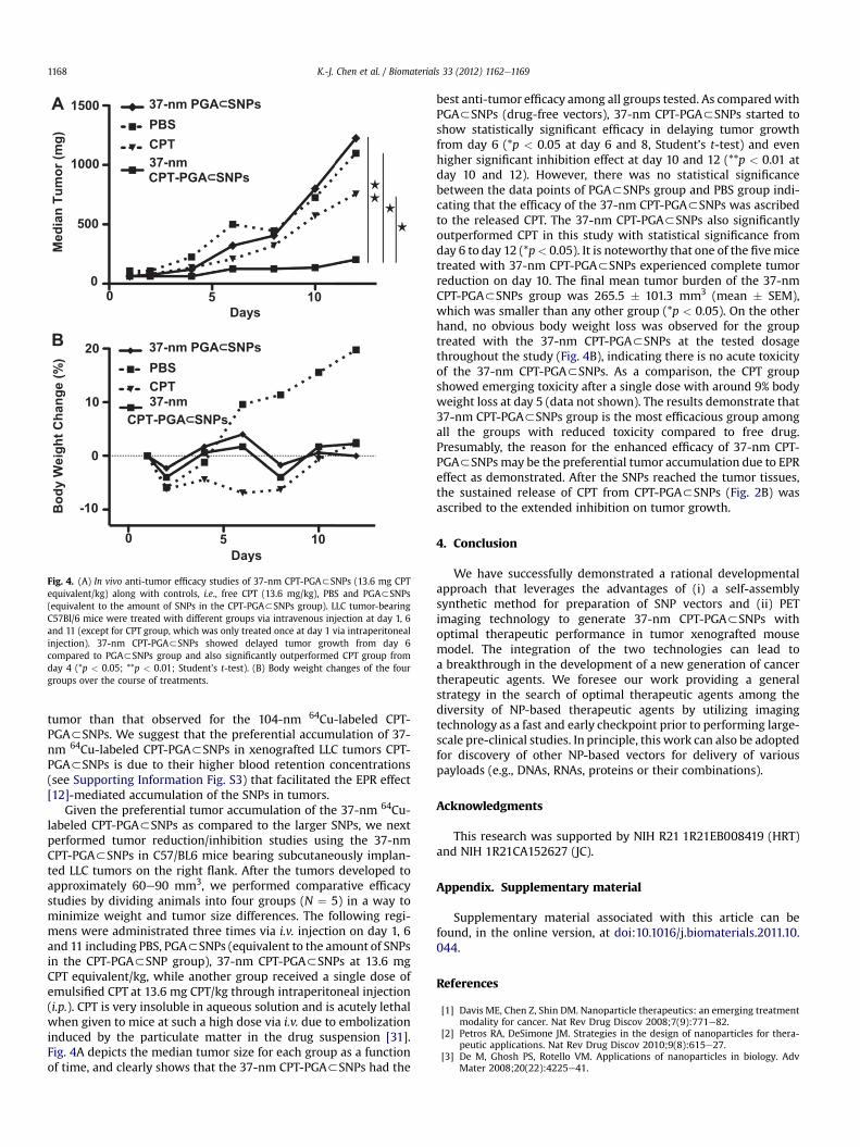
Fig. 4. (A) In vivo anti-tumor efficacy studies of 37-nm CPT-PGA3SNPs (13.6 mg CPTequivalent/kg) along with controls, i.e., free CPT (13.6 mg/kg), PBS and PGA3SNPs(equivalent to the amount of SNPs in the CPT-PGA3SNPs group). LLC tumor-bearingC57Bl/6 mice were treated with different groups via intravenous injection at day 1, 6and 11 (except for CPT group, which was only treated once at day 1 via intraperitonealinjection). 37-nm CPT-PGA3SNPs showed delayed tumor growth from day 6compared to PGA3SNPs group and also significantly outperformed CPT group fromday 4 (*p < 0.05; **p < 0.01; Student’s t-test). (B) Body weight changes of the fourgroups over the course of treatments.
K.-J. Chen et al. / Biomaterials 33 (2012) 1162e11691168
tumor than that observed for the 104-nm 64Cu-labeled CPT-PGA3SNPs. We suggest that the preferential accumulation of 37-nm 64Cu-labeled CPT-PGA3SNPs in xenografted LLC tumors CPT-PGA3SNPs is due to their higher blood retention concentrations(see Supporting Information Fig. S3) that facilitated the EPR effect[12]-mediated accumulation of the SNPs in tumors.
Given the preferential tumor accumulation of the 37-nm 64Cu-labeled CPT-PGA3SNPs as compared to the larger SNPs, we nextperformed tumor reduction/inhibition studies using the 37-nmCPT-PGA3SNPs in C57/BL6 mice bearing subcutaneously implan-ted LLC tumors on the right flank. After the tumors developed toapproximately 60e90 mm3, we performed comparative efficacystudies by dividing animals into four groups (N ¼ 5) in a way tominimize weight and tumor size differences. The following regi-mens were administrated three times via i.v. injection on day 1, 6and 11 including PBS, PGA3SNPs (equivalent to the amount of SNPsin the CPT-PGA3SNP group), 37-nm CPT-PGA3SNPs at 13.6 mgCPT equivalent/kg, while another group received a single dose ofemulsified CPT at 13.6 mg CPT/kg through intraperitoneal injection(i.p.). CPT is very insoluble in aqueous solution and is acutely lethalwhen given to mice at such a high dose via i.v. due to embolizationinduced by the particulate matter in the drug suspension [31].Fig. 4A depicts the median tumor size for each group as a functionof time, and clearly shows that the 37-nm CPT-PGA3SNPs had the
best anti-tumor efficacy among all groups tested. As comparedwithPGA3SNPs (drug-free vectors), 37-nm CPT-PGA3SNPs started toshow statistically significant efficacy in delaying tumor growthfrom day 6 (*p < 0.05 at day 6 and 8, Student’s t-test) and evenhigher significant inhibition effect at day 10 and 12 (**p < 0.01 atday 10 and 12). However, there was no statistical significancebetween the data points of PGA3SNPs group and PBS group indi-cating that the efficacy of the 37-nm CPT-PGA3SNPs was ascribedto the released CPT. The 37-nm CPT-PGA3SNPs also significantlyoutperformed CPT in this study with statistical significance fromday 6 to day 12 (*p< 0.05). It is noteworthy that one of the fivemicetreated with 37-nm CPT-PGA3SNPs experienced complete tumorreduction on day 10. The final mean tumor burden of the 37-nmCPT-PGA3SNPs group was 265.5 � 101.3 mm3 (mean � SEM),which was smaller than any other group (*p < 0.05). On the otherhand, no obvious body weight loss was observed for the grouptreated with the 37-nm CPT-PGA3SNPs at the tested dosagethroughout the study (Fig. 4B), indicating there is no acute toxicityof the 37-nm CPT-PGA3SNPs. As a comparison, the CPT groupshowed emerging toxicity after a single dose with around 9% bodyweight loss at day 5 (data not shown). The results demonstrate that37-nm CPT-PGA3SNPs group is the most efficacious group amongall the groups with reduced toxicity compared to free drug.Presumably, the reason for the enhanced efficacy of 37-nm CPT-PGA3SNPsmay be the preferential tumor accumulation due to EPReffect as demonstrated. After the SNPs reached the tumor tissues,the sustained release of CPT from CPT-PGA3SNPs (Fig. 2B) wasascribed to the extended inhibition on tumor growth.
4. Conclusion
We have successfully demonstrated a rational developmentalapproach that leverages the advantages of (i) a self-assemblysynthetic method for preparation of SNP vectors and (ii) PETimaging technology to generate 37-nm CPT-PGA3SNPs withoptimal therapeutic performance in tumor xenografted mousemodel. The integration of the two technologies can lead toa breakthrough in the development of a new generation of cancertherapeutic agents. We foresee our work providing a generalstrategy in the search of optimal therapeutic agents among thediversity of NP-based therapeutic agents by utilizing imagingtechnology as a fast and early checkpoint prior to performing large-scale pre-clinical studies. In principle, this work can also be adoptedfor discovery of other NP-based vectors for delivery of variouspayloads (e.g., DNAs, RNAs, proteins or their combinations).
Acknowledgments
This research was supported by NIH R21 1R21EB008419 (HRT)and NIH 1R21CA152627 (JC).
Appendix. Supplementary material
Supplementary material associated with this article can befound, in the online version, at doi:10.1016/j.biomaterials.2011.10.044.
References
[1] Davis ME, Chen Z, Shin DM. Nanoparticle therapeutics: an emerging treatmentmodality for cancer. Nat Rev Drug Discov 2008;7(9):771e82.
[2] Petros RA, DeSimone JM. Strategies in the design of nanoparticles for thera-peutic applications. Nat Rev Drug Discov 2010;9(8):615e27.
[3] De M, Ghosh PS, Rotello VM. Applications of nanoparticles in biology. AdvMater 2008;20(22):4225e41.
K.-J. Chen et al. / Biomaterials 33 (2012) 1162e1169 1169
[4] Bae Y, Kataoka K. Intelligent polymeric micelles from functional poly(ethyleneglycol)-poly(amino acid) block copolymers. Adv Drug Deliv Rev 2009;61(10):768e84.
[5] Wiradharma N, Zhang Y, Venkataraman S, Hedrick JL, Yang YY. Self-assembledpolymer nanostructures for delivery of anticancer therapeutics. NanoToday;4(4):302e317.
[6] Duncan B, Kim C, Rotello VM. Gold nanoparticle platforms as drug and bio-macromolecule delivery systems. J Control Release 2010;148(1):122e7.
[7] Meng HA, Liong M, Xia TA, Li ZX, Ji ZX, Zink JI, et al. Engineered design ofmesoporous silica nanoparticles to deliver doxorubicin and p-glycoproteinsiRNA to overcome drug resistance in a cancer cell line. ACS Nano 2010;4(8):4539e50.
[8] Nguyen HN, Wey SP, Juang JH, Sonaje K, Ho YC, Chuang EY, et al. The glucose-lowering potential of exendin-4 orally delivered via a pH-sensitive nano-particle vehicle and effects on subsequent insulin secretion in vivo. Bioma-terials 2011;32(10):2673e82.
[9] Sherlock SP, Tabakman SM, Xie LM, Dai HJ. Photothermally enhanced drugdelivery by ultrasmall multifunctional FeCo/graphitic shell nanocrystals. ACSNano 2011;5(2):1505e12.
[10] Chow EK, Zhang X-Q, Chen M, Lam R, Robinson E, Huang H, et al. Nano-diamond therapeutic delivery agents mediate enhanced chemoresistanttumor treatment. Sci Transl Med 2011;3:73.
[11] Peer D, Karp JM, Hong S, FaroKHzad OC, Margalit R, Langer R. Nanocarriers asan emerging platform for cancer therapy. Nat Nanotechnol 2007;2(12):751e60.
[12] Maeda H, Wu J, Sawa T, Matsumura Y, Hori K. Tumor vascular permeabilityand the EPR effect in macromolecular therapeutics: a review. J Control Release2000;65(1e2):271e84.
[13] Weissig V, Whiteman KR, Torchilin VP. Accumulation of protein-loaded long-circulating micelles and liposomes in subcutaneous Lewis lung carcinoma inmice. Pharm Res-Dordr 1998;15(10):1552e6.
[14] Decuzzi P, Godin B, Tanaka T, Lee SY, Chiappini C, Liu X, et al. Size and shapeeffects in the biodistribution of intravascularly injected particles. J ControlRelease 2010;141(3):320e7.
[15] Perrault SD, Walkey C, Jennings T, Fischer HC, Chan WCW. Mediating tumortargeting efficiency of nanoparticles through design. Nano Lett 2009;9(5):1909e15.
[16] Matsumura Y, Maeda H. A new concept for macromolecular therapeutics incancer-chemotherapy e mechanism of tumoritropic accumulation of proteinsand the antitumor agent smancs. Cancer Res 1986;46(12):6387e92.
[17] Goodman TT, Olive PL, Pun SH. Increased nanoparticle penetration incollagenase-treated multicellular spheroids. Int J Nanomedicine 2007;2:265.
[18] Wang H, Liu K, Chen K-J, Lu Y, Wang S, Lin WY, et al. A rapid pathway towarda superb gene delivery system: programming structural and functionaldiversity into a supramolecular nanoparticle library. ACS Nano 2010;4(10):6235e43.
[19] Wang H, Chen K-J, Wang S, Ohashi M, Kamei K-I, Sun J, et al. A small library ofDNA-encapsulated supramolecular nanoparticles for targeted gene delivery.Chem Commun 2010;45:1851.
[20] Liu Y, Wang H, Kamei K, Yan M, Chen K-J, Yuan Q, et al. Delivery of intacttranscription factor using self-assembled supramolecular nanoparticles.Angew Chem Int Ed 2011;50:3058.
[21] Wang S, Chen K-J, Wu T-H, Wang H, Lin W-Y, Ohashi M, et al. Photothermaleffects of supramolecularly assembled gold nanoparticles for the targetedtreatment of cancer cells. Angew Chem Int Ed 2010;49(22):3777e81.
[22] Wang H, Wang ST, Su H, Chen K-J, Armijo AL, Lin WY, et al. A supramolecularapproach for preparation of size-controlled nanoparticles. Angew Chem Int Ed2009;48(24):4344e8.
[23] Chen K-J, Wolahan MS, Wang H, Hsu C-H, Chang H-W, Durazo A, et al. A smallMRI contrast agent library of gadolinium(III)-encapsulated supramolecularnanoparticles for improved relaxivity and sensitivity. Biomaterials 2011;32(8):2160e5.
[24] Phelps ME. Positron emission tomography provides molecular imaging ofbiological processes. Proc Nat Acad Sci 2000;97(16):9226e33.
[25] Czernin J, Phelps ME. Positron emission tomography scanning: current andfuture applications. Annu Rev Med 2002;53:89e112.
[26] Phelps ME. PET: molecular imaging and its biological applications. New York:Springer; 2004.
[27] Li SD, Huang L. Pharmacokinetics and biodistribution of nanoparticles. MolPharmaceut 2008;5(4):496e504.
[28] Lu H, Cheng JJ. Hexamethyldisilazane-mediated controlled polymerization ofalpha-amino acid N-carboxyanhydrides. J Am Chem Soc 2007;129(46):14114e5.
[29] Lu H, Wang J, Bai Y, Lang J, Liu S, Lin Y, et al. Ionic polypeptides with unusualhelical stability. Nat Commun 2011;2:206.
[30] Bhatt RL, de Vries P, Tulinsky J, Bellamy G, Baker B, Singer JW, et al. Synthesisand in vivo antitumor activity of poly(L-glutamic acid) conjugates of 20(S)-camptothecin. J Med Chem 2003;46(1):190e3.
[31] Cheng J, Khin KT, Davis ME. Antitumor activity of beta-cyclodextrin polymer -camptothecin conjugates. Mol Pharmaceut 2004;1(3):183e93.