Embed Size (px)
Citation preview
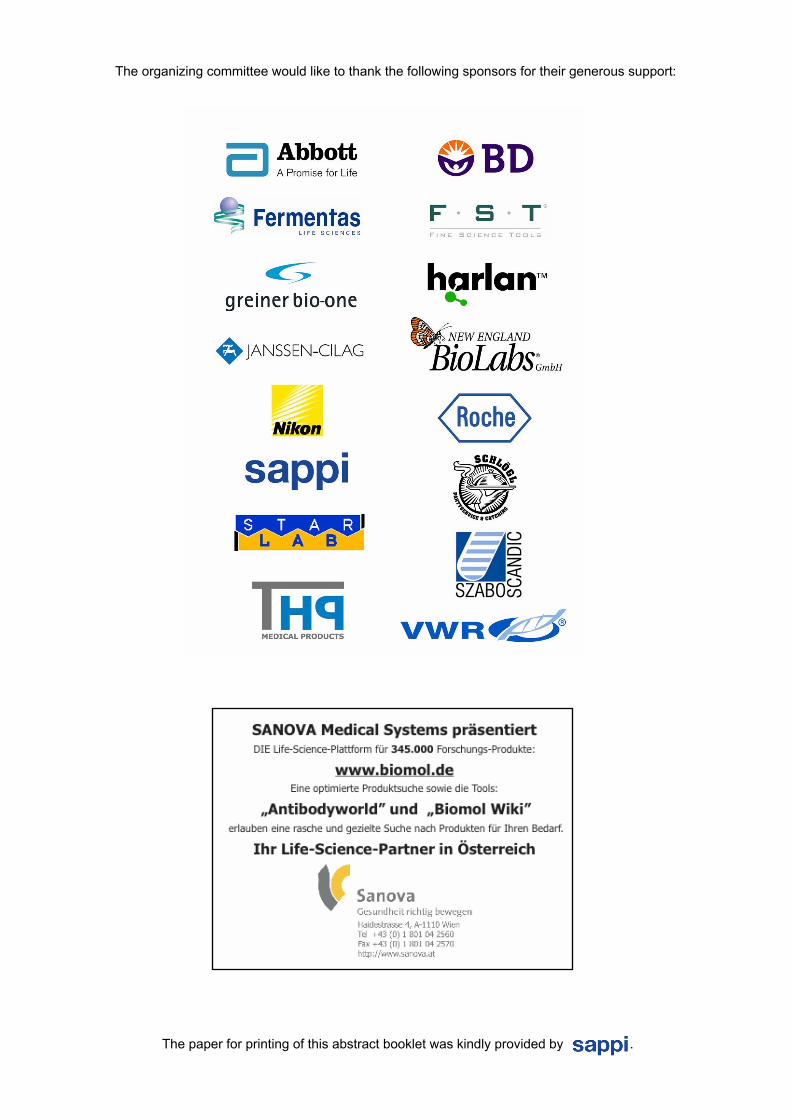
The organizing committee would like to thank the following sponsors for their generous support:
The paper for printing of this abstract booklet was kindly provided by .
acknowledgements
2
Organizing Committee
Anastasia Abramova
Thomas Bauer
Barbara Drobits
Wolf Henning Gebhardt
Madeleine Kalb
Afitap Derya Köprülü
Katarzyna Niespodziana
Haley Ramsey
Véronique Schulten
Oksana Vratskikh
Medical University of Vienna, Vienna, Austria
Speakers
Steve Anderton
Thomas Decker
Alexander von Gabain
Florian Greten
Roy Jefferis
Greg Lemke
Ravinder Maini
Frank Nestle
Martin Oft
Josef Penninger
Antal Rot
Georg Stingl
Erwin Wagner
Administration
Marianne Wang
Special thanks to
Maria Sibilia
TABLE OF CONTENTS
Acknowledgements p. 2
PhD Program Inflammation and Immunity p. 3
Greetings p. 4
Scientific Program p. 6
Abstracts p. 8
Congress Information p. 22
phd program inflammation and immunity
3
The PhD and MD/PhD Program Inflammation and Immunity (IAI) aims at revealing novel mechanisms
controlling the development and function of immune cells in health and disease and train excellent young
researchers with a new qualification profile in basic, translational and clinical research. The IAI PhD program is
closely linked to two Special Research Programs (SFB-F18 and SFB-F23) funded by the Austrian Science
Fund (FWF) and several Austrian (GEN-AU, CDL) and European (NoE, RTN, Strep) networks.
Our overall scientific objective is to understand the detailed events and molecular mechanisms
associated with inflammatory and immunological diseases. Genes, molecules, isolated cells and tissues as well
as whole organisms are all being exploited as model systems. Thereby mouse immunologists will closely
interact with human immunologists and establish a close interaction between basic and clinical science. We
provide synergistic expertise in the fields of molecular biology, cell biology, mouse genetics, immunology,
allergology, infectiology and immunopharmacology, also at the translational level. The major critical advantage
of working in this interactive network will be the transfer of molecular observations made in vitro to the clinical
treatment of immune and inflammatory diseases in patients. Access to patient material will be provided by the
clinical research groups present within the IAI program. This interface between basic and clinical research is of
particular importance for the success of the IAI PhD program.
The IAI PhD Program will concentrate its research efforts in the following four areas:
* Basic aspects of Immunity
* Inflammatory diseases
* Infectiology and Vaccinology
* Allergy, Hypersensitivity and Transplantation
Our scientific goals will be implemented by a state-of-the art career development plan which includes
comprehensive educational training in the field of IAI, special lectures on career awareness, collaborative
programs with international universities and research institutions, exchange of research tools and technologies,
teaching and special scientific workshops. This will offer IAI PhD students all the prerequisites for a future
successful career in academia, industry or any field of health care.
http://www.meduniwien.ac.at/phd-iai/ Email: [email protected]
greetings
4
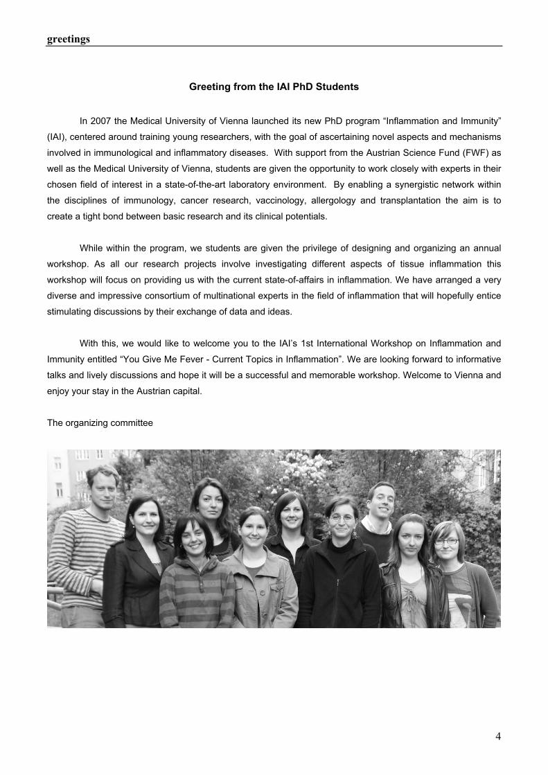
Greeting from the IAI PhD Students
In 2007 the Medical University of Vienna launched its new PhD program “Inflammation and Immunity”
(IAI), centered around training young researchers, with the goal of ascertaining novel aspects and mechanisms
involved in immunological and inflammatory diseases. With support from the Austrian Science Fund (FWF) as
well as the Medical University of Vienna, students are given the opportunity to work closely with experts in their
chosen field of interest in a state-of-the-art laboratory environment. By enabling a synergistic network within
the disciplines of immunology, cancer research, vaccinology, allergology and transplantation the aim is to
create a tight bond between basic research and its clinical potentials.
While within the program, we students are given the privilege of designing and organizing an annual
workshop. As all our research projects involve investigating different aspects of tissue inflammation this
workshop will focus on providing us with the current state-of-affairs in inflammation. We have arranged a very
diverse and impressive consortium of multinational experts in the field of inflammation that will hopefully entice
stimulating discussions by their exchange of data and ideas.
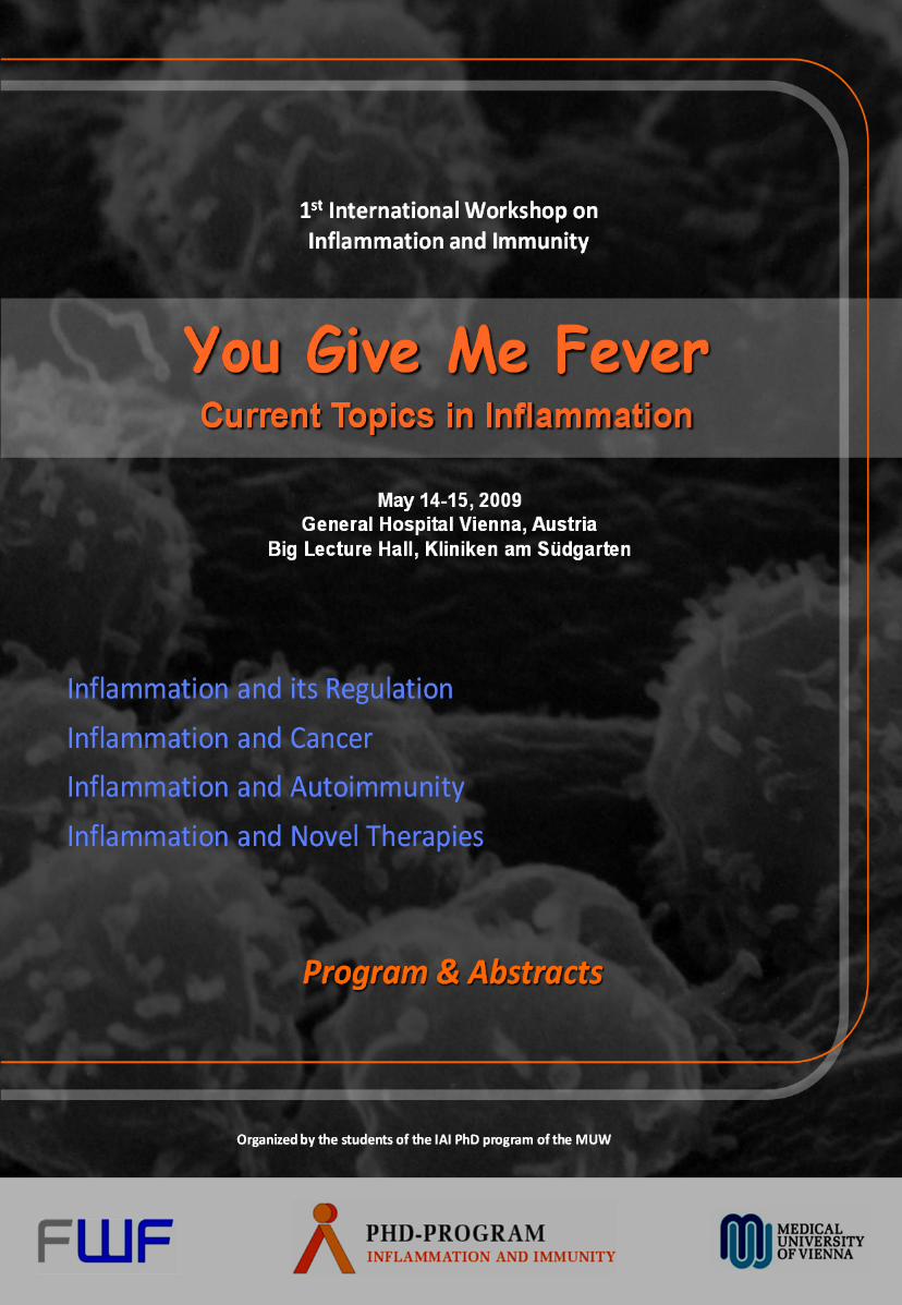
With this, we would like to welcome you to the IAI’s 1st International Workshop on Inflammation and
Immunity entitled “You Give Me Fever - Current Topics in Inflammation”. We are looking forward to informative
talks and lively discussions and hope it will be a successful and memorable workshop. Welcome to Vienna and
enjoy your stay in the Austrian capital.
The organizing committee
greetings
5
Welcome to the 1st International Workshop of the Doctoral Program Inflammation and Immunity (IAI)
"You Give Me Fever: Current Topics in Inflammation"
About two years ago the Doctoral Program “Inflammation and Immunity“ (IAI) has been implemented as
an educational excellence platform in basic medical research at the Medical University of Vienna (MUW). Since
then the MUW as a major European Center of biomedical research and education provides the infrastructure
for students from all over the world. Meanwhile 18 internationally recruited PhD students from 8 countries have
been accepted into the IAI program and they are conducting their research in the laboratories of the 8 Principal
Investigators (PI) that are part of the IAI PhD program. All the PIs hold positions as professors or associated
professors and are affiliated with six different Departments of the MUW. Our aim is to strengthen the research
interactions and to extend our collaborative network beyond laboratory walls and different research disciplines,
as well as language gaps and cultural differences. This workshop is part of our endeavor.
The topic chosen by our students for the 1st international workshop is Inflammation. The
research in this area is of worldwide medical need and has potential to accelerate the process from basic
discoveries to new therapeutic strategies leading to prevention, diagnosis and treatment of various diseases
including cancer, rheumatoid arthritis and psoriasis, just to name a few. The students have invited
internationally well-known experts and world leading scientists in the field and we are looking forward to an
active exchange of opinions, stimulating discussions and exciting scientific interactions for the next two days.
In the name of all PIs I would like to thank the students for putting together such an exciting
program and to all the speakers for accepting the invitation and traveling to Vienna to make this event possible.
I wish all the IAI students and all participants of the workshop two exciting days.
Finally, we would like to express our gratitude towards the Austrian Science Fund (FWF) and
the MUW for financing and supporting our PhD program and to all companies for co-sponsoring this workshop.
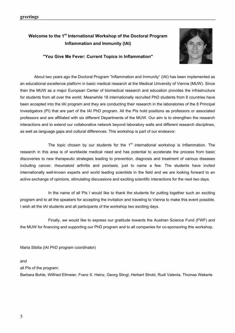
Maria Sibilia (IAI PhD program coordinator)
and
all PIs of the program:
Barbara Bohle, Wilfried Ellmeier, Franz X. Heinz, Georg Stingl, Herbert Strobl, Rudi Valenta, Thomas Wekerle

scientific program
6
“You Give Me Fever: Current Topics in Inflammation”
Thursday, May 14, 2009
08:30 – 09:00 Registration
09:00 – 09:15 Welcome
OPENING LECTURE Chair: Maria Sibilia
09:15 – 10:00 Inflammatory and neoplastic skin diseases: New insights – new therapies Georg Stingl
SESSION I: INFLAMMATION AND ITS REGULATION
Chair: Véronique Schulten, Afitap Derya Köprülü
10:00 – 10:45 Bones and fever regulation Josef Penninger
11:00 – 11:15 Coffee Break
11:15 – 12:00 Interferons and Stats in infection, inflammation and the onset of adaptive immunity Thomas Decker
12:15 – 13:00 Chemokine patterning by glycosaminoglycans and interceptors:
Chemokinese phrase and fable in vivo Antal Rot
13:15 – 14:30 Lunch Break (poster session: IAI-PhD students and speakers)
SESSION II: INFLAMMATION AND CANCER Chair: Barbara Drobits, Oksana Vratskikh
14:30 – 15:15 Molecular mechanisms linking inflammation & cancer Florian R. Greten
15:30 – 16:15 AP-1 (Fos/Jun) in inflammation and cancer development Erwin F. Wagner
16:30 – 16:45 Coffee Break
16:45 – 17:30 Friends or Foes - tumor promoting or tumor eliminating
types of inflammation Martin Oft
scientific program
7
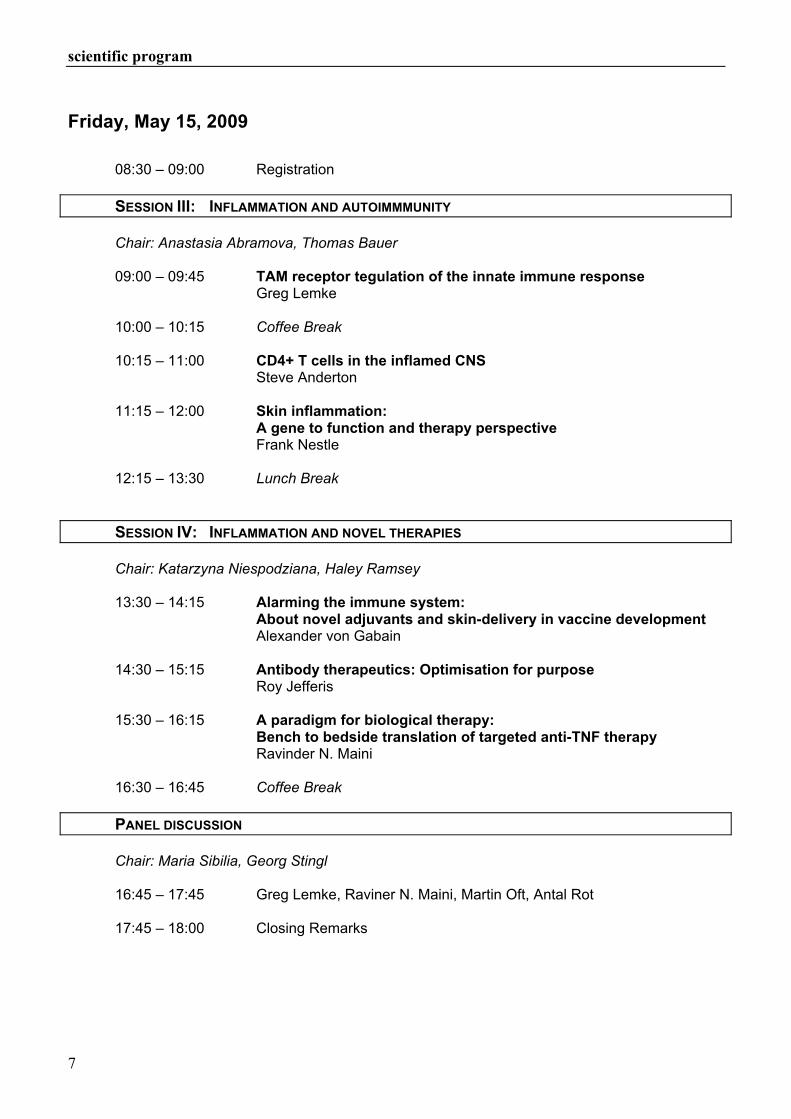
Friday, May 15, 2009
08:30 – 09:00 Registration
SESSION III: INFLAMMATION AND AUTOIMMMUNITY Chair: Anastasia Abramova, Thomas Bauer
09:00 – 09:45 TAM receptor tegulation of the innate immune response Greg Lemke
10:00 – 10:15 Coffee Break
10:15 – 11:00 CD4+ T cells in the inflamed CNS Steve Anderton
11:15 – 12:00 Skin inflammation:
A gene to function and therapy perspective Frank Nestle
12:15 – 13:30 Lunch Break
SESSION IV: INFLAMMATION AND NOVEL THERAPIES Chair: Katarzyna Niespodziana, Haley Ramsey
13:30 – 14:15 Alarming the immune system: About novel adjuvants and skin-delivery in vaccine development
Alexander von Gabain
14:30 – 15:15 Antibody therapeutics: Optimisation for purpose Roy Jefferis
15:30 – 16:15 A paradigm for biological therapy:
Bench to bedside translation of targeted anti-TNF therapy Ravinder N. Maini
16:30 – 16:45 Coffee Break
PANEL DISCUSSION
Chair: Maria Sibilia, Georg Stingl
16:45 – 17:45 Greg Lemke, Raviner N. Maini, Martin Oft, Antal Rot
17:45 – 18:00 Closing Remarks

abstracts
8
INFLAMMATORY AND NEOPLASTIC SKIN DISEASES:
NEW INSIGHTS – NEW THERAPIES
Georg Stingl
Department of Dermatology, Division of Immunology, Allergy and Infectious Diseases Medical University of Vienna, Vienna, Austria
The skin, the outermost layer of the body, is built in a way that allows it to protect the integrity of the
host and, at the same time, to serve as bridge and communication site between inside and outside. This barrier
function of the skin has a physical, a chemical, and an immunological component. Upon receipt of a danger
signal or under certain pathological conditions, the skin is quickly populated by cells of the innate and adaptive
immune system which, by the production of different types of mediators, give rise to inflammation and, as a
consequence, to tissue damage and repair. Research efforts in the last decade have provided insights into the pathogenesis of the most common
inflammatory skin diseases which, at least in the case of psoriasis, have provided the basis for entirely novel
treatment strategies that, in the meantime, have revolutionized our therapeutic armamentarium. The opposite situation is true in skin cancer. Not only do cancer cells proliferate per se in an uninhibited
fashion, but they are also not constrained by appropriate host defense mechanisms. We now have
immunostimulatory compounds available that, upon topical application, induce tissue inflammation and endow
immunocytes with cytodestructive effector functions that can contribute to cancer regression and perhaps also
to the elimination of virus-infected cells. This etiology- and pathogenesis-based approach to drug development will certainly result in the
increased production of therapeutic tools of high efficacy as well as good tolerability that are tailored to the
needs of the individualized patient.

abstracts
9
BONES AND FEVER REGULATION
Josef Penninger
IMBA, Institute for Molecular Biotechnology of the Austrian Academy of Sciences,
Vienna, Austria
RANKL and its receptor RANK are the key regulators of osteoclastogenesis also involved in lymph
node formation and development of thymic epithelial cells. Moreover, new medicine based on RANKL inhibition
is on the verge of wide-spread human use making it paramount to identify potential additional functions for
RANKL. I will discuss the discovery of RANKL, the basic principles of RANKL/RANK function in bone
metabolism and report a novel RANKL-RANK function in the brain
abstracts
10
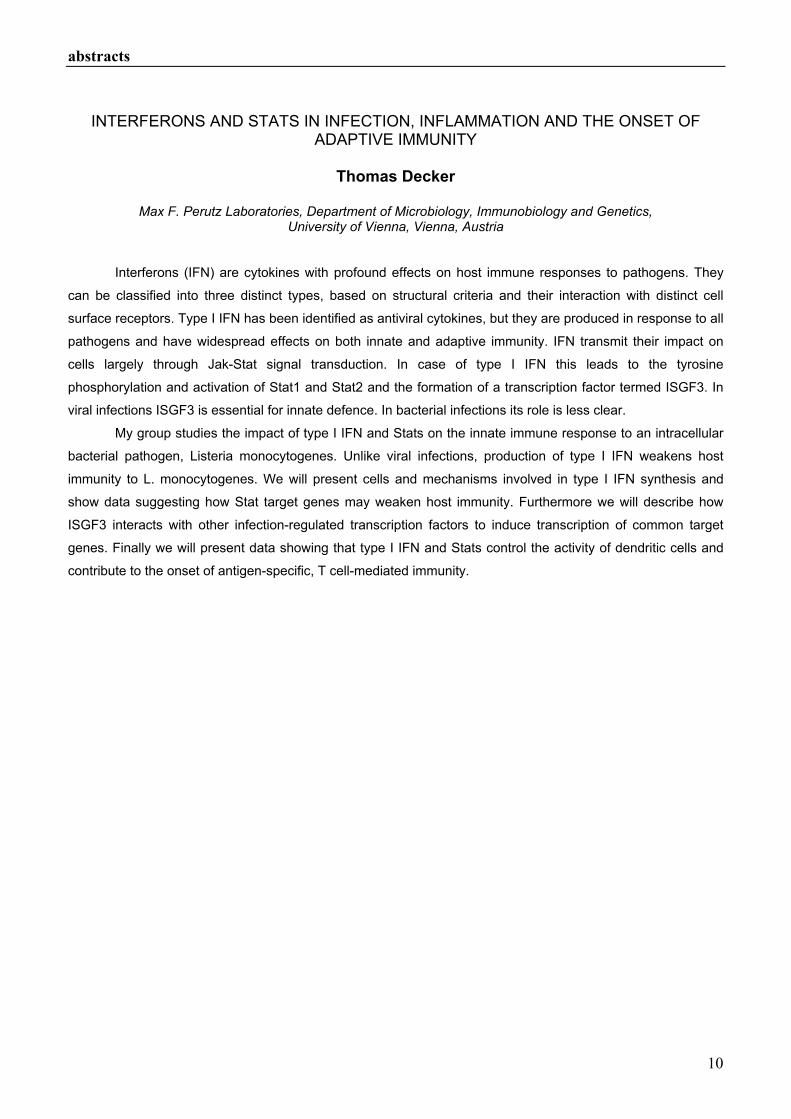
INTERFERONS AND STATS IN INFECTION, INFLAMMATION AND THE ONSET OF
ADAPTIVE IMMUNITY
Thomas Decker
Max F. Perutz Laboratories, Department of Microbiology, Immunobiology and Genetics, University of Vienna, Vienna, Austria
Interferons (IFN) are cytokines with profound effects on host immune responses to pathogens. They
can be classified into three distinct types, based on structural criteria and their interaction with distinct cell
surface receptors. Type I IFN has been identified as antiviral cytokines, but they are produced in response to all
pathogens and have widespread effects on both innate and adaptive immunity. IFN transmit their impact on
cells largely through Jak-Stat signal transduction. In case of type I IFN this leads to the tyrosine
phosphorylation and activation of Stat1 and Stat2 and the formation of a transcription factor termed ISGF3. In
viral infections ISGF3 is essential for innate defence. In bacterial infections its role is less clear.
My group studies the impact of type I IFN and Stats on the innate immune response to an intracellular
bacterial pathogen, Listeria monocytogenes. Unlike viral infections, production of type I IFN weakens host
immunity to L. monocytogenes. We will present cells and mechanisms involved in type I IFN synthesis and
show data suggesting how Stat target genes may weaken host immunity. Furthermore we will describe how
ISGF3 interacts with other infection-regulated transcription factors to induce transcription of common target
genes. Finally we will present data showing that type I IFN and Stats control the activity of dendritic cells and
contribute to the onset of antigen-specific, T cell-mediated immunity.
abstracts
11
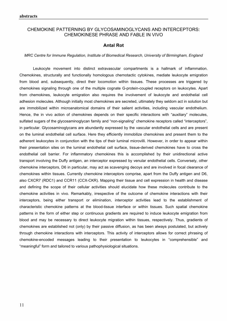
CHEMOKINE PATTERNING BY GLYCOSAMINOGLYCANS AND INTERCEPTORS: CHEMOKINESE PHRASE AND FABLE IN VIVO
Antal Rot
MRC Centre for Immune Regulation, Institute of Biomedical Research, University of Birmingham, England
Leukocyte movement into distinct extravascular compartments is a hallmark of inflammation.
Chemokines, structurally and functionally homologous chemotactic cytokines, mediate leukocyte emigration
from blood and, subsequently, direct their locomotion within tissues. These processes are triggered by
chemokines signaling through one of the multiple cognate G-protein-coupled receptors on leukocytes. Apart
from chemokines, leukocyte emigration also requires the involvement of leukocyte and endothelial cell
adhesion molecules. Although initially most chemokines are secreted, ultimately they seldom act in solution but
are immobilized within microanatomical domains of their salient activities, including vascular endothelium.
Hence, the in vivo action of chemokines depends on their specific interactions with “auxiliary” molecules,
sulfated sugars of the glycosaminoglycan family and “non-signaling” chemokine receptors called “interceptors”,
in particular. Glycosaminoglycans are abundantly expressed by the vascular endothelial cells and are present
on the luminal endothelial cell surface. Here they efficiently immobilize chemokines and present them to the
adherent leukocytes in conjunction with the tips of their luminal microvilli. However, in order to appear within
their presentation sites on the luminal endothelial cell surface, tissue-derived chemokines have to cross the
endothelial cell barrier. For inflammatory chemokines this is accomplished by their unidirectional active
transport involving the Duffy antigen, an interceptor expressed by venular endothelial cells. Conversely, other
chemokine interceptors, D6 in particular, may act as scavenging decoys and are involved in focal clearance of
chemokines within tissues. Currently chemokine interceptors comprise, apart from the Duffy antigen and D6,
also CXCR7 (RDC1) and CCR11 (CCX-CKR). Mapping their tissue and cell expression in health and disease
and defining the scope of their cellular activities should elucidate how these molecules contribute to the
chemokine activities in vivo. Remarkably, irrespective of the outcome of chemokine interactions with their
interceptors, being either transport or elimination, interceptor activities lead to the establishment of
characteristic chemokine patterns at the blood-tissue interface or within tissues. Such spatial chemokine
patterns in the form of either step or continuous gradients are required to induce leukocyte emigration from
blood and may be necessary to direct leukocyte migration within tissues, respectively. Thus, gradients of
chemokines are established not (only) by their passive diffusion, as has been always postulated, but actively
through chemokine interactions with interceptors. This activity of interceptors allows for correct phrasing of
chemokine-encoded messages leading to their presentation to leukocytes in “comprehensible” and
“meaningful” form and tailored to various pathophysiological situations.
abstracts
12
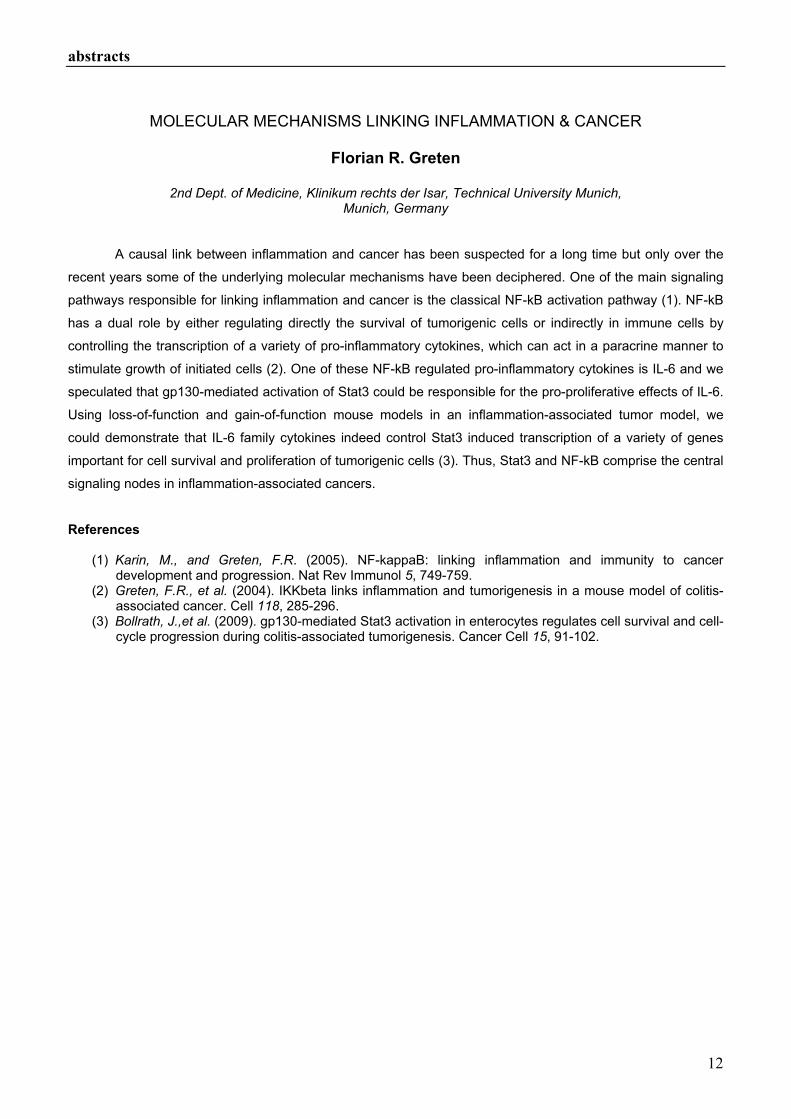
MOLECULAR MECHANISMS LINKING INFLAMMATION & CANCER
Florian R. Greten
2nd Dept. of Medicine, Klinikum rechts der Isar, Technical University Munich,
Munich, Germany
A causal link between inflammation and cancer has been suspected for a long time but only over the
recent years some of the underlying molecular mechanisms have been deciphered. One of the main signaling
pathways responsible for linking inflammation and cancer is the classical NF-kB activation pathway (1). NF-kB
has a dual role by either regulating directly the survival of tumorigenic cells or indirectly in immune cells by
controlling the transcription of a variety of pro-inflammatory cytokines, which can act in a paracrine manner to
stimulate growth of initiated cells (2). One of these NF-kB regulated pro-inflammatory cytokines is IL-6 and we
speculated that gp130-mediated activation of Stat3 could be responsible for the pro-proliferative effects of IL-6.
Using loss-of-function and gain-of-function mouse models in an inflammation-associated tumor model, we
could demonstrate that IL-6 family cytokines indeed control Stat3 induced transcription of a variety of genes
important for cell survival and proliferation of tumorigenic cells (3). Thus, Stat3 and NF-kB comprise the central
signaling nodes in inflammation-associated cancers.
References
(1) Karin, M., and Greten, F.R. (2005). NF-kappaB: linking inflammation and immunity to cancer development and progression. Nat Rev Immunol 5, 749-759.
(2) Greten, F.R., et al. (2004). IKKbeta links inflammation and tumorigenesis in a mouse model of colitis-associated cancer. Cell 118, 285-296.
(3) Bollrath, J.,et al. (2009). gp130-mediated Stat3 activation in enterocytes regulates cell survival and cell-cycle progression during colitis-associated tumorigenesis. Cancer Cell 15, 91-102.
abstracts
13
AP-1 (FOS/JUN) IN INFLAMMATION AND CANCER DEVELOPMENT
Erwin F. Wagner
CNIO, Spanish National Cancer Research Centre, E- 28029 Madrid, Spain
The analysis of the molecular factors determining cell survival or death in response to inflammatory
stimuli is essential for understanding the pathogenesis of inflammatory diseases including liver and skin cancer
and for identifying novel therapeutic approaches.
In hepatitis and liver cancer, but also during liver regeneration, the expression of Jun/AP-1 is critically
important. Using genetically modified alleles, we analyzed the Jun upstream kinase JNK and p38 as well as the
p53/TNFα pathway in liver inflammation and cancer. Jun N-terminal kinase (JNK) is a major mediator of
cytokine-induced cell death, whereas Jun functions as a survival gene in the Concanavalin A (ConA) model of
TNFα-dependent hepatitis(1). The functions of JNK1/2, p38α and Jun were investigated during liver cancer
development using the chemical-induced carcinogenesis model (2). Mice with liver-specific deletion of p38α
show enhanced tumor development, whereas increased proliferation of p38α-deficient hepatocytes and tumor
cells was suppressed by inactivation of JNK1 or c-Jun (3,4). These data demonstrate that while p38α MAPK
suppresses liver cancer development, JNK1 plays an oncogenic role. The impact on therapeutic applications as
well as the expression of components of this pathway in human hepatocellular carcinomas will be discussed.
In the second part I will illustrate how the mouse skin/epidermis has become an important model to
study the regulation and function of Fos and Jun subunits in physiological and disease processes. We
investigated the consequences of constitutive and inducible epidermal deletion of both Jun and JunB proteins
during development and in adult mice (5). Mice lacking Jun and JunB in epidermal cells die at post-natal day 1
with a phenotype similar to cachexia. Recent molecular analyses demonstrating that the mutant pups die from
a TNFα-dependent disease will be discussed, which contrasts to the psoriasis-like phenotype, when employing
an inducible, epidermal knock-out strategy (5). Overall, these data demonstrate that the stress-responsive AP-1
proteins are general regulators of innate inflammation control in the skin.
References
(1) Hasselblatt, P. et al. (2007) Hepatocyte survival during acute hepatitis is mediated by c-Jun/AP-1-dependent expression of inducible nitric oxide synthase PNAS 104, 17105-1710.
(2) Eferl, R. et al. (2003) Liver tumor development: c-Jun antagonizes the pro-apoptotic activity of p53. Cell 112, 181-192.
(3) Hui, L. et al. (2007) p38α suppresses normal and cancer cell proliferation by antagonizing the JNK-c-Jun pathway. Nature Genet.39, 741-749.
(4) Hui, L. et al. (2008) Proliferation of human HCC cells and chemically induced mouse liver cancers requires JNK1-dependent p21 downregulation. J Clin Invest.118, 3943-53.
(5) Zenz et al. (2005) Psoriasis-like skin disease and arthritis caused by inducible epidermal deletion of Jun proteins. Nature 437, 369-375.
abstracts
14
FRIENDS OR FOES - TUMOR PROMOTING OR TUMOR ELIMINATING
TYPES OF INFLAMMATION
Martin Oft
Department of Oncology, Schering-Plough Biopharma, Palo Alto, CA, USA
Chronic inflammation is epidemiologically associated with the cancer incidence in humans.
Inflammation also promotes tumor development in mouse models of human cancer. This tumor associated
inflammation has in its elements much in common with the deregulation of the immune system observed in
immune mediated inflammatory diseases. Inflammatory cytokines such as Interleukin 23, TNFα, IL-6 and IL-1
are highly expressed in human cancer, in particular at later stages of tumor progression. Human cytotoxic T
cells specific to oncogenes and onco-fetal antigens are present in human cancer patients but does not protect
from tumor progression. The presence of T cells in the tumor tissue however correlates with a favorable
prognosis for the patient. Yet, tumor infiltrating T cells frequently fail to express activation markers associated
with immune surveillance and MHC molecules are expressed at a low level in tumor cells and tumor infiltrating
dendritic cells. The immunological recognition of tumor antigens or the expansion of antigen specific T cell at
large might therefore not be as profoundly impaired in tumor patients as the correct polarization and the
effector function of tumor infiltrating T cells.
Mouse models of human cancer have recently shed some light on the molecular regulations for this
association of tumors with inflammation and a rationale how inflammation fuels tumor progression. Mice
deficient in IL-23, a cytokine essential for auto-inflammatory diseases, are also resistant to tumor induction by
carcinogens. The absence of IL-23 signaling not only suppresses the infiltration of inflammatory macrophages
and angiogenesis, but also increases the infiltration of cytotoxic CD8+ T cells and their activity in situ. Tumor
infiltrating T cells isolated from IL23R deficient are highly activated. Surprisingly, short term neutralization of IL-
23 repolarizes the deregulated tumor immunity, increasing the infiltration and activity of CD8 T cells.
We will present further data how the modulation of other pro-inflammatory cytokines can induce both:
the inhibition of tumor associated inflammation and the recovery of cytotoxic T cell responses.
abstracts
15
TAM RECEPTOR REGULATION OF THE INNATE IMMUNE RESPONSE
Greg Lemke
The Salk Institute, La Jolla, CA 92037, USA
Pathogen encounter by dendritic cells (DCs) and macrophages triggers a rapid inflammatory response
that is essential to combating infection. However, this response must be tightly regulated, since unrestrained
Toll-like receptor (TLR) and cytokine receptor signaling generates a chronic inflammatory milieu that often
leads to autoimmunity. We have found that the TAM receptor tyrosine kinases - Tyro3, Axl, and Mer - broadly
inhibit both TLR and TLR-induced cytokine receptor cascades (1,2). Remarkably, TAM inhibition of
inflammation is transduced through an essential stimulator of inflammation - the type I interferon receptor
(IFNAR) and its associated transcription factor STAT1. TLR induction of IFNAR-STAT1 signaling up-regulates
components of the TAM system, which in turn usurp the IFNAR-STAT1 cassette to induce the cytokine and
TLR suppressors SOCS1 and SOCS3. These results illuminate a self-regulating cycle of inflammation, in which
the obligatory, cytokine-dependent activation of TAM signaling hijacks a pro-inflammatory pathway to provide
an intrinsic feedback inhibitor of both TLR- and cytokine-driven immune responses. They have important
implications for both TAM regulation of viral infection and the use of TAM-based therapies in concert with type I
IFN treatment of autoimmune disease.
References
(1) Rothlin, C.V., Ghosh, S., Zuniga, E.I., Oldstone, M.B. and Lemke, G. (2007) Cell 131: 1124-36. (2) Lemke, G. and Rothlin, C.V. (2008) Nat. Rev. Immunol. 8: 327-36.
abstracts
16
CD4+ T CELLS IN THE INFLAMED CNS
Steve Anderton
Institute of Immunology and Infection Research, School of Biological Sciences
University of Edinburgh, Edinburgh, Scotland, UK
The inflammatory phase of multiple sclerosis (MS) appears to be driven autoimmune assault on CNS
myelin. The mouse model, experimental autoimmune encephalomyelitis (EAE), can be induced by
immunization with a variety of myelin autoantigens, and bears several of the immunopathological hallmarks of
MS (1). EAE is used widely as the primary pre-clinical model for the development of new disease-modifying MS
drugs. EAE is driven by CD4+ T cells (which can transfer disease upon passive-transfer to non-immunized
hosts). Beyond this fact, it is remarkable that we still do not understand the basic natural history of this disease
in terms of what do the autoaggressive T cells need to produce to drive the CNS inflammation and how do
those models that show natural recovery from disease achieve this? Historically, EAE was viewed as the
prototypic Th1-driven disease, mediated by T cells that produce IFN-γ (1). However, gene knockout studies
have led to a recent revision of this paradigm, with some now believing the Th17 cells are the chief
encephalitogenic cells (2-6).
Our interest was initially sparked by the natural recovery from EAE displayed by some models. Was
this in any way immune-mediated? We showed that the recovery phase correlates absolutely with a striking
increase in the frequency of CD4+foxp3+ T regulatory cells (Tregs) specifically within the inflamed CNS (7).
These cells were highly activated compared to their counterparts from the periphery, were rapidly proliferating,
were highly suppressive in vitro and could provide a degree protection against EAE when transferred to naïve
hosts (7, 8). Importantly, mice that had been depleted of Tregs failed to recover from EAE (7, 9). We developed
an in vitro readout of suppressive function for these CNS-derived Tregs and found that they could strongly
inhibit IFN-γ production by CNS effector T cells, but did not inhibit the low-level production of IL-17 by these
cells (8). This led us to compare the pathogenic potential of myelin-reactive Th1 versus Th17 populations; the
prediction being that Th17 cells would transfer chronic disease because they could not be suppressed by Tregs
within the CNS. Surprisingly, only Th1 cells could access the non-inflamed CNS to establish the EAE lesion.
Th17 cells on their own were not pathogenic (10). More recent work has been exploring this difference in an
attempt to identify the key T cell derived factors required for the initiation of EAE. We have also been exploring
the therapeutic potential of Tregs, and our observations on this will be discussed.
References
(1) Martin et al.Crit Rev Clinl Laby Sci. 32:121-182, 1995 (2) Cua et al., Nature 421:744-748, 2003 (3) Langrish et al, J Exp Med 201:233-240, 2005 (4) Bettelli et al., Nature 441:235-238, 2006 (5) Steinman et al., Nat Med 13:139-145, 2007 (6) Steinman et al., J Exp Med 205:1517-1522, 2008 (7) McGeachy et al., J Immunol 175:3025-3032, 2005 (8) O'Connor et al., J Immunol 179:958-966, 2007 (9) Stephens et al., PNAS 102:17418-17423, 2005 (10) O'Connor et al., J Immunol 181:3750-3754, 2008

abstracts
17
SKIN INFLAMMATION: A GENE TO FUNCTION AND THERAPY PERSPECTIVE
Frank O. Nestle
St. John's Institute of Dermatology, King's College London School of Medicine, London, UK
To identify genes relevant to disease pathogenesis we performed a genome-wide association study in
Psoriasis, one of the most common chronic inflammatory disorders. We detected a highly significant
association between psoriasis and genetic markers in the interleukin-23 receptor (IL-23R) gene on
chromosome 1p31, a finding replicated in an independent dataset. The most significantly associated
polymorphism results in an amino acid substitution (Arg381Gln) located in the IL-23R cytoplasmic domain. The
same variant has recently been implicated in the pathogenesis of inflammatory bowel disease, supporting a
critical role of IL-23 signaling in epithelial inflammation. Functional dissection of the IL-23 pathway using
immunosuppressed mice grafted with psoriatic skin revealed a key role for the IL-23 pathways in the disease
process. These data provide genetic and functional evidence for a crucial role of the IL-23 pathway in
cutaneous inflammation and lay the foundation for new treatment strategies in psoriasis and potentially other
chronic epithelial inflammatory disorders.
abstracts
18
ALARMING THE IMMUNE SYSTEM:
ABOUT NOVEL ADJUVANTS AND SKIN-DELIVERY IN VACCINE DEVELOPMENT
Alexander von Gabain
Intercell AG, Campus Vienna Biocenter 6, Vienna, Austria
Infectious diseases remain one of the greatest global challenges for both the developed and the less
developed parts of the world. Vaccines are the most promising hope to control infections worldwide. However,
induction of protective immunity often fails due to a lack of immunogenicity of vaccine antigens, in the absence
of proper adjuvants. Furthermore, traditional injectable vaccines are often not sufficiently targeting the frontline
of innate immunity. These shortcomings concern all kind of vaccines; the improvement of existing ones, the
development of novel ones, but also the launch of therapeutic ones.
Adjuvants have recently been recognized as means to overcome some difficulties to potentate existing
vaccines, but also to design novel vaccines, including therapeutic vaccines. At this stage only Alum and MF59
are in use as adjuvants in registered products. Lately, a new generation of adjuvants is moving forward that
target the so-called Toll-like receptors of antigen-presenting cells (APCs) that kick in also the cellular arm of the
immune system, e.g. TH1-driven immunity.
On the other hand, also new delivery technologies have the potential to facilitate the efficacy of existing
vaccines and to enable novel vaccines by the virtue to depose the antigens at sites where the concentration of
APCs is the highest; e. g. in the epidermis. A novel skin patch delivery technology has been developed and
demonstrated to facilitate the efficacy of a “panflu” vaccine upon injection, but also to enable the development
of an ETEC vaccine against traveler diarrhea. Thus, patch-derived skin delivery provides a tool to reduce the
number of needle injections, but also to improve the adjuvantation of vaccines.
I will discuss novel adjuvant and skin delivery technologies in context with the design of novel
prophylactic and therapeutic vaccines against infectious diseases, like TB, ETEC-mediated diarrhea and HCV.

abstracts
19
ANTIBODY THERAPEUTICS: OPTIMISATION FOR PURPOSE
Roy Jefferis
Division of Immunity & Infection, The Medical School, University of Birmingham, B15 2TT UK
Recombinant monoclonal antibodies (rMAbs) are exemplars of translational medicine; both in terms of
clinical benefit delivered and revenue generated within the biopharmaceutical industry. Additionally, it is
estimated that ~ 30 % of new drugs likely to be licensed during the next decade will be based on antibody
products. However, the demand for the manufacture of metric tonnes of product results in high “cost of goods”
(CoG) that can limit their availability, due to the strain it puts on national and private health budgets. This can
result in rigid patient selection and/or differences in drug availability.
To date all licensed antibodies have been based on the IgG class of immunoglobulins. This class
predominates in blood and equilibrates with the extra-vascular space. The formation IgG/antigen complexes
activate a wide range of effector functions resulting in the killing, removal and destruction of pathogens, i.e.
inflammatory cascades. Humans express four subclasses of IgG and each subclass exhibits a unique profile of
effector functions, therefore, the choice of IgG subclass is a vital decision point when developing an rMAb.
Uniquely, glycosylation within the IgG-Fc is essential to the expression of effector activities. The
manufacture of rMAbs requires, ideally, complete fidelity with the natural glycoprotein form; however, this is not
achieved with the Chinese hamster ovary (CHO) cell line; the predominant vehicle for rMAb production The
presentation will explore the relative attributes of natural IgG antibody proteins and new rMAb variants resulting
from protein and/or glycosylation engineering and novel production platforms that could contribute to lowering
the CoG.
References
(1) Jefferis, R. (2009). Glycosylation as a strategy to improve antibody-based therapeutics. Nature Reviews: Drug Discovery. 8, 226-234
(2) Jefferis, R. (2009). Glycosylation of antibody therapeutics: optimisation for purpose. Methods in Molecular Biology. 483, 223-38.
(3) Jefferis, R. (2009). Aglycosylated antibodies and the methods of making and using those antibodies: WO2008030564. Expert Opin. Ther. Pat. 19:101-105.
(4) Mimura, Y., Jefferis, R., et al. (2009). Glycosylation of therapeutic IgGs, In Therapeutic Antibodies: from theory to practice, (Ed) An, Z., Wiley, in press.
(5) Jefferis R. (2007). Antibody therapeutics: isotype and glycoform selection. Expert Opin. Biol. Ther.7, 1401-1413.
(6) Walsh, G., Jefferis R. (2006). Post-translational modifications in the context of therapeutic proteins. Nat Biotechnol. 24, 1241-1252

abstracts
20
A PARADIGM FOR BIOLOGICAL THERAPY:
BENCH TO BEDSIDE TRANSLATION OF TARGETED ANTI-TNF THERAPY
Ravinder N. Maini
The Kennedy Institute of Rheumatology Division, Imperial College London, W6 8LH, UK
In the 1980’s emerging recombinant DNA and monoclonal antibody technology stimulated a shift in the
molecular concepts of pathogenesis and development of biological targeted therapies for rheumatoid arthritis
(RA). During this period, at the Kennedy Institute of Rheumatology, London, Feldmann and I, together with our
talented research team, initiated laboratory investigations into the role of cytokines in RA.
In studies on tissues from joints of patients’ ex-vivo, we demonstrated expression of a number of pro-
inflammatory cytokines and their inhibitors at sites of inflammatory and structural damage. When cells obtained
from similar diseased tissues were cultured in vitro, it was found that the production of cytokines was
dysregulated, pointing to endogenous cellular ligand-receptor interactions. Whilst the chronic over production
of cytokines provided interesting insight into pathogenesis of disease, the known pleiotropy and redundancy of
their action suggested that therapeutic targeting of a single cytokine would prove ineffective.
In further experiments designed to examine the possibility of inter-connectivity in cytokine production,
the addition of a specific neutralising antibody to TNFα in the in vitro tissue culture model described above,
demonstrated the inhibition of IL-1, IL-6 and GM-CSF synthesis. This surprising finding established the concept
that TNFα was of pivotal importance in driving the inflammatory mechanism at the chronic stage of the disease.
Meanwhile, in the early 1990s, Centocor Inc had developed cA2, a mouse x human chimaeric anti-TNFα
specific neutralising antibody, subsequently known as infliximab (Remicade®), for the treatment of septic
shock, and agreed to support a proof concept study in standard drug-recalcitrant RA. Between 1992 and 1994
we demonstrated impressive therapeutic efficacy with excellent tolerability in observational and placebo-
controlled trials. Continuing studies established pharmacokinetics, and an efficacy threshold, and the need for
repeated therapy.
Subsequently, based on animal model studies demonstrating synergy of combination of monoclonal
antibodies to TNF and T cells, we designed randomised clinical trials which demonstrated superior
effectiveness of infliximab when added to the ‘gold standard’ drug methotrexate (MTX) for long term
suppression of disease activity and structural damage of joints. The co-therapy approach was verified by
others for 2 additional anti-TNF biological drugs, etanercept and adalimumab, and is now the regimen
commonly used in clinical practice.
Our further studies have focused on the biological effects of TNF blockade in patients. These have
demonstrated inhibition of production of pro-inflammatory cytokines, reduction in chemokine and adhesion
molecule expression and angiogenesis, associated with blockade of leukocyte trafficking, restoration of
haematological and immunological parameters and inhibition of cartilage and bone damage.
Today, 3 biological inhibitors of TNF have advanced the treatment of aggressive RA. Excellent
response is observed, including disease remission, especially in patients with intervention early in the disease
course. Adverse events including the incidence of serious infections remain a concern and mandate careful

abstracts
21
selection and monitoring of patients. Long-term benefit in 50-60% has been observed and over 1 million
patients have been exposed to anti-TNF biological drugs; however, high cost limits access.
Anti-TNF therapy is now a well established option for aggressive RA and over 1 million patients have
been exposed to anti-TNF biologicals with long-term benefit in 50-60%. Excellent responses are seen
especially in patients with intervention early in the disease course. Adverse events including the incidence of
serious infections remain a concern and mandate careful selection and monitoring of patients considered
suitable for this important therapeutic development.
Our work has stimulated the successful development of anti-TNF therapy for other inflammatory
rheumatic diseases, inflammatory bowel disease and psoriasis. New targeted biological therapies for
rheumatoid patients unresponsive to TNF-blockade and for other autoimmune diseases have now emerged
and more are in the pipe-line.
References
(1) Feldmann, M. Maini RN. (2001). Anti-TNF alpha therapy of rheumatoid arthritis: what have we learned? Annu Rev Immunol 19:163-96.
(2) Maini RN, Feldmann M. (2002). How does infliximab work in rheumatoid arthritis? Arthritis Res. 4 suppl 2: S22-8
(3) Feldmann M, Maini RN. (2003). TNF defined as a therapeutic target for rheumatoid arthritis and other autoimmune diseases. Nature Med; 9: 1245-1250
(4) Feldmann M, Maini SRN. (2008). Role of cytokines in rheumatoid arthritis: an education in pathopysiology and therapeutics. Immunol Rev. 223:7-19
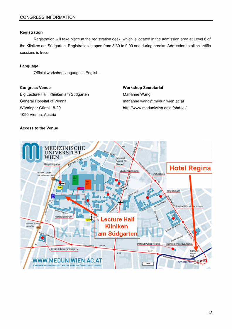
CONGRESS INFORMATION
22
Registration
Registration will take place at the registration desk, which is located in the admission area at Level 6 of
the Kliniken am Südgarten. Registration is open from 8:30 to 9:00 and during breaks. Admission to all scientific
sessions is free.
Language Official workshop language is English.
Congress Venue Big Lecture Hall, Kliniken am Südgarten
General Hospital of Vienna
Währinger Gürtel 18-20
1090 Vienna, Austria
Workshop Secretariat Marianne Wang
http://www.meduniwien.ac.at/phd-iai/
Access to the Venue