Embed Size (px)
Citation preview
636 Journal of Lipid Research Volume 54, 2013 This article is available online at http://www.jlr.org
Group IVA cytosolic phospholipase A 2 (cPLA 2 � ) is an 85 kDa enzyme, which liberates arachidonic acid (AA) from the sn -2 position of membrane phospholipids in response to infl ammatory agonists ( 1–3 ). cPLA 2 � consists of an N-terminal lipid binding C2 domain and a C-terminal catalytic or lipase domain that is separated by a fl exible linker ( 2, 4 ). The C2 domain is � 120 amino acid module, binds to zwitterionic lipids such as phosphatidylcholine (PC), and docks to PC-rich internal membranes in mam-malian cells ( 5–9 ) in a Ca 2+ -dependent manner. Following the membrane binding and penetration of the C2 domain ( 10, 11 ), the � 600 residue catalytic domain releases AA from zwitterionic lipids ( 3, 12 ). The generation of AA initiates pathways leading to eicosanoid synthesis, which has been implicated in heart disease ( 13 ), asthma ( 14 ), arthritis ( 15 ), cancers ( 16 ), and Alzheimer’s disease ( 17 ).
The spatial and temporal translocation of cPLA 2 � to the nuclear envelope, endoplasmic reticulum, and Golgi apparatus is controlled by both cell-specifi c and agonist-dependent events. Recently, two anionic lipids, ceramide-1-phosphate (C1P) ( 18, 19 ) and PI(4,5)P 2 ( 20–23 ) have been found to bind and activate cPLA 2 � . The molecular mecha-nisms regulating cPLA 2 � binding to C1P and PI(4,5)P 2 are only beginning to unravel with the C1P ( 24 ) and PI(4,5)P 2 ( 21, 25 ) binding sites being identifi ed in the C2 domain
Abstract Group IVA cytosolic phospholipase A 2 (cPLA 2 � ), which harbors an N-terminal lipid binding C2 domain and a C-terminal lipase domain, produces arachidonic acid from the sn -2 position of zwitterionic lipids such as phosphatidyl-choline. The C2 domain has been shown to bind zwitteri-onic lipids, but more recently, the anionic phosphomonoester sphingolipid metabolite ceramide-1-phosphate (C1P) has emerged as a potent bioactive lipid with high affi nity for a cationic patch in the C2 domain � -groove. To systematically analyze the role that C1P plays in promoting the binding of cPLA 2 � -C2 to biological membranes, we employed biophysical measurements and cellular translocation studies along with mutagenesis. Biophysical and cellular translocation studies demonstrate that C1P specifi city is mediated by Arg 59 , Arg 61 , and His 62 (an RxRH sequence) in the C2 domain. Computa-tional studies using molecular dynamics simulations con-fi rm the origin of C1P specifi city, which results in a spatial shift of the C2 domain upon membrane docking to coordi-nate the small C1P headgroup. Additionally, the hydroxyl group on the sphingosine backbone plays an important role in the interaction with the C2 domain, further demonstrat-ing the selectivity of the C2 domain for C1P over phosphatidic acid. Taken together, this is the fi rst study demonstrat-ing the molecular origin of C1P recognition. —Ward, K. E., N. Bhardwaj, M. Vora, C. E. Chalfant, H. Lu, and R. V. Stahelin. The molecular basis of ceramide-1-phosphate recognition by C2 domains. J. Lipid Res . 2013. 54: 636–648.
Supplementary key words calcium • cytosolic phospholipase A 2 � • eicosanoids • lipid binding • membrane binding
This work was supported by grants from the American Heart Association (SDG0735350N and GRNT12080254) to R.V.S.; NIH/HL-072925, NIH/CA-154314, VA Merit Award BX001792, VA Research Career Scientist Award, and a U.S.–Israel Binational Science Foundation/BSF#2011380 to C.E.C. K.E.W. is supported by an American Heart Association Predoctoral Fellowship (AHA 11PRE7640028) and a NIH CBBI Training Fellowship (T32GM075762). This work was also supported by the Indiana University School of Medicine-South Bend Imaging and Flow Cytometry Core Facility (R.V.S).
Manuscript received 11 August 2012 and in revised form 29 December 2012.
Published, JLR Papers in Press, December 31, 2012 DOI 10.1194/jlr.M031088
The molecular basis of ceramide-1-phosphate recognition by C2 domains
Katherine E. Ward , 1, * Nitin Bhardwaj , 1,† Mohsin Vora , § Charles E. Chalfant , ** Hui Lu , 2,† and Robert V. Stahelin 2, * , §
Department of Chemistry and Biochemistry and the Mike and Josie Harper Center for Cancer Research,* University of Notre Dame , Notre Dame, IN; Bioinformatics Program, Department of Bioengineering, † University of Illinois at Chicago , Chicago, IL; Department of Biochemistry and Molecular Biology, § Indiana University School of Medicine , South Bend, IN; and Department of Biochemistry,** Medical College of Virginia Campus, Virginia Commonwealth University, the Massey Cancer Center , and Research and Development, Hunter Holmes McGuire Veterans Administration Medical Center , Richmond, VA
Abbreviations: AA, arachidonic acid; BCA, bicinchoninic acid; BLAST, basic local alignment search tool; C1P, ceramide-1-phosphate; cPLA 2 � , group IVA cytosolic phospholipase A 2 ; deoxy-C1P, N -hexade-canoyl-3-deoxy-sphingosine-1-phosphate; MD, molecular dynamics; PA, phosphatidic acid; PC, phosphatidylcholine; POPC, 1-palmitoyl-2-oleoyl- sn -glycero-3-phosphocholine; POPE, 1-palmitoyl-2-oleoyl- sn -glycero-3-phosphoethanolamine; RGP3, regulator of G-protein signaling 3; SPR, surface plasmon resonance; TACE, TNF- � -converting enzyme; UVRAG, UV resistance-associated gene; WT, wild type.
1 K. E. Ward and N. Bhardwaj contributed equally to this work. 2 To whom correspondence should be addressed. e-mail: [email protected] ; [email protected]
The online version of this article (available at http://www.jlr.org) contains supplementary data in the form of two fi gures.
at Univ of IL at C
hicago, on March 4, 2013
ww
w.jlr.org
Dow
nloaded from
.html http://www.jlr.org/content/suppl/2012/12/31/jlr.M031088.DC1Supplemental Material can be found at:
The origin of C1P binding specifi city 637
(3-[3-cholamidopropyl) dimethylammonio]1-propane-sulfonate (CHAPS), and bicinchoninic acid (BCA) protein assay kit were from Fisher Scientifi c (Hampton, NH). L1 sensor chips were from GE Healthcare (Piscataway, NJ). Phospholipid concentra-tions were determined by a modifi ed Bartlett analysis ( 32 ). Restric-tion endonucleases and enzymes for molecular biology were obtained from New England Biolabs (Beverly, MA). A549 trans-fection reagents (PLUS™ reagent and Lipofectamine LTX) were from Life Technologies (Grand Island, NY).
DNA mutagenesis and protein purifi cation The QuikChange site-directed mutagenesis kit (Agilent Techno-
logies; Santa Clara, CA) was used to introduce mutations into the pET28a vector with a His 6 tag engineered into the N-terminus of the cPLA 2 � C2 domain gene. The manufacturer’s instructions were used to perform temperature cycling using Pfu DNA poly-merase. This replicates both strands with high fi delity without displacing the mutagenic primers. A mutated plasmid containing staggered nicks was generated and treated with DpnI endonu-clease. This enzyme specifi cally digests methylated and hemim-ethylated parental DNA templates and selects for mutations containing synthesized DNA. The nicked DNAs were then trans-formed into Escherichia coli XL-10 Gold cells. All mutated constructs were sequenced to ensure presence of the desired mutation. The C2 domain and respective mutations were expressed and puri-fi ed from E. coli BL21(DE3) cells as previously described ( 10 ). Protein concentrations were determined by the BCA method, and aliquots of 3 mg/ml were made using storage buffer (10 mM HEPES, pH 7.4, 0.16 M KCl).
Surface plasmon resonance measurements All surface plasmon resonance (SPR) measurements were per-
formed at 25°C. A detailed protocol for coating the L1 sensor chip has been described elsewhere ( 33, 34 ). Briefl y, after washing the sensor chip surface, 90 � l of vesicles containing either POPC or POPC-C1P (97:3) were injected at 5 � l/min to give a response of 6,200 resonance units. An uncoated fl ow channel was used as a control surface. Under our experimental conditions, no binding was detected to this control surface beyond the refractive index change for the C2 domain or cPLA 2 � as previously re-ported ( 9, 11, 34 ). Each lipid layer was stabilized by injecting 10 � l of 50 mM NaOH three times at 100 � l/min. SPR measure-ments were done at the fl ow rate of 5 � l/min. 50–90 � l of protein in 10 mM HEPES, pH 7.4, containing 0.16 M KCl and 10 � M Ca 2+ , was injected to give a suffi cient association time for each binding signal to reach saturation ( R eq ) ( Fig. 1A, C ). The lipid surface on the L1 chip, which is composed of intact lipid vesicles ( 34, 35 ), was regenerated using 10 � l of 50 mM NaOH. After sensorgrams were obtained for fi ve or more different concentra-tions of each protein within a 10-fold range of K d , each of the sensorgrams was corrected for refractive index change by sub-tracting the control surface response. R eq values were then plotted versus protein concentrations ( C ), and the K d value was deter-mined by a nonlinear least-squares analysis of the binding isotherm using an equation, R eq = R max /(1 + K d / C ) ( 36 ). Each data set was repeated three times to calculate a standard deviation value.
C1P stoichiometry measurements The cPLA 2 � -C2 domain contains only one endogenous trypto-
phan, Trp 71 , which lies beneath the cationic patch that has pre-viously ( 24, 31 ) and herein been shown to bind C1P. The stoichiometric ratio of C1P to cPLA 2 � -C2 was determined through modifi cation of the previously established methodology as de-scribed for the C2 domain of PKC � and PI(4,5)P 2 ( 37 ). HEPES, pH 7.4, 10 mM, containing 0.16 M KCl and 500 nM Ca 2+ , where
and catalytic domain, respectively. Although the cationic site in the catalytic domain is promiscuous in anionic lipid binding ( 21, 23, 26 ), C1P is the only membrane-embedded anionic lipid that has been shown to increase membrane affi nity of the cPLA 2 � C2 domain ( 19 ). Thus, it is thought that C1P acts as a coincidence detector ( 27 ) to promote the membrane docking of the C2 domain through elon-gating the membrane residence time ( 19, 24, 28 ). It is known that the C2 domain is responsible for the translocation of the catalytic domain to internal membranes; thus, C1P binding probably contributes to this functionality. Although the binding to C1P has not been observed at supraphysiologi-cal calcium levels, these types of binding experiments must be done at physiologically relevant cytoplasmic calcium concentrations, because C1P is completely shielded by calcium above 300 � M ( 29 ).
To date, cPLA 2 � ( 19 ) and TNF- � -converting enzyme (TACE) ( 30 ) are the only documented proteins that have been shown to bind to C1P selectively over other anionic lipids. Moreover, C1P can regulate the translocation of cPLA 2 � as well as increase its biological activity ( 31 ). These data are intriguing , inasmuch as one may expect other phosphomonoesters to bind with some affi nity. For instance, how does the C2 domain recognize C1P over phosphatidic acid (PA) (see Fig. 6A )? Mutagenesis of residues in the C2 domain cationic patch (Arg 57 , Lys 58 , Arg 59 ) (see Fig. 3 ) abrogate the C1P-dependent translocation and activity in cells ( 31 ), as well as the 10-fold increase in binding affi nity C1P provides when incorporated in lipid vesicles. Thus far, these data are a bit muddled, owing to the double and triple mutants that were employed in full-length cPLA 2 � ( 24, 31 ).
To clarify the basis of C1P binding, single mutations of all fi ve of the basic residues of the cationic patch (RKRTRH) were made in the C2 domain and full-length enzyme. To gain insight into the mechanism of the cPLA 2 � interaction with cell membranes containing C1P, single point mutations in the full-length protein as well as in the C2 domain were constructed to examine the impor-tance of each residue in the basic patch in vitro and in cells. Subsequently, molecular dynamics (MD) simulations were performed, with the C2 domain docking to a bilayer containing PC or PC-C1P to further elucidate the origin of C1P specifi city. Taken together, this experimental and computational investigation demonstrates that an RxRH sequence adjacent to the calcium binding loops of the C2 domain mediates C1P specifi city.
MATERIALS AND METHODS
Materials 1-Palmitoyl-2-oleoyl- sn -glycero-3-phosphocholine (POPC),
1-palmitoyl-2-oleoyl- sn -glycero-3-phosphoethanolamine (POPE), and N -palmitoyl-ceramide-1-phosphate (C1P) were purchased from Avanti Polar Lipids, Inc. (Alabaster, AL) and used with-out further purifi cation. N -hexadecanoyl-3-deoxy-sphingosine-1-phosphate (deoxy-C1P), synthesized by Avanti Polar Lipids, Inc. was a kind gift from Walt Shaw and Stephen Burgess. Octyl glucoside,
at Univ of IL at C
hicago, on March 4, 2013
ww
w.jlr.org
Dow
nloaded from
.html http://www.jlr.org/content/suppl/2012/12/31/jlr.M031088.DC1Supplemental Material can be found at:
638 Journal of Lipid Research Volume 54, 2013
Dodecane-ethanol lipid delivery in A459 cells Lipids were prepared as previously described ( 38 ). Briefl y,
lipid mixtures were prepared by drying the allotted volume of chloroform-methanol (2:1) solubilized lipids under N 2 gas to make a 2.5 mM stock solution. Lipids were resuspended in dodecane-ethanol (98:2) at 37°C and subsequently sonicated and heated to 37°C for 20 min. Lipid solutions were diluted to the indicated concentrations using the dodecane-ethanol mixture. Cells ex-pressing EGFP-cPLA 2 � were treated 24 h post transfection with 500 nM C1P or deoxy-C1P in dodecane-ethanol (98:2) for 2 h at 37°C and imaged with confocal microscopy.
Molecular dynamics set-up All simulations reported in this study were done on an all-atoms
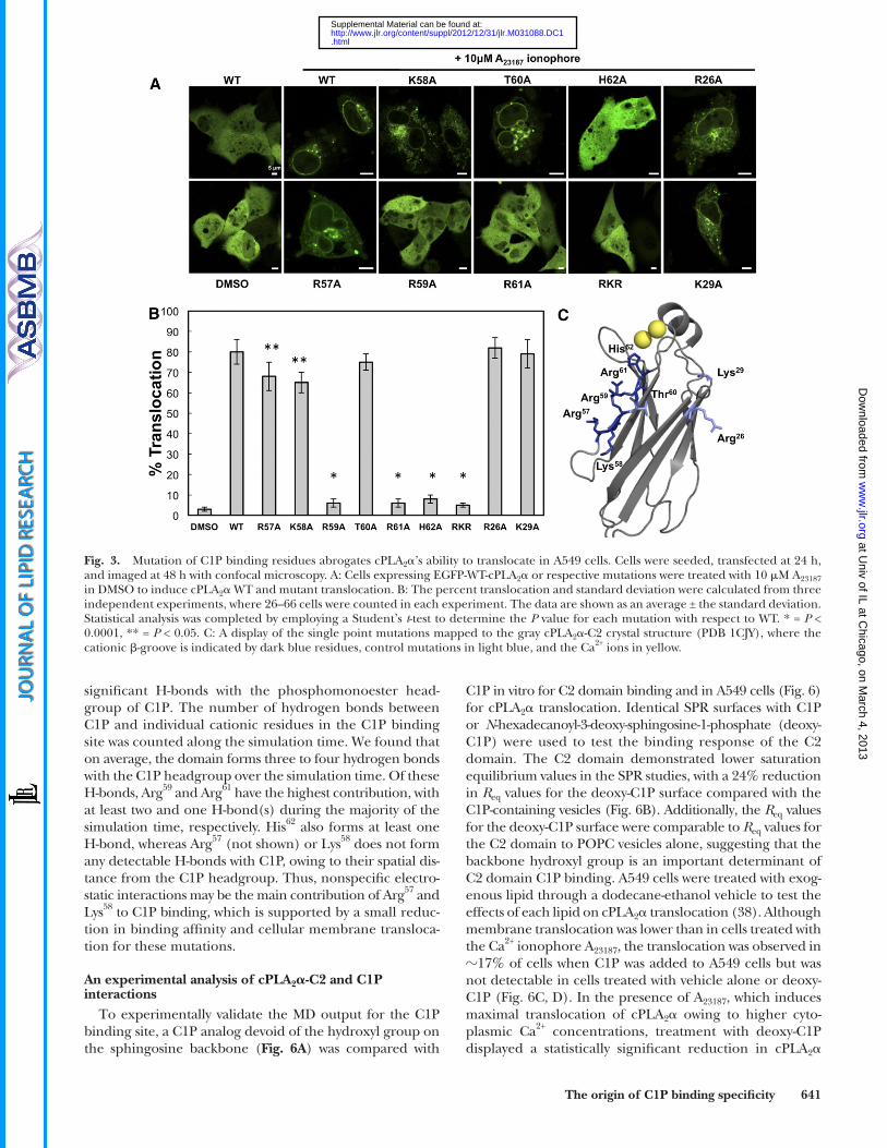
scheme. The fi rst set of simulations was performed without C1P (with only POPC) while reducing the number of Ca 2+ ions from 2 to 0 (system names according to the number of Ca 2+ ions present: PC 2Ca 2+ , PC 1Ca 2+ , PC) (see Fig. 4A ). In the second set of simula-tions, 1 C1P molecule was introduced with a different number of Ca 2+ ions bound (system names C1P 2Ca 2+ , C1P 1Ca 2+ , C1P).
System model First, a patch of POPC bilayer was created using the ‘mem-
brane’ plugin in visual molecular dynamics ( 39 ) in the xy plane such that the z-axis formed the membrane normal. The dimen-sion of the patch (100 Å × 100 Å) was suffi cient to cover the pro-tein’s area in the xy plane and to leave an additional margin of at least 15 Å on each side. The layer that interacted with the pro-tein was called the ‘positive layer.’ For the bilayer where C1P was introduced, one POPC molecule was replaced with one C1P mol-ecule. The C1P molecule was placed directly below the � -groove cat-ionic patch previously shown to bind C1P ( 24 ). The surrounding
the Ca 2+ /EGTA was calculated according to Maxchelator soft-ware (www.stanford.eu/~cpatton/-maxc.html) was used as the assay buffer. Briefl y, cPLA 2 � -C2 was held constant at 1 � M and measured for maximal tryptophan fl uorescence on an Aminco Bowman Series 2 luminescence spectrophotometer using excita-tion and emission wavelengths of 284 nm and 340 nm and band-widths of 4 and 8 nm, respectively. Subsequently, small unilamellar vesicles containing POPC-POPE-C1P (50:40:10) were added at increasing molar C1P concentrations until the fl uorescence was no longer quenched. The data were normalized, where the maxi-mum tryptophan quenched was set to 1, then the rise and satura-tion phases of the data were fi t using a linear least-squares equation ( 37 ). The intersection of the two lines defi nes the stoi-chiometry of cPLA 2 � -C2 to C1P. The stoichiometry experiment was performed in duplicate with 10 mol% C1P and confi rmed in triplicate using vesicles containing 5 mol% C1P [POPC-POPE-C1P (55:40:5)].
Cellular protein expression and confocal microscopy imaging
A549 lung adenocarcinoma cells were transfected with en-hanced green fl uorescent protein (EGFP) -cPLA 2 � wild type (WT) or mutants using PLUS™ reagent and lipofectamine LTX according to the manufacturers’ protocols (Life Technolo-gies; Grand Island, NY). Cells were grown in 50:50 DMEM-RPMI with 10% FBS and 1% penicillin-streptomycin as described previously ( 31 ). Cells were treat ed with 10 � M A 23187 Ca 2+ iono-phore to induce translocation to cellular membranes, then imaged via confocal microscopy (Zeiss LSM 710) on Nunc Lab-Tek II chambered cover glasses, 8-well (Thermo Fisher Scientifi c; Waltham, MA) using an oil 63× 1.4 numerical aper-ture objective.
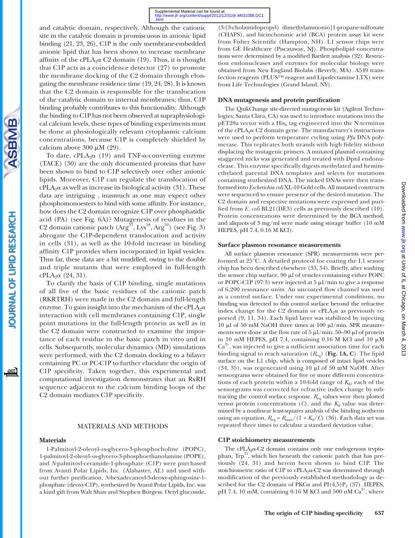
Fig. 1. Lipid binding properties of cPLA 2 � -C2 and mutations. Quantitative binding analysis was performed for the cPLA 2 � -C2 and respec-tive mutations to POPC or POPC-C1P (97:3) vesicles at 10 � M Ca 2+ . A: SPR sensorgrams are shown for 10 nM WT cPLA 2 � -C2 binding to POPC (light gray) or POPC:C1P (97:3) (black) vesicles. B: The equilibrium response ( R eq ) from WT cPLA 2 � -C2 binding at each respective protein concentration was plotted versus [cPLA 2 � -C2] to fi t with a nonlinear least-squares analysis of the binding isotherm ( R eq = R max /(1 + K d / C ) to determine the K d (see 1 E). C: SPR sensorgrams are shown for 100 nM WT cPLA 2 � -C2 (black) or 100 nM R59A binding to POPC-C1P (97:3) (gray) vesicles. D: Fold increase in K d was normalized for each respective protein to the K d value for WT cPLA 2 � -C2 for POPC-C1P (97:3) -containing vesicles. The fi lled bars depict binding to POPC vesicles, and the gray bars display binding to POPC-C1P (97:3) vesicles. E: K d values for WT and respective mutations binding to POPC or POPC-C1P (97:3) vesicles. The binding experiments were completed from independent experiments in triplicate and are listed with their respective standard deviation.
at Univ of IL at C
hicago, on March 4, 2013
ww
w.jlr.org
Dow
nloaded from
.html http://www.jlr.org/content/suppl/2012/12/31/jlr.M031088.DC1Supplemental Material can be found at:
The origin of C1P binding specifi city 639
does not require an artifi cial nonisotropic dielectric constant to remain stable and accurately represents all atoms over the course of the simulations.
Computing observables from MD simulation System coordinates were saved every 2 ps during the course
of the simulation to analyze the following properties under differ-ent conditions. To monitor the movement of the protein, ‘mem-brane surface’ was defi ned as the surface layer in the xy plane with z-coordinate equal to the average of z-coordinates of phosphorus atoms present in the positive layer of that system. The change in the difference of the z-coordinate of the center of mass of the pro-tein and the z-coordinate of the membrane surface was used to monitor the movement of the protein with respect to the mem-brane. For analysis, the initial value of this difference was trans-lated to a value of zero such that a difference in positive value would indicate the movement of the protein away from the layer as compared with the initial confi guration, and a negative value would indicate its movement toward the membrane. To compare the binding orientation of the domain with and without C1P, tilt-ing of the protein with respect to the membrane surface was also investigated. The fi rst principal axis of the domain was calculated with the C � coordinates of its atoms. Owing to the shape of the domain, its fi rst principal axis passes through the binding and the nonbinding loops, and its movement is around the x axis in the plane of the paper. Therefore, the rotation of the fi rst principal axis would directly demonstrate the rotation of the C2 domain.
Homology and sequence alignments The cPLA 2 � C2 domain sequence was defi ned as residues
1–130 for the searching parameters. Homology among other organisms was investigated using a nonredundant protein-protein basic local alignment search tool (BLAST) with no excluded organisms on the default search parameters. Percent identity was calculated for the proteins compared with residues 1–130 of the query search by BLAST. The range of organisms was selected based on their diversity and is not inclusive of all organisms con-taining the basic sequence highlighted. To locate other C2 domains containing the RxRH motif, the conserved domain database engine on National Center for Biotechnology Informa-tion was utilized to aggregate C2 domain sequences (cd00030). At this point, the search was not limited to any particular organism. Once a positive hit was found, the Homo sapiens analog was found using BLAST. Sequences also found in Homo sapiens containing the RxRH motif were input into Fig. 8F . The search parameters did not include all C2 domains, but rather are limited to the sub-set included in the CD model under cd00030 that contained a reasonable sequence in Homo sapiens as of July 2012.
RESULTS
cPLA 2 � -C2 domain C1P binding specifi city is determined by Arg 59 , Arg 61 , and His 62
The preferential binding of cPLA 2 � to zwitterionic lipids such as PC is well established ( 10, 45 ), as is the increased binding affi nity for C1P-containing vesicles over PC mem-branes ( 19, 24, 31 ). To quantify the strength and selectivity of binding to these lipids, the equilibrium binding constant ( K d ) for WT cPLA 2 � -C2 and single point mutations was determined using SPR for POPC or POPC-C1P (97:3) vesicles, which are tethered intact on a L1 sensor chip. The WT protein bound to C1P vesicles ten times stronger than to POPC vesicles alone ( Fig. 1D, E ), whereas R57A
POPC molecules were rotated and translated to remove any large steric clashes with C1P. A layer of 15 Å of water on the top and the bottom of the bilayer was used to ensure suffi cient hydration.
Statistical ensemble Periodic boundary conditions were applied in the xy direc-
tions to simulate an infi nite planar layer and in the z direction to simulate a multilayer system. First, the membrane system was equilibrated without the protein in both sets beginning with 10,000 steps of conjugate-gradient energy minimization to re-move remaining steric overlaps. This was followed by 100 ps of dynamical run. Due to a lack of homogeneity of the system (pres-ence of two types of lipids) and unavailability of experimental data about the area per lipid for C1P, simulations of the bilayers were performed with isothermal-isobaric ensemble with the tem-perature fi xed at 300° K. This method is appropriate for inho-mogeneous systems such as a lipid bilayer of heterogeneous composition, for two reasons. First, for such systems, no large temperature difference between the components of the system is created, and therefore it is not necessary to couple the differ-ent components to separate heat baths. Second, the method has the advantage of not being critically dependent on the choice of the piston parameters and allows the area per lipid to fl uctuate and stabilize to an optimum value ( 40 ). The Langevin piston method ( 41 ) was used to impose a constant pressure P = 1 atm with a damping coeffi cient of 5 ps –1 . The particle mesh Ewald (PME) method was used for computation of the electrostatic forces ( 42, 43 ) with the grid spacing below 1.0 Å. All hydrogen bonds were restrained, allowing a time step of 2 fs.
Docking of protein on the membrane surface In this study, the starting orientation of the protein with re-
spect to the membrane in both the sets was the same as the ex-perimentally validated binding orientation with POPC ( 44, 45 ). Ca 2+ ions were also kept intact, as found in the crystal structure ( 46 ). Ca 2+ ion 2 was removed in simulations involving one Ca 2+ ion, inasmuch as Ca 2+ ion 2 has been shown to regulate enzyme activity and Ca 2+ ion 1 has been shown to regulate membrane binding ( 11 ). The penetration of protein into the membrane was also carefully emulated by translation of the protein along the z-axis. Some translations of the lipids in the xy plane and rigid-body rotations around the z-axis were also performed to eliminate unfavorable contacts and atomic overlaps. Additional water was added on the top of the protein-lipid system to create a layer of 15 Å above the uppermost atom (with the highest z-coordinate) of the protein. Counter ions were added to ensure the electroneu-trality of the system. The remaining poor contacts or overlaps between the protein and lipid atoms were removed by minimiz-ing the energy of the system by 10,000 steps of conjugate-gradient minimization followed by a dynamical simulation run of 10 ns.
Force fi eld parameters The antechamber tool ( 47 ) from Amber 7 ( 48 ) was used to
parameterize C1P along with POPC. Antechamber is a set of auxil-iary programs that can be used to effi ciently identify bond and atom types, judge atomic equivalences, assign partial atomic charges, generate residue topology fi les, and, fi nally, create force fi eld parameters on the basis of the above information. Generalized amber force fi eld ( 49 ) parameters were used, and AM1-bond charge correction was used for generation of high-quality atomic charges. After the preparation of the input coordinate and topol-ogy fi les with Amber, NAMD 2.5 ( 50 ) was used for carrying out all molecular dynamics simulations. An explicit atom representa-tion for all atoms, including both heavy atoms and hydrogens, was implemented. This representation yields a force fi eld that
at Univ of IL at C
hicago, on March 4, 2013
ww
w.jlr.org
Dow
nloaded from
.html http://www.jlr.org/content/suppl/2012/12/31/jlr.M031088.DC1Supplemental Material can be found at:
640 Journal of Lipid Research Volume 54, 2013
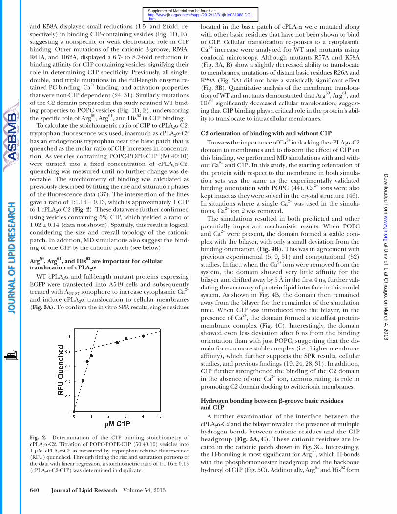
located in the basic patch of cPLA 2 � were mutated along with other basic residues that have not been shown to bind to C1P. Cellular translocation responses to a cytoplasmic Ca 2+ increase were analyzed for WT and mutants using confocal micro scopy. Although mutants R57A and K58A ( Fig. 3A, B ) show a slightly decreased ability to translocate to membranes, mutations of distant basic residues R26A and K29A ( Fig. 3A ) did not have a statistically signifi cant effect ( Fig. 3B ). Quantitative analysis of the membrane transloca-tion of WT and mutants demonstrated that Arg 59 , Arg 61 , and His 62 signifi cantly decreased cellular translocation, suggest-ing that C1P binding plays a critical role in the protein’s abil-ity to translocate to intracellular membranes.
C2 orientation of binding with and without C1P To assess the importance of Ca 2+ in docking the cPLA 2 � -C2
domain to membranes and to discern the effect of C1P on this binding, we performed MD simulations with and with-out Ca 2+ and C1P. In this study, the starting orientation of the protein with respect to the membrane in both simula-tion sets was the same as the experimentally validated binding orientation with POPC ( 44 ). Ca 2+ ions were also kept intact as they were solved in the crystal structure ( 46 ). In situations where a single Ca 2+ was used in the simula-tions, Ca 2+ ion 2 was removed.
The simulations resulted in both predicted and other potentially important mechanistic results. When POPC and Ca 2+ were present, the domain formed a stable com-plex with the bilayer, with only a small deviation from the binding orientation ( Fig. 4B ). This was in agreement with previous experimental ( 5, 9, 51 ) and computational ( 52 ) studies. In fact, when the Ca 2+ ions were removed from the system, the domain showed very little affi nity for the bilayer and drifted away by 5 Å in the fi rst 4 ns, further vali-dating the accuracy of protein-lipid interface in this model system. As shown in Fig. 4B , the domain then remained away from the bilayer for the remainder of the simulation time. When C1P was introduced into the bilayer, in the presence of Ca 2+ , the domain formed a steadfast protein-membrane complex ( Fig. 4C ). Interestingly, the domain showed even less deviation after 6 ns from the binding orientation than with just POPC, suggesting that the do-main forms a more-stable complex (i.e., higher membrane affi nity), which further supports the SPR results, cellular studies, and previous fi ndings ( 19, 24, 28, 31 ). In addition, C1P further strengthened the binding of the C2 domain in the absence of one Ca 2+ ion, demonstrating its role in promoting C2 domain docking to zwitterionic membranes.
Hydrogen bonding between � -groove basic residues and C1P
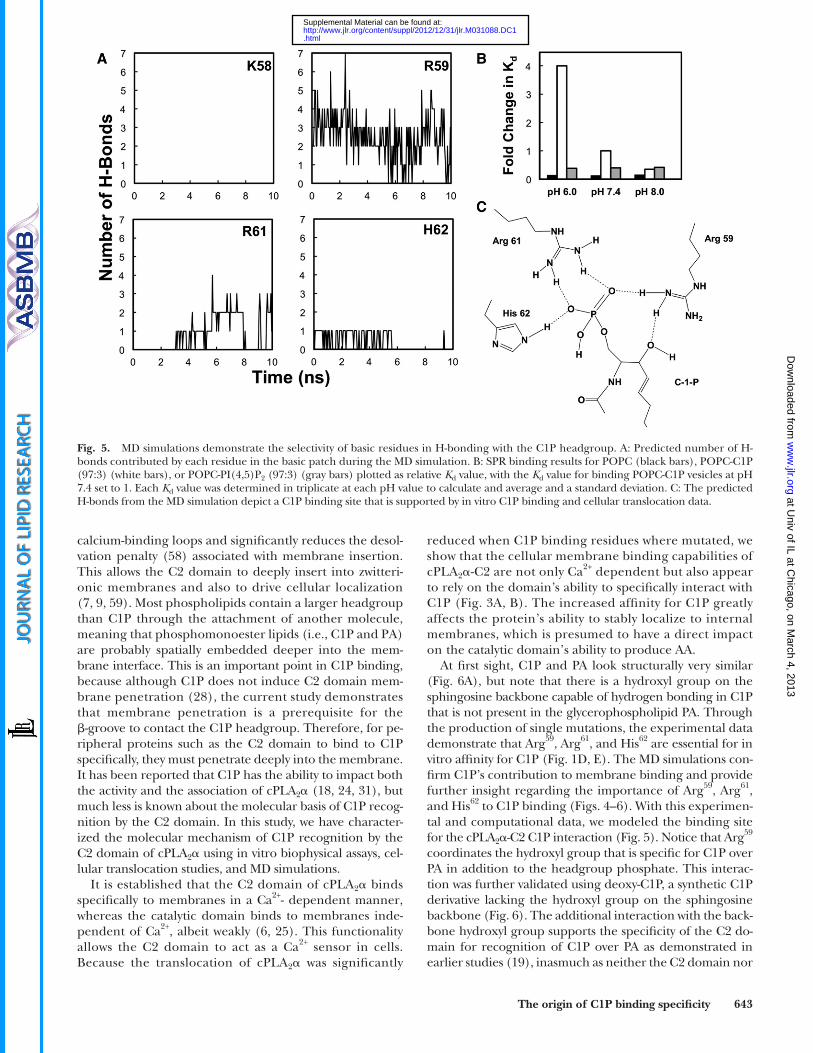
A further examination of the interface between the cPLA 2 � -C2 and the bilayer revealed the presence of multiple hydrogen bonds between cationic residues and the C1P headgroup ( Fig. 5A, C ). These cationic residues are lo-cated in the cationic patch shown in Fig. 3C . Interestingly, the H-bonding is most signifi cant for Arg 59 , which H-bonds with the phosphomonoester headgroup and the backbone hydroxyl of C1P ( Fig. 5C ). Additionally, Arg 61 and His 62 form
and K58A displayed small reductions (1.5- and 2-fold, re-spectively) in binding C1P-containing vesicles ( Fig. 1D, E ), suggesting a nonspecifi c or weak electrostatic role in C1P binding. Other mutations of the cationic � -groove, R59A, R61A, and H62A, displayed a 6.7- to 8.7-fold reduction in binding affi nity for C1P-containing vesicles, signifying their role in determining C1P specifi city. Previously, all single, double, and triple mutations in the full-length enzyme re-tained PC binding, Ca 2+ binding, and activation properties that were non-C1P dependent ( 24, 31 ). Similarly, mutations of the C2 domain prepared in this study retained WT bind-ing properties to POPC vesicles ( Fig. 1D, E ), underscoring the specifi c role of Arg 59 , Arg 61 , and His 62 in C1P binding.
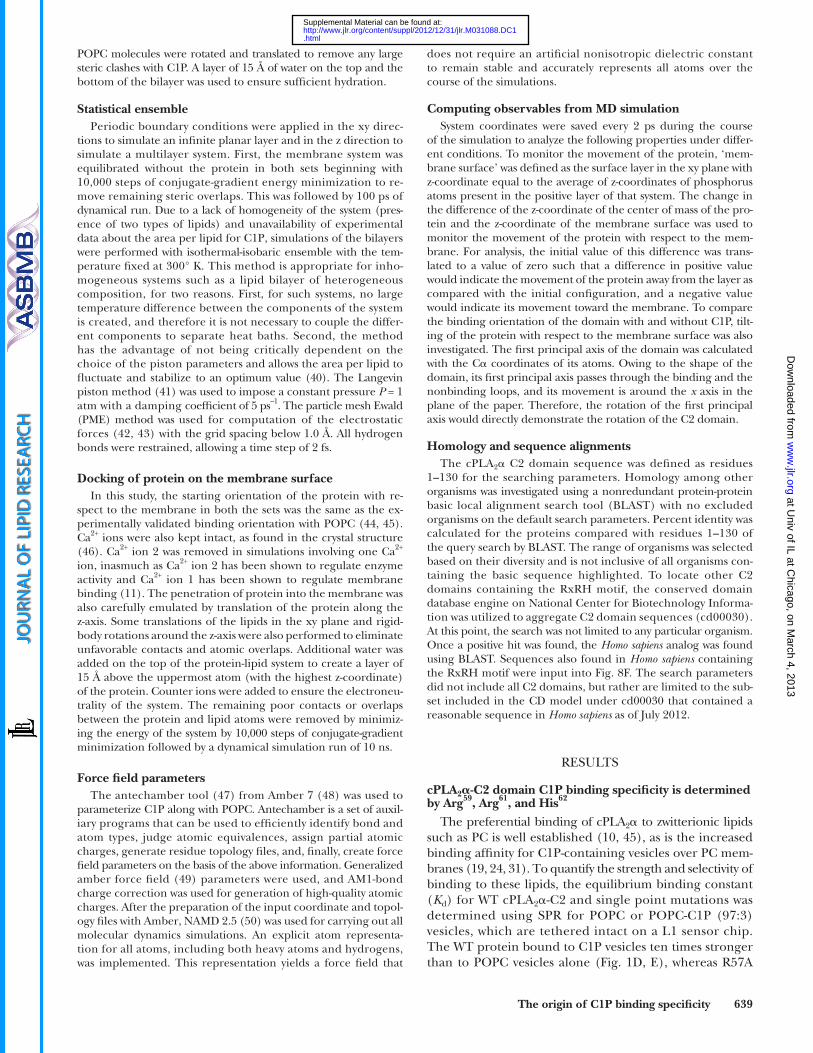
To calculate the stoichiometric ratio of C1P to cPLA 2 � -C2, tryptophan fl uorescence was used, inasmuch as cPLA 2 � -C2 has an endogenous tryptophan near the basic patch that is quenched as the molar ratio of C1P increases in concentra-tion. As vesicles containing POPC-POPE-C1P (50:40:10) were titrated into a fi xed concentration of cPLA 2 � -C2, quenching was measured until no further change was de-tectable. The stoichiometry of binding was calculated as previously described by fi tting the rise and saturation phases of the fl uorescence data ( 37 ). The intersection of the lines gave a ratio of 1:1.16 ± 0.13, which is approximately 1 C1P to 1 cPLA 2 � -C2 ( Fig. 2 ). These data were further confi rmed using vesicles containing 5% C1P, which yielded a ratio of 1.02 ± 0.14 (data not shown). Spatially, this result is logical, considering the size and overall topology of the cationic patch. In addition, MD simulations also suggest the bind-ing of one C1P by the cationic patch (see below).
Arg 59 , Arg 61 , and His 62 are important for cellular translocation of cPLA 2 �
WT cPLA 2 � and full-length mutant proteins expressing EGFP were transfected into A549 cells and subsequently treated with A 23187 ionophore to increase cytoplasmic Ca 2+ and induce cPLA 2 � translocation to cellular membranes ( Fig. 3A ). To confi rm the in vitro SPR results, single residues
Fig. 2. Determination of the C1P binding stoichiometry of cPLA 2 � -C2. Titration of POPC-POPE-C1P (50:40:10) vesicles into 1 � M cPLA 2 � -C2 as measured by tryptophan relative fl uorescence (RFU) quenched. Through fi tting the rise and saturation portions of the data with linear regression, a stoichiometric ratio of 1:1.16 ± 0.13 (cPLA 2 � -C2-C1P) was determined in duplicate.
at Univ of IL at C
hicago, on March 4, 2013
ww
w.jlr.org
Dow
nloaded from
.html http://www.jlr.org/content/suppl/2012/12/31/jlr.M031088.DC1Supplemental Material can be found at:
The origin of C1P binding specifi city 641
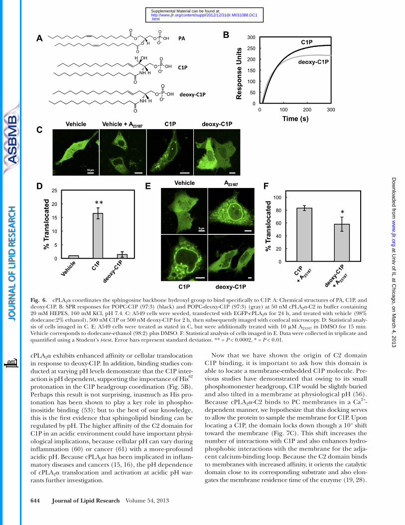
C1P in vitro for C2 domain binding and in A549 cells ( Fig. 6 ) for cPLA 2 � translocation. Identical SPR surfaces with C1P or N -hexadecanoyl-3-deoxy-sphingosine-1-phosphate (deoxy-C1P) were used to test the binding response of the C2 domain. The C2 domain demonstrated lower saturation equilibrium values in the SPR studies, with a 24% reduction in R eq values for the deoxy-C1P surface compared with the C1P-containing vesicles ( Fig. 6B ). Additionally, the R eq values for the deoxy-C1P surface were comparable to R eq values for the C2 domain to POPC vesicles alone, suggesting that the backbone hydroxyl group is an important determinant of C2 domain C1P binding. A549 cells were treated with exog-enous lipid through a dodecane-ethanol vehicle to test the effects of each lipid on cPLA 2 � translocation ( 38 ). Although membrane translocation was lower than in cells treated with the Ca 2+ ionophore A 23187 , the translocation was observed in � 17% of cells when C1P was added to A549 cells but was not detectable in cells treated with vehicle alone or deoxy-C1P ( Fig. 6C, D ). In the presence of A 23187 , which induces maximal translocation of cPLA 2 � owing to higher cyto-plasmic Ca 2+ concentrations, treatment with deoxy-C1P displayed a statistically signifi cant reduction in cPLA 2 �
signifi cant H-bonds with the phosphomonoester head-group of C1P. The number of hydrogen bonds between C1P and individual cationic residues in the C1P binding site was counted along the simulation time. We found that on average, the domain forms three to four hydrogen bonds with the C1P headgroup over the simulation time. Of these H-bonds, Arg 59 and Arg 61 have the highest contribution, with at least two and one H-bond(s) during the majority of the simulation time, respectively. His 62 also forms at least one H-bond, whereas Arg 57 (not shown) or Lys 58 does not form any detectable H-bonds with C1P, owing to their spatial dis-tance from the C1P headgroup. Thus, nonspecifi c electro-static interactions may be the main contribution of Arg 57 and Lys 58 to C1P binding, which is supported by a small reduc-tion in binding affi nity and cellular membrane transloca-tion for these mutations.
An experimental analysis of cPLA 2 � -C2 and C1P interactions
To experimentally validate the MD output for the C1P binding site, a C1P analog devoid of the hydroxyl group on the sphingosine backbone ( Fig. 6A ) was compared with
Fig. 3. Mutation of C1P binding residues abrogates cPLA 2 � ’s ability to translocate in A549 cells. Cells were seeded, transfected at 24 h, and imaged at 48 h with confocal microscopy. A: Cells expressing EGFP-WT-cPLA 2 � or respective mutations were treated with 10 � M A 23187 in DMSO to induce cPLA 2 � WT and mutant translocation. B: The percent translocation and standard deviation were calculated from three independent experiments, where 26–66 cells were counted in each experiment. The data are shown as an average ± the standard deviation. Statistical analysis was completed by employing a Student’s t -test to determine the P value for each mutation with respect to WT. * = P < 0.0001, ** = P < 0.05. C: A display of the single point mutations mapped to the gray cPLA 2 � -C2 crystal structure (PDB 1CJY), where the cationic � -groove is indicated by dark blue residues, control mutations in light blue, and the Ca 2+ ions in yellow.
at Univ of IL at C
hicago, on March 4, 2013
ww
w.jlr.org
Dow
nloaded from
.html http://www.jlr.org/content/suppl/2012/12/31/jlr.M031088.DC1Supplemental Material can be found at:
642 Journal of Lipid Research Volume 54, 2013
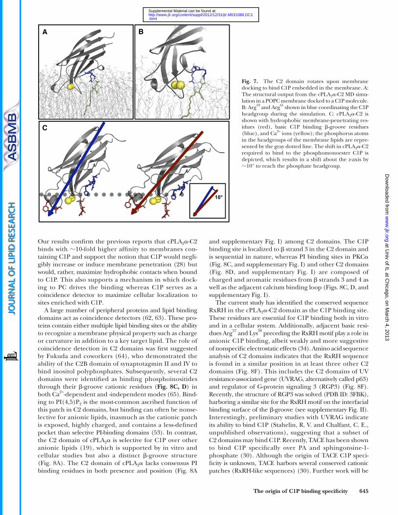
of C1P, we also examined the binding orientation in the absence and presence of C1P. Recently, electron paramag-netic resonance ( 44, 54, 55 ) and X-ray refl ectivity ( 45 ) studies have elucidated the cPLA 2 � -C2 domain orienta-tion in the presence of POPC lipids; however, the orienta-tion when C1P is present in the membrane is still unknown. We found that starting with the same orientation ( 44 ) as that with only POPC, the domain tilts by � 10° toward the bilayer over the course of the simulation in the presence of C1P ( Fig. 7 ). This tilting facilitates formation of stable H-bonds between cationic residues and the C1P head-group. This observation is further supported by the fact that the C1P headgroup is much smaller in size than the POPC headgroup and is thought to be more deeply bur-ied, and at physiological pH, the C1P acyl chains have been found to tilt away from the surface normal of the monolayer ( 56 ). Thus, Arg 59 , Arg 61 , and His 62 may further facilitate formation of hydrogen bonds by extending their side chains toward the bilayer (for instance Arg 59 in Fig. 7B ), which has previously been shown for Lys and Arg residues in peptides that bind PA ( 57 ).
DISCUSSION
cPLA 2 � -C2 has previously been shown to deeply pen-etrate PC membranes in a Ca 2+ - dependent manner ( 10, 44, 45 ). Ca 2+ has been shown to act as an electrostatic switch, which reduces the negative charge surrounding
translocation, suggesting that deoxy-C1P may act as a domi-nant negative by masking the endogenous C1P present in A549 cells ( Fig. 6E, F ). Treatment with additional C1P had no statistical effect in the presence of A 23187 , as shown by 83% translocation, because C1P binding is probably satu-rated at the higher calcium concentration created by the ionophore ( Fig. 6E, F ). These results provide experimental evidence regarding the importance of the sphingosine backbone hydroxyl group of C1P as an interaction determi-nant of the C2 domain ( Fig. 5C ).
Because His 62 was shown to hydrogen bond to C1P in the MD simulations, we sought to experimentally assess the pH dependency of this interaction compared with PC and PI(4,5)P 2 . Previously, His protonation at acidic pH was shown to enhance the affi nity of a number of phos-phoinositide binding domains for phosphoinositides ( 53 ). Using SPR, K d values at pH 6.0, 7.4, and 8.0 were deter-mined for POPC, POPC-C1P (97:3), or POPC-PI(4,5)P 2 (97:3) vesicles and demonstrate a strong relationship between pH and affi nity for C1P-containing vesicles ( Fig. 5B ). The interaction is 4-fold stronger at pH 6.0 than at pH 7.4 and is further reduced at pH 8.0. In contrast, binding af-fi nity to POPC or POPC-PI(4,5)P 2 vesicles was not infl u-enced by pH. The pH dependence of selective binding provided additional experimental evidence to validate the importance of His 62 in the cPLA 2 � -C1P interaction.
The C2 domain tilts toward the membrane to bind C1P To further characterize the interfacial membrane inter-
action that the cPLA 2 � -C2 domain forms in the presence
Fig. 4. MD simulations of cPLA 2 � -C2 docking PC- and C1P-containing bilayers. A: The MD simulations were performed on six different systems: POPC with both Ca 2+ ions present (PC 2Ca 2+ ), POPC and one Ca 2+ ion present (PC 1Ca 2+ ), POPC without Ca 2+ present (PC), POPC-C1P with both Ca 2+ ions present (C1P 2Ca 2+ ), POPC-C1P with one Ca 2+ ion present (C1P 1Ca 2+ ), and POPC-C1P without Ca 2+ present (C1P). B: The MD output of cPLA 2 � -C2 docked to a POPC bilayer; or C: a POPC-C1P bilayer under variable conditions as defi ned in A.
at Univ of IL at C
hicago, on March 4, 2013
ww
w.jlr.org
Dow
nloaded from
.html http://www.jlr.org/content/suppl/2012/12/31/jlr.M031088.DC1Supplemental Material can be found at:
The origin of C1P binding specifi city 643
reduced when C1P binding residues where mutated, we show that the cellular membrane binding capabilities of cPLA 2 � -C2 are not only Ca 2+ dependent but also appear to rely on the domain’s ability to specifi cally interact with C1P ( Fig. 3A, B ). The increased affi nity for C1P greatly affects the protein’s ability to stably localize to internal membranes, which is presumed to have a direct impact on the catalytic domain’s ability to produce AA.
At fi rst sight, C1P and PA look structurally very similar ( Fig. 6A ), but note that there is a hydroxyl group on the sphingosine backbone capable of hydrogen bonding in C1P that is not present in the glycerophospholipid PA. Through the production of single mutations, the experimental data demonstrate that Arg 59 , Arg 61 , and His 62 are essential for in vitro affi nity for C1P ( Fig. 1D, E ). The MD simulations con-fi rm C1P’s contribution to membrane binding and provide further insight regarding the importance of Arg 59 , Arg 61 , and His 62 to C1P binding ( Figs. 4–6 ). With this experimen-tal and computational data, we modeled the binding site for the cPLA 2 � -C2 C1P interaction ( Fig. 5 ). Notice that Arg 59 coordinates the hydroxyl group that is specifi c for C1P over PA in addition to the headgroup phosphate. This interac-tion was further validated using deoxy-C1P, a synthetic C1P derivative lacking the hydroxyl group on the sphingosine backbone ( Fig. 6 ). The additional interaction with the back-bone hydroxyl group supports the specifi city of the C2 do-main for recognition of C1P over PA as demonstrated in earlier studies ( 19 ), inasmuch as neither the C2 domain nor
calcium-binding loops and signifi cantly reduces the desol-vation penalty ( 58 ) associated with membrane insertion. This allows the C2 domain to deeply insert into zwitteri-onic membranes and also to drive cellular localization ( 7, 9, 59 ). Most phospholipids contain a larger headgroup than C1P through the attachment of another molecule, meaning that phosphomonoester lipids (i.e., C1P and PA) are probably spatially embedded deeper into the mem-brane interface. This is an important point in C1P binding, because although C1P does not induce C2 domain mem-brane penetration ( 28 ), the current study demonstrates that membrane penetration is a prerequisite for the � -groove to contact the C1P headgroup. Therefore, for pe-ripheral proteins such as the C2 domain to bind to C1P specifi cally, they must penetrate deeply into the membrane. It has been reported that C1P has the ability to impact both the activity and the association of cPLA 2 � ( 18, 24, 31 ), but much less is known about the molecular basis of C1P recog-nition by the C2 domain. In this study, we have character-ized the molecular mechanism of C1P recognition by the C2 domain of cPLA 2 � using in vitro biophysical assays, cel-lular translocation studies, and MD simulations.
It is established that the C2 domain of cPLA 2 � binds specifi cally to membranes in a Ca 2+ - dependent manner, whereas the catalytic domain binds to membranes inde-pendent of Ca 2+ , albeit weakly ( 6, 25 ). This functionality allows the C2 domain to act as a Ca 2+ sensor in cells. Because the translocation of cPLA 2 � was signifi cantly
Fig. 5. MD simulations demonstrate the selectivity of basic residues in H-bonding with the C1P headgroup. A: Predicted number of H-bonds contributed by each residue in the basic patch during the MD simulation. B: SPR binding results for POPC (black bars), POPC-C1P (97:3) (white bars), or POPC-PI(4,5)P 2 (97:3) (gray bars) plotted as relative K d value, with the K d value for binding POPC-C1P vesicles at pH 7.4 set to 1. Each K d value was determined in triplicate at each pH value to calculate and average and a standard deviation. C: The predicted H-bonds from the MD simulation depict a C1P binding site that is supported by in vitro C1P binding and cellular translocation data.
at Univ of IL at C
hicago, on March 4, 2013
ww
w.jlr.org
Dow
nloaded from
.html http://www.jlr.org/content/suppl/2012/12/31/jlr.M031088.DC1Supplemental Material can be found at:
644 Journal of Lipid Research Volume 54, 2013
Now that we have shown the origin of C2 domain C1P binding, it is important to ask how this domain is able to locate a membrane-embedded C1P molecule. Pre-vious studies have demonstrated that owing to its small phosphomonester headgroup, C1P would be slightly buried and also tilted in a membrane at physiological pH ( 56 ). Because cPLA 2 � -C2 binds to PC membranes in a Ca 2+ - dependent manner, we hypothesize that this docking serves to allow the protein to sample the membrane for C1P. Upon locating a C1P, the domain locks down though a 10° shift toward the membrane ( Fig. 7C ). This shift increases the number of interactions with C1P and also enhances hydro-phophobic interactions with the membrane for the adja-cent calcium-binding loop. Because the C2 domain binds to membranes with increased affi nity, it orients the catalytic domain close to its corresponding substrate and also elon-gates the membrane residence time of the enzyme ( 19, 28 ).
cPLA 2 � exhibits enhanced affi nity or cellular translocation in response to deoxy-C1P. In addition, binding studies con-ducted at varying pH levels demonstrate that the C1P inter-action is pH dependent, supporting the importance of His 62 protonation in the C1P headgroup coordination ( Fig. 5B ). Perhaps this result is not surprising, inasmuch as His pro-tonation has been shown to play a key role in phospho-inositide binding ( 53 ); but to the best of our knowledge, this is the fi rst evidence that sphingolipid binding can be regulated by pH. The higher affi nity of the C2 domain for C1P in an acidic environment could have important physi-ological implications, because cellular pH can vary during infl ammation ( 60 ) or cancer ( 61 ) with a more-profound acidic pH. Because cPLA 2 � has been implicated in infl am-matory diseases and cancers ( 15, 16 ), the pH dependence of cPLA 2 � translocation and activation at acidic pH war-rants further investigation.
Fig. 6. cPLA 2 � coordinates the sphingosine backbone hydroxyl group to bind specifi cally to C1P. A: Chemical structures of PA, C1P, and deoxy-C1P. B: SPR responses for POPC-C1P (97:3) (black) and POPC-deoxy-C1P (97:3) (gray) at 50 nM cPLA 2 � -C2 in buffer containing 20 mM HEPES, 160 mM KCl, pH 7.4. C: A549 cells were seeded, transfected with EGFP-cPLA 2 � for 24 h, and treated with vehicle (98% dodecane:2% ethanol), 500 nM C1P or 500 nM deoxy-C1P for 2 h, then subsequently imaged with confocal microscopy. D: Statistical analy-sis of cells imaged in C. E: A549 cells were treated as stated in C, but were additionally treated with 10 � M A 23187 in DMSO for 15 min. Vehicle corresponds to dodecane-ethanol (98:2) plus DMSO. F: Statistical analysis of cells imaged in E. Data were collected in triplicate and quantifi ed using a Student’s t -test. Error bars represent standard deviation. ** = P < 0.0002, * = P < 0.01.
at Univ of IL at C
hicago, on March 4, 2013
ww
w.jlr.org
Dow
nloaded from
.html http://www.jlr.org/content/suppl/2012/12/31/jlr.M031088.DC1Supplemental Material can be found at:
The origin of C1P binding specifi city 645
and supplementary Fig. I) among C2 domains. The C1P binding site is localized to � strand 3 in the C2 domain and is sequential in nature, whereas PI binding sites in PKC � ( Fig. 8C, and supplementary Fig. I ) and other C2 domains ( Fig. 8D, and supplementary Fig. I) are composed of charged and aromatic residues from � strands 3 and 4 as well as the adjacent calcium binding loop ( Figs. 8C, D, and supplementary Fig. I).
The current study has identifi ed the conserved sequence RxRH in the cPLA 2 � -C2 domain as the C1P binding site. These residues are essential for C1P binding both in vitro and in a cellular system. Additionally, adjacent basic resi-dues Arg 57 and Lys 58 preceding the RxRH motif play a role in anionic C1P binding, albeit weakly and more suggestive of nonspecifi c electrostatic effects ( 34 ). Amino acid sequence analysis of C2 domains indicates that the RxRH sequence is found in a similar position in at least three other C2 domains ( Fig. 8F ). This includes the C2 domains of UV resistance-associated gene (UVRAG, alternatively called p63) and regulator of G-protein signaling 3 (RGP3) ( Fig. 8F ). Recently, the structure of RGP3 was solved (PDB ID: 3FBK), harboring a similar site for the RxRH motif on the interfacial binding surface of the � -groove (see supplementary Fig. II). Interestingly, preliminary studies with UVRAG indicate its ability to bind C1P (Stahelin, R. V. and Chalfant, C. E., unpublished observations), suggesting that a subset of C2 domains may bind C1P. Recently, TACE has been shown to bind C1P specifi cally over PA and sphingonsine-1-phosphate ( 30 ). Although the origin of TACE C1P speci-fi city is unknown, TACE harbors several conserved cationic patches (RxRH-like sequences) ( 30 ). Further work will be
Our results confi rm the previous reports that cPLA 2 � -C2 binds with � 10-fold higher affi nity to membranes con-taining C1P and support the notion that C1P would negli-gibly increase or induce membrane penetration ( 28 ) but would, rather, maximize hydrophobic contacts when bound to C1P. This also supports a mechanism in which dock-ing to PC drives the binding whereas C1P serves as a coincidence detector to maximize cellular localization to sites enriched with C1P.
A large number of peripheral proteins and lipid binding domains act as coincidence detectors ( 62, 63 ). These pro-teins contain either multiple lipid binding sites or the ability to recognize a membrane physical property such as charge or curvature in addition to a key target lipid. The role of coincidence detection in C2 domains was fi rst suggested by Fukuda and coworkers ( 64 ), who demonstrated the ability of the C2B domain of synaptotagmin II and IV to bind inositol polyphosphates. Subsequently, several C2 domains were identifi ed as binding phosphoinositides through their � -groove cationic residues ( Fig. 8C, D ) in both Ca 2+ -dependent and -independent modes ( 65 ). Bind-ing to PI(4,5)P 2 is the most-common ascribed function of this patch in C2 domains, but binding can often be nonse-lective for anionic lipids, inasmuch as the cationic patch is exposed, highly charged, and contains a less-defi ned pocket than selective PI-binding domains ( 53 ). In contrast, the C2 domain of cPLA 2 � is selective for C1P over other anionic lipids ( 19 ), which is supported by in vitro and cellular studies but also a distinct � -groove structure ( Fig. 8A ). The C2 domain of cPLA 2 � lacks consensus PI binding residues in both presence and position ( Fig. 8A
Fig. 7. The C2 domain rotates upon membrane docking to bind C1P embedded in the membrane. A: The structural output from the cPLA 2 � -C2 MD simu-lation in a POPC membrane docked to a C1P molecule. B: Arg 59 and Arg 61 shown in blue coordinating the C1P headgroup during the simulation. C: cPLA 2 � -C2 is shown with hydrophobic membrane-penetrating res-idues (red), basic C1P binding � -groove residues (blue), and Ca 2+ ions (yellow); the phosphorus atoms in the headgroups of the membrane lipids are repre-sented by the gray dotted line. The shift in cPLA 2 � -C2 required to bind to the phosphomonoester C1P is depicted, which results in a shift about the z-axis by � 10° to reach the phosphate headgroup.
at Univ of IL at C
hicago, on March 4, 2013
ww
w.jlr.org
Dow
nloaded from
.html http://www.jlr.org/content/suppl/2012/12/31/jlr.M031088.DC1Supplemental Material can be found at:
646 Journal of Lipid Research Volume 54, 2013
7 . Evans , J. H. , D. M. Spencer , A. Zweifach , and C. C. Leslie . 2001 . Intracelluar calcium signals regulating cytosolic phospholipase A2 translocation to internal membranes. J. Biol. Chem. 276 : 30150 – 30160 .
8 . Nalefski , E. A. , and J. J. Falke . 1998 . Location of the membrane-docking face on the Ca2+-activated C2 domain of cytosolic phos-pholipase A2. Biochemistry . 37 : 17642 – 17650 .
9 . Stahelin , R. V. , J. D. Rafter , S. Das , and W. Cho . 2003 . The molecu-lar basis of differential subcellular localization of C2 domains of protein kinase C-alpha and group IVA cytosolic phospholipase A2. J. Biol. Chem. 278 : 12452 – 12460 .
10 . Bittova , L. , M. Sumandea , and W. Cho . 1999 . A structure-function study of the C2 domain of cytosolic phospholipase A2. Identifi cation of essential calcium ligands and hydrophobic membrane binding residues. J. Biol. Chem. 274 : 9665 – 9672 .
11 . Stahelin , R. V. , and W. Cho . 2001 . Roles of calcium ions in the membrane binding of C2 domains. Biochem. J. 359 : 679 – 685 .
12 . Shimizu , T. , T. Ohto , and Y. Kita . 2006 . Cytosolic phospholipase A2: biochemical properties and physiological roles. IUBMB Life . 58 : 328 – 333 .
13 . Kerkelä , R. , M. Boucher , R. Zaka , E. Gao , D. Harris , J. Piuhola , J. Song , R. Serpi , K. C. Woulfe , J. Y. Cheung , et al . 2011 . Cytosolic phospholipase A(2) � protects against ischemia/reperfusion injury in the heart. Clin. Transl. Sci. 4 : 236 – 242 .
14 . Hewson , C. A. , S. Patel , L. Calzetta , H. Campwala , S. Havard , E. Luscombe , P. A. Clarke , P. T. Peachell , M. G. Matera , M. Cazzola , et al . 2012 . Preclinical evaluation of an inhibitor of cytosolic phos-pholipase A2 � for the treatment of asthma. J. Pharmacol. Exp. Ther. 340 : 656 – 665 .
15 . Tai , N. , K. Kuwabara , M. Kobayashi , K. Yamada , T. Ono , K. Seno , Y. Gahara , J. Ishizaki , and Y. Hori . 2010 . Cytosolic phospholipase A2 alpha inhibitor, pyrroxyphene, displays anti-arthritic and anti-bone destructive action in a murine arthritis model. Infl amm. Res. 59 : 53 – 62 .
16 . Sundarraj , S. , S. Kannan , R. Thangam , and P. Gunasekaran . 2012 . Effects of the inhibition of cytosolic phospholipase A(2) �
needed to characterize proteins such as TACE and will aid in the full characterization of the molecular variability in the cationic cluster of selective C1P binding proteins. To date, the origin of specifi city has not been shown for proteins or cationic peptides that bind PA ( 66, 67 ); how-ever, in many cases, binding to C1P has not been tested. In closing, this study should provide a framework to discover and characterize new C1P binding proteins to better un-derstand binding to the small phosphomonoester head-group.
REFERENCES
1 . Clark , J. D. , A. R. Schievella , E. A. Nalefski , and L. L. Lin . 1995 . Cytosolic phospholipase A2. J. Lipid Mediat. Cell Signal. 12 : 83 – 117 .
2 . Dessen , A. , J. Tang , H. Schmidt , M. Stahl , J. D. Clark , J. Seehra , and W. S. Somers . 1999 . Crystal structure of human cytosolic phospho-lipase A2 reveals a novel topology and catalytic mechanism. Cell . 97 : 349 – 360 .
3 . Leslie , C. C. 1997 . Properties and regulation of cytosolic phospho-lipase A2. J. Biol. Chem. 272 : 16709 – 16712 .
4 . Leslie , C. C. , T. A. Gangelhoff , and M. H. Gelb . 2010 . Localization and function of cytosolic phospholipase A2 at the Golgi. Biochimie . 92 : 620 – 626 .
5 . Corbin , J. A. , J. H. Evans , K. E. Landgraf , and J. J. Falke . 2007 . Mechanism of specifi c membrane targeting by C2 domains: local-ized pools of target lipids enhance Ca2+ affi nity. Biochemistry . 46 : 4322 – 4336 .
6 . Evans , J. H. , and C. C. Leslie . 2004 . The cytosolic phospholipase A2 catalytic domain modulates association and residence time at the Golgi membranes. J. Biol. Chem. 279 : 6005 – 6016 .
Fig. 8. C2 domains bind different anionic lipids through their � -groove cationic residues. A: A model of cPLA 2 � -C2 with a C1P molecule docked into the C1P binding site. The cPLA 2 � -C2 crystal structure is depicted in gray, Ca 2+ in yellow, cationic C1P binding site in blue, and C1P in green; the model displays the cPLA 2 � -C2 binding C1P. B: Higher magnifi cation shows more clearly the specifi c interactions between Arg 59 , Arg 61 , and His 62 and C1P in the C2 domain of cPLA 2 � . C: The PKC � C2 domain is shown (3GPE) coordinated to Ins(1,4,5)P 3 with highlighted PI(4,5)P 2 binding residues. Basic residues are shown in dark blue, and hydrophobic residues are shown in teal. D: The rabphi-lin 3A C2 domain (2K3H) is shown with purported PI(4,5)P 2 binding residues in dark blue. E: The RxRH motif is conserved in cPLA 2 C2 domains from diverse organisms. Asterisks indicate identical residues, and the � -groove residues are bolded in blue. F: The RxRH motif is found in the � -groove of other C2 domains, including UVRAG, which binds C1P.
at Univ of IL at C
hicago, on March 4, 2013
ww
w.jlr.org
Dow
nloaded from
.html http://www.jlr.org/content/suppl/2012/12/31/jlr.M031088.DC1Supplemental Material can be found at:
The origin of C1P binding specifi city 647
Biacore chip and their permeabilization by a eukaryotic pore-form-ing toxin. Anal. Biochem. 344 : 43 – 52 .
36 . Stahelin , R. V. , F. Long , K. Diraviyam , K. S. Bruzik , D. Murray , and W. Cho . 2002 . Phosphatidylinositol 3-phosphate induces the mem-brane penetration of the FYVE domains of Vps27p and Hrs. J. Biol. Chem. 277 : 26379 – 26388 .
37 . Lai , C. L. , K. E. Landgraf , G. A. Voth , and J. J. Falke . 2010 . Membrane docking geometry and target lipid stoichiometry of membrane-bound PKC � C2 domain: a combined molecular dy-namics and experimental study. J. Mol. Biol. 402 : 301 – 310 .
38 . Wijesinghe , D. S. , P. Subramanian , N. F. Lamour , L. B. Gentile , M. H. Granado , A. Bielawska , Z. Szulc , A. Gomez-Munoz , and C. E. Chalfant . 2009 . Chain length specifi city for activation of cPLA2alpha by C1P: use of the dodecane delivery system to determine lipid-specifi c effects. J. Lipid Res. 50 : 1986 – 1995 .
39 . Humphrey , W. , A. Dalke , and K. Schulten . 1996 . VMD: visual molecular dynamics. J. Mol. Graphics. 14: 33–38, 27–28.
40 . Mihailescu , D. , and J. C. Smith . 2000 . Atomic detail peptide-membrane interactions: molecular dynamics simulation of grami-cidin S in a DMPC bilayer. Biophys. J. 79 : 1718 – 1730 .
41 . Feller , S. E. , Y. H. Zhang , R. W. Pastor , and B. R. Brooks . 1995 . Constant pressure molecular dynamics simulation: the Langevin piston method. J. Chem. Phys. 103 : 4613 – 4621 .
42 . Darden , T. , D. York , and L. Pedersen . 1993 . Particle mesh Ewald: An N•log(N) method for Ewald sums in large systems. J. Chem. Phys. 98 : 10089 – 10092 .
43 . Essmann , U. , L. Perera , M. L. Berkowitz , T. Darden , H. Lee , and L. G. Pedersen . 1995 . A smooth particle mesh Ewald method. J. Chem. Phys. 103 : 8577 – 8593 .
44 . Frazier , A. A. , M. A. Wisner , N. J. Malmberg , K. G. Victor , G. E. Fanucci , E. A. Nalefski , J. J. Falke , and D. S. Cafi so . 2002 . Membrane orientation and position of the C2 domain from cPLA2 by site-directed spin labeling. Biochemistry . 41 : 6282 – 6292 .
45 . Málková , S. , F. Long , R. V. Stahelin , S. V. Pingali , D. Murray , W. Cho , and M. L. Schlossman . 2005 . X-ray refl ectivity studies of cPLA2{alpha}-C2 domains adsorbed onto Langmuir monolayers of SOPC. Biophys. J. 89 : 1861 – 1873 .
46 . Perisic , O. , S. Fong , D. E. Lynch , M. Bycroft , and R. L. Williams . 1998 . Crystal structure of a calcium-phospholipid binding domain from cytosolic phospholipase A2. J. Biol. Chem. 273 : 1596 – 1604 .
47 . Wang , J. , W. Wang , P. A. Kollman , and D. A. Case . 2006 . Automatic atom type and bond type perception in molecular mechanical calculations. J. Mol. Graph. Model. 25 : 247 – 260 .
48 . Case , D. A. , T. E. Cheatham III , T. Darden , H. Gohlke , R. Luo , K. M. Merz , Jr ., A. Onufriev , C. Simmerling , B. Wang , and R. J. Woods . 2005 . The Amber biomolecular simulation programs. J. Comput. Chem. 26 : 1668 – 1688 .
49 . Wang , J. , R. M. Wolf , J. W. Caldwell , P. A. Kollman , and D. A. Case . 2004 . Development and testing of a general amber force fi eld. J. Comput. Chem. 25 : 1157 – 1174 .
50 . Phillips , J. C. , R. Braun , W. Wang , J. Gumbart , E. Tajkhorshid , E. Villa , C. Chipot , R. D. Skeel , L. Kale , and K. Schulten . 2005 . Scalable molecular dynamics with NAMD. J. Comput. Chem. 26 : 1781 – 1802 .
51 . Nalefski , E. A. , M. A. Wisner , J. Z. Chen , S. R. Sprang , M. Fukuda , K. Mikoshiba , and J. J. Falke . 2001 . C2 domains from different Ca2+ signaling pathways display functional and mechanistic diversity. Biochemistry . 40 : 3089 – 3100 .
52 . Jaud , S. , D. J. Tobias , J. J. Falke , and S. H. White . 2007 . Self-induced docking site of a deeply embedded peripheral membrane protein. Biophys. J. 92 : 517 – 524 .
53 . Kutateladze , T. G. 2010 . Translation of the phosphoinositide code by PI effectors. Nat. Chem. Biol. 6 : 507 – 513 .
54 . Malmberg , N. J. , and J. J. Falke . 2005 . Use of EPR power saturation to analyze the membrane-docking geometries of peripheral pro-teins: applications to C2 domains. Annu. Rev. Biophys. Biomol. Struct. 34 : 71 – 90 .
55 . Malmberg , N. J. , D. R. Van Buskirk , and J. J. Falke . 2003 . Membrane-docking loops of the cPLA2 C2 domain: detailed structural analysis of the protein-membrane interface via site-directed spin-labelling. Biochemistry . 42 : 13227 – 13240 .
56 . Kooijman , E. E. , D. Vaknin , W. Bu , L. Joshi , S. W. Kang , A. Gericke , E. K. Mann , and S. Kumar . 2009 . Structure of ceramide-1-phosphate at the air-water solution interface in the absence and presence of Ca2+. Biophys. J. 96 : 2204 – 2215 .
57 . Kooijman , E. E. , D. P. Tieleman , C. Testerink , T. Munnik , D. T. Rijkers , K. N. Burger , and B. de Kruijff . 2007 . An electrostatic/
in non-small cell lung cancer cells. J. Cancer Res. Clin. Oncol. 138 : 827 – 835 .
17 . Gentile , M. T. , M. G. Reccia , P. P. Sorrentino , E. Vitale , G. Sorrentino , A. A. Puca , and L. Colucci-D’Amato . 2012 . Role of cyto-solic calcium-dependent phospholipase A2 in Alzheimer’s disease pathogenesis. Mol. Neurobiol. 45 : 596 – 604 .
18 . Pettus , B. J. , A. Bielawska , P. Subramanian , D. S. Wijesinghe , M. Maceyka , C. C. Leslie , J. H. Evans , J. Freiberg , P. Roddy , Y. A. Hannun , et al . 2004 . Ceramide 1-phosphate is a direct activator of cytosolic phospholipase A2. J. Biol. Chem. 279 : 11320 – 11326 .
19 . Subramanian , P. , R. V. Stahelin , Z. Szulc , A. Bielawska , W. Cho , and C. E. Chalfant . 2005 . Ceramide 1-phosphate acts as a positive allos-teric activator of group IVA cytosolic phospholipase A2 alpha and enhances the interaction of the enzyme with phosphatidylcholine. J. Biol. Chem. 280 : 17601 – 17607 .
20 . Balsinde , J. , M. A. Balboa , W. H. Li , J. Llopis , and E. A. Dennis . 2000 . Cellular regulation of cytosolic group IV phospholipase A2 by phosphatidylinositol bisphosphate levels. J. Immunol. 164 : 5398 – 5402 .
21 . Casas , J. , M. A. Gijon , A. G. Vigo , M. S. Crespo , J. Balsinde , and M. A. Balboa . 2006 . Phosphatidylinositol 4,5-bisphosphate anchors cytosolic group IVA phospholipase A2 to perinuclear membranes and decreases its calcium requirement for translocation in live cells. Mol. Biol. Cell . 17 : 155 – 162 .
22 . Leslie , C. C. , and J. Y. Channon . 1990 . Anionic phospholipids stimu-late arachidonyl-hydrolyzing phospholipase A2 from macrophages and reduce the calcium requirement for activity. Biochim. Biophys. Acta . 1045 : 261 – 270 .
23 . Mosior , M. , D. A. Six , and E. A. Dennis . 1998 . Group IV cytosolic phospholipase A2 binds with high affi nity and specifi city to phos-phatidylinositol 4,5-bisphosphate resulting in dramatic increases in activity. J. Biol. Chem. 273 : 2184 – 2191 .
24 . Stahelin , R. V. , P. Subramanian , M. Vora , W. Cho , and C. E. Chalfant . 2007 . Ceramide-1-phosphate binds group IVA cytosolic phospholipase a2 via a novel site in the C2 domain. J. Biol. Chem. 282 : 20467 – 20474 .
25 . Das , S. , and W. Cho . 2002 . Roles of catalytic domain residues in interfacial binding and activation of group IV cytosolic phospho-lipase A2. J. Biol. Chem. 277 : 23838 – 23846 .
26 . Tucker , D. E. , M. Ghosh , F. Ghomashchi , R. Loper , S. Suram , B. S. John , M. Girotti , J. G. Bollinger , M. H. Gelb , and C. C. Leslie . 2009 . Role of phosphorylation and basic residues in the catalytic domain of cytosolic phospholipase A2alpha in regulating inter-facial kinetics and binding and cellular function. J. Biol. Chem. 284 : 9596 – 9611 .
27 . Falke , J. J. 2012 . Lipid targeting domain with dual-membrane speci-fi city that expands the diversity of intracellular targeting reactions. Proc. Natl. Acad. Sci. USA . 109 : 1816 – 1817 .
28 . Subramanian , P. , M. Vora , L. B. Gentile , R. V. Stahelin , and C. E. Chalfant . 2007 . Anionic lipids activate group IVA cytosolic phos-pholipase A2 via distinct and separate mechanisms. J. Lipid Res. 48 : 2701 – 2708 .
29 . Kooijman , E. E. , J. Sot , L. R. Montes , A. Alonso , A. Gericke , B. de Kruijff , S. Kumar , and F. M. Goni . 2008 . Membrane organization and ionization behavior of the minor but crucial lipid ceramide-1-phosphate. Biophys. J. 94 : 4320 – 4330 .
30 . Lamour , N. F. , D. S. Wijesinghe , J. A. Mietla , K. E. Ward , R. V. Stahelin , and C. E. Chalfant . 2011 . Ceramide kinase regulates the production of tumor necrosis factor � (TNF � ) via inhibition of TNF- � converting enzyme. J. Biol. Chem. 286 : 42808 – 42817 .
31 . Lamour , N. F. , P. Subramanian , D. S. Wijesinghe , R. V. Stahelin , J. V. Bonventre , and C. E. Chalfant . 2009 . Ceramide 1-phosphate is required for the translocation of group IVA cytosolic phos-pholipase A2 and prostaglandin synthesis. J. Biol. Chem. 284 : 26897 – 26907 .
32 . Kates , M. 1986 . Techniques of Lipidology. Burdon , R.H. and P.H. van Knippenberg, editors. 2 nd edition. Elsevier Science Publishers B.V., Amsterdam. 114–115.
33 . Bittova , L. , R. V. Stahelin , and W. Cho . 2001 . Roles of ionic residues of the C1 domain in protein kinase C-alpha activation and the origin of phosphatidylserine specifi city. J. Biol. Chem. 276 : 4218 – 4226 .
34 . Stahelin , R. V. , and W. Cho . 2001 . Differential roles of ionic, aliphatic, and aromatic residues in membrane-protein interac-tions: a surface plasmon resonance study on phospholipases A2. Biochemistry . 40 : 4672 – 4678 .
35 . Anderluh , G. , M. Besenicar , A. Kladnik , J. H. Lakey , and P. Macek . 2005 . Properties of nonfused liposomes immobilized on an L1
at Univ of IL at C
hicago, on March 4, 2013
ww
w.jlr.org
Dow
nloaded from
.html http://www.jlr.org/content/suppl/2012/12/31/jlr.M031088.DC1Supplemental Material can be found at:

648 Journal of Lipid Research Volume 54, 2013
hydrogen bond switch as the basis for the specific interaction of phosphatidic acid with proteins. J. Biol. Chem. 282 : 11356 – 11364 .
58 . Murray , D. , and B. Honig . 2002 . Electrostatic control of the mem-brane targeting of C2 domains. Mol. Cell . 9 : 145 – 154 .
59 . Evans , J. H. , S. H. Gerber , D. Murray , and C. C. Leslie . 2004 . The calcium binding loops of the cytosolic phospholipase A2 C2 do-main specify targeting to Golgi and ER in live cells. Mol. Biol. Cell . 15 : 371 – 383 .
60 . Punnia-Moorthy , A. 1987 . Evaluation of pH changes in infl amma-tion of the subcutaneous air pouch lining in the rat, induced by carrageenan, dextran, and Staphylococcus aureus. J. Oral Pathol. 16 : 36 – 44 .
61 . Gerweck , L. E. , and K. Seetharaman . 1996 . Cellular pH gradient in tumor versus normal tissue: potential exploitation for the treat-ment of cancer. Cancer Res. 56 : 1194 – 1198 .
62 . Moravcevic , K. , C. L. Oxley , and M. A. Lemmon . 2012 . Conditional peripheral membrane proteins: facing up to limited specifi city. Structure . 20 : 15 – 27 .
63 . Scott , J. L. , C. A. Musselman , E. Adu-Gyamfi , T. G. Kutateladze , and R. V. Stahelin . 2012 . Emerging methodologies to investigate lipid-protein interactions. Integr. Biol. (Camb) . 4 : 247 – 258 .
64 . Fukuda , M. , J. Aruga , M. Niinobe , S. Aimoto , and K. Mikoshiba . 1994 . Inositol-1,3,4,5-tetrakisphosphate binding to C2B domain of IP4BP/synaptotagmin II. J. Biol. Chem. 269 : 29206 – 29211 .
65 . Cho , W. , and R. V. Stahelin . 2006 . Membrane binding and subcellu-lar targeting of C2 domains. Biochim. Biophys. Acta . 1761 : 838 – 849 .
66 . Kooijman , E. E. , and K. N. Burger . 2009 . Biophysics and function of phosphatidic acid: a molecular perspective. Biochim. Biophys. Acta . 1791 : 881 – 888 .
67 . Stace , C. L. , and N. T. Ktistakis . 2006 . Phosphatidic acid- and phos-phatidylserine-binding proteins. Biochim. Biophys. Acta . 1761 : 913 – 926 .
at Univ of IL at C
hicago, on March 4, 2013
ww
w.jlr.org
Dow
nloaded from
.html http://www.jlr.org/content/suppl/2012/12/31/jlr.M031088.DC1Supplemental Material can be found at:


![The Role of Sphingosine-1-Phosphate and Ceramide-1 ...downloads.hindawi.com/journals/mi/2017/4806541.pdf · 2 (PP2), which dephosphorylates AKT [18], decreases survival, and activates](https://img.dokumen.tips/doc/110x75/5fc2e10c43eb520d2616e22d/the-role-of-sphingosine-1-phosphate-and-ceramide-1-2-pp2-which-dephosphorylates.jpg)