Embed Size (px)
Citation preview
The Practice Building BULLETIN VOLUME IVISSUE XIX
» DESCRIPTION OF NORMAL GROWTH:
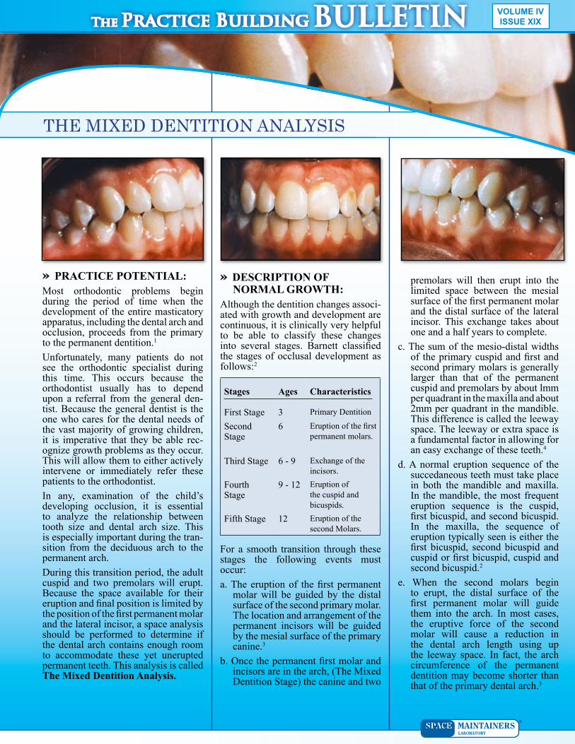
Although the dentition changes associated with growth and development are continuous, it is clinically very helpful to be able to classify these changes into several stages. Barnett classified the stages of occlusal development as follows:2
Stages Ages Characteristics
First Stage 3 Primary Dentition
Second Stage
6 Eruption of the first permanent molars.
Third Stage 6 9 Exchange of the incisors.
Fourth Stage
9 12 Eruption of the cuspid and bicuspids.
Fifth Stage 12 Eruption of the second Molars.
For a smooth transition through these stages the following events must occur:a. The eruption of the first permanent
molar will be guided by the distal surface of the second primary molar. The location and arrangement of the permanent incisors will be guided by the mesial surface of the primary canine.3
b. Once the permanent first molar and incisors are in the arch, (The Mixed Dentition Stage) the canine and two
» PRACTICE POTENTIAL:Most orthodontic problems begin during the period of time when the development of the entire masticatory apparatus, including the dental arch and occlusion, proceeds from the primary to the permanent dentition.1
Unfortunately, many patients do not see the orthodontic specialist during this time. This occurs because the orthodontist usually has to depend upon a referral from the general dentist. Because the general dentist is the one who cares for the dental needs of the vast majority of growing children, it is imperative that they be able recognize growth problems as they occur. This will allow them to either actively intervene or immediately refer these patients to the orthodontist.In any, examination of the child’s developing occlusion, it is essential to analyze the relationship between tooth size and dental arch size. This is especially important during the transition from the deciduous arch to the permanent arch.During this transition period, the adult cuspid and two premolars will erupt. Because the space available for their eruption and final position is limited by the position of the first permanent molar and the lateral incisor, a space analysis should be performed to determine if the dental arch contains enough room to accommodate these yet unerupted permanent teeth. This analysis is called The Mixed Dentition Analysis.
premolars will then erupt into the limited space between the mesial surface of the first permanent molar and the distal surface of the lateral incisor. This exchange takes about one and a half years to complete.
c. The sum of the mesiodistal widths of the primary cuspid and first and second primary molars is generally larger than that of the permanent cuspid and premolars by about lmm per quadrant in the maxilla and about 2mm per quadrant in the mandible. This difference is called the leeway space. The leeway or extra space is a fundamental factor in allowing for an easy exchange of these teeth.4
d. A normal eruption sequence of the succedaneous teeth must take place in both the mandible and maxilla. In the mandible, the most frequent eruption sequence is the cuspid, first bicuspid, and second bicuspid. In the maxilla, the sequence of eruption typically seen is either the first bicuspid, second bicuspid and cuspid or first bicuspid, cuspid and second bicuspid.2
e. When the second molars begin to erupt, the distal surface of the first permanent molar will guide them into the arch. In most cases, the eruptive force of the second molar will cause a reduction in the dental arch length using up the leeway space. In fact, the arch circumference of the permanent dentition may become shorter than that of the primary dental arch.3
THE MIXED DENTITION ANALYSIS
The Practice Building BULLETIN
The Practice Building BULLETINThe Practice Building BULLETIN• An Abnormal Eruption Sequence
normally the greater mesiodistal width of the second primary molar will permit easy eruption of the second bicuspid and provide space anteriorly for the cuspid. Should the arch length become shortened due to an unfavorable sequence of eruption, the cuspid will have insufficient space for its final positioning and will be forced to erupt in labioversion with a decided mesial inclination.3
Also in most cases, the eruptive force of the second molar will cause a reduction in the dental arch length and use up this leeway space. If this occurs before the second bicuspid erupts there may be insufficient space for it in the arch.3
When any of these problems exist, you must have the ability to determine whether or not you will have enough space to allow normal growth to continue. To this end, we use the Mixed Dentition Analysis.» THE MIXED DENTITION
ANALYSISThe purpose of the Mixed Dentition Analysis is to evaluate the amount of space available in the arch for the succeeding permanent teeth. Although many methods have been suggested, none of them are as precise as one might like. For example, radiographs have often been used to help estimate the size of the succedaneous teeth. However, distortions, enlargements, and rotations of the tooth germs make this method ineffective.The method presented here is advocated for the following reasons:1) it has minimal systemic error and the range of such error is known, 2) it can be done with equal reliability by the beginner and the expert, 3) it is not time consuming, 4) it requires no special equipment or radiographic projections, 5) it is easily done in the mouth or on dental casts, 6) it may be used for both dental arches.3
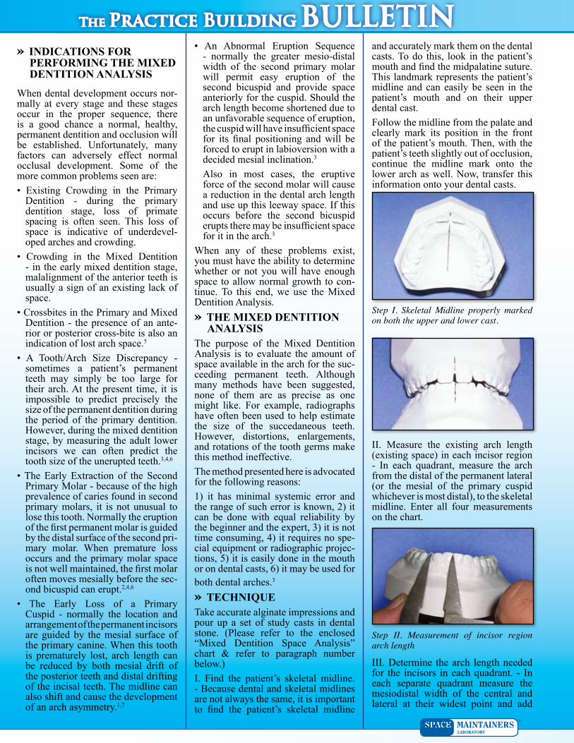
» TECHNIQUETake accurate alginate impressions and pour up a set of study casts in dental stone. (Please refer to the enclosed “Mixed Dentition Space Analysis” chart & refer to paragraph number below.)I. Find the patient’s skeletal midline. Because dental and skeletal midlines are not always the same, it is important to find the patient’s skeletal midline
and accurately mark them on the dental casts. To do this, look in the patient’s mouth and find the midpalatine suture. This landmark represents the patient’s midline and can easily be seen in the patient’s mouth and on their upper dental cast.Follow the midline from the palate and clearly mark its position in the front of the patient’s mouth. Then, with the patient’s teeth slightly out of occlusion, continue the midline mark onto the lower arch as well. Now, transfer this information onto your dental casts.
» INDICATIONS FOR PERFORMING THE MIXED DENTITION ANALYSIS
When dental development occurs normally at every stage and these stages occur in the proper sequence, there is a good chance a normal, healthy, permanent dentition and occlusion will be established. Unfortunately, many factors can adversely effect normal occlusal development. Some of the more common problems seen are:• Existing Crowding in the Primary
Dentition during the primary dentition stage, loss of primate spacing is often seen. This loss of space is indicative of underdeveloped arches and crowding.
• Crowding in the Mixed Dentition in the early mixed dentition stage, malalignment of the anterior teeth is usually a sign of an existing lack of space.
• Crossbites in the Primary and Mixed Dentition the presence of an anterior or posterior crossbite is also an indication of lost arch space.5
• A Tooth/Arch Size Discrepancy sometimes a patient’s permanent teeth may simply be too large for their arch. At the present time, it is impossible to predict precisely the size of the permanent dentition during the period of the primary dentition. However, during the mixed dentition stage, by measuring the adult lower incisors we can often predict the tooth size of the unerupted teeth.3,4,6
• The Early Extraction of the Second Primary Molar because of the high prevalence of caries found in second primary molars, it is not unusual to lose this tooth. Normally the eruption of the first permanent molar is guided by the distal surface of the second primary molar. When premature loss occurs and the primary molar space is not well maintained, the first molar often moves mesially before the second bicuspid can erupt.2,4,6
• The Early Loss of a Primary Cuspid normally the location and arrangement of the permanent incisors are guided by the mesial surface of the primary canine. When this tooth is prematurely lost, arch length can be reduced by both mesial drift of the posterior teeth and distal drifting of the incisal teeth. The midline can also shift and cause the development of an arch asymmetry.1,7
II. Measure the existing arch length (existing space) in each incisor region In each quadrant, measure the arch from the distal of the permanent lateral (or the mesial of the primary cuspid whichever is most distal), to the skeletal midline. Enter all four measurements on the chart.
III. Determine the arch length needed for the incisors in each quadrant. In each separate quadrant measure the mesiodistal width of the central and lateral at their widest point and add
Step I. Skeletal Midline properly marked on both the upper and lower cast.
Step II. Measurement of incisor region arch length
The Practice Building BULLETINThe Practice Building BULLETIN
cuspids and premolars in both arches. We use the Tanaka and Johnson System to make this prediction. Simply take one-half the sum of the mesiodistal width of the four lower incisors and add 10.5mm to it for the lower quadrants and 11.0mm or the upper quadrants. (e.g. lower - 6+5+5+6 = 22; 1/2 of 22 = 11; 11+10.5 = 21.5)
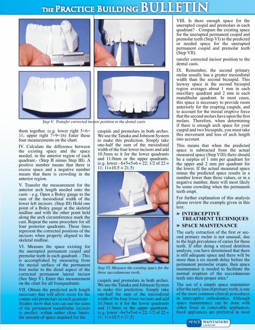
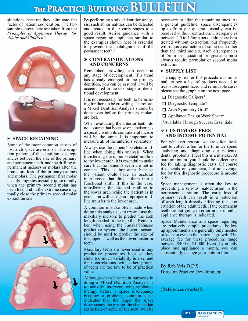
them together. (e.g. lower right 5+6= 11; upper right 7+9=16) Enter these four measurements on the chart.IV. Calculate the difference between the existing space and the space needed, in the anterior region of each quadrant.- (Step II minus Step III). A positive number means that there is excess space and a negative number means that there is crowding in the anterior region.V. Transfer the measurement for the anterior arch length needed onto the casts - e.g. Open a Boley gauge to the sum of the mesiodistal width of the lower left incisors. (Step III) Hold one point of a Boley gauge at the skeletal midline and with the other point held along the arch circumference mark the cast. Repeat the same procedure for all four posterior quadrants. These lines represent the corrected positions of the incisors when properly aligned to the skeletal midline.VI. Measure the space existing for the unerupted permanent cuspid and premolar teeth in each quadrant. - This is accomplished by measuring from the mesial surface of the permanent first molar to the distal aspect of the corrected permanent lateral incisor (See Step V). Enter this measurement on the chart for all fourquadrants.VII. Obtain the predicted arch length necessary that will allow room for the canine and premolars in each quadrant - Studies show that you can use the sizes of the permanent mandibular incisors to predict, within rather close limits, the amount of space required for the
VIII. Is there enough space for the unerupted cuspid and premolars in each quadrant? - Compare the existing space for the unerupted permanent cuspid and premolar teeth (Step VI) to the predicted or needed space for the unerupted permanent cuspid and premolar teeth (Step VII).ransfer corrected incisor position to the dental casts.IX. Remember, the second primary molar usually has a greater mesiodistal width than the second bicuspid. This leeway space in the second bicuspid region averages about 1 mm in each maxillary quadrant and 2 mm in each mandibular quadrant. In most cases, this space is necessary to provide room anteriorly for the erupting cuspids, and to account for the mesial eruptive force that the second molars have upon the first molars. Therefore, when determining if there is enough arch space for your cuspid and two bicuspids, you must take this movement and loss of arch length into account.This means that when the predicted space is subtracted from the actual measured space (Step VIII) there should be a surplus of 1 mm per quadrant for the upper and 2 mm per quadrant for the lower. If the actual measured space minus the predicted space results in a number lower than these values, or in a negative number, there will most likely be some crowding when the permanent teeth erupt.For further explanation of this analysis please review the example given in this text.» INTERCEPTIVE
TREATMENT TECHNIQUES» SPACE MAINTENANCEThe early extraction of the first or second primary molar is not unusual, due to the high prevalence of caries for these teeth. If after doing a mixed dentition analysis, you have determined that there is still adequate space and there will be more than a six month delay before the permanent premolar erupts, then space maintenance is needed to facilitate the normal eruption of the succedaneous teeth into their proper sites.8
The use of a simple space maintainer after the early loss of primary teeth, is one of the most common clinical procedures in interceptive orthodontics. Although space maintenance can be done with either fixed or removable appliances, fixed appliances are preferred in most
cuspids and premolars in both arches. We use the Tanaka and Johnson System to make this prediction. Simply take one-half the sum of the mesiodistal width of the four lower incisors and add 10.5mm to it for the lower quadrants and 11.0mm or the upper quadrants. (e.g. lower - 6+5+5+6 = 22; 1/2 of 22 = 11; 11+10.5 = 21.5)
Step V. Transfer corrected incisor position to the dental casts
Step VI. Measure the existing space for the three succedaneous teeth.
The Practice Building BULLETINThe Practice Building BULLETINsituations because they eliminate the factor of patient cooperation. The two samples shown here are taken from the Principles of Appliance Therapy for Adults and Children.8
» SPACE REGAININGSome of the more common causes of lost arch space are errors in the eruption pattern of the dentition, discrepancies between the size of the primary and permanent teeth, and the drifting of permanent incisors or molars after the premature loss of the primary canines and molars. The permanent first molar usually migrates mesially quite rapidly when the primary second molar has been lost, and in the extreme case may totally close the primary second molar extraction site.
By performing a mixed dentition analysis, such abnormalities can be detected and treated in their early stages to a good result. Active guidance with a space regaining appliance similar to the examples shown here is essential to prevent the malalignment of the permanent teeth.8
» CONTRAINDICATIONS AND CONCERNS
Remember, crowding can occur at any stage of development. If a trend has already emerged in the primary dentition, you can be assured it will be accentuated in the next stage of dentitional development.It is not necessary for teeth to be missing for there to be crowding. Therefore, a Mixed Dentition Analysis should be done even before the primary molars are lost.When evaluating the anterior teeth, do not assume that because one incisor has a specific width its contralateral incisor will be the same. It is important to measure all of the anteriors separately.Always use the patient’s skeletal midline when doing this analysis. When transferring the upper skeletal midline to the lower arch, it is essential to make sure that the patient’s teeth are not in contact. This is important because the patient could have an occlusal interference that directs them into a functional shift. If this is the case, transferring the skeletal midline to the lower arch while the patient is in occlusion will cause an improper midline transfer to the lower arch.A common mistake often made when doing this analysis is to try and use the maxillary incisors to predict the arch length needed in the maxilla. Remember, when using the Tanaka/Johnson predictive system, the lower incisors should be used to predict the size of the upper as well as the lower posterior teeth.Maxillary teeth are never used in any predictive procedures because they show too much variability in size, and their correlations with other groups of teeth are too low to be of practical value.Although one of the main purposes in doing a Mixed Dentition Analysis is to actively intervene with appliance therapy before a space discrepancy becomes a problem, common sense indicates that the larger the space discrepancy the greater the chance that extraction of some of the teeth will be
necessary to align the remaining ones. As a general guideline, space discrepancies up to 2mm per quadrant usually can be resolved without extraction. Discrepancies between 2.5 to 4.5mm per quadrant are best treated without extraction, but frequently will require extraction of some teeth other than the third molars. Arch discrepancies of 5mm per quadrant or greater almost always require premolar or second molar extractions.1
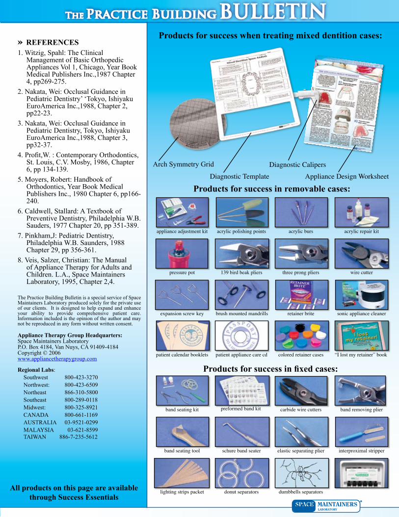
» SUPPLY LISTThe supply list for this procedure is minimal, to see a list of products needed to treat subsequent fixed and removable cases please see the graphic on the next page.
Diagnostic Calipers*Diagnostic Template*Arch Symmetry Grid*Appliance Design Work Sheet*
(*Available Through Success Essentials)
» CUSTOMARY FEES AND INCOME POTENTIALFor whatever reason, we are often hesitant to collect a fee for the time we spend analyzing and diagnosing our patients’ dental problems. I feel this is a mistake. At bare minimum, you should be collecting a fee for taking diagnostic casts. Of course it depends on your area, but an average fee for this diagnostic procedure is around $100.Space management is often the key to preventing a serious malocclusion in the permanent dentition. The early loss of primary teeth can result in a reduction of arch length directly effecting the later eruption of the adult teeth. If the permanent teeth are not going to erupt in six months, appliance therapy is indicated.Space Maintenance and space regaining are relatively simple procedures. Follow up appointments are generally only needed to keep an eye on the patients’ growth. The average fee for these procedures range between $400 to $1,000. Even if you only place one appliance a month, you can substantially change your bottom line.
By Rob Veis D.D.S. DirectorPractice Development
(References overleaf)
The Practice Building BULLETINThe Practice Building BULLETIN» REFERENCES1. Witzig, Spahl: The Clinical
Management of Basic Orthopedic Appliances Vol 1, Chicago, Year Book Medical Publishers Inc.,1987 Chapter 4, pp269275.
2. Nakata, Wei: Occlusal Guidance in Pediatric Dentistry’ ‘Tokyo, Ishiyaku EuroAmerica Inc.,1988, Chapter 2, pp2223.
3. Nakata, Wei: Occlusal Guidance in Pediatric Dentistry, Tokyo, Ishiyaku EuroAmerica Inc.,1988, Chapter 3, pp3237.
4. Profit,W. : Contemporary Orthodontics, St. Louis, C.V. Mosby, 1986, Chapter 6, pp 134139.
5. Moyers, Robert: Handbook of Orthodontics, Year Book Medical Publishers Inc., 1980 Chapter 6, pp166240.
6. Caldwell, Stallard: A Textbook of Preventive Dentistry, Philadelphia W.B. Sauders, 1977 Chapter 20, pp 351389.
7. Pinkham,J: Pediatric Dentistry, Philadelphia W.B. Saunders, 1988 Chapter 29, pp 356361.
8. Veis, Salzer, Christian: The Manual of Appliance Therapy for Adults and Children. L.A., Space Maintainers Laboratory, 1995, Chapter 2,4.
The Practice Building Bulletin is a special service of Space Maintainers Laboratory produced solely for the private use of our clients. It is designed to help expand and enhance your ability to provide comprehensive patient care. Information included is the opinion of the author and may not be reproduced in any form without written consent.
Appliance Therapy Group Headquarters:Space Maintainers LaboratoryP.O. Box 4184, Van Nuys, CA 914094184Copyright © 2006www.appliancetherapygroup.com
Regional Labs:Southwest 8004233270Northwest: 8004236509Northeast 8663105800Southeast 8002890118Midwest: 8003258921CANADA 8006611169AUSTRALIA 0395210299MALAYSIA 036218599TAIWAN 88672355612
Products for success when treating mixed dentition cases:
Arch Symmetry Grid
Diagnostic TemplateDiagnostic Calipers
Appliance Design Worksheet
Products for success in removable cases:
Products for success in fixed cases:
appliance adjustment kit acrylic polishing points acrylic burs acrylic repair kit
pressure pot 139 bird beak pliers three prong pliers wire cutter
expansion screw key retainer brite sonic appliance cleaner
patient calendar booklets patient appliance care cd colored retainer cases
band seating kit preformed band kit carbide wire cutters band removing plier
band seating tool schure band seater elastic separating plier interproximal stripper
lighting strips packet donut separators dumbbells separators
brush mounted mandrills
“I lost my retainer” book
All products on this page are available through Success Essentials
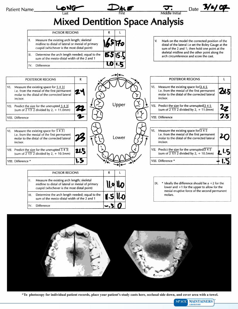
*To photocopy for individual patient records, place your patient’s study casts here, occlusal side down, and cover area with a towel.