Embed Size (px)
Citation preview

16 Microgram Journal, Volume 7, Number 1 (March 2010)
The Mass Spectrum of Cocaine:
Deuterium Labeling and MS/MS Studies
R. Martin Smith*
rMS Workshops, LLC
6442 Dylyn Drive
Madison, WI 53719
[email: rmsworkshopsllc -at- msn.com]
John F. Casale U.S. Department of Justice
Drug Enforcement Administration
Special Testing and Research Laboratory
22624 Dulles Summit Court
Dulles, VA 20166
[email address withheld at author’s request]
ABSTRACT: Seven derivatives of cocaine (l-2-exo-carbomethoxy-3-exo-benzoyloxytropane; Ia) were synthesized in which the
hydrogen (H) atoms at various positions were replaced by deuterium (2H = D) – specifically, at the N-methyl, O-methyl, phenyl, 2-,
3-, and 4- positions, as well as at the combined 1-, 5-, 6-, and 7- positions (Ib - Ih, respectively). The mass spectra of these com-
pounds were recorded. Elemental compositions for selected ions in the spectrum of Ia were determined using high-resolution mass
spectrometry, and precursor and product ion spectra for many of these ions were studied using MS/MS. Mechanisms for many pre-
viously proposed fragmentation pathways were either confirmed or clarified, and new insights were gained into the fragmentation of
Ia. Reasons for variations in relative intensities of the m/z 94 and 152 peaks between the spectra of the cocaine diastereomers are
proposed.
KEYWORDS: forensic science, cocaine, mass spectrometry, fragmentation mechanisms, deuterium-labeled derivatives, high-
resolution MS, MS/MS, product and precursor ions, pseudococaine.
The electron ionization (EI) mass spectra of tropane deriva-
tives were first studied by Budzikiewicz, Djerassi, and cowork-
ers, using the spectra of deuterated derivatives and the examina-
tion of metastable ion spectra [1,2]. Cocaine (l-2-exo-
carbomethoxy-3-exo-benzoyloxytropane; Ia), a molecule of
considerable forensic interest, was not included in the original
studies. Subsequent workers elucidated similar basic fragmen-
tation pathways for Ia (Figures 1-3) [3,4,5]. Many questions
remain, however, regarding the nature of some fragmentations
and the formation of less abundant ions. In particular, previous
proposals concerning the relative intensities of the m/z 152 peak
and the m/z 94/96 pair of peaks in the spectra of the cocaine
diastereomers seem unsatisfactory. The present work attempts
to expand upon this knowledge.
Seven labeled derivatives of Ia were prepared in which the
hydrogen (H) atoms at specific positions were replaced by deu-
terium (2H = D; structures Ib-Ih). Mass spectra for these com-
pounds were recorded and examined to determine the presence
or absence of the labels for many of the ions represented in the
cocaine spectrum. In addition, MS/MS data were collected in
both the precursor (“parent”) and product (“daughter”) ion
modes in order to ascertain relationships between the various
ions.
Experimental Procedures
Solvents, Chemicals, and Materials
All solvents were distilled-in-glass products of Burdick and
Jackson Laboratories (Muskegon, MI). N-Methyl-N-trimethyl-
silyltrifluoroacetamide (MSTFA) was obtained from Pierce
Chemical (Rockford, IL). All other chemicals were
reagent grade quality products of Sigma-Aldrich Chemical
(Milwaukee, WI). Alumina (basic) was deactivated slightly by
adjusting the water content to 4% (w/w). Cocaine (Ia), pseu-
dococaine, ecgonine methyl ester, N-trideuteriomethylcocaine
(Ic), and -cocaine were from the authentic reference collection
of the DEA Special Testing and Research Laboratory.
Cocaine (Ia: All H = H)
O-CD3 (Ib: O-CH3 = OCD3; all other H = H)
NCD3 (Ic: N-CH3 = N-CD3; all other H = H)
Phenyl-d5 (Id: C6H5 = C6D5; all other H = H)
2-d1 (Ie: H2 = D; all other H = H)
3-d1 (If: H3 = D; all other H = H)
4,4-d2 (Ig: H4 = D; all other H = H)
1,5,6,6,7,7-d6 (Ih: H1 = H5 = H6 = H7 = D; all other H = H)

Microgram Journal, Volume 7, Number 1 (March 2010) 17
Figure 1 - alpha-Cleavage fragmentations of the cocaine molecular ion after initial ionization at N.
Synthesis
All syntheses were performed in flame-dried glassware and
protected from moisture. Ie, If, Ig, Ih, and 2-d1-, 3-d1-, 4,4-d2-,
1,5,6,6,7,7-d6-pseudococaine were synthesized as previously
described [6]. Unlabelled 2-carbomethoxy-3-tropinone was
also synthesized as previously described [7]. Yields for the
following syntheses were not optimized.
Cocaine (OCD3) (Ib): Anhydrous benzoylecgonine (110 mg,
0.380 mmol) was combined with CH2Cl2 (4 mL) and
1’,1’-carbonyldiimidazole (63.5 mg, 0.391 mmol) in a 15 mL
centrifuge tube. The reaction was allowed to stand overnight.
Trideuteriomethanol (CD3OH; 100 μL) was added and the mix-
ture allowed to stand for one day. The reaction mixture was
evaporated to dryness under a stream of nitrogen at 75oC and
treated with 7 mL of hot hexane. The hexane was decanted to a
new tube and allowed to cool, precipitating the imidazole by-
product. The hexane was filtered, evaporated in vacuo, recrys-
tallized from hexane, and dried to provide a white powder
(42 mg, 36% yield).
Cocaine (phenyl-d5) (Id): Ecgonine methyl ester hydrochlo-
ride (500 mg, 2.12 mmol) was suspended in 7 mL of dry pyri-
dine in a 100 mL round bottom flask, to which pentadeute-
riobenzoyl chloride (437 mg, 3.00 mmol) was added. After
stirring for 5 days, the reaction was diluted with 50 mL of dry
acetone to precipitate the product. The crude product was cap-
tured by suction filtration, dissolved into water (4 mL), ren-
dered alkaline (pH = 9) with aqueous Na2CO3, extracted with
CHCl3 (1 x 8 mL), dried over anhydrous Na2SO4, filtered, and
evaporated in vacuo to a crystalline mass. The product was
recrystallized from hexane to provide a white powder (114 mg,
17% yield).
Anhydroecgonine methyl ester: Cocaine base (7.1 g, 23.4
mmol) was refluxed in 6N HCl overnight, and then evaporated
to a residue on a steam bath. The residue was transferred to a
flask containing MeOH (50 mL) and boron trifluoride diethyl
etherate (1.0 g, 7.05 mmol) and refluxed for 2 hours. The reac-
tion was evaporated in vacuo, quenched with water (40 mL),
adjusted to pH 9 with Na2CO3, extracted with CHCl3 (2 x 100
mL), dried over anhydrous Na2SO4, filtered, and evaporated in
vacuo to a semi-crystalline mass. Approximately 1.5 g of the
crude product was dissolved in a minimal amount of Et2O and
loaded onto a glass chromatographic column (1 x 22 cm) con-
taining 15 g of basic alumina (150 mesh). The column was
eluted sequentially with 20 mL each of the following solvents:
1) Et2O, 2) Et2O/CHCl3 (1:1), and 3) CHCl3. Ten mL fractions
were collected and examined by GC/MS, both underivatized
and following derivatization with MSTFA. The first three frac-
tions were combined and evaporated to dryness to give a clear
oil (655 mg, 15% yield).
2,3-Dehydrococaine: 2-Carbomethoxy-3-tropinone (153 mg,
0.776 mmol) was combined with dry pyridine (2 mL) and ben-
zoyl chloride (300 mg, 2.13 mmol) in a 15 mL centrifuge tube
and left to stand for 1 hour. Et2O (13 mL) was added to pre-
cipitate the crude product, which was washed with additional
Et2O (10 mL). The semi-crystalline material was dissolved in
0.36N H2SO4 (1 mL), washed with Et2O (2 x 14 mL), adjusted
to pH 9 with Na2CO3, extracted with CHCl3 (1 x 10 mL), dried
over anhydrous Na2SO4, filtered, and evaporated in vacuo to
give an off-white powder (118 mg, 50% yield).
Gas Chromatography/Low Resolution-Mass Spectrometry
(GC/LR-MS)
GC/LR-MS analyses were performed using an Agilent (Palo
Alto, CA) Model 5973 quadrupole mass-selective detector
(MSD) interfaced with an Agilent Model 6890 gas chromato-
graph. The GC system was fitted with a 30 m x 0.25 mm ID
fused-silica capillary column coated with DB-1 (0.25 m)
(J & W Scientific, Rancho Cordova, CA). The oven

18 Microgram Journal, Volume 7, Number 1 (March 2010)
Figure 2 - Previously proposed fragmentations of Ia following initial alpha-cleavage next to the N atom.

Microgram Journal, Volume 7, Number 1 (March 2010) 19
Figure 3 - Previously proposed fragmentations of Ia following initial ionization at the ester O atoms.

20 Microgram Journal, Volume 7, Number 1 (March 2010)
temperature was programmed as follows: Initial temperature,
100oC; initial hold, 0.0 min; program rate, 6oC/min; final tem-
perature, 300oC; final hold, 5.67 min. The injector was oper-
ated in the split mode (21.5:1) at a temperature of 280oC. The
MSD was operated in the electron ionization (EI) mode with an
ionization energy (I.E.) of 70 eV and a scan range of 34-700
m/z units at 1.34 scans/s. The auxiliary transfer line to the
MSD and the source were maintained at 280oC and 230oC, re-
spectively.
High Resolution-Mass Spectrometry (HR-MS)
HR-MS data were obtained on two instruments. The first
was a Finnigan MAT Model 8230 system (Bremen, Germany)
operating at an I.E. of 70 eV. The source temperature was
175oC. The second was an AEI Scientific Apparatus Model
MS-902 system (Manchester, UK) operating at an I.E. of 70
eV. The source temperature was maintained at 120-150oC.
Sample introduction was accomplished with a solids probe on
both systems, and data was acquired at a resolution of ca.
10,000 (5% valley).
Precursor/Product Ion Mass Spectrometry (MS/MS)
Precursor and product ion data were obtained on a Thermo
Scientific TSQ-7000 triple quadrupole MS system (Bremen,
Germany). Ionization energies were varied from 5-70 eV.
Sample introduction was accomplished with solids probe. Un-
fortunately, these data were obtained several years ago without
details of the conditions used being recorded. These instru-
ments are no longer in the authors’ laboratories.
Results and Discussion
Electron ionization mass spectra of Ia and of the seven D-
labeled derivatives (Ib-Ih) are shown in Figures 4-6. Careful
examination and comparison of these spectra reveal intensity
patterns that are reasonably consistent from one spectrum to the
next. The positions of individual peaks within the spectra,
however, vary depending upon the presence or absence of the D
label(s) in the ion(s) represented. When some or all the D label
is retained in a particular ion, the position of the peak corre-
sponding to this ion moves to a higher m/z value in the spec-
trum of the D-labeled compound (the “shift technique”) [8].
The number of D atoms retained is determined by the differ-
ence in m/z value between the peak in the cocaine spectrum and
that in the spectrum of the D-labeled derivative.
A compilation of m/z values for selected peaks in the spec-
trum of Ia and for the corresponding peaks in the spectrum of
each of the derivatives is shown in Table I. In many cases, each
peak in the spectrum of Ia can be correlated to a single peak in
the spectrum of each derivative. When this occurs, a single
fragmentation mechanism may account for formation of that
ion.
In some cases, especially for lower m/z peaks, a single peak
in the spectrum of Ia is represented by two or more peaks in a
derivative spectrum. This indicates that two or more mecha-
nisms may be operative in forming that ion, or that a single
peak in the low-resolution spectrum may represent two or more
ions. Given the multiplicity of potential precursors for many
low m/z ions, this is not surprising. In a few instances, the peak
correspondences between spectra are either too complex to be
determined with confidence or suffer from interferences from
Figure 4 - Electron ionization mass spectra of a) cocaine (Ia);
b) N-CD3-norcocaine (Ic); and c) benzoylecgonine
trideuteriomethylester (Ib).
nearby peaks. These instances are noted in Table I.
Table II summarizes the elemental compositions of ions
formed during the fragmentation of Ia as determined by
HR-MS. The elemental compositions confirm those deter-
mined by Shapiro, et al. [9], although the list presented here is
more extensive. Table II also lists precursor and product ions
for these ions as identified by MS/MS. These data represent a
substantial condensation of the raw data. In order to best iden-
tify precursor-product ion relationships, emphasis was placed
on scans at the lowest collision energies (5-10 eV) because
product ions formed under these conditions are the least likely
to undergo additional fragmentation. A schematic representa-
tion of these relationships is shown in Figure 7. Although it is
tempting to use the information in Figure 7 to define EIMS
fragmentation pathways for Ia, it must be remembered that both
precursor and product ions formed by collisional activation in
the MS/MS experiments may have different structures than
those formed during EIMS [10].
The molecular ion (M+·) of Ia (C17H21NO4+·, m/z 303) frag-
ments at collision energies of 5-20 eV to produce at least nine
stable product ions that apparently form without the detectable
intermediacy of other ions. These include the listed ions having
m/z 272, 222, 198, 182, 181, 122, 97, 83, and 82 (Table III).
Although product ion scans for m/z 303 did not produce peaks
at m/z 288, 275, 274, 259, 244, and 155 under these conditions,
other data indicate that in the EI spectrum these ions may also
Microgram Journal, Volume 7, Number 1 (March 2010) 21
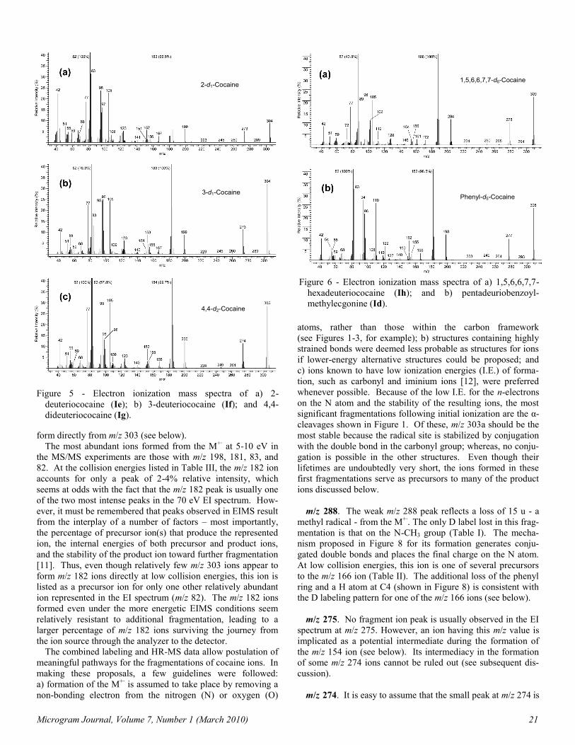
Figure 5 - Electron ionization mass spectra of a) 2-
deuteriococaine (Ie); b) 3-deuteriococaine (If); and 4,4-
dideuteriococaine (Ig).
Figure 6 - Electron ionization mass spectra of a) 1,5,6,6,7,7-
hexadeuteriococaine (Ih); and b) pentadeuriobenzoyl-
methylecgonine (Id).
form directly from m/z 303 (see below).
The most abundant ions formed from the M+· at 5-10 eV in
the MS/MS experiments are those with m/z 198, 181, 83, and
82. At the collision energies listed in Table III, the m/z 182 ion
accounts for only a peak of 2-4% relative intensity, which
seems at odds with the fact that the m/z 182 peak is usually one
of the two most intense peaks in the 70 eV EI spectrum. How-
ever, it must be remembered that peaks observed in EIMS result
from the interplay of a number of factors – most importantly,
the percentage of precursor ion(s) that produce the represented
ion, the internal energies of both precursor and product ions,
and the stability of the product ion toward further fragmentation
[11]. Thus, even though relatively few m/z 303 ions appear to
form m/z 182 ions directly at low collision energies, this ion is
listed as a precursor ion for only one other relatively abundant
ion represented in the EI spectrum (m/z 82). The m/z 182 ions
formed even under the more energetic EIMS conditions seem
relatively resistant to additional fragmentation, leading to a
larger percentage of m/z 182 ions surviving the journey from
the ion source through the analyzer to the detector.
The combined labeling and HR-MS data allow postulation of
meaningful pathways for the fragmentations of cocaine ions. In
making these proposals, a few guidelines were followed:
a) formation of the M+· is assumed to take place by removing a
non-bonding electron from the nitrogen (N) or oxygen (O)
atoms, rather than those within the carbon framework
(see Figures 1-3, for example); b) structures containing highly
strained bonds were deemed less probable as structures for ions
if lower-energy alternative structures could be proposed; and
c) ions known to have low ionization energies (I.E.) of forma-
tion, such as carbonyl and iminium ions [12], were preferred
whenever possible. Because of the low I.E. for the n-electrons
on the N atom and the stability of the resulting ions, the most
significant fragmentations following initial ionization are the α-
cleavages shown in Figure 1. Of these, m/z 303a should be the
most stable because the radical site is stabilized by conjugation
with the double bond in the carbonyl group; whereas, no conju-
gation is possible in the other structures. Even though their
lifetimes are undoubtedly very short, the ions formed in these
first fragmentations serve as precursors to many of the product
ions discussed below.
m/z 288. The weak m/z 288 peak reflects a loss of 15 u - a
methyl radical - from the M+·. The only D label lost in this frag-
mentation is that on the N-CH3 group (Table I). The mecha-
nism proposed in Figure 8 for its formation generates conju-
gated double bonds and places the final charge on the N atom.
At low collision energies, this ion is one of several precursors
to the m/z 166 ion (Table II). The additional loss of the phenyl
ring and a H atom at C4 (shown in Figure 8) is consistent with
the D labeling pattern for one of the m/z 166 ions (see below).
m/z 275. No fragment ion peak is usually observed in the EI
spectrum at m/z 275. However, an ion having this m/z value is
implicated as a potential intermediate during the formation of
the m/z 154 ion (see below). Its intermediacy in the formation
of some m/z 274 ions cannot be ruled out (see subsequent dis-
cussion).
m/z 274. It is easy to assume that the small peak at m/z 274 is
2-d1-Cocaine
3-d1-Cocaine
4,4-d2-Cocaine
1,5,6,6,7,7-d6-Cocaine
Phenyl-d5-Cocaine

22 Microgram Journal, Volume 7, Number 1 (March 2010)
Cocaine peak m/z
O-CD3 m/z
N-CD3 m/z
Phenyl-d5 m/z
2-d1 m/z
3-d1 m/z
4,4-d2 m/z
1,5,6,6,7,7-d6 m/z
303 306 306 308 304 304 305 309
288 291 288 293 289 289 290 294
274 277 277 279 275 275 275,276 276
272 272 275 277 273 273 274 278
259 262 262 264 260 260 261 265
244 244 247 249 245 245 246 250
222 225 222 227 223 223 224 224
198 201 201 198 199 199 200 204
182 185 185 182 183 183 184 188
181 184 184 181 181,182 182 182,183 187
166 166,169 (166),169 166 (166),167 166,167 (167),168 (171),172
155 158 158 155 156 155 155,156 161
154 157 157 154 155 154? 154+ 155?, 156?
152 155 155 152 152 153 153 154
150 150 153 150 151? 151 151,152 155,156
140 140 143 140 141 140 140 144,145,146
138 138 141 138 139? 139 139,140 140,141?
122a 122 125 122 123 123 (123),124 127,128
122b 122 122 127 122 122 122 122
108 108 111 108 108,109 109 109,110 112,113
105 105 105 110 105 105 105 105
100 103 100? 100 101 101 101,102? ?
97 97 100 97 97? 97 98,99? 102,103?
96 96 99 96 96(-97?) 96 96,98? 96?,101?
94 94 97 94 95 95 95 96?
83 83 86 83 83 83,84? 83,84 88,89
82 82 85 82 82 82 82,83? 87,88?
77 77 77 82 77 77 77 77
68 68 ? + 71 68 68 68 68 69-73?
67 67 67 + ? 67 67 67 67 + ? 68-73?
59 62 59 59 59 59 59 59
55 55 58? 55 55 55 55 57-61?
51 51 51 54 51 51 51 51
42 42 45 42 42 42 42 43
Table I - Peak Correspondence Table.
Key: -Boldface indicates partial or total loss of deuterium label at that position.
-Numbers in parentheses indicate a peak of much smaller intensity.
-Question mark (?) indicates that data cannot be interpreted with confidence.

Microgram Journal, Volume 7, Number 1 (March 2010) 23
Figure 7 - Relationships between various cocaine fragment ions as determined by MS/MS.
an isotope peak in the m/z 272 ion cluster. Alerted by the
MS/MS data that an ion having m/z 274 is a precursor to the m/z
152 ion (Table II), careful analysis reveals that this peak is con-
sistently about 0.3-0.6% too large to be due to isotope contribu-
tions alone. The spectra of the D-labeled derivatives shows
partial loss of one D at C4 for this peak, plus the loss of four D
atoms from positions 1, 5, 6, and 7. Loss of the D label at C2
could not be determined with certainty. Because an ion having
m/z 274 reflects the loss of 29 u from the M+·, and five of these
mass units are due to H atoms, the remainder must consist of
two C atoms. This loss is most easily envisioned as a H atom
from either C2 or C4 plus C6 and C7 with their four attached H
atoms (Figures 9 and 10). This may occur via H rearrangement
followed by ethyl radical loss [1,13], or by loss of ethylene to
produce m/z 275 and subsequent H radical loss from either
C2 or C4. In 2-carbomethoxy-3-tropanone (II), 2,3-
anhydrococaine (III), and other tropanes having some unsatura-
tion at C2 and/or C3, peaks are observed at both M-28 and M-
29, indicating that, at least in those compounds, both pathways
occur [1, 2, 14]. In those cases, loss of 29 u leads to an ion hav-
ing aromatic character (Figure 11). That is not the case here.
Loss of benzoic acid from m/z 274 produces the m/z 152 ion
with additional loss of the D labels on the phenyl ring and
either C2 or C4 (see below).
m/z 272. The m/z 272 ion is formed by loss of a methoxy
radical from the methyl ester group in the M+·. This is supported
by the elemental composition and labeling data in Tables I and
II. As expected, the only D label lost is the one on the OCH3
group. Two mechanisms for formation are possible that cannot
be distinguished using available data (Figures 2 and 3). Both
have been proposed previously [3,4,15].
m/z 259. No HR-MS or MS/MS data were recorded for this
ion during this study. The loss from the M+· is 44 u, the mass of
CO2. All D labels are retained, indicating that no H atoms are
lost during this fragmentation. It is impossible to tell from the
available data which of the two carboxyl groups is lost. The
mechanism in Figure 12 shows loss of the alkyl carboxyl group.
Although a stepwise mechanism is shown, a nearly concerted
elimination of CO2 with methyl group migration is possible.
m/z 244. The elemental composition determined for this ion
by HR-MS shows loss of C2H3O2 from the M+·. The only D
label lost is that on the OCH3 group, consistent with loss of a
carbomethoxy radical.
Formation of m/z 244 is most easily depicted as resulting
from m/z 303c or 303d (Figures 9 and 10). Its formation from
m/z 303a cannot occur without H rearrangement, and formation
from m/z 303b would likely result in either a cyclopropane ring
or a diradical. “Backside” attack at C2 by the radical site at C7
in m/z 303c causes elimination of a carbomethoxy radical and
generates a bicyclo[2.2.2] ion (Figure 9) [5]. Alternatively, a
similar attack by a radical site at C6 produces a bicyclo[4.2.0]
ion (Figure 10).
Both proposed m/z 244 ions have fragmentation options that
can lead to product ions listed in Table II. Loss of ethylene by
a cycloelimination reaction forms m/z 216 (identified as a prod-
uct ion in the MS/MS data, even though the corresponding peak
is not observed in the spectrum). The m/z 244 ion is a possible
precursor to the m/z 105 ion, as well as to one or more of the
m/z 122 ions [5]. Cyclic loss of benzoic acid or its correspond-
ing radical ion produces m/z 122a1 and 122a2 or 122b, respec-
tively; and migration of the charge to the benzoyl O atom gen-
erates the benzoyl ion (m/z 105).
m/z 222. This ion, which is usually represented by a peak of
very low intensity in the EI spectrum, is formed directly from

24 Microgram Journal, Volume 7, Number 1 (March 2010)
Table II - Elemental Composition and MS/MS Table.
Cocaine
Ion m/z
Elemental Composition
Fragment Lost Precursor Ion(s) At 5-15
eV (m/z)a,b
Important Product Ion(s) At 5-15 eV (m/z)a,c
303 C17H21NO4 None None 272,222,198,182,181,83,82
288 Not Determined (CH3) (303) (166)
274 Not Determined (C2H5) (303) (152)
272 C16H18NO3 OCH3 (303) 150, 122, 105, 82
244 C15H18NO2 CH3CO2 (303) 216, 122, 105
222 Not Determined (C5H7N) (303) (100)
198 C10H16NO3 C6H5CO 303 166, 82
182 C10H16NO2 C6H5CO2 303 150, 108, 82
181 C10H15NO2 C6H5CO2H (303) 180,166,152,122,108,94
166 C9H12NO2 C8H9O2 303,288,198,181 138, 134, 110, 82
155 C8H13NO2 C9H8O2 (303) 140, 96, 82
154 C8H12NO2 C9H9O2 (275), (155) (152), 122, 94
152 C8H10NO2 C9H11O2 274,181,154 122, 108, 93, 92, 59
150 C9H12NO C8H9O3 (272), (182) 122, 119, 93, 91, 82
140 C7H10NO2 C10H11O2 155 Not Determined
138 C7H8NO2 C10H13O2 (166) Not Determined
122a C8H12N C9H9O4 181, 150 107, 94, 91, 81
122b C7H6O2 C10H15NO2 303,272,244,222 105, 77
108 C7H10N C10H11O4 (182),(181),(152) 93, 42
105 C7H5O C10H16NO3 272,244,122 77, 51
100 C5H8O C12H13NO3 303, 222 Not Determined
97 C6H11N C11H10O4 303,156 96, 82, 55
96 C6H10N C11H11O4 303,155,97 94, 81, 79, 68, 42
94 C6H8N C11H13O4 303,181,154,122,96 93, 78
83 C5H9N C12H12O4 (303) 82, 42
82 C5H8N C12H13O4 303,182,97 80, 67
77 C6H5 C11H16NO4 (105) Not Determined
68 C4H6N C13H15O4 (97), (96) Not Determined
67 Not Det. Not Det. (96),(94),(82) Not Determined
59 C2H3O C15H18NO2 (152) Not Determined
55 C3H5N C14H16O4 (166),(97),(82) Not Determined
51 (C4H3) - (105), (77) Not Determined
42 C2H4N C15H17O4 (122),(108),(96) Not Determined
aBoldface indicates the most prominent precursor or product ion(s) of the group. bValues in parentheses are proposals based on other information, most often data from product ion scans. cValues in parentheses are proposals based on other information, often data from precursor ion scans.

Microgram Journal, Volume 7, Number 1 (March 2010) 25
the M+· at low internal energies (Table III). The loss (81 u) is
an unusual one, considering the functional groups present in the
molecule. The spectra of the D-labeled derivatives show losses
of the N-CD3 label as well as four of the six D atoms on C1,
C5, C6, and C7. These losses strongly suggest that the entire
5-membered ring, consisting of the N atom and C1, C5, C6, and
C7, is lost as a unit, with two H atoms being transferred back to
the remaining portion of the molecule. Because peaks repre-
senting ions having similar m/z values are very prominent in the
spectrum (m/z 82 and 83), loss of this portion of the M+· as a
neutral species is not unreasonable. Loss of the N atom, in
addition to the even molecular mass, dictates that m/z 222 is an
odd-electron ion; therefore the lost neutral species is either a
molecule or a diradical.
Two fragment ions associated with m/z 222 by MS/MS are
those at m/z 122 and 100. The m/z 122 ion must be
C6H5CO2H+· because it cannot contain the N atom. The mecha-
nisms shown in Figure 13 account for these observations.
Figure 8 - Proposed formation of m/z 288 and m/z 166 from m/z 303b.
Table III. Tabulated Product Ion Spectrum of the Molecular
Ion of Ia at Various Collision Energies.
Product Ion (m/z)
Rel. Int. (-20 eV)
Rel. Int. (-13 eV)
Rel. Int. (-5 eV)
272 - 0.5 2.0
222 - - 5.0
198 16.0 32.0 40.0
182 4.0 3.0 2.0
181 4.0 10.0 50.0
180 1.0 - -
166 2.0 1.0 -
122 3.0 2.0 2.0
108 5.0 4.0 2.0
107 0.5 1.0 -
100 1.0 2.0 -
97 1.0 1.0 2.0
96 1.0 0.5 -
95 1.0 2.0 5.0
94 1.0 1.0 -
83 38.0 72.0 100.0
82 100.0 100.0 60.0
81 9.0 13.0 30.0

26 Microgram Journal, Volume 7, Number 1 (March 2010)
Figure 9 - Proposed formation of m/z 275a and m/z 244a after initial cleavage of the C1-C7 bond (m/z 303c). Subsequent fragmenta-
tions of m/z 275a and m/z 244a include formation of m/z 274a, m/z 216, m/z 152, m/z 122a1, and m/z 105.
m/z 182. The M+· loses C7H5O2 – benzoate radical – to form
this ion (Tables II and III). None of the D labels except those
on the phenyl ring are lost. A straightforward mechanism for
its formation is shown in Figure 2 [15,16].
m/z 181. Loss of benzoic acid (C6H5CO2H) directly from the
M+· produces this ion (Tables II and III). The D labels on the
aromatic ring are clearly lost in this fragmentation, and loss of a
H atom from either C2 or C4 is supported. The m/z 181 ion is
implicated as an intermediate in forming ions having m/z 166,
152, 122a (it cannot be C6H5CO2H+·) and 108. Generation of
m/z 181 involves a McLafferty-type rearrangement after initial
m/z 198. The combined HR-MS and MS/MS data show loss
of C7H5O directly from the M+· (Tables II and III). The only D
labels lost are those on the aromatic ring, thus the neutral frag-
ment must be a benzoyl radical (C6H5CO·). In the spectra of
α- and β-cocaine (IVa and IVb, respectively; Figure 14a – the
spectra of these two compounds are virtually identical), the
peak associated with this ion is quite small, indicating that for-
mation of this ion may require the presence of a H atom at C3,
which is not present in either α- or β-cocaine. The m/z 198 ion
is an important precursor to one of the m/z 166 ions, which can
occur by loss of a molecule of methanol (Figure 2) [4].

Microgram Journal, Volume 7, Number 1 (March 2010) 27
Figure 10 - Proposed formation of m/z 275b and m/z 244b after initial cleavage of the C5-C6 bond (m/z 303d). Subsequent fragmen-
tations of m/z 275b and m/z 244b include formation of m/z 274b, m/z 216, m/z 152, m/z 122a2, and m/z 105.

28 Microgram Journal, Volume 7, Number 1 (March 2010)
Figure 11 - Losses of C2H5 from 2-carbomethoxy-3-tropanone (II) and 2,3-anhydrococaine (III) produce ions stabilized by aromatic
conjugation.

Microgram Journal, Volume 7, Number 1 (March 2010) 29
Figure 12 - Proposed formation of the m/z 259 ion.
Figure 13 - Proposed formation of m/z 222 and its subsequent fragmentation to give the m/z 122 and m/z 100 ions.

30 Microgram Journal, Volume 7, Number 1 (March 2010)
Figure 14 - Electron ionization mass spectra of a) alpha-cocaine
(IVa); and b) pseudococaine.
ionization at the carbonyl O atom of the benzoyloxy group [5].
In this case, the charge ends up on the olefin product. Two
isomeric structures for m/z 181 are possible; they appear to suf-
fer different fates (Figures 15 and 16).
m/z 166. Determining the origin of the m/z 166 peak illus-
trates the danger of assuming there is a one-to-one correlation
between ions and peaks in a low-resolution EI mass spectrum.
The MS/MS data indicates that the m/z 166 ions might have at
least three precursor ions – particularly the ones having m/z
288, 198, and 181. Formation of m/z 166 from m/z 288 and 181
involves loss of benzoic acid and a methyl radical, whereas
production of this ion from m/z 198 involves loss of benzoyl
radical and a molecule of methanol. In each case, the total loss
is C8H9O2, consistent with the elemental composition (Table II).
As can be seen from the previously proposed structures for the
precursor ions, it seems likely that there are at least three
isomeric m/z 166 ions.
The D labeling data is understandably complex. Although
the labels on the aromatic ring are lost, none of the other deriva-
tives show complete loss or retention of individual labels. Both
the OCD3 and NCD3 derivatives show partial loss of their
respective labels, with somewhat more OCD3 label lost than
NCD3. Fragmentation of D-labeled 2,3-anhydroecgonine
methyl ester (methylecgonidine; V), whose M+· is isoelectronic
with m/z 181b, shows similar behavior [17]. This means that
the methyl radical is lost to some extent from the N atom, and
the methyl group in the ester can be lost as either a methyl
radical or a molecule of methanol.
At the remaining atoms, the H atom at C3, one H atom at C4,
and one H atom from C1, C5, C6, and C7, are all lost to some
degree, but only the loss at C3 is notably significant. The m/z
166 ion may fragment to produce the ions having m/z 138 and
82. The individual mechanisms shown in Figures 2 (losses at
C3 and O-CH3), 8 (losses at C2, C4, and N-CH3), and 17 (losses
at C4 and O-CH3 or N-CH3) explain various aspects of the data
[18]. Still other mechanisms are possible.
m/z 155. Although precursor ion spectra for this ion were not
recorded, its most likely source is M+· because it can be formed
in a single step from m/z 303b (Figure 18) [1,2,16]. Both D
labels are lost from C4, as are those from the aromatic ring and
from C3, indicating that C3, C4, and the functional groups at-
tached to C3 are lost as a single unit. This is consistent with the
size of the lost neutral fragment (C9H8O2 = C6H5CO2 +
CH2=CH). The m/z 155 peak is absent from the spectra of both
α- and β-cocaine because in these compounds both the benzoate
and carbomethoxy groups are located on C3, necessitating the
loss of both functional groups in this fragmentation. Important
product ions of m/z 155 in the MS/MS experiments include m/z
140, 96, and 82. Fragmentations that might produce m/z 140
and 82 are shown in Figure 18.
m/z 154. The m/z 154 peak can easily be overlooked in the
group of peaks between m/z 150 and 155. The elemental com-
position of this ion indicates loss of C9H9O2 from the M+·. The
spectra of the D-labeled derivatives show loss of the labels on
the phenyl ring, and at least four D atoms are lost from C1, C5,
C6, and C7. Loss of the benzoate group (C7H5O2) along with
the C6-C7 bridge (C2H4) is consistent with these data.
The only precursor identified for m/z 154 by MS/MS was the
m/z 155 ion; however, the structure for this ion must be differ-
ent from the one indicated by the D labeling data because, as
just discussed, m/z 155 no longer contains C3 and C4, rather
than C6 and C7 (Figure 18). A more reasonable precursor for
the ion represented in the EI spectrum is one having m/z 275,
which is not represented in the spectrum but is a realistic inter-
mediate to the m/z 274 ion (Figures 9 and 10).
From the MS/MS data, it appears that the m/z 154 ion may
fragment to give m/z 152, 122, and 94. The structure of the m/z
122 ion that is formed under these conditions is not clear, but it
seems certain that it is neither m/z 122a nor 122b because the
formal loss of CH3OH is indicated (thus the resultant m/z 122
ion must contain an O atom) and the benzoate group was
already lost in forming m/z 154. Although a third m/z 122 ion
containing both N and O is represented in the spectra of the
other cocaine diastereomers at low abundance, it is not
observed in the cocaine spectrum.
m/z 152. The elemental composition for this ion indicates the
loss of C9H11O2 from the M+·. Both the N-CD3 and O-CD3
labels are retained, but the phenyl label is lost. Losses of D
from the tropane skeleton are complex, but losses of D at C2,
one D atom at C4 and four D atoms from the C1, C5, C6, and
C7 (consistent with loss of the C6-C7 bridge) are supported.
Other D losses occur, however, indicating that formation of this
ion must follow more than one pathway. These data, along (V)
alpha-Cocaine
Pseudococaine

Microgram Journal, Volume 7, Number 1 (March 2010) 31
Figure 15 - Proposed formation of m/z 181a (by loss of the H atom at C4) and its subsequent fragmentations.

32 Microgram Journal, Volume 7, Number 1 (March 2010)
Figure 16 - Proposed formation of m/z 181b (by loss of the H atom at C2) and its subsequent fragmentations.

Microgram Journal, Volume 7, Number 1 (March 2010) 33
Figure 17 - Possible mechanisms for formation of m/z 166a and m/z 166c from m/z 181a.
with the elemental composition, primarily indicate formal loss
of a molecule of benzoic acid plus an ethyl radical. The most
likely structure is the aromatic 3-carbomethoxy-N-
methylpyridinium ion [5,13,15,19].
Although some m/z 152 ions result from fragmentation of m/z
154 at very low collision energies (Table II), two more impor-
tant precursors appear to be the m/z 274 and 181 ions. At colli-
sion energies of 6-20 eV, both are significant contributors.
Both these ions are reasonable intermediates for the EI frag-
mentations leading to m/z 152. Formation of m/z 152 via m/z
274 involves α-cleavage from the M+· with subsequent loss of
the C6-C7 bridge along with a H atom from either C4 or C2,
followed by cyclic loss of a molecule of benzoic acid involving
a H atom at whichever C atom (C2 or C4) is still saturated
(Figures 9 and 10). When m/z 152 is formed via m/z 181, this
process is formally reversed (Figures 15 and 16). Although one

34 Microgram Journal, Volume 7, Number 1 (March 2010)
Figure 18 - Proposed formation of m/z 155 and its subsequent fragmentations.
might assume that the double bond in either process would
form initially between C2 and C3 because it would be conju-
gated with the double bond in the carbonyl group at C2, there is
no clear evidence that this is the case.
The intensity of the m/z 152 peak for cocaine (0.4% of total
peak intensities; Table IV) is significantly smaller than it is in
the spectra of pseudococaine (Figure 14b), allococaine, and α-
and β-cocaine (approximately 2%). The intensity of m/z 152 in
the spectrum of pseudoallococaine has an intermediate value.
The relative size of this peak is often used to distinguish the
spectrum of cocaine from those of the other diastereomers
[3,5,7,9,20]. Trying to explain why this is so has been a goal of
forensic chemists for many years.
Both of the fragmentation pathways to m/z 152 proposed here
involve steps that are potentially sensitive to the relative stereo-
chemistry at C2 and C3. First, loss of the C6-C7 bridge as an
ethyl radical involves a 5-center H rearrangement from either
C2 or C4 to the radical site at C6 or C7 (Figures 9, 10, 15, and
16). Although this rearrangement might be influenced by the
relative position of the migrating H atom, it is clear that when
the H atom is initially endo to the ring system the rearrange-
ment occurs with consummate facility; in the spectrum of
2,3-anhydroecgonine methyl ester (V), this fragmentation
accounts for the base peak in the spectrum (Figure 16). Even
when the migrating H atom is initially exo to the ring system
and becomes trans to the C6-C7 bridge, molecular models

Microgram Journal, Volume 7, Number 1 (March 2010) 35
Figure 19 - Schematic representations of stereochemical rela-
tionships present in the loss of benzoic acid from the M+· in
the four cocaine diastereomers.
Figure 20 - Schematic representations of stereochemical rela-
tionships present in the loss of benzoic acid from m/z 274 in
the four cocaine diastereomers.
indicate that it can be approached by the radical site nearly as
well as when the H atom is cis to the bridge.
On the other hand, cyclic loss of benzoic acid – whether from
the M+· via the McLafferty (γ-hydrogen) rearrangement or from
the m/z 274 ion by a superficially similar type of rearrangement
– should proceed readily only when the carbonyl O atom and
the migrating H atom can approach one another easily. This
can occur by migration of a H atom from either C2 or C4,
depending upon which H atoms are available (Figures 9, 10, 15,
and 16). Access to an available H atom at C4 should be equally
facile for all of the cocaine diastereomers in both fragmentation
schemes, since one of the two available H atoms at C4 will
necessarily be “cis” to the benzoate group (Figures 10 and 16).
At C2, the benzoate group and single available H atom are “cis”
to one another only in pseudo- and allococaine (Figure 19; also
Figure 16, indicated by *; and Table IV).
When m/z 152 is formed via m/z 274, the six-membered ring
in the m/z 274 ion has two π-bonds that force rigidity (Figure
20, indicated by *). Again the 2-H and 3-benzoyloxy groups
are cis to one another in pseudo- and allococaine, but trans to
one another in cocaine and pseudoallococaine. With α- and β-
cocaine, in which both functional groups are located on C3, a
choice of migrating H atoms is always offered at both C2 and
C4, so that stereochemical relationships are not an issue, and a
more intense m/z 152 peak is observed.
This rationalization does not explain the subtle differences
that are observed – especially that the m/z 152 peak is larger in
the pseudoallococaine spectrum than in that of cocaine. How-
ever, it should be apparent that because more than one pathway
exists for formation of m/z 152, and because of the probable
difference in conformation of the ring between the 3-exo and
3-endo isomers (Figure 19), a more nuanced analysis is not
possible on the basis of the data presented here.
m/z 150. Two ions give the m/z 150 ion as a significant prod-
uct ion in the MS/MS experiments – those having m/z 272 and
182. The first instance involves overall loss of a methoxy radi-
cal plus a molecule of benzoic acid; the latter, loss of a benzo-
ate radical and a molecule of methanol. Loss of both the OCD3
and phenyl labels in this ion is consistent with this interpreta-
tion. The D labels in the tropane skeleton are difficult to inter-
pret because of interference by other peaks in this region of the
spectrum. Nonetheless, it appears that there may be loss of D
from C4 or from the C1, C5, C6, and C7 portion of the mole-
cule. At low collision energies, m/z 150 loses CO to produce
m/z 122a.
Mechanisms that are consistent with these data are shown in
Figures 3 and 21, although additional ones are possible. Both
mechanisms show the H atom involved in the cyclic losses of
benzoic acid and methanol coming from C4. The two-
dimensional diagrams in Figure 21 do not adequately show that
the methoxy O atom and the H atom on C4 can approach each
other as close as about 1.5 Å.
m/z 140. This ion appears to be formed by loss of a methyl
radical from the m/z 155 ion (Table II). The pattern of D loss
from the tropane skeleton is the same as that for m/z 155, but
the additional loss of the O-methyl group is seen. Figure 18
depicts a mechanism that is consistent with this observation.
m/z 138. The only precursor identified for m/z 138 in the
MS/MS experiments is an ion having m/z 166, but it is not clear
which one. From the elemental composition, it is apparent that
the lost fragment is CH2=CH2, not CO. Both the methyl ester
and phenyl groups are lost, but the N-methyl group is retained.
Losses from the tropane skeleton are harder to discern.
Although the D atoms at C2 and C3 appear to be retained, three
or four D atoms are lost from the d6 derivative. This could indi-
cate loss of the C6-C7 bridge. A simple mechanism involving a
reverse cycloaddition fragmentation from m/z 166c is consistent
with these data (Figure 17).

36 Microgram Journal, Volume 7, Number 1 (March 2010)
Table IV - Stereochemical Constraints to Rearrangement at C2 (m/z 152).
Diastereomer Intensity of m/z 152a C2 H to Benzoyl O Distance (Å)b
Via m/z 274 Via m/z 181
Cocaine 0.4% 2.0 2.0 (chair)
Pseudococaine 1.6% 1.5 1.6 (chair)
Allococaine 2.1% 1.5 1.4 (boat)
Pseudoallococaine 0.7% 2.0 1.8 (boat)
aAs percentage of sum of intensities measured for all peaks between m/z 35 and 310. Ref. 5. bApproximate interatomic distances determined from framework molecular models.
m/z 122. There are two ions having different elemental com-
positions recorded at m/z 122. High-resolution MS shows that
one of these ions, designated as m/z 122a, has the formula
C8H12N. The second (m/z 122b) is the benzoic acid radical ion.
It is important to remember that the MS/MS studies cannot dis-
tinguish between these ions.
(a) In precursor MS/MS spectra, both m/z 181 and 150 pro-
duce m/z 122a. Mass-analyzed ion kinetic energy studies done
by Shapiro, et al. also identified the m/z 181 ion as precursor
[9]. These fragmentations involve loss of 59 u (the carbometh-
oxy group) from m/z 181 and CO from m/z 150.
The D labeling data must be analyzed with caution because
of the presence of the two m/z 122 ions. Both our own (0.8%
vs. 5.8%) and Shapiro’s HR-MS work [9] show that m/z 122a is
significantly more abundant than m/z 122b. This is also re-
flected in the spectra of the D-labeled derivatives Ic and Id, in
that the m/z 125 peak (reflecting the presence of the N-CD3
label) is much more intense than m/z 122 in Figure 4b, but m/z
127 is only a small peak in Figure 6b (reflecting retention of the
phenyl D labels). Therefore, most of the observable shifts in
the spectra of the deuterated derivatives are due to m/z 122a,
not m/z 122b.
Although the methyl ester and aromatic ring are absent in m/z
122a and the N-methyl group is retained, the remaining D label-
ing data are complex. The other derivatives show a pattern in
which the D atoms at C2 are lost about 40% of the time; 25% of
those at C3 are lost; and two D atoms are lost from C4 about
30% of the time, but only one D is lost about another 30% of
the time. The d6-derivative exhibits retention of all six D atoms
(35%), loss of one D (35%), loss of five D (15%), and loss of
all 6 D (15%). In this case, the losses of 5-6 D atoms are more
logically associated with the m/z 122b ion or perhaps even with
the m/z 119 ion, indicating that the C6-C7 bridge is probably
retained in m/z 122a.
One cannot reasonably postulate a single, simple mechanism
that accounts for this pattern of losses. Figures 9, 10, 15, and
21 show several possible mechanisms that together are consis-
tent with the predominant retention of H atoms at all skeletal
positions except C2 and C4 [16]. Note that m/z 122a cannot be
formed easily from m/z 181b (Figure 16).
(b) Because of the elemental composition and presence of the
aromatic ring, the m/z 122b ion is assigned the benzoic acid
radical ion structure. An important precursor appears to be the
M+·, although other ions undoubtedly also play a role (Table II).
Initial ionization at the benzoate group, followed by McLafferty
rearrangement involving removal of a H atom either from C2 or
C4, leads to m/z 122b (Figure 3). Fragmentation of the m/z
122b ion leads to the benzoyl ion (m/z 105) and its known frag-
ment ions (m/z 77 and 51) [21]. The patterns of D losses
observed for m/z 105, 77, and 51 are all consistent with this
assignment.
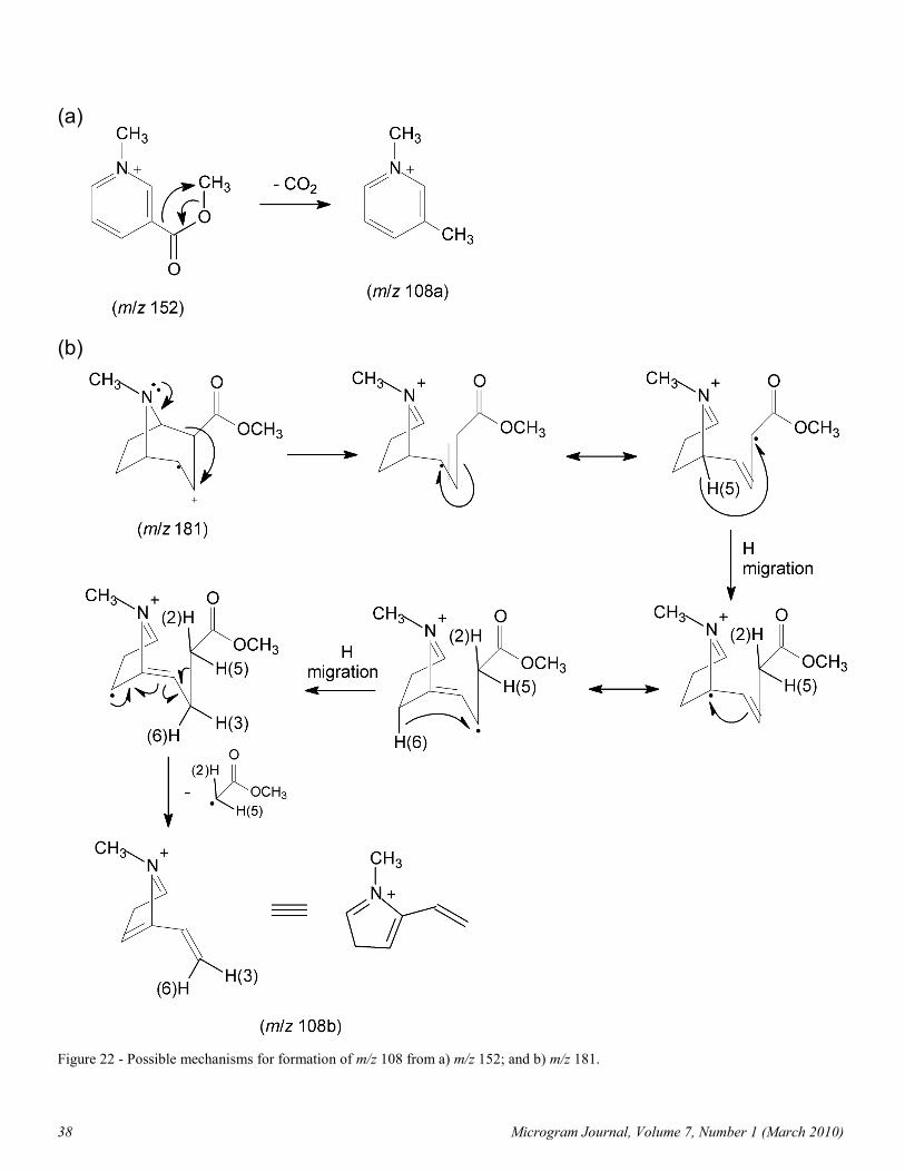
m/z 108. Taken as a whole, the data for the m/z 108 ion con-
tain a contradiction that cannot be resolved without invoking
two separate mechanisms. The N-CH3 group is clearly retained
because of the labeling and elemental composition data, but
both ester groups are lost. On the tropane skeleton most of the
D label at C2 is lost, most of the label at C3 is retained, and
about 50% of one label at C4 is lost, as are one or two D atoms
from C1, C5, C6, and C7. This strongly indicates that the C6-
C7 bridge remains intact in this ion and implies that the seven
C atoms in the ion consist of the N-methyl group plus all the
atoms in the tropane skeleton except C2. However, the MS/MS
data show that the ions having m/z 182, 181, and 152 can all act
as precursors to m/z 108. In the last case, the C6-C7 bridge is
no longer present. This indicates that the m/z 108 ion is formed
from different precursors under EIMS and MS/MS conditions.
Formation of m/z 108 from m/z 152 involves the loss of a
molecule of CO2, leading to a stable aromatic ion (Table II and
Figure 22a). However, production of m/z 108 from either m/z
181 or 182 seems to involve loss of the carbomethoxy group,
C2, and a H atom from C5 (Figure 22b). Other fragmentation
schemes, as well as other structures for m/z 108, are possible.
m/z 105. See discussion under m/z 122b above and Figure 3.
m/z 100. The m/z 222 ion appears to fragment to m/z 122b
and m/z 100 at lower collision energies. Analysis of the spectra
of the D-labeled derivatives for the m/z 100 ion shows retention
of the O-CD3 group, as well as the D atoms at C2, C3, and one
D atom at C4. The N-CD3 and the phenyl groups are lost.
Although the pattern of losses in the d6-derivative could not be
determined with certainty, loss of the five-membered ring
consisting of C1, the N atom, C5, C6, and C7 is likely simply
because the retained mass indicated by the apparent presence of
the carbomethoxy group, C2, C3, and C4 is sufficient. A
mechanism for formation of this ion that both accounts for
these data and is consistent with the elemental composition in
Table II is shown in Figure 13.

Microgram Journal, Volume 7, Number 1 (March 2010) 37
Figure 21 - Proposed formation of m/z 150, m/z 122, m/z 94, and m/z 82 from m/z 182.
38 Microgram Journal, Volume 7, Number 1 (March 2010)
Figure 22 - Possible mechanisms for formation of m/z 108 from a) m/z 152; and b) m/z 181.
(b)
(a)

Microgram Journal, Volume 7, Number 1 (March 2010) 39
Figure 23 - Proposed formation of m/z 96 and its fragments from m/z 155.
Figure 24 - Proposed fragmentations of m/z 82 to give m/z 67 and m/z 55.

40 Microgram Journal, Volume 7, Number 1 (March 2010)
m/z 97. The principal precursor to this ion is the M+·,
especially at low collision energies (Table III). Deuterium loss
patterns are difficult to interpret because of the proximity of
other more intense peaks in this area of the spectrum. Nonethe-
less, it appears that the D atoms at C2 and C3, as well as one D
atom from C4, may be lost. No more than 1 D atom is lost
from the remainder of the tropane skeleton, indicating that the
C6-C7 bridge is retained. The N-methyl group is retained, but
both ester groups are lost. A simple mechanism that explains
most of these data is shown in Figure 2 [1,2,13].
m/z 96. Major precursors to the m/z 96 ion at low collision
energies are the ones having m/z 155 and 97. This strongly
suggests that this ion, like those of its precursors, contains the
five-membered ring portion of the tropane skeleton, rather than
the six-membered ring. Losses from the D-labeled derivatives
bear this out. Although there is loss of D at C2 and significant
loss at C3, no more than one D atom is lost from C1, C5, C6,
and C7, showing that the C6-C7 bridge is retained. The loss of
the label from C4 could not be determined with certainty. The
N-methyl group is retained, but both ester groups are lost. Two
mechanisms are shown: the one in Figure 2 starts with m/z 97
[1,2,13]; the other (Figure 23) begins with m/z 155. Additional
mechanisms are possible.
m/z 94. In contrast to the m/z 96 ion, many of the m/z 94 ions
appear to have an N-methylpyridinium structure [1,5,13,16].
The most important precursors for this ion at low collision ener-
gies are m/z 181 and 122a. The N-methyl group is retained, as
are the H atoms at C2 and C3. Both ester groups are absent, as
are one H atom from C4 and four H atoms from the C1, C5, C6,
and C7 portion of the molecule. This is similar to the pattern of
losses exhibited by m/z 152 and is consistent with loss of the
C6-C7 bridge. Formation of this ion from m/z 122a can occur
readily via a reverse cycloaddition loss of ethylene (Figures 3,
9, 10, 15, and 21). All reasonable pathways to m/z 94 from m/z
181 appear to utilize m/z 122a as an intermediate.
An undetermined percentage of m/z 94 ions have m/z 96 as
their precursor, indicating that they probably have a structure in
which the five-membered ring is intact (Figure 2).
Like the intensity of the m/z 152 peak, the relative intensities
of the m/z 94 and 96 peaks also distinguish the spectrum of
cocaine from those of the other diastereomers. In the spectra of
cocaine and pseudoallococaine, the peak at m/z 94 is more
intense than the one at m/z 96; in the spectra of the other two
isomers it is smaller than m/z 96 (compare Figures 4a and 14b).
Spectra of D-labeled derivatives of cocaine and pseudococaine
strongly indicate that this phenomenon is not just a simple
reversal of intensities. The m/z 94 peak in the cocaine spectrum
is significantly larger than that produced by pseudococaine,
while the m/z 96 peaks in both spectra have similar intensities
relative to those of other nearby peaks. In addition, the m/z 94
peak in the cocaine spectrum represents a greater proportion of
the overall ion current (4.8%) than it does for the other isomers
(2.9-3.3%) [5]. This implies that m/z 94 forms more easily with
cocaine than it does with the other diastereomers.
None of the individual steps involved in generation of m/z
94b via either m/z 155 or 97 should be sensitive to stereochemi-
cal differences in the precursor ions because C2 and C3 are lost
in forming this ion (Figures 2 and 23). However, a careful
examination of the other fragmentation pathways leading to m/z
94a show that formation of this ion is in competition with
formation of m/z 152 via common intermediates: Pathways
a + b vs. pathway c in Figures 9 and 10; and pathways a + c vs.
pathway b in Figure 15. It is therefore tempting to conclude
that the same factors that discourage formation of m/z 152
should encourage formation of m/z 94, and vice versa [5]. In-
deed, the percent of total ion current for the m/z 94 peak in the
spectra of the four diastereomers is virtually opposite of what is
seen for the m/z 152 peak – that is, cocaine and pseudoalloco-
caine produce the smallest m/z 152 peaks and the largest m/z 94
peaks, while for pseudococaine and allococaine the situation is
reversed. One may conclude, then, that the relative rates of
cyclic loss of benzoic acid for these compounds directly affect
the ease of forming m/z 152 and thereby indirectly affect forma-
tion of m/z 94.
m/z 83. The m/z 83 ion was identified as the most abundant
fragment ion produced by the M+· at low collision energies,
indicating that it is formed directly in one step (Table II). The
N-methyl group is retained, as are most of the H atoms in the
five-membered ring portion of the molecule. Carbon atoms 2,
3, and 4 and their substituents are lost in forming this ion. This
ion fragments almost exclusively to give m/z 82 (Figure 2)
[1,2,13].
m/z 82. Several important precursors to m/z 82 were identi-
fied by MS/MS, so that a number of pathways for its formation
are likely. At low collision energies, the M+·, m/z 182, m/z 97,
and m/z 83 all can produce this ion, although m/z 303 and
m/z 182 appear to be the most important. The pattern of D
losses is similar to that seen with m/z 83, indicating that the
structure of this ion consists of the five-membered ring of the
tropane skeleton (Figure 2) [1,2,13]. A possible mechanism for
formation directly from the M+· is shown in Figure 24. Forma-
tion from m/z 182 is shown in Figure 21.
m/z 77. This ion is a known fragment of m/z 105 (Figure 3).
See discussion under m/z 122b above.
m/z 68. The ion having m/z 96 is the only one that produces
m/z 68 as a major fragment in the MS/MS experiments. The
structure of m/z 68 appears to lack C2 and C3 and their sub-
stituents, which is consistent with the structure proposed for m/z
96. The only label that is clearly retained is the one on the
N-methyl group, although it appears that several of the D atoms
on C1, C5, C6, and C7 are retained as well (the observed cluster
of peaks between m/z 68 and 73 in the spectrum of the
d6-derivative shows no single important peak). Cyclic loss of
ethylene from m/z 96 could account for formation of this ion,
which most likely has a methylene-azacyclopropenium struc-
ture (Figure 23).
m/z 67. The peaks at m/z 67 and m/z 68 often move together
in the spectra of the D-labeled derivatives, which gives the
impression that they are related to each other in a simple
manner. Although the elemental composition for this ion was
not determined, it appears as an important product ion in the
MS/MS spectra of m/z 94 and m/z 82, indicating that it may
have a different structure from m/z 68. It also appears that the

Microgram Journal, Volume 7, Number 1 (March 2010) 41
N-methyl label is lost, although in this area the spectrum of the
N-CD3 derivative shows more complexity than that simple
analysis implies. On the other hand, formation of m/z 67 from
m/z 82 can occur by loss of a methyl radical – presumably that
on the N atom (Figure 24). The m/z 67 ion could form from m/z
94 by loss of HCN by a complex mechanism involving transfer
of H atoms from the N-methyl group back onto the remaining C
atoms (structure and mechanism not shown). This ion has a
different elemental composition than that of the one formed
from m/z 82. However, the presence of two separate mecha-
nisms is not inconsistent with the D loss pattern.
m/z 59. Only two of the studied ions give m/z 59 as a product
ion – m/z 182 at higher, and m/z 152 at lower, collision ener-
gies. The only D label retained is on the O-CD3 group. That, in
combination with the elemental composition, indicates that this
ion is the carbomethoxy ion. It is difficult to write a mecha-
nism for its formation from either of these ions without invok-
ing high-energy intermediates or significant rearrangement.
Other precursors to this ion are possible.
m/z 55. Of the listed precursors to the m/z 55 ion, only
m/z 82 is important at low collision energies. This involves the
loss of 27 u as C2H3. The spectra of D-labeled derivatives indi-
cate loss of C2, C3, and C4. The pattern of loss shown by the
d6-derivative is complex and suffers from interference by
the surrounding peaks, but at least some (and perhaps most) of
the labels are retained. This is consistent with the fact that m/z
82 is the precursor. One possible mechanism is shown in
Figure 24.
m/z 51. This ion is a known fragment of m/z 105 and m/z 77
(Figure 3). See discussion under m/z 122b above.
m/z 42. The only D labels retained by this ion are in the
N-CD3 group and one of the D atoms on C1, C5, C6, or C7.
The m/z 96 ion is the only one studied that gives m/z 42 as a
significant product ion at low collision energies. A possible
mechanism is shown in Figure 23.
Conclusions
Although previously proposed mechanisms for formation of
many of the more abundant ions in the cocaine spectrum were
confirmed in this study, details about other fragmentations were
revealed. Of particular interest are explanations clarifying the
subtle differences between the spectra of the cocaine
diastereomers – formation of m/z 152 from m/z 274 and 181 via
pathways that are sensitive to the relative stereochemistry at C2
and C3, and the apparent inverse relationship between ease of
formation of m/z 152 and m/z 94. Also of interest are a) the
complexities that underlie the m/z 166 peak; b) the importance
of the N-containing ion having m/z 122; and c) details regarding
the formation of lower abundance ions, especially those above
m/z 200.
Acknowledgements
The authors are deeply indebted to Donald A. Cooper, Senior
Forensic Chemist, DEA Special Testing and Research Labora-
tory, ret., for obtaining the MS/MS and HR-MS data and for his
ideas and comments on the manuscript.
References
1. Blossey EC, Budzikiewicz H, Obashi M, Fodor G, Djerassi
C. Mass spectrometry in structural and stereochemical
problems – XXXIX: Tropane alkaloids. Tetrahedron
1964;20:585-93.
2. Smith DH, Duffield AM, Djerassi C. Mass spectrometry in
structural and stereochemical problems – CCXXII: De-
lineation of competing fragmentation pathways of complex
molecules from a study of metastable ion transitions in
deuterated derivatives. Org Mass Spectrom 1973;7:367-
81.
3. Smith RM. The mass spectrum of cocaine. J Forensic Sci
1977;42(3):475-80.
4. Smith RM. Understanding mass spectra – A basic ap-
proach, 2nd ed., John Wiley and Sons, New York, NY
2004:262-74.
5. Cooper DA. Mass spectrometry in drug research. Spectra
1985;10(3):14-20.
6. Casale JF, Lewin AH, Raney HT, Cooper DA. Synthesis
of deuterium labeled cocaine and pseudococaine.
J Labelled Compd Radiopharm 1991;29(3),327-35.
7. Casale JF. A practical total synthesis of cocaine's enanti-
omers. Forensic Sci Int 1987;33(3):275-98.
8. McLafferty FW, Turecek F. Interpretation of Mass
Spectra, 4th ed., University Science Books, Mill Valley, CA
1993:112-14.
9. Shapiro RH, Amenta DS, Kinter MT, Tomer KB. Mass
spectral analysis of cocaine and pseudococaine. Spectros
Int J 1983;2:227-31.
10. Rose ME. A caution on the use of metastable ions to assign
fragmentation pathways. Org Mass Spectrom 1981;
16:323-24.
11. McLafferty FW, Turecek F. op. cit. 1993:115-20.
12. McLafferty FW, Turecek F. op. cit. 1993: 343-5.
13. Dewhurst JE, Kaminski JJ, Supple JH. Mass spectra of
some tropane and tropidine derivatives. J Heterocycl
Chem 1972;9(3):507-11.
14. Casale JF, Smith RM, unpublished results.
15. Jindal SP, Lutz T. Ion cluster techniques in drug metabo-
lism: Use of a mixture of labeled and unlabeled cocaine to
facilitate metabolite identification. J Anal Toxicol 1986;10
(4):150-5.
16. Ethier JC, Neville GA. Quadrupole EI and CI mass spectra
of some principal tropane alkaloids. Can J Spectrom
1986;31:81-8.
17. Casale JF. Methyl esters of ecgonine: Injection-port
produced artifacts from cocaine base (crack) exhibits.
J Forensic Sci 1992;37(5):1295-310.
18. Vollmer D, Rempel DL, Gross ML, Williams F. Isomeri-
zation of 4-vinylcyclohexene radical cation: A tandem
mass spectrometry study. J Am Chem Soc 1995;117:1669-
70.
19. Zhang JY, Foltz RL. Cocaine metabolism in man: Identi-
fication of four previously unreported cocaine metabolites
in human urine. J Anal Toxicol 1990;14(4):201-5.
20. Allen AC, Cooper DA, Kiser WO, Cottrell RC. The
cocaine diastereomers. J Forensic Sci 1981;26(1):12-26.
21. Beynon JH, Saunders RA, Williams AE. The Mass
Spectra of Organic Molecules, Elsevier, New York, NY,
1968:223.