Embed Size (px)
Citation preview
Ann. rheum. Dis. (1955), 14, 162.
THE LIVER IN RHEUMATOID ARTHRITISBY
AARON M. LEFKOVITS AND IRVING J. FARROWFrom the General Medicine and Rheumatology Section of the Medical Service,
Veterans Administration Medical Teaching Group Hospital (Kennedy), Memphis, Tennessee
(RECEIVED FOR PUBLICATION JANUARY 18, 1955)
It is generally recognized that rheumatoidarthritis is a systemic disease characterized byprofound and widespread disturbances in the con-nective tissues throughout the body, but having aspecial predilection for the periarticular and articularstructures. Because the supporting framework ofparenchymal organs is made up of connective tissueelements, it has been suspected that the functionof these organs is altered in patients with rheumatoidarthritis as a result of direct involvement of thesupporting framework and/or by secondary morpho-logical alterations of the parenchymatous elements.Thus, the liver has been the subject of investigationby several observers, and its function has beenstudied to determine whether this organ is involvedin patients with rheumatoid arthritis. With thenewer techniques of biopsy, liver tissue can beobtained with greater ease, so that the morphologicalterations in the liver may be studied and correlatedwith liver function tests in patients with variousdiseases.The purpose of this report is to present our
experience with some laboratory procedures designedto test some of the functions of the liver in onehundred patients with rheumatoid arthritis, and toreport the results of examination of liver tissueobtained by needle biopsy in twelve of these patients,and the post-mortem findings in the liver of anadditional three of these patients.
Review of the LiteratureA review of the existing literature relating to the
involvement of the liver in rheumatoid arthritis revealsinadequate or contradictory information.
Davis (1936) found a high incidence of increased levelsof globulin and decreased levels of albumin in the plasmaof patients with rheumatoid arthritis. The bloodfibrinogen level was also found to be increased in thesepatients and there was an inverse relationship betweenthe fibrinogen and globulin levels. Rawls and others(1937) used the azorubin S test as an index of liverfunction in one hundred patients who had "severe","moderately severe", or "mild" rheumatoid arthritis:27 of 34 severe cases (73 per cent.), 21 of 44 moderately
severe cases (47 5 per cent.), and seven of 22 mild cases(25 25 per cent.), amounting to 55 per cent. of the entiregroup, showed liver dysfunction. In addition, theseauthors also found a slight reduction of the serumproteins and a reversal of the A/G ratio in most of thosecases (number not mentioned) that showed liver dys-function. They found no correlation between the dura-tion of the disease and impairment of liver function, butliver dysfunction was found more frequently in patientswho had more severe rheumatoid arthritis.Rawls and others (1939) reported on the results of
several liver function tests in fifty unselected cases ofrheumatoid arthritis. The azorubin S test showed someevidence of liver dysfunction in 60 per cent.; the hippuricacid excretion (Quick method) was abnormal in 62 percent. The bilirubin excretion (Soffer and Paulsonmethod) was abnormal in twelve of 25 cases (48 per cent.);the galactose tolerance test (excretion of 2 g. or more) wasabnormal in seven of 25 patients (14 per cent.); theicteric index (index 8 considered normal) was abnormalin 22 cases (44 per cent.); the serum albumin level wasabnormal (4 g./100 ml. or less) in 34 cases (68 per cent.);the A/G ratio was abnormal (less than 2) in 38 cases(76 per cent.). The azorubin S test agreed more closelywith the clinical evidence of liver damage than any othersingle test.
Baggenstoss and Rosenberg (1943) reported on thepost-mortem findings in thirty patients with rheumatoidarthritis whose death was due to various causes. Themost frequently observed hepatic lesions were grossatrophy or hypertrophy, chronic passive congestion,fatty changes, "serous hepatitis", and central necrosis.These changes were associated with heart disease and/orinanition. They found no evidence to suggest that thereticulo-endothelial system was stimulated in thesepatients, nor did they find "any hepatic lesion whichcould be considered specific".Perlmann and Kaufman (1946) investigated the serum
protein electrophoresis in 23 patients with rheumatoidarthritis. The results suggested an elevation of aglobulins during the early course of the disease followedby a predominance in y globulins in later stages.
Carter and Maclagan (1946) found the serum colloidalgold test to be positive in 76 per cent. and the thymolturbidity test to be positive in 38 per cent. of 34 patientswith "atrophic type" of arthritis.Kalbak (1951) found positive thymol reactions in
75 per cent. of 21 patients with rheumatoid arthritis;162
copyright. on 17 F
ebruary 2019 by guest. Protected by
http://ard.bmj.com
/A
nn Rheum
Dis: first published as 10.1136/ard.14.2.162 on 1 June 1955. D
ownloaded from
THE LIVER IN RHEUMATOID ARTHRITIS
in four, the positive thymol reaction became normalduring treatment with cortisone or ACTH. He thoughtthat the positive thymol reaction was due to change inserum proteins.
Archer (1951) found the cephalin flocculation andthymol turbidity tests to be consistently negative in tenpatients with rheumatoid arthritis, and in two otherpatients with both rheumatoid arthritis and degenerativejoint disease; the total serum proteins and serum albuminwere normal, and the serum globulin tended to be raised.He emphasized that the lowering of the A/G ratio in'non-specific" arthritis was the result of a raised globulinlevel alone and not of a diminished albumin level.
Schmengler (1952) reported on the findings of liverinvolvement in thirty cases of "chronic rheumatism";one of which was examined post mortem. The liver waspalpable in 21; the Takata reaction was positive infifteen of the twenty patients; the thymol turbidity testwas positive in "most cases". Electrophoretic partitionof serum proteins was done in 23: the y globulin wasincreased in 70 per cent., the globulin in 26 per cent.,and the X globulin in 35 per cent. Liver tissue was
examined in the one autopsied case and in 27 patientsobtained by biopsy through the peritoneoscope. In twopatients, attempt at biopsy was unsuccessful. All liverspecimens were abnormal. Grossly, the liver appearedgreyish-red with loss of glistening; the surface was
irregular and presented scars, fibrous bands, or reticulararrangement. Microscopically, the cells showed vari-ations in size, fatty degeneration, "reactions of Kupffercells", haemosiderin accumulations here and there, attimes necrosis, perivascular round cell infiltrations,increase in fibrous tissue, and transformation into"rheumatic hepatitis" and cirrhosis.
Movitt and Davis (1953) found no significant abnor-malities in the serum albumin level, cephalin flocculationtests, and prothrombin concentrations, or in the mor-phology of the liver obtained by needle biopsy in seven-teen male patients with rheumatoid arthritis, but theincidence of raised serum globulin was high.The observations relating to the ameliorating influence
of liver disease with or without jaundice, pregnancy, andseveral other diseases upon the manifestations of rheuma-toid arthritis are of interest in this connexion. Theseare adequately discussed by Hench (1940) and will notbe repeated here. They led indirectly to the investiga-tions which resulted in the discovery of the beneficial,although temporary, effect of adrenal steroids and ACTHupon the manifestations of rheumatic diseases.
It is readily seen from the foregoing that theresults obtained by these investigators have notgiven us a definite answer to the question whetherthe liver is involved by the rheumatoid diseaseprocess? Our attempts at resolving this problemare hindered mainly by the following difficulties:
(I) Non-hepatic diseases altering the results of so-calledliver function tests;
(2) Our inability to distinguish between the effects of
systemic disease processes upon the liver and intrinsicliver disease by the available tests of liver function;
(3) The inadequacy of the available histologicalmethods of examination of liver tissue obtained, eitherby biopsy or post mortem, to reveal functional alterationsor impairment.
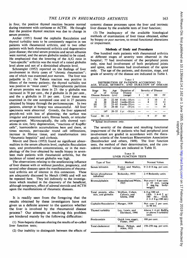
Methods of Study and ProceduresOne hundred male patients with rheumatoid arthritis
at different stages of severity were observed in thehospital; 77 had involvement of the peripheral jointsonly, nine had involvement of both peripheral jointsand spine, and fourteen had involvement of the spineonly. The age of the patients, and duration, stage, andgrade of severity of the disease are indicated in Table I.
TABLE IDISTRIBUTION OF PATIENTS ACCORDING TO
AGE, STAGE, SEVERITY, AND DURATION OF DISEASE
Stage No. Age Duration of Severity of Diseaseof of Group Disease
Disease Patients (yrs) (range) I II III IV
1 20 22-64 6 wks -l0yrs - 5 10 5II 30 24-63 3 mths-20 yrs - 4 13 13
III 22 25-65 8 mths-35 yrs - - 14 8IV 14 28-66 1 yr -35 yrs - 14
14* 20-55 1 yr -16yrs - -
Total .. 86+14 - 9 37 60
* Spinal involvement only.
The severity of the disease and resulting functionalimpairment of the 86 patients who had peripheral jointinvolvement are graded in accordance with the thera-peutic criteria of the American Rheumatism Association(Steinbrocker and others, 1949). The liver functiontests, the method of their determination, and the con-sidered normal values are indicated in Table If.
TABLE IILIVER FUNCTION TESTS
Type of Test Method Normal Values
Serum bilirubin Evelyn and Malloy, 0 2-0 8 mg. per cent.1937
Serum phosphatase Bodansky, 1933 2-4 Bodansky units(alkaline)
Bromsulphalein Rosenthal and White, Not over 1 6 per cent.1925 in 45 min. (using
5 mg./kg. body-weight)
Total protein, albu- Wolfson, Cohen, 6-8 g./100 ml.min, and globulin Calvary, and (Alb. 3-4-4 5 g./
Ichiba, 1948 100 ml.; Glob.2-3-3 6 g./100 ml.)
Cephalin flocculation Hanger, 1939 Not over 2 per cent.in 48 hrs
Thymol turbidity Ley, Lewis, and Above 1-4 ml. BaSO4Davidson, 1946 or above 5 turbidity
units
Prothrombin Quick (one stage), 100 per cent.Fisher, 1947
Total cholesterol Bloor, Pelkan, and 150-250 mg. per cent.Allen, 1922
163
copyright. on 17 F
ebruary 2019 by guest. Protected by
http://ard.bmj.com
/A
nn Rheum
Dis: first published as 10.1136/ard.14.2.162 on 1 June 1955. D
ownloaded from

ANNALS OF THE RHEUMATIC DISEASESTable III shows the patients in whom each type of
test was determined, and the patients with peripheraljoint involvement are grouped according to the stage oftheir disease. Twelve patients with involvement of
TABLE III
TYPE AND TOTAL NUMBER OF LIVERFUNCTION TESTS DETERMINED
I~~~~~~~~~~~~~~~~~~~~~~~~~~~~~~~~~~~~~~~~~Peripheral
Joint InvolvementType ofTest Stage of DiseaseOnly
I II III IV
Serum bilirubin 9 22 18 9
Alkaline phos-phatase 7 14
Bromsulphalein. 9 25
10 11
22 12
Protein 18 27 21
Albumin.. 18 27 21
Globulin..
Cephalinflocculation
Thymol turbidity
18
10
11
13
13
27 21 13
26 22 12
28 19 12
-SpinalInvolve-
- mentOnly
13
5 47z 7A
peripheral joints had Vim-Silverman needle biopsies ofthe liver performed by the transthoracic approach.* Inan additional three patients, liver tissue was obtainedpost mortem; two of these patients had severe cripplingdeformities of the peripheral joints with extensiveosteoporosis: one had compression of the spinal cord bycollapsed osteoporotic cervical vertebrae and died as aresult of pneumonia; the other also died of pneumoniabut had, in addition, severe coronary artery disease.The third patient had spinal involvement only andpresented the characteristic appearance of "bamboo-spine"; he had severe hypertensive cardiovasculardisease and died as a result of rupture of dissectinganeurysm of the arch of the aorta. The pertinentclinical and laboratory data of these fifteen patients areindicated in Table IV.
Resultst' "_/ Table V (overleaf) indicates the type and number
12 91 of abnormal results grouped by stage of disease.
12 91 It is readily seen that there is a direct relationship12 91 between the severity of the disease, as indicated
by the stage of the disease, and the number of11 81 abnormal results. Of twenty patients with Stage I
12 83 rheumatoid arthritis, thirteen (65 per cent.) hadProthrombin 9
Cholesterol 9
27 21 11 7 75
20 15 10 1 11 65
* These biopsies were performed by the members of the Gastro-intestinal Section of the hospital. The liver tissue was fixed in 10per cent. formalin and stained with haematoxylin and eosin in theusual manner.
SUMMARY OF PERTINENT CLINICAL AND LABORATORY DATA AND MORPHOLOGICAL ALTERATIONS IN
Pat t Ae
Patient AgeNo. (yrs)
1 46
ErythrocyteSeverity Duration of Sedimentation
of Disease RateDisease (yrs) (mm./hr)
(Wintrobe)
II 6 34
AlkalineSerum Phosphatase
Bilirubin (Bodanskyunits)
05 i1 3
BromoSulphalein(per cent.)
Neg.
Albumin Glot
3 8 3.
2* 35 II 5/ 12 36 0 39 3 35 20 3 3 5.
6 28 0-48
233436
0 70 5
35 0 530 0 54
32 0 7
3537
23
0.1
0 5
17 Neg.
Neg.1 8 20
Neg.
1-55 Neg.1-2 15
Neg.
- 75Neg.
3 8
46 4.
3.9 3-3 5 3.3*8 3.
3 4 4-3 6 4.
4-2 2
3-1 3.3.4 3.
Neg.
* Liver illustrated in photomicrograph. t Died.
164
3
45
6*
28
335857
75
17
7 328 54
9 44
10 5511 44
It
II
1111
IIIIII
III
IIIIII
IV
84
5
48
12 62 21
13*t i 58 IV 18 28 0-39 1 2 1 8 2 9 3
14*t 55 IV 6 36 - 5 1 75 3 6 4-
15t 55 - 26 28 0 8 - - 3 7 2
I 1. I
copyright. on 17 F
ebruary 2019 by guest. Protected by
http://ard.bmj.com
/A
nn Rheum
Dis: first published as 10.1136/ard.14.2.162 on 1 June 1955. D
ownloaded from

THE LIVER IN RHEUMATOID ARTHRITIS16
abnormal results. Of thirty patients in Stage II,
26 (86- 66 per cent.) had abnormal results. Of
22 patients in Stage III, 21 (95-45 per cent.) had
abnormal results. All fourteen patients in Stage IV
had abnormal results.
Depression of the serum albumin level was the most
frequently found abnormality in Stage I patients. In
.Stage patients a raised serum globulin level was the
most frequent abnormnality (seen in 12), and the serum
albumin level was depressed in ten. Similar changes in
the albumin and globulin levels were found in Stage
III and IV patients.
The cephalin flocculation test was abnormal in five of
26 Stage patients (19 per cent.). The thymol turbiditytest was abnormal in five of 28 Stage patients (1 7-8
per cent.). The cephalin flocculation test was abnormal
in seven of 22 Stage III patients (31 -8 per cent.), and the
thymol turbidity in six of nineteen Stage III patients
(31-5 per cent.). The cephalin flocculation test was
abnormal in six of twelve Stage IV patients (50 per cent.),but thymol turbidity test was abnormal only in one of
thirteen Stage IV patients.
The serum cholesterol level was raised in approxi-
mately 50 per cent. of Stage I and patients, but in onlythree of fifteen patients (20 per cent.) and in only one
of ten Stage IV patients.
The serum bilirubin level was normal in all the patients
(58 with peripheral joint involvement and thirteen with
spinal involvement) in whom it was determined.
The bromosulphalein test was normal in all Stage I
patients; it was abnormal in six Stage patients (24 per
cent.), five Stage III patients (22-7 per cent.), and three
Stage IV patients (25 per cent.).
Of the fourteen patients who had only spinal involve-
ment, six showed abnormalities in a total of eleven
abnormnal results, as indicated in Table V.
Microscopic examination of tissue obtained by
biopsy in the twelve patients in whom this was per-
formed revealed only minor alterations, chieflyvariations in the size and pigmentation of the nuclei
of the liver cells, slight prominence of the peripheral
fibrous tissue with slight but varying degrees of
round cell infiltration, and, occasionally, slight
increase of fat. In two of these specimens, these
alterations were more pronounced; Figs and 2
(overleaf) depict the most marked of these changes.
In none of these biopsies was the pathologist* able
to discern definite evidence of cirrhosis.
*Dr. Joseph M. Young, Pathologist, Veterans Administration
Medical Teaching Group (Kennedy) Hospital.
)F PATIENTS IN WHOM LIVER TISSUE WAS AVAILABLE FOR EXAMINATION BY BIOPSY OR AT POST-MORTEM
ialin Thymol Prothrombin Cholesterol Weight of Morphological Cause ofnation Turbidity (per cent.) (mg. per cent.) Liver Changes in Liver Death
(g.)
!g. 0-6 100 -Slight prominence of fibrous tissue at portal triadswith few round cells
Variations in size and pigmentation of nuclei2-8 57 340 Moderate increase of fibrous tissue at portal triads
with round cell infiltrationFatty metamorphosis
175 100 220 Slight prominence of fibrous tissue at portal triadswith few round cells
175 100 215 No significant abnormalities3 25 100 Fatty metamorphosis4.3 45 - Moderate increase of fibrous tissue at portal triads
with round cell infiltrationVariations in size and pigmentation of nuclei
1.0 100 - No significant abnormalities1 75 100 - Slight prominence of fibrous tissue at portal triads
with few round cellsVariations in size and pigmentation of nuclei
t0-75 100 187 Slight prominence of fibrous tissue at portal triadswith few round cells
Variations in size and pigmentation of nuclei5-75 100 182 Variations in size and pigmentation of nuclei3-5 100 235 Slight prominence of fibrous tissue at portal triads
with few round cellsFatty metamorphosis
3 1-75 100 - Slight prominence of fibrous tissue at portal triadswith few round cells
3 2-7 100 245 2,250 Slight prominence of fibrous tissue at portal triads Pneumonia33.5 - ~~~~~~~~~~~~~~~~withfew round cells335 ~~~~~~~~~1,375 Central zone congestion and infiltration of poly- Pneumonia
morphs--- 240 1,830 Slight prominence of fibrous tissue at portal triads Aneurysm
with few round cells of aorta
165
copyright. on 17 F
ebruary 2019 by guest. Protected by
http://ard.bmj.com
/A
nn Rheum
Dis: first published as 10.1136/ard.14.2.162 on 1 June 1955. D
ownloaded from

ANNALS OF THE RHEUMATIC DISEASESTABLE V
ABNORMAL LIVER FUNCTION RESULTS, SHOWN WITH NUMBER OF PATIENTS TESTED
Type of Test
Serum bilirubin ..
Alkaline phosphatase..
Bromsulphalein ..
Total Protein ..
Albumin
Globulin
Peripheral Joint Involvement
Stage of Disease
0/9
0/7
0/9
Depressed 0/18
Elevated .. 0/18
Depressed 4/18
Elevated .. 0/18
Depressed 2/18
Elevated .. 2/18 12/27
Cephalin flocculation .
Thymol turbidity
Prothrombin ..
DepressedCholesterol ..
1/10
0/11
2/9
1/9Elevated .. 4/9
Total Abnormal Results
Total Patients with Abnormal ResultsNo. 13
% 65
The liver obtained at autopsy in the three patientsthat died likewise failed to show significant morpho-logical abnormalities. One specimen showed con-
gestion and polymorphonuclear infiltration of thecentral zone areas; this change was thought to berelated to the bilateral pneumonic process withsmall abscesses rather than to the rheumatoidarthritis. Figs 3 and 4 (overleaf) show the micro-scopic appearance of two of these livers.
Comment
The results suggest that there is a relationshipbetween the severity of rheumatoid arthritis and thehepatic dysfunction indicated by certain liverfunction tests. Elevation of the serum globulin anddepression of the serum albumin were the mostfrequent abnormalities, closely followed by abnormalcephalin flocculation and thymol turbidity tests.The other tests did not indicate abnormalities in a
sufficient number of patients to be of statisticalsignificance. Abnormal liver function tests were
found infrequently in patients who had only spinalinvolvement. Histological examination of the avail-able liver tissue in fifteen of these patients showedonly minor alterations. None of the three liver
specimens obtained post mortem from the threepatients who died (disease of Stage IV severity, butinactive, and of 18, 6, and 26 years' duration,respectively) showed any significant abnormalities.Perhaps, however, although haematoxylin-eosinstains did not depict abnormalities, other more
refined methods of examination, such as electronmicroscopy, special staining, and histochemicalstudies, would have revealed a closer correlation withthe liver function tests.
It is difficult to evaluate the significance of theseminor morphological alterations. Such changes are
seen not infrequently in liver specimens obtained atautopsy from individuals who die of diverse con-
ditions in whom no evidence of liver disease could befound. At the most, these abnormalities couldprobably be designated by what Popper (1954)termed "non-specific reactive hepatitis". There wasno strict correlation between the functions of theliver as appraised by the liver function tests used hereand the histological appearance of the liver speci-mens. Other investigators (Franklin and others,1948; Popper and others, 1950; Lichtman, 1942)have also commented upon the lack of consistentcorrelation between the clinical and laboratoryfindings in liver disease and the histological picture
166
II
0/22
0/14
6/25
3/27
4/27
10/27
2/27
2/27
III
0/18
1/10
5/22
0/21
3/21
9/21
2/21
0/21
12/21
7/22
6/19
3/21
3/15
51
21
95 4
SpinalInvolvement
Only
0/13
95
0/6
1/12
1/12
0/12
1/121/12
2/122/11
0/12
117
0/11
11
6
IV
0/9
211
3/12
1/13
1/13
9!13
0/13
0/13
11 13
6/12
1 /12
3/11
0/10
1/10
38
14
100
5/26
5/28
6/27
1/20
10/20
66
26
86 6
copyright. on 17 F
ebruary 2019 by guest. Protected by
http://ard.bmj.com
/A
nn Rheum
Dis: first published as 10.1136/ard.14.2.162 on 1 June 1955. D
ownloaded from

THE LIVER IN RHEUMATOID ARTHRITIS
4<.
u~~~~sWsts*r .r s*1|s X
Fig. 1.-Case 2, showing moderate increase of periportal fibroustissue, round cell infiltration, and slight fatty change.
of liver tissue. Popper (1954) noted that severehepatic failure was at times associated with rela-tively few histological alterations, and, conversely,apparently intact liver function was found in patientsshowing widespread changes in the hepatic paren-chyma. These contradictory findings are generallyattributed to the fact that the haematoxylin-eosinstained liver sections probably do not depict thefunctional aberrations of the liver parenchyma.Moreover, the regenerative ability of the liver is sogreat that impairment of its function can be demon-strated only after extensive liver damage.The utilization of the so-called liver function tests
in assessing the functions of the liver is further com-plicated by the observation that these tests are non-specific and are not infrequently altered in anychronic disease of whatever aetiology. The abnor-mal values obtained in these tests could, therefore,be considered to reflect the chronic wasting state socommon in rheumatoid arthritics rather than thedirect consequence of the rheumatoid process itself.But many observers believe that, although thesetests are in a strict sense not liver function tests, theycan, nevertheless, be employed to test the functionsof the liver. Some of these, notably the proteinfractionation, cephalin flocculation, and thymolturbidity tests, reflect a qualitative and probably
Fig. 2.-Case 6, showing variations in size and staining of nuclei,moderate increase of periportal fibrous tissue, and round cell
infiltration.
a quantitative alteration in the serum proteins.Since the liver plays an important role in the elabora-tion of these proteins, and the serum albumin isthought to be formed only in the liver (Wright,1952), it can be stated that abnormal values obtainedby these tests can be considered to indicate dis-turbances in liver function. Though our results donot permit an unequivocal answer to the problem"of liver involvement in the rheumatoid process",they appear to justify the opinion that some of thefunctions of the liver are pathologically altered inpatients suffering from rheumatoid disease.
Summary(1) An attempt has been made to ascertain
whether the liver is involved in patients withrheumatoid arthritis.
(2) Liver function tests were performed in 86patients with rheumatoid arthritis, who had peri-pheral joint involvement, and in an additionalfourteen patients who had only spinal involvement.Of the 86 patients with peripheral joint involvement,nine had spinal involvement also.
(3) Three patients died and liver tissue wasobtained for post-mortem examination. In twelve
167
copyright. on 17 F
ebruary 2019 by guest. Protected by
http://ard.bmj.com
/A
nn Rheum
Dis: first published as 10.1136/ard.14.2.162 on 1 June 1955. D
ownloaded from

ANNALS OF THE RHEUMATIC DISEASES
q O
ig3tt
Fig.e pherdi pora le trand sh slightenc fibro usa
oterut paient liverfntissu forts Eexaiation wasth
~~te
serum globulin level and depression of the serum
albumin level were the most frequently found
abnormalities, closely followed by abnormalities in
cholesterol, cephalin flocculation, and thymol tur-
bidity.
(5) Histological study of liver tissue revealed
definite abnormal morphological alterations only in
two out of the fifteen available specimens. These
changes are briefly described. The lack of cor-
relation between the clinical and laboratory evidence
of liver involvement and the histological appearance
of liver tissue is briefly discussed.
(6) Although the results obtained do not allow
a definite answer to the problem proposed, the
authors believe them to justify the opinion that the
functions of the liver are impaired in persons
affected by rheumatoid disease.
We express our grateful appreciation to Dr. Joseph M.
Young, pathologist, for examining the slides, and to the
Medical Illustration Laboratory, Veterans Administra-
S~~~~~~~~ ;Tit*%'1~~.. CON .'4 r .is
04, ~4
Fig. 4.-Case 14, showing congestion and polymorphonuclear
infiltration of central zone area.
tion Medical Teaching Group Hospital, Memphis, 15,
Tennessee, for the preparation of the illustrations.
REFERENCES
Archer, B. H. (1951). Ann. intern. Med., 34, 1173.
Baggenstoss, A. H., and Rosenberg, E. F. (1943). Arch. Path.
(Chicago), 35, 503.
Bloor, W. R., Pelkan, K. F., and Allen, D. M. (1922). J. biol. Chems.,52, 91.
Bodansky, A. (1933). Ibid., 101, 93.
Carter, A. B., and Maclagan, N. F. (1946). Brit. med. J., 2, 80.
Davis, J. 5., Jr. (1936). J. Lab. c/in. Med., 21, 478.
Evelyn, K. A., and Malloy, H. T. (1937). J. biol. Chem., 119, 481.
Fisher, B. (1947). Amer. J. c/in. Path., 17, 471.
Franklin, M., Popper, H., Steigmann, F., and Kozoll, D. D. (1948).
J. Lab. c/in. Med., 33, 435.
Hanger, F. M. (1939). J. c/in. Invest., 18, 261.
Hench, P. 5. (1940). Med. C/in. N. Amer., 24, 1209.
Kalbak. K. (1951). Anna/s of the Rheumatic Diseases, 10, 182.
Ley, A. B., Lewis, J. H., and Davidson, C. 5. (1946). J. Lab. c/in.
Med., 31, 910.
Lichtman, 5. 5. (1942). "Diseases of the Liver, Gallbladder, and
Bile Ducts", 1st ed., p. 250. Lea and Febiger, Philadelphia.
Movitt, E. R., and Davis, A. E. (1953). Amer. J. med. Sci., 226, 516.
Perlmann, G. E., and Kaufman, D. (1946). J. c/in. Invest., 25, 931.
Popper, H. (1954). Amer. J. Med., 16, 98.
,Waldatein, 5. 5., and Szanto, P. B. (1950). Amer. J1. c/in. Path.,
20, 724.
Rawls, W. B., Weiss, 5., and Collins, V. L. (1937). Ann. intern. Med.,
10, 1021.
-, , (1939). Ibid., 12, 1455.
Rosenthal, S. M., and White, E. C. (1925). J. Amer. med. Ass.,
84, 1112.
Schmengler, F. E. (1952). Medizinische, 1, 1553.
Steinbrocker, O., Traeger, C. H., and Batterman, R. C. (1949).
J. Amer. med. Ass., 140, 659.
Wolfson, W. Q., Cohn, E., Calvary, E., and Ichiba, F. (1948). Tech.Bull. Reg. Med. Tech., 18, 723.
Wright, B. (1952). "Applied Physiology", 9th ed., p. 831. Oxford
University Press, London.
168
copyright. on 17 F
ebruary 2019 by guest. Protected by
http://ard.bmj.com
/A
nn Rheum
Dis: first published as 10.1136/ard.14.2.162 on 1 June 1955. D
ownloaded from

THE LIVER IN RHEUMATOID ARTHRITISLe foie dans l'arthrite rhumatismale
RtSUMt(1) On a tache de trouver si le foie etait implique chez
les malades atteints d'arthrite rhumatismale.(2) On fit des tests de la fonction hepatique chez 86
rhumatisants avec atteinte articulaire peripherique etchez 14 autres avec atteinte vertebrale seule. Chez 9 surles 86 rhumatisants polyarticulaires il y eut une atteintevertebrale concomitant.
(3) On effectua un prelevement de tissu hepatique al'autopsie de 3 malades decedes et chez 12 malades vivants.
(4) On nota un rapport bien determine entre laseverite de la maladie et la frequence des resultatsanormaux des tests de la fonction hepatique. L'augmen-tation du taux de la globulin serique et la baisse de celuide l'albumine se virent le plus souvent, suivies de prespar des chiffres anormaux du cholesterol, de la reactionde floculation de la cephaline et de la turbidity du thymol.
(5) A l'examen histologique, des alterations morpho-logiques du tissu hepatique ne furent trouvees que dansdeux pieces sur les quinze disponibles. On presente unebreve description de ces alterations. On discute som-mairement l'absence d'un rapport entre les manifes-tations cliniques et de laboratoire de l'atteinte du foie etl'apparence histologique du tissu hepatique.
(6) Bien que les resultats obtenus n'offrent pas desolution claire du probleme pose, les auteurs croientqu'ils justifient l'opinion que les fonctions du foie seraientalterees dans la maladie rhumatismale.
El higado en la artritis reumatoideSUMARIO
(1) Se trat6 de comprobar si el higado estaba implicadoen los enfermos con artritis reumatoide.
(2) Se hicieron valoraciones de la funci6n hepaticaen 86 enfermos con implicaci6n articular periferica y en14 con implicaci6n vertebral sola. En 9 de los 86 conimplicaci6n periferica la espina participaba del procesoreumatico.
(3) Se obtuvo tejido hepatico de autopsies al morir3 enfermos y de biopsias en doce casos.
(4) Se not6 una relaci6n determinada entre la severidadde la enfermedad y la frecuencia de los resultadosanormales de las determinaciones de la funci6n hepAtica.HallAronse mAs frecuentemente cifras altas de globulinserica y bajas de albumina, seguidas de cerca por valoresanormales del colesterol, de la reacci6n de floculaci6nde la cefalina y de la turbiedad del timol.
(5) Histol6gicamente, alteraciones morfol6gicas deltejido hepAtico encontrAronse tan solo en dos prepara-ciones de las quince obtenidas. Se presenta una brevedescripci6n de estas alteraciones. Se discute brevementela falta de correlaci6n entre las manifestaciones clinicasy de laboratorio de la implicaci6n del higado y el aspectohistol6gico del tejido hepAtico.
(6) Aunque los resultados obtenidos no ofrecen unasoluci6n clara del problema puesto, los autores creenjustificada la opinion de que las funciones del higadoestan alteradas en la enfermedad reumatica.
169
copyright. on 17 F
ebruary 2019 by guest. Protected by
http://ard.bmj.com
/A
nn Rheum
Dis: first published as 10.1136/ard.14.2.162 on 1 June 1955. D
ownloaded from