Embed Size (px)
Citation preview

Chapter 19
G. Messerli, J. Ménétrey Place of navigation in anterior
cruciate ligament reconstruction
Introduction
A nterior cruciate ligament (ACL) reconstruc-tion is one of the most frequently performed major orthopedic procedures in the young
adult population. In North America, the incidence of acute ACL tears is around 1/3000 per annum, and more than 100,000 ACL repairs are performed annually (1). Th e main goal of this surgery is to pro-Thvide an excellent functional stability and a durable reconstruction for an active and high-demanding population.Computer-assisted orthopedic surgery (CAOS) is arecent concept defi ned as the ability to utilize com-fiputer algorithms to allow the surgeon to determinethree-dimensional (3-D) placement of implants in situ. Misplacement of the femoral and tibial tun-nels, which might cause graft failure or limited knee range of motion, is the main reason for tech-nical failure after ACL repair (2–7). Therefore, the Thmajor aim of CAOS is to avoid inconsistent place-ment of graft tunnels during ACL reconstruction by increasing the precision of these procedures.
Defi nitionsfi
Computer-assisted surgery (CAS) techniques havebeen developed on the principle of stereotaxis,which permits the location of a structure in a plane coordinate system. First, neurosurgeons have usedCAS system for the location of brain tumor.Computer-assisted orthopedic surgery (CAOS)was an application of CAS to spine surgery in thefi rst era and then has been extended to all other fifi elds of orthopedic surgery, in particular to hip fiand knee. CAOS techniques are no more a simple stereostaxis system and now cover a large base of applications in surgical practice, such as planning, simulation, guidance, assistance, and training.The CAOS systems can be classifiTh ed into active andfipassive systems. Th e active system refers to a robot Thable to perform alone a procedure under control of the surgeon. In these cases, preoperative imagingand intraoperative data are required, with rigid fix-fi
ation of the robot and limb to the operating table (8–10). Th ere are also some systems where the sur-Thgeons initiate the action of the robot and then the system guided or restricted the surgeons’action as planned on the preoperative imaging (11). ThoseThsystems do not perform autonomously the surgical act and can be named semi-active.Passive systems do not perform any surgical action by themselves. Th ey are also called navigation sys-Thtems, and their main goal is to provide information to improve the conventional techniques or to guide the surgeon during a more demanding procedure. A new development of passive CAOS system is “thecustom templating technique” (Fig. 1). This tech-Thnology use preoperative planning to produce 3-D template model specifi cs to one patient with rapid fiprototyping machines (12).
History
We must give the credit to Claudius Galen (131 Pergamum, 205 Rome) for fi rst describing the fianatomy and the nature of the ACL. He mentions them as “genu cruciata,” serving to stabilize joint and to limit abnormal motion (13).
Fig. 1 – Knee prototyping.
et al., The Knee Joint © Springer-Verlag France, Paris 2012M. Bonnin
218 The Traumatic Knee
is now widely accepted around the world. If more than 100,000 ACL repairs are realized annually (1)in the United States, this popular procedure is per-formed in the great majority by surgeons who do less than 20 ACL reconstructions per year (24).Function of the ACL plays a major role in the kine-matics of the knee by stabilizing it in a wide range of movements. Th e ACL injury usually generates Thinstability of the knee that will prevent a return to the previous activity and change this cinematic in consequence of the adaptation of other structures to compensate the absence of the LCA, as demon-strated by Berchuck and Andriacchi (25). Despite this adjustment, mainly due to the muscles, this modifi cation of the movement may, in time, result fiin meniscus injury and cartilage lesion, motivating a reconstruction surgery, as would tend to prove the study by Dunn et al. (26). Th e choice of graft Thand the precision of its location determine the bio-mechanical qualities and the stability of the recon-structed knee, as well as evolution and return to previous activity level.Although current techniques of ACL reconstruc-tion can achieve an improvement in function, the physiologic and biologic features of the normal ACL are not fully restored (27). In a 10-year review of the literature, Beasley et al. (28) report similar subjective scores regardless of the reconstructive technique, with an average of 80–95% of patients reporting the feeling of a normal or nearly normal knee. Th e unsatisfactory results are due to a wide Thrange of reasons, including recurrent pain, loss of motion, and persistent instability (29–31).Failed ACL reconstructions have been classifi ed by fiHarner (32) and Fu (31) into three groups based on the cause of the recurrent instability:– Traumatic failure due to high-energy trauma and
rupture of a functional graft– Biological failure due to infection, absence of
graft incorporation, or biomechanical tissue fail-ure
– Technical errors (about 2/3 of failure are due to technical errors)
Technical errors
It is important to understand the structural prop-erties of the intact ACL, as replacement grafts should have similar tensile, mechanical, biological, and dimensional properties as those of the intact ACL to best reproduce its in vivo function. Cur-rently, the main technical errors concern:– poor graft selection;– inadequate graft harvest;– improper fi xation of the graft in the bony tun-fi
nels;– improper tensioning of the graft;
In 1850 Stark treated two patients with casts and gave the description of an ACL rupture (14).TheThfi rst report of an ACL repair performed in 1898 isfipublished by Battle, in 1900 (15), but Mayo Robsonreported in 1903 an 8-year follow-up of an anterior and posterior cruciate ligament suture using catgut (16). Th e same year, Lange performs the fiTh rst recon-fistruction with the semi-tendinosus tendon (17).Th e 1960s signed the beginning of the modern eraThfor the ACL reconstruction. Kenneth Jones (KJ) used the medial third of the patellar tendon. TheThgraft was not detached from the tibia, and a plug of patellar bone was set in a femoral tunnel (18). Major disadvantage of this technique was shortness and anterior placement of the graft. But Bruckner (19) and Franke (20) improved it, drilling a tibial tunnel and using a free transplant of patellar tendon end-ing with tibial and patellar bone plugs. The bone-Thpatellar tendon-bone (BPTB) autograft was borne.In the 1980s, the modified KJ was back with the fiaccession of the arthroscopic surgery. Sixty-threeyears after Takagi fi rst introduced a cystoscope fiin a knee, Dandy performed, in 1981, the firstfiarthroscopic ACL reconstruction (21).Concerning CAS, the phenomenal progress in med-ical imaging, especially the CT scan, introduced the possibility to image-guided surgery. The fiTh rstfiapplication was for brain surgery, and by exten-sion, spine surgeons were quickly involved (22).Then, hip and knee were gradually implicated. ThTh eThfi rst generation of CAS was active robots, and thefiROBODOC® system conceived in 1986 by Bargarand Paul was used to assist surgeons in performing part of a total hip replacement in 1992 (10). TheThfi rst generation of CAS application for ACL recon-fistruction started 2 years later with the method introduced by Dessenne (8) and Klos (9). CASPAR (Computer Assisted Surgical Planning and Robot-ics, ortho Maquet GmbH Co. KG, Rastatt, Ger-many) was the first active robot to drill tunnels fiduring ACL reconstruction procedures (23).Active robots were progressively replaced by naviga-tion systems also called passive systems. If the firstfinavigation systems were based on imaging and used CT scan, the second generation of CAS was image-free system allowing real-time assistance surgery. Th e next generation of CAS will be image-free sys-Thtem with intelligent guidance and active robot.
ACL reconstruction analysis
Generality
Th e fact that ACL reconstruction is becoming theThfi rst choice of treatment for ACL-defifi cient knee fi

Place of navigation in anterior cruciate ligament reconstruction 219
Agenda and duties of a navigation system
Specificationsfi
Anatomy and biomechanics are very complex in the knee. Th e closed interactions between soft-Thtissue structures, ligaments, tendons, and bones require accurate surgical procedure to restore nor-mal function of the knee joint and to avoid adverse repercussion on the knee kinematics.Using a navigation system in ACL reconstruction should permit to enhance the accuracy of the pro-cedure, especially placement of the femoral and tibial tunnels, in several ways. If arthroscopy can visualize inside a joint cavity, bone structures and 3-D perspective are diffi cult to evaluate. A naviga-ffition system can integrate preoperative planning, allow intraoperative simulation, and have the capability of providing information and perfor-mance assessment in real time. Individual varia-tion in joint geometry is also a frequent cause of tunnel misplacement. Th e navigation system This able to consider anatomical difference andffffto adjust calculation to each individual patient (Figs. 2–4). Moreover, CAS can improve reproducibility intunnel placement inter- and intra-operators. TheThdevelopment of new techniques and minimally invasive surgery need better training methods, and navigation is a readily available tool for this. It is also an excellent educational tool to teach junior colleague or senior surgeon not trained to this sur-gery (Figs. 5–7).Furthermore, navigation offers objective kine-ffffmatic data before leaving the operating room and is a tremendous research tool giving objective mea-sures to evaluate surgical performance and clinical outcome.In practice, a navigation system should be easy to install and to use. More than 15 min of additional time for installation is not acceptable. A wireless system should become the gold standard. Eventu-ally, the navigation system must be not too expen-sive in terms of charge for the health system.For all these reasons, the navigation system should achieve better ACL reconstruction and therefore better long-term results, even with less experi-enced surgeons.
Validations
Introduction of a CAS must be made after exten-sive testing as for any other new technique intro-duced in medicine. CAOS needs objective data obtained after clinical trials to validate accuracy of the system and to standardize the procedure.
– failure to recognize laxity in secondary con-straints;
– inadequate notchplasty;– non-anatomic tunnel placement.Tunnel position in both the femur and the tibia iscrucial in ACL reconstruction (4,6). Several studieshave shown that improper tunnel placement is the main cause of reconstruction failure (33,34).
Tunnel placement
Th e majority of improper tunnel placements wereThrelated to the femoral tunnel (30,31). The notion of Thisometric position (4), a location in which the graftdoes not undergo change in length and tension dur-ing the movement of flexion-extension, has been flabandoned. It is now generally accepted that tunnel placement should reproduce the anatomy (35).ACL graft can only withstand a small amount of strain prior to sustained plastic deformation. That This why tunnels should be placed in such a way as tolimit an elongation of the graft above 2.5 mm (36).Th e anterior placement of the femoral tunnel may Thlead to graft lengthening in fl exion and may pro-flmote the rupture (2,4,6,7,37). Placing the graft in a too posterior position increases tension inextension and leads to an excessive graft laxity infl exion. In addition, the tunnel placement on the flsagittal plane should be in a more oblique positionrather than vertical to restore a better rotationalstability at the knee joint (38,39).Less crucial but still very important, a tibial tun-nel too far anterior will increase the tension on thegraft in both fl exion and extension (36) and may flcause an impingement between the graft and the intercondylar notch roof. This will lead to a loss of Thextension, anterior pain, instability, and graft fail-ure (5,37,40,41).Th e incidence of tunnel misplacement may be as Thhigh as 40% in ACL reconstruction (42).
How improving tunnel positioning
Although the anatomic landmarks of the ACL havebeen well documented (32,43–45), the identifica-fition under arthroscopy of the insertion sites of the graft can be diffi cult, especially on the femoral ffiside, even for a confi rmed surgeon (46).fiACL reconstruction concerns a majority of youngadults who need more than 80% of good results. CAS is expected to give us the precision we need to place the graft in the most accurate position tolimit graft impingement, laxity, or graft failure.At present, navigation systems, image-based or image-free, are privileged to facilitate ACL recon-struction.

220 The Traumatic Knee
Fig. 2 – Virtual graft reconstruction in the virtual knee with anatomical landmarks of the femur and tibia and attachmentof the tunnels before drilling. Using a drill guided by the com-puter and after navigation of the tunnel position, a K-wire isprecisely positioned at the center of the virtual graft withinthe joint.
Fig. 3 – Arthroscopic visualisation of K-wires after drilling under computer guidance.
Fig. 4 – Final tunnel placement with dilators mimicking the size of the implanted graft.
Fig. 5 – High variability, intra- and inter-surgeon. Intra-sur-geon variability: 3.1 (±1.4) mm on the femur and 3.4 (±0.8)mm on the tibia (unpublished data). Inter-surgeon variability:6.3 (±3) mm on the femur and 5.6 (±1.6) mm on the tibia (unpublished data). You see here two grafts placed by two dif-ferent surgeons illustrating the high inter-surgeon variability.
Fig. 6 – We conducted a study in which we asked surgeons of diff erent experi-ences to place under arthroscopy their ideal femoral and tibial tunnel. Theyperformed their placement fi ve timeswith and without CAS. Central point represents the ideal point of the femo-ral tunnel position. Yellow line, withoutCAS; red line, with CAS (unpublished data): numbers 1–4, fellows; numbers 5–10, general orthopaedic surgeons;numbers 10–14, knee surgeons.
Fig. 7 – Central point represents the ideal point of the tibial tunnel position. Yellow line, without CAS; red line, with CAS (unpublished data): numbers 1–4, fellows; numbers 5–10, general orthopaedic surgeons; numbers 10–14, knee surgeons.

Place of navigation in anterior cruciate ligament reconstruction 221
magnifi cation calibration of the flfi uoroscopic sys-fltem during surgery. CT scan gives accurate 3-D anatomical landmarks of the knee, but requirespreoperative CT scanning and templating of the images obtained (9).During the surgical procedure, these systems indi-cate the location of the surgical tools on the images obtained before surgery with either fl uoroscopy or flCT scan. Th us, virtual images of bone tunnels and Thgraft are expected, allowing the surgeon to place the graft in the optimal position (47–49).
Image-free system (Fig. 8A–E)
In this technique, the surgeon digitizes bony landmarks and ligament attachment sites under arthroscopic control. Th en, using generic model, Ththe computer records and displays in real-time vir-tual femur, tibia, and ligament in 3-D space, allow-ing the surgeon to find the best insertion site to fiminimize elongation and notching of the virtual graft. When the best implantation has been found, tunnels can be drilled in the real knee under com-puter assistance (42).
Surgical technique and navigation material
Both systems described above are technically simi-lar. Th e standard procedure is an endoscopic recon-Thstruction using autologous tissues.First, arthroscopy evaluation is performed and menisci are treated if necessary. Th en, graft isThharvested, generally bone-patellar tendon-bone (BPTB) or hamstring tendons (HT), and prepared before to measure the diameter and to register itin the system.Dynamic reference bases (DRB) using the LED technology are securely anchored to the femur and tibia, facing the camera and allowing tracking throughout knee flexion.flImage-guided systems use a C-arm fluoroscope flthat can be localized by the digital camera. Antero-posterior and lateral view fl uoroscopic images of flthe knee with the DRB and a calibration target are obtained simultaneously by the camera. The LED Thdrill guide is then registered to the navigation sys-tem. Th e virtual bone tunnel and graft route are Thtraced on the navigating system, and the tibial guide can be positioned in real time to implant the tunnel in the most accurate position (49).Image-free systems use also LED DRB as well as digital camera and a palpation hook that permitto digitize anatomic landmarks under direct visual endoscopic control. Anatomic axes of femur and tibia are determined and registered; then surfaces of the tibial plateau, femoral condyle, and intercon-
At present, no minimal clinical accuracy is required to CAOS. If the acetabular component in total hip arthroplasty accepts at least 5 degrees of versionerror, 1 degree of error in pedicle screw insertioncan be a problem in spine surgery. In ACL reconstruction, it is a question of millime-ter. Validation and assessment of accuracy are nec-essary to provide CAOS permitting surgical proce-dure within an error of 1 mm or less.
Navigation system in ACL reconstruction
Introduction
Navigation is considered as a passive CAS. The sur-Thgeon keeps full control on the computer. Although methods of CAOS are evolving, currently availablesystems allow navigation with either image-based or image-free techniques.
Technical steps
Th e technical steps depend on the system of navi-Thgation. It is beyond the goal of this chapter todescribe every system individually, but some stepsare common to each navigation system:1. Image acquisition2. Registration3. TrackingAcquisition of image can be either preoperative,using a CT scan with 3-D preoperative planning, or peroperative, using fl uoroscopy-based navigation flor image-free systems.Registration, for image-based navigation, meanscollecting data of the anatomy to match themwith the images obtained preoperatively with aCT or peroperatively with fl uoroscopy. Image-free flsystems lack the 3-D patient-specific data and firely patient anatomy to generic model based on statistical data with key anatomic landmarks and joint rotation center obtained through kinematic testing.Tracking is the control in real time of the naviga-tion tool’s position and bone orientation. Infraredlight-emitting diodes (LEDs) are a very common system used in navigation because it is a fast and accurate system.
Image-based system
Fluoroscopic navigation is based on a 2-D imag-ing obtained with preoperative x-ray or withperoperative fl uoroscopy and needs a specififl c fi

222 The Traumatic Knee
test for anterior laxity or pivot shift for anteriorand rotational laxity, is made in real time.When the accurate and ideal graft positioning is obtained, tunnels planned can be drilled using adrill guide piloted by the computer. Th e graft is thenThfi xed and tensed in a standard fashion. Thfi is tech-Thnique has been validated in our laboratory and has been shown to accurate to less than 1 mm (50).
dylar notch are digitized with the virtual potential ACL attachment sites. Th e computer provides a Thvirtual 3-D representation of the planned graft and virtual drill tunnel. Impingement and elongation of this virtual planned ligament (42) are evaluated during knee extension and flexion. Thfl e attachmentThpoint of the planned ligament can be changed at any time. Functional graft evaluation, as Lachman
Fig. 8 – Data and testing, tunnel positioning and graft variation. (Reprintedwith permission: courtesy of Dr P. Colombet and Praxim.) (A) 90° Drawer test, anteroposterior laxity measurement. (B) Data from the diff erent laxitytests: drawer, Lachman, pivot-shift, and varus-valgus. (C) Graft impinge-ment in the notch. Graphic of the predicted isometry profi le for the selected tunnel positions. (D) Selection of the femoral tunnel placement, eff ect on notch impingement, graft anisometry, and graft length variation. (E) Isom-etry map displayed on the lateral wall of the intercondylar notch for a given tibial tunnel placement. Green indicates the most isometric femoral tunnel position, and yellow the less. AM tunnel is in grey, and PL tunnel in dark green.
A B
C D
E
Place of navigation in anterior cruciate ligament reconstruction 223
image-based navigation, even with experienced surgeons in ACL reconstruction but inexperi-enced in CAS navigation technique. Mauch (58), in a clinical trial of 53 patients randomized into a conventional (n = 29) and a navigation (n = 24)group, examines the tibial tunnel placement by evaluating the lateral radiograph. Comparing both groups, no significant difffi erence was foundffffin terms of location and variability of the tibial tunnel placement. However, when comparing the centers of the tibial tunnels with the optimal 44% found in previous studies (2,32), the value for the conventional group varied signifi cantly while thefinavigation group did not.Th ere are also some recent studies with clinical Thoutcomes. Hart (52) randomize 80 patients in a clinical trial, 40 patients treated with navigation, and 40 conventionally. Stability was evaluated with the KT-1000 arthrometer. The maximumThmanual side-to-side KT-1000 results were com-parable with 2 mm in 26 patients in each group (P < 0.41). The mean diffTh erence in anterior laxity ffffcompared with the contralateral side was 1.88 mmin the navigated group and 1.93 mm in the stan-dard group. The diffTh erence was not signififfff cant (fi P <0.52). No diff erence between the pivot-shift tests ffffpreoperatively and postoperatively were found between the two groups. The same observation Thwas made for the IKDC score that improved from 41.3 ± 4 points in each group preoperatively to76.5 (± 10.3) points in the navigated group and 73.1 (± 10.3) points in the standard group postop-eratively (P < 0.87). Th e tunnels positioning was Thanalyzed using the technique described by Harner (32). If tibial tunnel position was comparable in both groups, they found that navigated femoral tunnels were more accurate (P < 0.01). Plaweski et al. (55) in a randomized series of 60 patients (30 treated with and 30 without navigation) find fithat 23.3% of the conventional group had more than 3 mm side-to-side diff erence in Lachman test ffffin contrast to 13% of the navigated group. More-over, 23.4% of the conventional group presented mild pivot-shift glide compared with 13% in the navigated group. Although, stability seems to be improved in the navigated, those results are not statistically significant. Furthermore, no absolute filaxity was noted after dynamic stress radiographs.A signifi cant difffi erence was observed in the vari-ffffability of laxity with 96.7% with less than 2 mm laxity in the navigated group compared with 83% in the standard group (P = 0.003).Th e author’s experience in a prospective non-Thrandomized series of 30 navigated ACL recon-structions is described here (50). Th irty patients Thunderwent clinical and radiological examinationat a minimum follow-up period of 12 months. TheThclinical outcomes were evaluated using the IKDC-
Results in ACL navigated
Introduction
Although experience with navigated ACL recon-struction is still limited, some studies (47,48,51–55)have reported that a navigation system enhances accuracy of tunnel placements and good functional results are expected.Nevertheless, no consensus has been found to usethe same method of measure to determine the position of these tunnels. Moreover, radiographsare probably not the best way to analyze tunnel positioning because of the absence of x-ray stan-dardization or the diffi culty to identify the tunnels ffithemselves. In these conditions, accuracy is diffi-fficult to evaluate or to compare.There is also no consensus to appreciate clinicalThdata and functional results.
Clinical results
In his study, comparing conventional tunnel drill-ing and fl uoroscopic navigation, Klos (47) fifl nd fithat graft placement variability from radiographic measurement is signifi cantly reduced when usingficomputer assistance. Th e standard deviation of Ththe anteroposterior graft location decreases from6% without assistance to less than 3% with naviga-tion for the tibial tunnel and from 9% to 3% for the femoral graft location.Eichhorn (56), in a series of 300 navigated recon-structions compared with 300 standard recon-structions, fi nd a better tibial and femoral tunnelfipositioning in the navigated group. The diffTh erenceffffwas not statistically significant, but tibial tunnelsfiwere too posterior and femoral tunnels too vertical in the standard group.In his series of 23 patients, Julliard (57) uses thenavigation system to register intraoperative dataafter a standard procedure and retrospectively fi nds that using the computer would allow a bet-fiter placement of the graft with a more physiologic elongation (2.3 ± 0.6 mm with navigation vs. 4.7 ± 3.2 mm with standard procedure).More recently, randomized clinical trials have been published. Picard (54) compares accuracy in tunnel placement performed with traditional arthroscopic ACL reconstruction and navigation technique on 20 foam knees in each group. Dis-tances to ideal tibial and femoral tunnel placementwere 4.2 ± 1.8 mm and 4.9 ± 2.3 mm, respectively, for the standard technique and 2.7 ± 1.9 mm and3.4 ± 2.3 mm for the navigated group, respectively. Th e diffTh erences were statistically signififfff cant, sug-figesting a more accurate tunnel placement with
224 The Traumatic Knee
of good results (28). A navigation system is prob-ably a tool not only to improve the accuracy of less experienced surgeon but also to educate a junior colleague. It is also a research tool and provides a source of data to better appreciate knee kinematics and factors that infl uenced long-term ACL recon-flstruction outcomes.Long-term follow-ups are missing to evaluate the clinical benefi t of using a navigated system. Thfi eThrandomized studies from Plaweski (55) and Hart(52) have a minimum follow-up of 24 months only. In terms of laxity, those trials lack to show evidence of improvement despite the better graftposition, and IKDC scores are not signifi cantly fibetter after navigation. Nevertheless, these seriesseem to demonstrate a better reproducibility in tunnel and graft position, probably the first stepfito have more homogeneity in clinical long-term results. Th e next step is to improve potential Therrors of the navigations systems themselves, dueto data collection, tracking, and registration proce-dures. Surgeons must remember that navigations systems cannot determine whether the collected inputs are correct or incorrect. If the tracking device is not well fi xed or the registration inac-ficurate, even the best anatomic landmarks will notbe able to save the operators. Th e actual comput-Thers are not intelligent, and every system follows the “garbage in, garbage out” law. Next advancemust be focused on improving stability of the reconstructed knee. Tunnel placement is only one part of the problem. Avoiding rotational instabil-ity could be the next revolution of the procedure. Double-bundle reconstructions are supposed to improve the rotational stability, and first resultsfiare encouraging (59,60). Again, CAOS is certainly an eff ective tool to evaluate global performance ffffof these reconstructions and is able to provide measures of the anteroposterior and rotational displacement. Analyzing these data should permitbetter correlation with clinical outcomes in the future. Finally, only prospective randomized stud-ies, using a standard navigation system and proce-dure, with at least 10-year follow-up, and standard clinical evaluation criteria, will give critical infor-mation to perform better long-term results in ACL reconstruction.
Perspective
Th e future development of navigation systems Thshould follow two major axes: first, improvement fiof the device itself and, second, improvement of the clinical application and enhancement of the biomechanical data to better understand the knee kinematics.
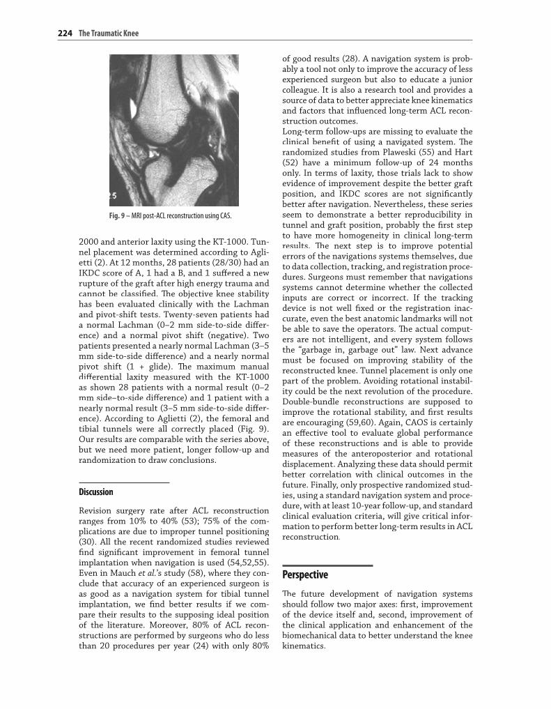
2000 and anterior laxity using the KT-1000. Tun-nel placement was determined according to Agli-etti (2). At 12 months, 28 patients (28/30) had an IKDC score of A, 1 had a B, and 1 suffered a new ffffrupture of the graft after high energy trauma and cannot be classified. Thfi e objective knee stability Thhas been evaluated clinically with the Lachmanand pivot-shift tests. Twenty-seven patients had a normal Lachman (0–2 mm side-to-side differ-ffffence) and a normal pivot shift (negative). Two patients presented a nearly normal Lachman (3–5 mm side-to-side diff erence) and a nearly normalffffpivot shift (1 + glide). The maximum manual Thdifferential laxity measured with the KT-1000 ffffas shown 28 patients with a normal result (0–2mm side–to-side diff erence) and 1 patient with a ffffnearly normal result (3–5 mm side-to-side differ-ffffence). According to Aglietti (2), the femoral andtibial tunnels were all correctly placed (Fig. 9). Our results are comparable with the series above, but we need more patient, longer follow-up and randomization to draw conclusions.
Discussion
Revision surgery rate after ACL reconstructionranges from 10% to 40% (53); 75% of the com-plications are due to improper tunnel positioning(30). All the recent randomized studies reviewed fi nd signififi cant improvement in femoral tunnel fiimplantation when navigation is used (54,52,55). Even in Mauch et al.’s study (58), where they con-clude that accuracy of an experienced surgeon is as good as a navigation system for tibial tunnel implantation, we find better results if we com-fipare their results to the supposing ideal positionof the literature. Moreover, 80% of ACL recon-structions are performed by surgeons who do lessthan 20 procedures per year (24) with only 80%
Fig. 9 – MRI post-ACL reconstruction using CAS.

Place of navigation in anterior cruciate ligament reconstruction 225
a torn anterior cruciate ligament. Knee Surg Sports Trau-matol Arthrosc 6(Suppl 1): S49–S55Khalfayan EE, Sharkey PF, Alexander AH,6. et al. (1996).Th e relationship between tunnel placement and clinical Thresults after anterior cruciate ligament reconstruction.Am J Sports Med 24:335–341Sommer C, Friederich NF, Müller W (2000) Improp-7. erly placed anterior cruciate ligament grafts: correlation between radiological parameters and clinical results. KneeSurg Sports Traumatol Arthrosc 8:207–213Dessenne V, Lavallée S, Julliard R, 8. et al. (1995) Computer-assisted knee anterior cruciate ligament reconstruction:fi rst clinical tests. J Image Guid Surg 1:59–64fiKlos TVS, Banks AZ, Banks SA,9. et al. (1995) Computer and radiographic assisted anterior cruciate ligament reconstruction of the knee. In: Anderson J, editor. Pro-ceedings of the second symposium on medical roboticsand computer assisted surgery. New York: Wiley:252–255Paul HA, Bargar WL, Mittlesstadt B,10. et al. (1992) Develop-ment of a surgical robot for cementless total hip arthro-plasty. Clin Orthop 285:57–66Cobb J, Henckel J, Richards R,11. et al. (2004) Robot assisted minimally invasive unicompartmental knee arthroplasty results of fi rst clinical trial. Comput Aided Surg 9:88fiMcGurk M, Amis AA, Potamianos P, 12. et al. (1997) Rapidprototyping techniques for anatomical modeling in medi-cine. Ann R Coll Surg Engl 79:169–174Galen C (1968) On the usefulness of the parts of the body. 13. May MT (trans). Ithaca, NY: Cornell University PressStark J (1850) Two cases of ruptured crucial ligaments of 14. the knee joint. Edinburg Med Surg 74:267–271Battle WH (1900) A case after open section of the knee-15. joint for irreductible traumatic dislocation. Clin Soc Lon-don Trans 33:232Mayo Robson AW (1903) Ruptured crucial ligaments and 16. their repair by operation. Am Surg 37:716–718Lange F (1903) Uber die Sehnenplastik. Verh Dtsch Othop 17. Ges 2:10–12Jones K (1963) Reconstruction of the anterior cruciate 18. ligament: a technique using the central one-third of the patellar ligament. J Bone Joint Surg 45A:925–932Bruckner H (1966) Eine neue Methode zur Kreuzband-19. plastik. Chirurg 37:413–414Franke K (1976) Clinical experience in 130 cruciate liga-20. ment reconstructions. Orthop Clin North Am 7:191–193Dandy DJ (1981) Arthroscopic surgery of the knee. Edin-21. burg: Churchill LivingstoneSoni AH, Gudavalli MR, Herndon WA,22. et al. (1986) Appli-cation of passive robot in spine surgery. Presented at the Eighth Annual Conference of the IEEE/Engineer Biology and Medical Society, 1186–1191Petermann J, Kober R, Heinze P (2000) Computer-as-23. sisted planning and robot-assisted surgery in anterior cruciate ligament reconstruction. Operat Tech Orthoped 10:50Eichhorn J, Girdano N (2005) Computerassistierte Rekon-24. struktion des vorderen Kreuzbandes mit dem Navigation-ssystem.. Arthroskopie 18:24–26Berchuck M, Andriacchi TP, Bach BR (1990) Gait adapta-25. tions by patients who have a deficient anterior cruciate filigament. J Bone Joint Surg 72A:871–877Dunn WR, Lyman S, Lincolm AE, 26. et al. (2004) The effTh ect ffffof ACL reconstruction on the risk of knee re-injury: an outcome study of 6567 cases. Am J Sports Med 32:1906–1914Herrington L, Wrapson C, Matthews M, 27. et al. (2005) Ante-rior cruciate ligament reconstruction, hamstring versus bone-patella tendon-bone grafts: a systematic literaturereview of outcome from surgery. The Knee 12:41–50Th
Th e navigation systems should be not too expen-Thsive, providing a simple and intuitive system.Although, an image-free system is more desirable,systems using MRI that are generally used rou-tinely as a diagnostic procedure, or ultra-sonog-raphy to do echo-morphing (61), could provideextra information without extra imaging and then better accuracy and reproducibility in the recon-struction. Optical trackers could be replaced by electromagnetic trackers to avoid invasive inser-tion of rigid pins into bone and the need for a con-tentious line of sight. Ultrasounds are also under investigation for tracking purpose. Hybrid systemsare in development, combining robotic and naviga-tion to improve accuracy of the surgeon and lim-iting his or her action to what has been planned preoperatively. Improvements in data collectionwith more intelligent systems able to evaluateand to correct inappropriate information are also in development. Moreover, we need more power-ful computers to calculate the ideal graft position from a single anatomic point. This will allow 3-D Thsimulation before to implant the graft in the abso-lute ideal position. Finally, navigation systemsthat can be used in the operating room and during the follow-up would provide all the information to compare knee kinematics before, during, and after surgery. Th is would close the loop in surgical Thpractice by measuring and directly relating surgical techniques to patient outcome.
Conclusion
Navigation in ACL reconstruction has a tremen-dous amount of potential. It is not only a tool to improve tunnel positioning but also a learning and a research tool that will give us the information and measures about knee kinematics to approach the perfect reconstruction and the best clinicaloutcome in the long term.
References
Griffi n LY, Agel J, Alholm MJ, ffi1. et al. (2000) Noncontact anterior cruciate ligament injuries: risk factors and pre-vention strategies. J Am Acad Orthop Surg 8:141–150Aglietti P, Buzzi R, Giron F,2. et al. (1997) Arthroscopic-as-sisted anterior cruciate ligament reconstruction with the central third patellar tendon: A 5-8-year follow-up. KneeSurg Sports Traumatol, Arthrosc 5:138–144Goble EM, Downey DJ, Wilcox TD (1995) Positioning of 3. the tibial tunnel for anterior cruciate ligament reconstruc-tion. Arthroscopy 11:688–695Hefzy MS, Grood ES (1986) Sensitivity of insertion loca-4. tions on length patterns of anterior cruciate ligament fi bers. J Biomech Eng 108:73–82fiHowell SM (1998) Principles for placing the tibial tunnel 5. and avoiding roof impingement during reconstruction of

226 The Traumatic Knee
Kohn D, Beusche T, Caris J (1998) Drill hole position in 46. endoscopic anterior cruciate ligament reconstruction.Results on an advanced arthroscopy course. Knee SurgSports Traumatol Arthrosc 6:13–15Klos TVS, Habets RJE, Banks AZ, 47. et al. (1998) Computerassistance in arthroscopic anterior cruciate ligament reconstruction. Clin Orthop 354:65–69Hiraoka H, Kuribayashi S, Fukuda A, 48. et al. (2006) Endo-scopic anterior cruciate ligament reconstruction usinga computer-assisted fl uoroscopic navigation system. J flOrthop Sci 11:159–166Burkart A, Debski RE, McMahon PJ,49. et al. (2001) Precision of ACL tunnel placement using traditional and robotic techniques. Comp Aid Surg 6:270–278Messerli G, Ménétrey J (2007) Reconstruction du liga-50. ment croisé antérieur assistée par ordinateur: Etude prospective non randomisée avec résultats à 12 mois des 30 premiers cas. Thèse n 10503, Université de GenèveChouteau J, Benareau I, Testa R, 51. et al. (2007) Compara-tive study of knee anterior cruciate ligament reconstruc-tion with or without fl uoroscopic assistance: a prospectiveflstudy of 73 cases. Arch Orthop Trauma Surg (Epub aheadof print)Hart R, Krejzla J, Svab P,52. et al. (2008) Outcomes after con-ventional versus computer-navigated anterior cruciateligament reconstruction. Arthroscopy 24:569–578Kodali P, Yang S, Koh J (2008) Computer-assisted Surgery 53. for anterior cruciate ligament reconstruction. Sports MedArthrosc Rev 16:67–76Picard F, DiGioia AM, Moody J, 54. et al. (2001) Accuracy intunnel placement for ACL reconstruction. Comparison of traditional arthroscopic and computer-assisted navigationtechniques. Comp Aid Surg 6:279–289Plaweski S, Cazal J, Rosell Ph, Merloz Ph (2006) Ante-55. rior cruciate ligament reconstruction using navigation.A comparative study on 60 patients. Am J Sports Med34:542–552Eichhorn J (1998) Th ree years of experience with com-Th56. puter navigation-assisted positioning of drilling tunnels in anterior cruciate ligament replacements (S67), Arthros-copy 20:31–32Julliard R, Lavallée S, Dessenne V (1998) Computer57. assisted reconstruction of the nterior cruciate ligament.Clin Orthop Relat Res 354:57–64Mauch F, Apic G, Becker U, Bauer G (2007) Differencesffff58. in the placement of the tibial tunnel during reconstruc-tion of the anterior cruciate ligament with and with-out computer-assisted navigation. Am J Sports Med35:1824–1832Ishibashi Y, Tsuda E, Fukuda A,59. et al. (2008) Stability eval-uation of single-bundle and double-bundle reconstruc-tion during navigated ACL reconstruction. Sports Med Arthrosc Rev 16:77–8360.Monaco E, Labianca L, Conteduca F, 60. et al. (2007) Double bundle or single bundle plus extraarticular teno-desis in ACL reconstruction? Knee Surg Sports Traumatol Arthrosc 15:1168–1174Stindel E, Briard JL, Lavallee S, 61. et al. (2004) Bone mor-phing: 3-D reconstruction without pre or intraoperativeimaging – concept and application. In: Stiehl JB, Kiner-mann WH, Haaker RG, editors. Navigation and robotics in total joint and spine surgery. Berlin: Springer: 39–45
Beasley LS, 28. et al. (2005) Anterior cruciate ligament recon-struction: a literature review of the anatomic, biomechan-ics, surgical consideration and clinical outcomes. Oper Tech Orthop 15:5–19Greis PE, Johnson DJ, Fu FH (1993) Revision anterior29.cruciate ligament surgery: Cause of graft failure and tech-nical considerations of revision surgery. Clin Sports Med 12:839–852
Harner CD (1995) Revision anterior cruciate ligament30. reconstruction using fresh-frozen allograft tissue. Instruc-tional Course at the 62nd Annual AAOS Meeting, Orlando, FL. Maday MG, Harner CD, Fu FH (1994) Revision ACL sur-31.gery: evaluation and treatment, In: Feagin JA, editor. TheThcrucial ligaments. New York, NY: Churchill Livingstone: 711–723Harner CD, Baek GH, Vogrin TM, 32. et al. (1999) Quanti-tative analysis of human cruciate ligament insertions. Arthroscopy 15:741–749Paessler H (1997) Revisionseingriffe nach vorderer Kreuz-ffff33.bandoperation und neuerlicher Instabilität: Ursache-nanalyse und taktische Vorgehen. Hefte Unfallchirurg268:447–450Shelbourne KD, Klootwyk TE, Wilckens JH, 34. et al. (1995)Ligament stability two to six years after anterior cruciate ligament reconstruction with autogenous patellar tendon graft and participation in accelerated rehabilitation pro-gram. Am J Sports Med 23:575–579Fu FH, Bennett CH, Lattermann C, 35. et al. (1999) Currenttrends in anterior cruciate ligament reconstruction. Part I:Biology and biomechanics of reconstruction. Am J Sports Med 27:821–830Fineberg MS, Zarins B, Sherman OH (2000) Practical con-36. siderations in anterior cruciate ligament replacement sur-gery. Arthroscopy 16:715–724Fu FH, Bennett CH, Ma CB, 37. et al. (2000) Current trends inanterior cruciate ligament reconstruction. Part II. Opera-tive procedures and clinical correlations. Am J Sports Med28:124–130Lee MC, Seong SC, Lee S,38. et al. (2007) Vertical femoral tunnel placement results in rotational knee laxity afteranterior cruciate ligament reconstruction. Arthroscopy 23:771–778Scopp JM, Jasper LE, Belkoff SM, ff39. et al. (2004) The effTh ectffffof oblique femoral tunnel placement on rotational con-straint of the knee reconstructed using patellar tendon autografts. Arthroscopy 20:294–299Jackson DW, Gasser SI (1994) Tibial tunnel placement in40.ACL reconstruction. Arthroscopy 10:124–131Watanabe BM, Howell SM (1995) Arthroscopic fi ndings fi41.associated with roof impingement of anterior cruciate ligament graft. Am J Sports Med 23:616–625Sati M, Stäubli HU, Bourquin Y,42. et al. (2002) Real-time computer in situ guidance system for ACL graft place-ment. Computer aided surgery 7:25–40Dienst M, Burks RT, Greis PE (2002) Anatomy and bio-43. mechanics of the anterior cruciate ligament. Orthop Clin North Am 33:605–620Girgis FG, Marshall JL, Monajem A (1975) The cruciateTh44. ligaments of the knee joint. Anatomical, functional and experimental analysis. Clin Orthop 106:216–231Odensten M, Gillquist J (1985) Functional anatomy of the45. anterior cruciate ligament and a rationale for reconstruc-tion. J Bone Joint Surg 67A:257–262