Embed Size (px)
Citation preview
Acta Poloniae Pharmaceutica ñ Drug Research, Vol. 70 No. 3 pp. 523ñ531, 2013 ISSN 0001-6837Polish Pharmaceutical Society
Natural products, especially plant extracts,have been used for millennia in the treatment ofmany diseases. More attention is now being devotedto the curative properties of plant extracts and thesearch for new natural substances which can be usedin the treatment of different diseases.
Potentilla L. (Rosaceae) species have beenused for a long time in traditional medicine in Asia,Europe and Northern America. Many of them areknown for their therapeutic properties and are con-sidered to be among the safest natural astringents inthe treatment of diarrhoea, dysentery and sorethroat. Recent investigations have also shown thatsome extracts of different parts of plants fromPotentilla species exhibit potential antioxidant,hypoglycemic, anti-inflammatory, antitumor and
anti-ulcerogenic properties (1). Phytochemical stud-ies, of both the aerial and underground parts ofplants, have concentrated on the isolation of manydifferent classes of compounds. Among them, triter-penoids, condensed and hydrolyzable tannins, andflavonoids, as well as other ingredients including aseries of organic acids, phenol carboxylic acids,sterols, sugars, amino acids and fatty acids havebeen detected in Potentilla species (1, 2). Recentpharmacological in vitro studies on Potentillaspecies have confirmed their anti-secretory, anti-inflammatory, antimicrobial and antioxidativeeffects. Application in in vivo test models alsoshowed their different biological activities (1, 3, 4).
The aim of our study was to analyze the influ-ence of aqueous extracts from selected Potentilla
THE INFLUENCE OF AQUEOUS EXTRACTS OF SELECTED POTENTILLA SPECIES ON NORMAL HUMAN COLON CELLS
MICHA£ TOMCZYK1*, ROMAN PADUCH2, ADRIAN WIATER3,MA£GORZATA PLESZCZY—SKA3, MARTYNA KANDEFER-SZERSZE—2
and JANUSZ SZCZODRAK3
1Department of Pharmacognosy, Faculty of Pharmacy, Medical University of Bia≥ystok, Mickiewicza 2a, 15-230 Bia≥ystok, Poland
2Department of Virology and Immunology, 3Department of Industrial Microbiology, Faculty of Biology and Biotechnology, Institute of Microbiology and Biotechnology,
Maria Curie-Sk≥odowska University, Akademicka 19, 20-033 Lublin, Poland
Abstract: Potentilla L. (Rosaceae) species have been used in traditional medicine in Asia, Europe and NorthernAmerica. This study analyzed the biological activity of aqueous extracts of Potentilla species (Rosaceae):Dasiphora fruticosa (syn. P. fruticosa), P. norvegica, P. pensylvanica, P. thuringiaca, P. crantzii and P. nepalen-sis. The activities were tested using MTT, NR and DPPH assays on normal human colon epithelium (CCD 841CoTr) and colon myofibroblast (CCD-18Co) cells. Moreover, cell morphology using the May-Gr¸nwald-Giemsa method, IL-6 by ELISA, and nitric oxide (NO) analysis with the Griess method in culture supernatantswere performed after 24 h. Extracts were tested at dose levels between 25 and 250 µg/mL. For ELISA, 15µg/mL was chosen. All extracts suppressed the metabolism of myofibroblasts, while epithelial cellsí mito-chondrial dehydrogenase activity decreased after incubation with extracts. All extracts showed a free radicalscavenging (DPPH) effect in a concentration-dependent manner. The most potent was the extract from D. fru-ticosa, while the least action was observed for P. thuringiaca. Potentilla extracts stimulated IL-6 production intested cells but the level of the cytokine was found to decrease in epithelial cells. Pre-incubation of cells withLPS resulted in increased IL-6 secretion. Modulation of NO production after extract addition and cell pre-incu-bation with LPS was also observed. Potentilla extracts may be interesting natural factors modulating the mainfeatures of cells forming the colon wall, and thus may be potentially useful in the prophylaxis or healing ofcolon disorders
Keywords: Potentilla L., Rosaceae, cytotoxicity, normal human colon cells
523
* Corresponding author: e-mail: [email protected]
524 MICHA£ TOMCZYK et al.
species: Dasiphora fruticosa (L.) Rydb. (syn.Potentilla fruticosa L.), Potentilla norvegica L.,Potentilla pensylvanica L., Potentilla thuringiacaBernh. Ex Link, Potentilla crantzii (Crantz) J. Beckex Fritsch and Potentilla nepalensis Hook. Var.ëMiss Willmottí on the viability and proliferation ofnormal human colon cells, lines: CCD 841 CoTr(normal colon epithelium) and CCD-18Co (colonmyofibroblasts). Additionally, the subject of thisstudy was the determination whether tested extractshave immunostimulatory, anti-inflammatory andreactive oxygen radical-scavenging properties.Therefore, the principal objective was to assess in invitro studies whether Potentilla species used as foodsupplements may influence (and eventually to whatdegree may affect) normal human colon cell walllining, and to evaluate their potential health benefitsin humans.
Plant material
Seeds of six species (D. fruticosa ñ index semi-num /ind. sem./ 2566, P. norvegica ñ ind. sem. 303,P. thuringiaca ñ ind. sem. 1551, P. pensylvanica ñind. sem. 1546, P. crantzii ñ ind. sem. 1534, P.nepalensis ñ ind. sem. 1542) were requested fromthe Hortus Botanicus Universitatis Masarykianae,Brno, Czech Republic; the Botanical Garden ofVilnius University, Lithuania; Hortus BotanicusUniversitatis Posnaniensis, PoznaÒ, Poland, and theGiardino Botanico Alpino, Cogne, Italy. Plants werecultivated in common plots at the Medicinal PlantsGarden, near the Medical University of Bia≥ystok,Poland. Voucher specimens of plants were deposit-ed in the Herbarium of the Department ofPharmacognosy, Medical University of Bia≥ystok,Poland and carefully authenticated by one of theauthors (MT), based on morphological characteris-
tics by comparison with literature data. Collectedplant materials (aerial parts of plants includingleaves, stem and flowers), during June-July 2007-2010, were air-dried under shade at room tempera-ture and then ground with an electric grinder intofine powders which were stored in air tight contain-ers at room temperature. Processing of the driedplant material (preparation of aqueous extracts) wascarried out after four years of harvest in 2010. Thelist of taxons studied is given in Table 1.
Preparation of extracts
Powdered plant material (2.0 g) was separatelyextracted with water (2 ◊ 150 mL) in an ultrasonica-tor bath (Sonic-5, POLSONIC, Poland) at a con-trolled temperature (40 ± 2OC) for 45 min.Supernatants were filtered through a funnel withglass wool, which was washed with 5 mL of solventand concentrated to dryness under vacuum (B¸chiSystem, Switzerland) at a controlled temperature(40 ± 2OC) and subjected to lyophilization using aLymph-Lock 1.0 (Labconco, USA) vacuum concen-trator until a constant weight was obtained. Yields:(1, 2.05%; 2, 2.46%; 3, 5.03%; 4, 3.01%; 5, 2.69%;6, 1.98%) ñ numbering according to Table 1.
Phytochemical profile
Total phenolic contents in extracts were deter-mined spectrophotometrically at 765 nm (Specord40, Analytik Jena, Germany) after the reaction withFolin-Ciocalteuís phenol reagent as gallic acidequivalents GAE/100 g in mg/g of dry weight (d.w.)according to the manual colorimetric methoddescribed by Tawaha et al. (5). Total phenolic acidscontent in plant material was determined by usingthe spectrophotometric method with Arnovísreagent according to the procedure described in the
Table 1. Plant material from the selected Potentilla L. species.
Sample Parts Voucherno.
Plant sources
used specimen no.Scientific name Common name
1. Dasiphora fruticosa (L.) Rydb.(syn. Potentilla fruticosa L.)a Shrubby cinquefoil herbs PFR-06018
2. Potentilla norvegica L. Norwegian cinquefoil herbs PNO-08024
3. Potentilla thuringiaca Bernh. ex Link European cinquefoil herbs PTH-06022
4. Potentilla pensylvanica L. Pennsylvania cinquefoil herbs PPS-08025
5. Potentilla crantzii (Crantz) J. Beck ex Fritsch Alpine cinquefoil herbs PCR-09026
6. Potentilla nepalensis Hook. var. ëMiss Willmottí Nepal cinquefoil herbs PNE-06023
a botanical nomenclature in accordance with the taxonomy of USDA Natural Resources Conservation Service (NRCS).
The influence of aqueous extracts of selected Potentilla species... 525
European Pharmacopoeia 6.0 (6). The total contentof flavonoids was determined by the spectrophoto-metric method according to Christ and M¸ller andfollowed the procedure described in the EuropeanPharmacopoeia 6.0 (6). The total tannin content wasdetermined by the weight method with hide powderaccording to the DAB 10 (7). The total proanto-cyanidin content was measured according to theEuropean Pharmacopoeia 6.0 (6).
Cell cultures
Normal human colon myofibroblasts CCD-18Co (ATCC No. CRL-1459) and normal humancolon epithelial cells CCD 841 CoTr (ATCC No.CRL-1807) were cultured in RPMI 1640 medium(Sigma) supplemented with 10% FCS at 37OC(CCD-18Co) or 34OC (CCD 841 CoTr) in a 5%CO2/95% air atmosphere. For experiments, cellsconcentration at 1 ◊ 105 cells/mL was used.
1,1-Diphenyl-2-picrylhydrazyl (DPPHï) test
Free radical scavenging activity of extracts wasmeasured by the 1,1-diphenyl-2-picrylhydrazyl(DPPHï) assay. This method is based on the abilityof antioxidants to reduce the stable dark violet radi-cal DPPHï (Sigma) to the yellow coloreddiphenylpicrylhydrazine. Briefly, 100 µL of DPPHï
solution (0.2 mg/mL in ethanol) was added to 100µL of extract concentrations (25-250 µg/mL) andstandards. Trolox (Sigma) at increasing concentra-tions (1-50 µg/mL) was used as a standard for thefree radical scavenging activity. After 20 min ofincubation at room temperature, the absorbance ofthe solution was measured at 515 nm. The lower theabsorbance, the higher the free radical scavengingactivity of the extracts. The activity of each extractwas determined by comparison of its absorbancewith that of a control solution (reagents withoutextract) and standard.
The capability to scavenge DPPHï radical wascalculated by the following formula:
DPPHï scavenging effect (%) == [(Xcontrol ñ Xextract)/Xcontrol)] ◊ 100
Xcontrol is the absorbance of the control and Xextract isthe absorbance in the presence of extracts (8).
Neutral red (NR) uptake assay
The NR cytotoxicity assay is based on theuptake and lysosomal accumulation of the supravitaldye, neutral red. Dead or damaged cells do not takeup the dye (9).
Cells were grown in 96-well multiplates in 100mL of RPMI 1640 culture medium with 2% FCS(fetal calf serum) and Potentilla extracts (25-250
µg/mL). Subsequently, the medium was discardedand 0.4% NR (Sigma) in culture medium was addedto each well. The plate was incubated for 3 h at 37OCin humidified 5% CO2. After incubation, the dyecontaining medium was removed, cells were fixedwith 1% CaCl2 in 4% paraformaldehyde, and there-after, the incorporated dye was solubilized using 1%acetic acetate in 50% ethanol solution (100 mL).The plates were gently shaken for 20 min at roomtemperature and the extracted dye absorbance wasmeasured spectrophotometrically at 540 nm using amicroplate reader (Molecular Devices Corp., Emax,Menlo Park, CA, USA).
MTT assay
Cells sensitivity to Potentilla extracts wasdetermined in a standard spectrophotometric 3-(4,5-dimethylthiazole-2-yl)-2,5-diphenyltetrazolium bro-mide (MTT) assay according to Mosmann (10). TheMTT test is based on the conversion of yellow tetra-zolium salt by viable cells to purple crystals of for-mazan. The reaction is catalyzed by mitochondrialsuccinyl dehydrogenase.
Cells grown in 96-well multiplates in 100 µLof culture medium were incubated for 3 h with MTTsolution (5 mg/mL, 25 µL/well) (Sigma). The yel-low tetrazolium salt was metabolized by viable cellsto formazan purple crystals. The reaction was cat-alyzed by mitochondrial succinyl dehydrogenase.The crystals were solubilized overnight in a 10%sodium dodecyl sulfate (SDS) in 0.01 M HCl mix-ture. The product was quantified spectrophotometri-cally by absorbance measurement at 570 nm wave-length using an E-max Microplate Reader(Molecular Devices Corporation, Menlo Park, CA,USA).
Nitric oxide (NO) measurement
Nitrite, a stable end product of NO, was deter-mined in culture supernatants by a spectrophotomet-ric method based on the Griess reaction. The level ofnitrite reflects NO production (11). Briefly, cellswere incubated for 24 h with the 15 µg/mL ofPotentilla extract concentration and then culturesupernatants were collected. The cell cultures,before the addition of extracts, were also pre-incu-bated with LPS (10 µg/mL) for 2 h. Next, 100 µL ofsupernatant was plated in 96-well flat-bottomedplates in triplicate and incubated with 100 µL ofGriess reagent (1% sulfanilamide/0.1% N-(1-naph-thyl)ethylenediamine dihydrochloride) (Sigma) in3% H3PO4 (POCH Gliwice, Poland) at room tem-perature for 10 min. The optical density was meas-ured at 550 nm using a microplate reader (Molecular
526 MICHA£ TOMCZYK et al.
Devices Corp., Emax, Menlo Park, CA, USA). Astandard curve was plotted using 0.5-25 µM sodiumnitrite (NaNO2) for calibration.
ELISA
The level of human IL-6 was tested immu-noenzymatically (ELISA) in culture supernatantsusing commercially available kits (Diaclone)according to the manufacturerís instructions. Theoptical density at 450 nm with the correction wave-length of 570 nm of each ELISA sample was deter-mined using a microplate reader. The IL-6 concen-tration was calculated on the basis of a standardcurve: 2 pg/mL (IL-6) was the detection limit.
Statistical analysis
Results are presented as the means ± SD fromthree experiments. Data were analyzed using a one-way ANOVA test. Differences of p ≤ 0.05 were con-sidered significant. Only the results with a signifi-cance of p ≤ 0.05 are reported. All experiments wererepeated three times.
RESULTS AND DISCUSSION
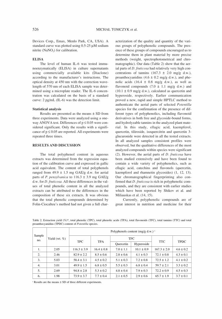
The total polyphenol content in aqueousextracts was determined from the regression equa-tion of the calibration curve and expressed in gallicacid equivalent. The content of total polyphenolsranged from 49.9 ± 1.5 mg GAE/g d.w. for aerialparts of P. pensylvanica to 116.3 ± 3.9 mg GAE/gd.w. for D. fruticosa. All these differences in the val-ues of total phenolic content in all the analyzedextracts can be attributed to the differences in thecomposition of these six extracts. It was obviousthat the total phenolic compounds determined byFolin-Ciocalteuís method had not given a full char-
acterization of the quality and quantity of the vari-ous groups of polyphenolic compounds. The pres-ence of these groups of compounds encouraged us todetermine them in plant material by more precisemethods (weight, spectrophotometrical and chro-matographic). Our data (Table 2) show that the aer-ial parts of D. fruticosa had relatively very high con-centrations of tannins (167.3 ± 2.0 mg/g d.w.),proanthocyanidins (4.6 ± 0.2 mg/g d.w.), and phe-nolic acids (16.4 ± 0.8 mg/g d.w.), as well asflavonoid compounds (7.0 ± 1.1 mg/g d.w.) and(10.1 ± 0.9 mg/g d.w.), calculated as quercetin andhyperoside, respectively. Earlier communicationproved a new, rapid and simple HPTLC method toauthenticate the aerial parts of selected Potentillaspecies for the confirmation of the presence of dif-ferent types of polyphenolics, including flavonoidderivatives in both free and glycoside-bound forms,and hydrolyzable tannins in the analyzed plant mate-rial. In this study, ellagic acid, kaempferol,quercetin, tiliroside, isoquercitrin and quercetin 3-glucuronide were detected in all the tested extracts.In all analyzed samples consistent profiles wereobserved, but the qualitative differences of the mostanalyzed compounds within species were significant(2). However, the aerial parts of D. fruticosa havebeen studied extensively and have been found tocontain a wide variety of polyphenolics, such asellagic acid, catechins and flavonols (quercetin,kaempferol and rhamnetin glycosides) (1, 12, 13).Our chromatographical fingerprinting also con-firmed that D. fruticosa is rich in polyphenolic com-pounds, and they are consistent with earlier studieswhich have been reported by Shikov et al. andMiliauskas et al. (14, 15).
Currently, polyphenolic compounds are ofgreat interest in nutrition and medicine for their
Table 2. Extraction yield (%)*, total phenolic (TPC), total phenolic acids (TPA), total flavonoids (TFC), total tannins (TTC) and totalproanthocyanidins (TPDC) content of Potentilla species.
Polyphenols content (mg/g d.w.) a
SampleYield (wt. %)
TPC TPATFC
TTC TPDCno. Quercetin Hyperoside
1. 2.05 116.3 ± 3.9 16.4 ± 0.8 7.0 ± 1.1 10.1 ± 0.9 167.3 ± 2.0 4.6 ± 0.2
2. 2.46 82.9 ± 2.2 8.5 ± 0.6 2.8 ± 0.6 4.1 ± 0.3 72.1 ± 0.8 4.3 ± 0.1
3. 5.03 58.4 ± 3.1 4.5 ± 0.2 5.1 ± 0.3 7.2 ± 0.8 72.5 ± 1.2 4.1 ± 0.2
4. 3.01 49.9 ± 1.5 6.8 ± 0.5 5.5 ± 0.3 6.8 ± 0.4 59.7 ± 2.1 3.3 ± 0.2
5. 2.69 94.8 ± 2.8 5.3 ± 0.2 4.8 ± 0.4 7.9 ± 0.3 72.2 ± 0.9 4.5 ± 0.3
6. 1.98 73.9 ± 3.7 7.7 ± 0.4 2.1 ± 0.5 2.9 ± 0.6 65.7 ± 1.9 3.7 ± 0.1
a Results are the means ± SD of three different experiments.
The influence of aqueous extracts of selected Potentilla species... 527
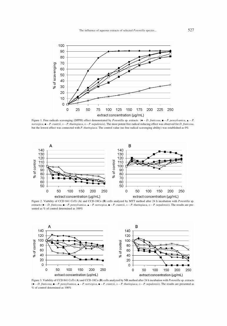
Figure 1. Free radicals scavenging (DPPH) effect demonstrated by Potentilla sp. extracts (◆ ñ D. fruticosa, ■ ñ P. pensylvanica, ▲ ñ P.norvegica, ● ñ P. crantzii, ✻ ñ P. thuringiaca, Ο ñ P. nepalensis). The most potent free radical reducing effect was observed for D. fruticosa,but the lowest effect was connected with P. thuringiaca. The control value (no free radical scavenging ability) was established as 0%
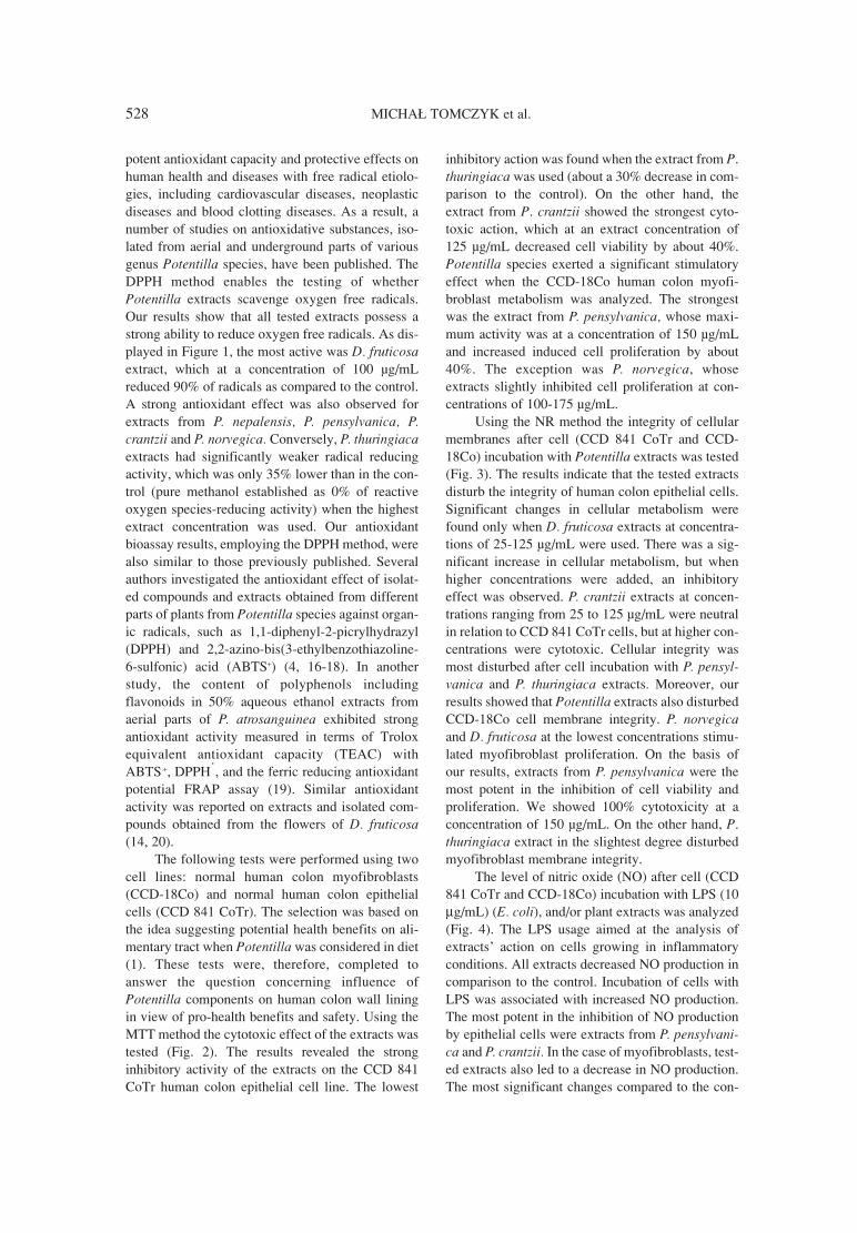
Figure 2. Viability of CCD 841 CoTr (A) and CCD-18Co (B) cells analyzed by MTT method after 24 h incubation with Potentilla sp.extracts (◆ ñ D. fruticosa, ■ ñ P. pensylvanica, ▲ ñ P. norvegica, ● ñ P. crantzii, ✻ ñ P. thuringiaca, Ο ñ P. nepalensis). The results are pre-sented as % of control determined as 100%
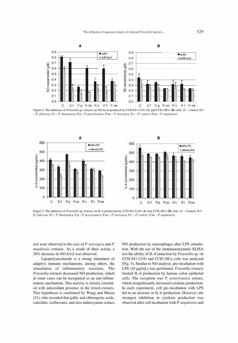
Figure 3. Viability of CCD 841 CoTr (A) and CCD-18Co (B) cells analyzed by NR method after 24 h incubation with Potentilla sp. extracts(◆ ñ D. fruticosa, ■ ñ P. pensylvanica, ▲ ñ P. norvegica, ● ñ P. crantzii, ✻ ñ P. thuringiaca, Ο ñ P. nepalensis). The results are presented as% of control determined as 100%
528 MICHA£ TOMCZYK et al.
potent antioxidant capacity and protective effects onhuman health and diseases with free radical etiolo-gies, including cardiovascular diseases, neoplasticdiseases and blood clotting diseases. As a result, anumber of studies on antioxidative substances, iso-lated from aerial and underground parts of variousgenus Potentilla species, have been published. TheDPPH method enables the testing of whetherPotentilla extracts scavenge oxygen free radicals.Our results show that all tested extracts possess astrong ability to reduce oxygen free radicals. As dis-played in Figure 1, the most active was D. fruticosaextract, which at a concentration of 100 µg/mLreduced 90% of radicals as compared to the control.A strong antioxidant effect was also observed forextracts from P. nepalensis, P. pensylvanica, P.crantzii and P. norvegica. Conversely, P. thuringiacaextracts had significantly weaker radical reducingactivity, which was only 35% lower than in the con-trol (pure methanol established as 0% of reactiveoxygen species-reducing activity) when the highestextract concentration was used. Our antioxidantbioassay results, employing the DPPH method, werealso similar to those previously published. Severalauthors investigated the antioxidant effect of isolat-ed compounds and extracts obtained from differentparts of plants from Potentilla species against organ-ic radicals, such as 1,1-diphenyl-2-picrylhydrazyl(DPPH) and 2,2-azino-bis(3-ethylbenzothiazoline-6-sulfonic) acid (ABTS+) (4, 16-18). In anotherstudy, the content of polyphenols includingflavonoids in 50% aqueous ethanol extracts fromaerial parts of P. atrosanguinea exhibited strongantioxidant activity measured in terms of Troloxequivalent antioxidant capacity (TEAC) withABTS.+, DPPHˇ, and the ferric reducing antioxidantpotential FRAP assay (19). Similar antioxidantactivity was reported on extracts and isolated com-pounds obtained from the flowers of D. fruticosa(14, 20).
The following tests were performed using twocell lines: normal human colon myofibroblasts(CCD-18Co) and normal human colon epithelialcells (CCD 841 CoTr). The selection was based onthe idea suggesting potential health benefits on ali-mentary tract when Potentilla was considered in diet(1). These tests were, therefore, completed toanswer the question concerning influence ofPotentilla components on human colon wall liningin view of pro-health benefits and safety. Using theMTT method the cytotoxic effect of the extracts wastested (Fig. 2). The results revealed the stronginhibitory activity of the extracts on the CCD 841CoTr human colon epithelial cell line. The lowest
inhibitory action was found when the extract from P.thuringiaca was used (about a 30% decrease in com-parison to the control). On the other hand, theextract from P. crantzii showed the strongest cyto-toxic action, which at an extract concentration of125 µg/mL decreased cell viability by about 40%.Potentilla species exerted a significant stimulatoryeffect when the CCD-18Co human colon myofi-broblast metabolism was analyzed. The strongestwas the extract from P. pensylvanica, whose maxi-mum activity was at a concentration of 150 µg/mLand increased induced cell proliferation by about40%. The exception was P. norvegica, whoseextracts slightly inhibited cell proliferation at con-centrations of 100-175 µg/mL.
Using the NR method the integrity of cellularmembranes after cell (CCD 841 CoTr and CCD-18Co) incubation with Potentilla extracts was tested(Fig. 3). The results indicate that the tested extractsdisturb the integrity of human colon epithelial cells.Significant changes in cellular metabolism werefound only when D. fruticosa extracts at concentra-tions of 25-125 µg/mL were used. There was a sig-nificant increase in cellular metabolism, but whenhigher concentrations were added, an inhibitoryeffect was observed. P. crantzii extracts at concen-trations ranging from 25 to 125 µg/mL were neutralin relation to CCD 841 CoTr cells, but at higher con-centrations were cytotoxic. Cellular integrity wasmost disturbed after cell incubation with P. pensyl-vanica and P. thuringiaca extracts. Moreover, ourresults showed that Potentilla extracts also disturbedCCD-18Co cell membrane integrity. P. norvegicaand D. fruticosa at the lowest concentrations stimu-lated myofibroblast proliferation. On the basis ofour results, extracts from P. pensylvanica were themost potent in the inhibition of cell viability andproliferation. We showed 100% cytotoxicity at aconcentration of 150 µg/mL. On the other hand, P.thuringiaca extract in the slightest degree disturbedmyofibroblast membrane integrity.
The level of nitric oxide (NO) after cell (CCD841 CoTr and CCD-18Co) incubation with LPS (10µg/mL) (E. coli), and/or plant extracts was analyzed(Fig. 4). The LPS usage aimed at the analysis ofextractsí action on cells growing in inflammatoryconditions. All extracts decreased NO production incomparison to the control. Incubation of cells withLPS was associated with increased NO production.The most potent in the inhibition of NO productionby epithelial cells were extracts from P. pensylvani-ca and P. crantzii. In the case of myofibroblasts, test-ed extracts also led to a decrease in NO production.The most significant changes compared to the con-
The influence of aqueous extracts of selected Potentilla species... 529
Figure 4. The influence of Potentilla sp. extracts on NO level produced by CCD 841 CoTr (A) and CCD-18Co (B) cells. (C ñ control, D.fñ D. fruticosa, P.t ñ P. thuringiaca, P.p ñ P. pensylvanica, P.no ñ P. norvegica, P.c ñ P. crantzii, P.ne ñ P. nepalensis)
Figure 5. The influence of Potentilla sp. extracts on IL-6 production by CCD 841 CoTr (A) and CCD-18Co (B) cells. (C ñ Control, D.f ñD. fruticosa, P.t ñ P. thuringiaca, P.p ñ P. pensylvanica, P.no ñ P. norvegica, P.c ñ P. crantzii, P.ne ñ P. nepalensis)
trol were observed in the case of P. norvegica and P.nepalensis extracts. As a result of their action, a30% decrease in NO level was observed.
Lipopolysaccharide is a strong stimulator ofadaptive immune mechanisms, among others, thestimulation of inflammatory reactions. ThePotentilla extracts decreased NO production, whichin some cases can be recognized as an anti-inflam-matory mechanism. This activity is closely correlat-ed with antioxidant presence in the tested extracts.This hypothesis is confirmed by Wang and Mazza(21), who revealed that gallic and chlorogenic acids,catechins, isoflavones, and also anthocyanins reduce
NO production by macrophages after LPS stimula-tion. With the use of the immunoenzymatic ELISAtest the ability of IL-6 induction by Potentilla sp. onCCD 841 CoTr and CCD-18Co cells was analyzed(Fig. 5). Similar to NO analysis, pre-incubation withLPS (10 µg/mL) was performed. Potentilla extractslimited IL-6 production by human colon epithelialcells. The exception was P. pensylvanica extract,which insignificantly increased cytokine production.In each experiment, cell pre-incubation with LPSled to an increase in IL-6 production. However, thestrongest inhibition in cytokine production wasobserved after cell incubation with P. nepalensis and
A B
A B
530 MICHA£ TOMCZYK et al.
D. fruticosa extracts. The studied extracts had a veryweak inhibitory effect on IL-6 production by myofi-broblasts. The most potent were extracts from P.thuringiaca and P. nepalensis. Interleukin-6 (IL-6) isa pro-inflammatory cytokine with diverse activity. Ittakes part in immune reactions, or induces aninflammatory state. Moreover, this cytokine is veryimportant in the pathogenesis, development and dis-semination of tumors (22). Lipopolysaccharide acti-vates the membrane complex of the differentiationCD14 and TLR4 receptors on the outer surface ofthe cell membrane. Thereafter, signal transductionby MAPK and NF-κB is undergone. The effect isthat many genes are activated, among them genesfor IL-6 (23). In the presented experiments, weshowed that Potentilla extracts limited IL-6 produc-tion by tested cells after LPS stimulation. This activ-ity may be connected with antioxidant activity,which is present in large amounts in this plant.Based on available data, antioxidants influenceinflammatory factor production by changing cellularredox potential. Some of the factors which belong tothis group inhibit NF-κB translocation to the nucle-
us, which implies limitation of the gene expression,e.g., for IL-6 (24).
Similar results were presented by Fang andcolleagues (25), who tested extracts fromPhyllanthus urinaria. They showed free radical-scavenging activity produced by macrophage-inhib-ited NO, TNF-α and IL-6 production. Moreover,extracts from Cinammomum camphora and Opuntiahumifusa reduced free radicals and inhibited IL-6production by RAW264.7 cells pre-incubated withLPS (26, 27).
In the May-Gr¸nwald-Giemsa (MGG) method,the morphology of cells was observed (Fig. 6). Inthe images the cytotoxic action of the extracts onhuman colon epithelial cells can be seen. The resultsare in agreement with those obtained in the NRmethod. The extract from D. fruticosa demonstratedthe strongest action on CCD 841 CoTr cell mor-phology. On the other hand, the lowest activity oncells was found when extracts from P. crantzii wasapplicated. As compared to the control, cells treatedwith Potentilla extracts begun to separate away.Moreover, depending on extract used, cells tended
Figure 6. Morphology of cells: line CCD 841 CoTr after incubation with Potentilla sp. extracts (May-Gr¸nwald-Giemsa staining). (A ñ D.fruticosa, B ñ P. thuringiaca, C ñ P. pensylvanica, D ñ P. norvegica, E ñ P. crantzii, F ñ P. nepalensis)
The influence of aqueous extracts of selected Potentilla species... 531
rather to contract (after P. thuringiaca, P. pensylvan-ica, P. nepalensis) or to streatch (mainly after D. fru-ticosa, D ñ P. norvegica, E ñ P. crantzii). It can besupposed that this effect may be closely linked withextract components influence on cytoskeleton ele-ments like actin filaments. However, more exacttests should be performed to find out which compo-nents are responsible for such different influence oncells growth. Moreover, it could be also possiblethat these components influence on adhesive mole-cules enabling close contact among cells and cellsadhere with solid layer. These suppositions needfurther tests. Myofibroblasts staining also revealedthe disadvantageous, similar to epithelial cells,effect of the extracts on cells morphology and via-bility.
CONCLUSIONS
The results of the study suggest that aqueousextracts from selected Potentilla species containhigh concentrations of polyphenols, such as tannins(proanthocyanidins) and phenolic acids, as well asflavonoids. All analyzed extracts from Potentillaspecies express strong free radical-reducing effects.The influence of extracts on cell viability variesdepending on the Potentilla species they wereobtained from and the experimental method used.Tested extracts reduce IL-6 and NO production bycolon cells. Analyzed extracts influence the mor-phology of tested colon cells. In limited amountsPotentilla may be used in human diet demonstratingpro-health benefits.
REFERENCES
1. Tomczyk M., LattÈ K.P.: J. Ethnopharmacol.122, 184 (2009).
2. Bazylko A., Tomczyk M., FlaziÒska A., LÍgasA.: J. Planar Chromatogr. 24, 441 (2011).
3. Shushunov S., Balashov L., Kravtsova A.,Krasnogorsky I., LattÈ K.P., Vasiliev A.: J.Med. Food 12, 1173 (2009).
4. Damien Dorman H.J., Shikov A.N.,Pozharitskaya O.N., Hiltunen R.: Chem.Biodivers. 8, 1344 (2011).
5. Tawaha K., Alali F.Q., Gharaibeh M.,Mohammad M., El-Elimat T.: Food Chem. 104,1372 (2007).
6. European Pharmacopoeia (Ph. Eur.), 6th edn.Council of Europe, Strasbourg (2007).
7. DAB 10. Deutsches Arzeneibuch, AmtlicheAusgabe, Deutcher Apotheker Verlag, Stuttgart1998.
8. Paduch R., Matysik G., WÛjciak-Kosior M.,Kandefer-SzerszeÒ M., Skalska-KamiÒska A.,Nowak-Kryska M, Niedziela P.: Pol. J. Environ.Stud. 17, 569 (2008).
9. Ganbold M., Barker J., Ma R., Jones L., CarewM.: J. Ethnopharmacol. 131, 396 (2010).
10. Mosmann T.: J. Immunol. Methods 65, 55(1983).
11. Muzitano M.F., Bergonzi M.C., De Melo G.O.,Lage C.L.S., Bilia A.R., Vincieri F.F., B. Rossi-Bergmann B., Costa S.S.: J. Ethnopharmacol.133, 132 (2011).
12. Tomczyk M., Bazylko A., Staszewska A.:Phytochem. Anal. 21, 174 (2010).
13. Tomczyk M., Wiater A., PleszczyÒska M.:Molecules 15, 4639 (2010).
14. Shikov A., Poltanov E., Pozharitskaya O.,Dorman H.J.D., Makarov V., Tikhonov V.,Hiltunen R.: Planta Med. 73, 916 (2007).
15. Miliauskas G., van Beek T.A., VenskutonisP.R., Linssen J.P.H., de Waard P., SudhˆlterJ.R.E.: J. Sci. Food Agric. 84, 1997 (2004).
16. åwiπder K., ålusarczyk S., Matkowski A.,OszmiaÒski J.: Herba Polon. 52, 126 (2006).(Polish).
17. OszmiaÒski J., Wojdy≥o A., Lamer-ZarawskaE., åwiπder K.: Food Chem. 100, 579 (2007).
18. Jaitak V., Sharma K., Kalia K., Kumar N.,Singh H.P., Kaul V.K., Singh B.: J. Food Comp.Anal. 23, 142 (2010).
19. Kalia K., Sharma K., Singh H.P., Singh B.: J.Agric. Food Chem. 56, 10129 (2008).
20. Miliauskas G., Venskutonis P.R., van BeekT.A.: Food Chem. 85, 231 (2004).
21. Wang J., Mazza G.: J. Agric. Food Chem. 50,850 (2002).
22. £ukaszewicz M., Mroczko B., Szmitkowski M.:Pol. Arch. Med. Wewn. 117, 247 (2007)(Polish).
23. Zapa≥a £., Lasek W.: Post. Biol. Kom. 34, 581(2007) (Polish).
24. Guz J., Dziaman T., Szpila A.: Postepy Hig.Med. Dosw. 61, 185 (2007) (Polish).
25. Fang S.H., Rao Y.K., Tzeng Y.M.: J.Ethnopharmacol. 116, 333 (2008).
26. Lee H.J., Hyun E.A., Yoon W.J., Kim B.H.,Rhee M.H., Kang H.K., Cho J.Y., Yoo E.S.: J.Ethnopharmacol. 103, 208 (2006).
27. Cho J.Y., Park S.C., Kim T.W., Kim K.S., SongJ.C., Kim S.K., Lee H.M. et al.: J. Pharm.Pharmacol. 58, 113 (2006).
Received: 05. 07. 2012